Cortico-subcortical metabolic correlates
of olfactory processing in healthy resting
subjects
Marco Alessandrini
1, Alessandro Micarelli
1, Agostino Chiaravalloti
2, Matteo Candidi
3, Ernesto Bruno
1,
Barbara Di Pietro
2, Orazio Schillaci
2& Marco Pagani
4,51‘‘Tor Vergata’’ University – Department of Clinical Sciences and Translational Medicine – Rome, Italy,2‘‘Tor Vergata’’ University – Department of Biopathology and Diagnostic Imaging – Rome, Italy,3‘‘La Sapienza’’ University - Department of Psychology, Rome, Italy,4Institute of Cognitive Sciences and Technologies - CNR – Rome, Italy,5Karolinska University Hospital - Department of Nuclear Medicine, Stockholm, Sweden.
A wide network of interconnected areas was previously found in neuroimaging studies involving normal as
well as pathological subjects; however literature seems to suffer from a lack of investigation in glucose
metabolism behaviour under olfactory condition. Thus, the present work describe for the first time a pure
olfactory related brain response of metabolism by using
18F-fluorodeoxyglucose-Positron Emission
Tomography/Computer Tomography in eleven resting subjects undergoing a neutral and a pure olfactory
condition. By contrasting these experimental phases, it was possible to depict a re-organization pattern of
default mode network structures in a relatively ecological environment. Moreover, by correlating such
pattern with a battery of validated olfactory and neuropsychological tests, our work allowed in showing
peculiar correlation data that could cluster the subjects sample in a certain range of normality. We believe
the present study could integrate the current knowledge in olfactory research and could be a start-up for
future contributions.
T
he existing literature on brain activation changes during olfactory tasks is mainly based on neuroimaging
techniques that unify the stimulation and the acquisition phases in the camera gantry often perceived as an
unfriendly, noisy and not odorless environment (i.e. functional magnetic resonance imaging - fMRI - shows
strong unintended auditory and olfactory stimulation through the period of experimental stimulation and data
acquisition), constituting a common bias to the interpretation of the majority of existing data
1,2. Furthermore
MRI examinations are often difficult if not impossible to stand for many patients bearers of neurodegenerative,
psychiatric and emotional disorders. Conversely,
18F-fluorodeoxyglucose-Positron Emission Tomography/
Computer Tomography (FDG-PET/CT) is a functional neuroimaging methodology that allows to investigate
the biochemical changes coupled to the cerebral glucose metabolism in relatively ecological environments
1,3- i.e.
in a comfortable quiet, light and airy room - avoiding possible biases resulting from physical and psychological
discomfort for the patient
1,2. For these reasons it seems important to report the metabolic changes associated to an
olfactory stimulation under these conditions. Furthermore the wide availability of this facility and the relatively
standardized image acquisition protocols favour the reproducibility of FDG-PET/CT studies. To the best of our
knowledge there is a noteworthy lack of investigation of olfaction neural correlates in resting-state by using
FDG-PET/CT. In fact, in the majority of imaging studies the neural correlates of olfactory stimulation have been
indirectly related to cerebral blood flow using techniques as fMRI and H
215O-PET
4–8, being in the latter
com-pulsory the presence of an in-house cyclotron along with a complicated and sensitive methodology.
The literature on olfaction neural correlates highlights two other important issues which may benefit by the use
of FDG-PET/CT. The first one concerns the perception and the sensation of smell: in fact these two phenomena
are largely dependent on sniffing behaviors which may be modulated by attentional mechanisms in order to
increase the probability of detecting odors. Indeed the subsystems controlling sniffing and smelling are separate in
the human olfactory cortex and the different airflows they produce could result in asymmetries in cerebral
activations
9. This imposes a limitation to fMRI studies where these two behaviours strongly affect the on-line
metabolic activity while this is not the case for FDG-PET/CT studies where the metabolic activity is delayed with
respect to the moment of inhalation and any attentional mechanism and specific sniffing behaviour.
SUBJECT AREAS:
OLFACTORY CORTEX TRANSLATIONAL RESEARCHReceived
26 February 2014
Accepted
2 May 2014
Published
3 June 2014
Correspondence and requests for materials should be addressed to A.M. (alessandromicarelli@ yahoo.it)The second important issue concerns the fact that the
chemosen-sory inputs can automatically induce an unwanted hedonic response,
implying an involvement of the limbic system. In fact, in studies
where a pure odorant compound such as vanilline (VAN) was used,
H
215O-PET scanning showed metabolic changes not only in the
olfactory bulb but also in the limbic system, e.g., in the piriform,
orbito-frontal, and anterior cingular cortices and the agranular
insular region
5.
In the present study we aimed at assessing the cortical and
sub-cortical metabolic involvement related to a pure olfactory
stimu-lation by using FDG-PET/CT in normal resting subjects.
Moreover, in order to integrate data resulting from the functional
imaging literature on olfactory neurobiology we correlated
neuroi-maging data resulting from neural glucose consumption with a
bat-tery of validated questionnaires (VQ).
Results
FDG-PET/CT.
A significantly higher glucose metabolism in Neutral
Condition (NC) was found in left superior, inferior, middle, medial
frontal and orbital gyrus (orbitofrontal cortex, OFC) (Cluster A) as
well as in anterior cingulate cortex (ACC) (Table 1, Figure 1) as
compared to Olfactory Condition (OC). In the opposite
compari-son a relative higher glucose metabolism was found in cerebellum
declive (DoC), cuneus (Cu), lingual gyrus (LG) and
parahippocam-pal gyrus (PHG), mainly in the left hemisphere (Table 2, Figure 2).
VQ and Correlation Analysis.
Olfactory and VQ results are shown
as means 6 SD in Table 3 and figure 3.
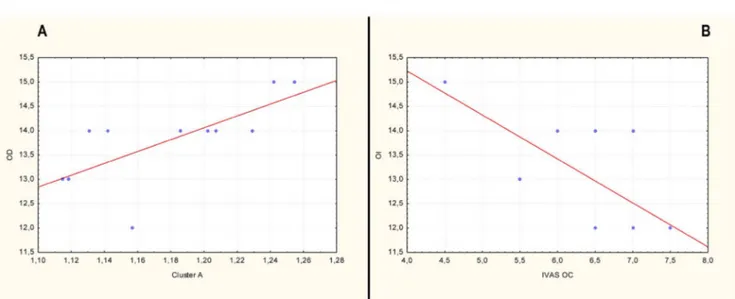
The correlation analysis resulted in significant (p , 0.05) positive
correlation (r 5 0.78) between Cluster A and the scores of Odor
Discrimination (OD) (Figure 4A). Moreover, a significant (p ,
0.05) negative correlation (r 5 20.65) was found between Odor
Identification (OI) and Intensity Visuo-Analogue Scale scores after
the OC (IVAS OC) (Figure 4B).
Discussion
A review of the literature since the original work of Zatorre in 1992
10to now, shows that many functional and structural imaging
tech-niques were applied to study the olfactory system using a number of
different olfactory tasks. Thus, due to the heterogeneity of methods
used across the studies the interpretation and comparison of results
from these experiments is often difficult
4–17. Furthermore, many
results support the idea that the neural correlates of odor processing
are strongly task dependent, involving a distributed network of
struc-tures - even outside olfactory core regions - determined by the nature
of the context and of the olfactory and sensory task
17.
Although as compared to fMRI and H
215O-PET/CT the temporal
resolution of brain activation as recorded by FDG-PET is inferior, in
our study we could replicate for the first time - due to the robust
within subjects experimental model allowing fairly good statistics
-the results previously found by -these two techniques.
In particular, completing data from previous neuroimaging
stud-ies, the first interesting finding is that OC might increase the activity
in the entorhinal cortex and hippocampus, as well as in PHG
8,
sug-gesting that the visual cortex may be involved in visual-olfactory
interaction
12,18–20and addressing the decreased glucose uptake found
in OFC to the ‘on-off switch’ behavior
16. This region was similarly
activated in response to all odor mixtures and deactivated in response
to single odorants. Interestingly, by means of the found positive
correlation (r 5 0.78; Figure 4A) between the OD scores and the
relative regional cerebral glucose metabolism (rCGM) increase in
OFC during the NC (Cluster A), the present data add further novelty
in understanding the OFC-related role in olfactory discrimination.
In fact, this aspect suggest that a relative OFC rCGM increase, during
resting condition, is always present when the cortical olfactory
sys-tem is trying to discriminate odors – even if they are not present as in
Table 1 | Numerical results of SPM comparisons between
18F-FDG uptake in NC and OC (N 5 11)
Comparison 1. Cluster level 2. Voxel level
Cluster extent Corrected pvalue Cortical Region maximumZ score of MNI coordinates Cortical region BA
NC - OC 2060 0.041 L Frontal Lobe 4.15 230,48, 24 Middle Frontal Gyrus 10
L Frontal Lobe 3.62 224,40, 26 Middle Frontal Gyrus 11 L Frontal Lobe 3.43 228,30, 222 Inferior Frontal Gyrus 11
L Frontal Lobe 3.25 212,52,14 Medial Frontal Gyrus 10
L Frontal Lobe 2.96 218,28, 226 Orbital Gyrus 47
L Limbic Lobe 2.93 220,44,6 Anterior Cingulate 32
L Frontal Lobe 2.80 222,66,8 Superior Frontal Gyrus 10 L Frontal Lobe 2.55 236,38, 22 Middle Frontal Gyrus 47 L Frontal Lobe 2.54 214,46, 212 Superior Frontal Gyrus 11
Table 1. In the ‘cluster level’ section on left, the number of voxels, the corrected P value of significance and the cortical region where the voxel is found, are all reported for each significant cluster. In the ‘voxel level’ section, all of the coordinates of the correlation sites (with the Z-score of the maximum correlation point), the corresponding cortical region and BA are reported for each significant cluster. L, left; R, right; BA, Brodmann’s area. In the case that the maximum correlation is achieved outside the grey matter, the nearest grey matter (within a range of 5 mm) is indicated with the corresponding BA.
Figure 1
|
3D rendering showing the cluster of voxels in the left orbitofrontal and anterior cingulate cortex in which FDG uptake was significantly higher at NC (n 5 11) as compared to OC (n 5 11). The first row represents the medial aspect of left (on the left) and right (on the right) hemispheres; the second row represents the anterior (on the left) and posterior (on the right) aspect on the brain; the third row represents the lateral aspect of the right (on the left) and of the left (on the right) hemispheres; the fourth row represents the inferior (on the left) and the superior (on the right) aspects of the brain. Coordinates and regional details are presented in Table 1.NC - and that its relative rCGM decrease, achieved by means of a
single and pure odor condition (as is OC) could be accompanied by a
decrease in OD. Moreover the found OFC activation/deactivation
pattern and the relative correlation with OD confirm and further
expand data previously shown by Boyle and colleagues
16.
In addition, for the first time, an rCGM increase in DoC was found
when comparing OC to NC. According to the
sensorimotor-cognit-ive dichotomy
21,22and to comparative anatomy of cerebellum for
which the Larsell’ DoC
23is actually thought to be overlapped with
Lobule VI, it appears feasible that a OC-related rCGM increase,
found for the first time in the present within-subject pure and passive
olfactory condition devoid of attentional tasks, could be not only
related to attentional processes and maintenance of the sniffing
mag-nitude
24but also to the olfactory condition ‘per se’. This intriguing
data could not only confirm the role of DoC in estimating the valence
of salient, emotional cues and selecting appropriate behavioral
res-ponses
25, but add further contributions in unraveling many roles
postulated regarding the olfactory-related cerebellar involvement
24.
Noticeably, the present study found a global leftward asymmetry
both when comparing OC to NC and viceversa. Previous olfactory
neuroimaging studies have demonstrated a bilateral activation in
primary olfactory cortex and greater activation in the right than in
the left orbitofrontal cortex
17,26,27. Possible explanation for this
incon-sistency could be related to the experimental model setting in which
any retrieval/recognition task of the incoming stimulus was
gener-ated. Regarding these aspects, a switching of lateralization from right
to the left hemisphere associated to a switch from recognition to
semantic processing of the on-line information, for both verbal
and non-verbal materials, has been widely assessed by the
hemi-spheric encoding/retrieval asymmetry model
28.
Furthermore, the improved sensitivity of the state-of-the-art PET
cameras allows to detect reliable signal changes also under
sub-optimal conditions. The possibility to create a defined ecologic
base-line condition to the exploration of olfactory neural underpinnings
helped in avoiding possible cortical activation related to unwanted
attentional processes and odour sensitivity enhancement
29due to the
examination environment.
This increases the dynamic range of brain metabolism as an
informative correlate of neural activity. Thus, this notion of baseline
implies that during a particular task not only activation is observed
but also deactivation in certain areas is found
3and depicts an
orga-nized default mode network (DMN)
30in which some regions are
most active during resting state
30–32. These regions have been thought
to be involved in monitoring the internal and external milieu
30–34and
an emerging point is that the DMN is composed of a group of
rela-tively large and interconnected areas, including the ACC and
ven-tromedial prefrontal cortex (BA 10)
30. Hence, when introducing a
pure odor compound in the present experimental model a cortical
reorganization pattern from anterior (OFC and ACC in NC) to
posterior (Cu, LG and PHG in OC) and subcortical (DoC) regions
was in line with Magistretti’s observations. This specific neural
beha-vior could suggest, for the first time, a possible rearrangement of
cortical activity when experimenting a pure and passive olfactory
stimulation condition.
To this end, Visual, Somatosensory and Cerebellum hub networks,
including PHG, Cu, LG and DoC have been previously found to be
negatively correlated with DMN activity, suggesting that an
antic-orrelated activity between this network and the former regions could
be engaged by sensory processing tasks
35.
Finally, by using a panel of the above-mentioned VQ we had the
opportunity to cluster the subjects sample in a normal range and,
thus, to correlate them each other. Interestingly a significant negative
correlation (r 5 20.65; Figure 4B) between a quantitative aspect of a
pure odorant (IVAS OC) with a qualitative one (OI) was found in the
present study by using validated tests
36. In particular this trend of
correlation could depict a daily life aspect in which an increase in
subjective intensity of a pure odorant target is commonly associated
to reduction in general odor identification. On the other side, it could
partially offer an explanatory model, at normal subjects level, of the
peculiar pathological behavior for which a higher level of intensity
score with a less percentage of identification was found in some
mood affected patients
37. However, literature is still lacking of a
neurobiological correlate of these aspects in homogeneous
experi-mental conditions. Nevertheless, this pattern of correlation could
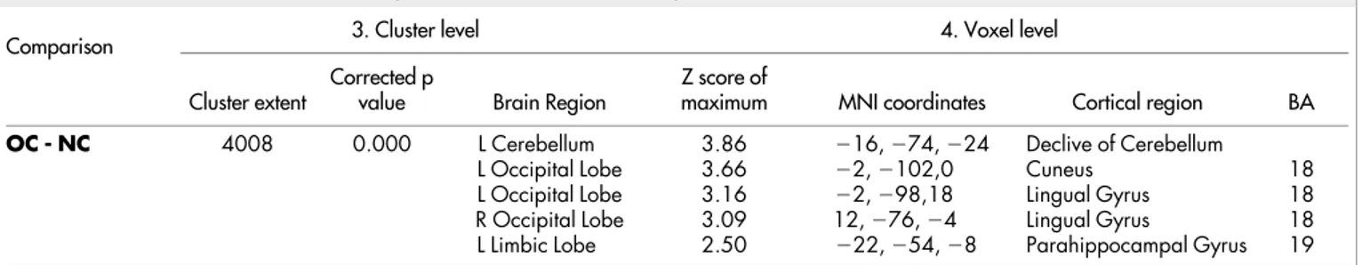
Table 2 | Numerical results of SPM comparisons between
18F-FDG uptake in OC and NC (N 5 11)
Comparison 3. Cluster level 4. Voxel level
Cluster extent Corrected pvalue Brain Region Z score ofmaximum MNI coordinates Cortical region BA
OC - NC 4008 0.000 L Cerebellum 3.86 216, 274, 224 Declive of Cerebellum
L Occipital Lobe 3.66 22, 2102,0 Cuneus 18
L Occipital Lobe 3.16 22, 298,18 Lingual Gyrus 18
R Occipital Lobe 3.09 12, 276, 24 Lingual Gyrus 18
L Limbic Lobe 2.50 222, 254, 28 Parahippocampal Gyrus 19
Table 2. In the ‘cluster level’ section on left, the number of voxels, the corrected P value of significance and the cortical region where the voxel is found, are all reported for each significant cluster. In the ‘voxel level’ section, all of the coordinates of the correlation sites (with the Z-score of the maximum correlation point), the corresponding cortical region and BA are reported for each significant cluster. L, left; R, right; BA, Brodmann’s area. In the case that the maximum correlation is achieved outside the grey matter, the nearest grey matter (within a range of 5 mm) is indicated with the corresponding BA.
Figure 2
|
3D rendering showing the cluster of voxels in the cuneus, lingual gyrus, parahippocampal gyrus and declive of cerebellum mainly in the left hemisphere, in which FDG uptake was significantly higher at OC (n 5 11) as compared to NC (n 5 11). The first row represents the medial aspect of left (on the left) and right (on the right) hemispheres; the second row represents the anterior (on the left) and posterior (on the right) aspect on the brain; the third row represents the lateral aspect of the right (on the left) and of the left (on the right) hemispheres; the fourth row represents the inferior (on the left) and the superior (on the right) aspects of the brain. Coordinates and regional details are presented in Table 2.suggest a connected sway of these tests and could advantage future
investigations regarding psychophysical dimensions of olfaction, by
starting out a defined cluster of psychological and neurobiological
results.
Methods
Subjects.Eleven right-handed individuals (six women and five men; mean age 45.7 6 11 years) without otorhinolaryngologic or neurological diseases were enrolled in the study. All of them were assessed as normosmic when evaluated with the multiple-forced choice Sniffin’ Sticks Screeinng test38in four main domains: Odor Threshold
(OT), Odor Discrimination (OD), Odor Identification (OI) and their sum (TDI). A detailed case history was collected for all subjects who underwent Ear-Nose-Throat examination with fiberoptic check of the upper airways. Neurological diseases were excluded with the Mini-Mental State Examination and MRI. All those condi-tions that could potentially develop an olfactory dysfunction were considered as exclusion criteria. Thus, patients with sino-nasal disorders or surgery history, head trauma, neuro-psychiatric disorders (Parkinson’s disease, Alzheimer’s disease, schizophrenia, multiple sclerosis, depression and Multiple Chemical Sensitivity/ Idiopatic Environmental Intolerance), lower airways and/or lung diseases, active hepatitis, cirrhosis, chronic renal failure, Vitamin B12 deficiency, alcohol, tobacco or drug abuse, cerebral vascular accidents, insulin dependent diabetes mellitus, hypothyroidism and Cushing Syndrome, were not included in the study.
Moreover, we excluded all patients in treatment with drugs that could interfere with18F -FDG uptake and distribution in the brain39. No patients were pregnant or
breastfeeding.
The Ethics Committee of the ‘‘Tor Vergata’’ University School of Medicine approved the protocol research. The study adhered to the principles of the Declaration of Helsinki and all of the participants provided written informed consent after receiving a detailed explanation of the study.
Experimental Procedure.All subjects underwent FDG-PET/CT after a neutral olfactory stimulation by using a common aerosol facial mask containing in its ampoule only 5 ml of saline sodium chloride (NaCl) 0.9% (Neutral Condition – NC). After one month, the same sample of individuals underwent a second FDG-PET/CT scan after a pure olfactory stimulation by using the same aerosol facial mask containing in its ampoule a solution of 1.5 ml of VAN 100% (Dacor Ltd j Sarandrea Ltd) and 5 ml of saline sodium chloride (NaCl) 0.9% (Olfactory Condition – OC). In both conditions oxygen flow rate was conveyed at 3.5 liters per minute and the stimulation consisted of one continuous 9 minutes block without any sniffing generated instructions. At the end of the third minute, each subject was injected with 3 MBq/Kg (210–350 MBq) of FDG i.v. and the olfactory stimulation continued for six more minutes. After both NC and OC all subjects laid down in a semi-darkened, noiseless and odorless room, without any artificial stimulation, with their eyes closed for 20 minutes.
PET/CT scanning.The PET/CT system Discovery ST16 (GE Medical Systems, TN, USA) was used for the whole population in exam. This system combines a high-speed ultra 16-detector-row (912 detectors per row) CT unit and a PET scanner with 10080 bismuth germanate (BGO) crystals in 24 rings with a 128 3 128 matrix. Crystal size 6,2 3 6,2 3 30 mm, coincidence window 11,7 nsec, system sensitivity 9,3 cps/kBq in 3D mode, dispersion fraction 44%, maximum count rate in cps at 50% dead time 63 kcps @ 12 kBq/mL (3-D), axial FWHM 1 cm radius 5,2 mm in 3D mode, axial FOV 157 mm.
Before and after the FDG injection, hydratation (500 ml of i.v. NaCl) 0.9%) was performed in order to reduce the pooling of the radiotracer in the kidneys. All subjects had normal serum glucose level and fasted for at least 5 h before the18F -FDG
injection.
Validated questionnaires (VQ).Before undergoing to olfactory stimulation all subjects completed the following VQ:
. Questionnaire of Olfactory Disorders (QOD) completed only before NC phase -to assess daily life problems due -to olfac-tory impairment, we used a short modi-fied version of an olfactory-specific QOD developed by Frasnelli and Hummel40
which has recently been used by many researchers41. The modified short version
of the QOD consists of 25 statements that are divided into three general domains: 17 negative statements (QOD-NS), two positive statements (QOD-PS), and six socially desired statements (QOD-SD);
. Zung Self-Rating Anxiety Scale42(Anxiety) - completed only before NC phase – is
a 20-item scale with some of the items keyed positively and some negatively on a four-point scale ranging from 1 5 none or a little of the time to 4 5 most or all of the time. The final score ranges from 20–80, a score between 20 and 44 is con-sidered in the normality range, 45–59 is mild to moderate anxiety, 60–74 is severe, and 75–80 is very severe;
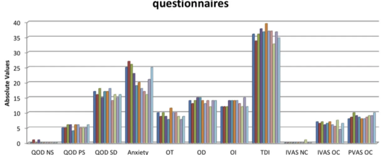
Table 3 | Mean and Standard Deviations (SD) of olfactory and
Validated Questionnaires scores
TESTS MEAN SD QOD NS 0.18182 0.404520 QOD PS 5.36364 0.674200 QOD SD 16.27273 1.272078 Anxiety 21.54545 3.856518 OT 9.27273 1.131672 OD 13.81818 0.873863 OI 13.09091 1.136182 TDI 36.18182 1.877801 IVAS NC 0.09091 0.301511 IVAS OC 6.36364 0.839372 PVAS OC 8.77273 0.719848
Table 3. QOD NS: questionnaire of olfactory disorders - negative statements; QOD PS: questionnaire of olfactory disorders – postitive statements; QOD SD: questionnaire of olfactory disorders – socially desired statement; Anxiety: self-rating anxiety scale; OT: odor threshold; OD: odor discrimination; OI: odor identification; TDI: sum of OT, OD and OI; IVAS NC: intensity visuo-analogue scale after neutral condition; IVAS OC: intensity visuo-visuo-analogue scale after olfactory condition; PVAS OC: pleasantness visuo-analogue scale after olfactory condition.
Figure 3
|
Subjects’ scores of olfactory tests and validated questionnaires. QOD NS: questionnaire of olfactory disorders - negative statements; QOD PS: questionnaire of olfactory disorders – postitive statements; QOD SD: questionnaire of olfactory disorders – socially desired statement; Anxiety: self-rating anxiety scale; OT: odor threshold; OD: odor discrimination; OI: odor identification; TDI: sum of OT, OD and OI; IVAS NC: intensity visuo-analogue scale after neutral condition; IVAS OC: intensity visuo-analogue scale after olfactory condition; PVAS OC: pleasantness visuo-analogue scale after olfactory condition.. Intensity Visuo-Analogue Scale (IVAS), in which each subject may score from 0 to 10 the subjective intensity of olfactory perception after both NC and OC37;
. Pleasantness Visuo-Analogue Scale (PVAS), in which each subject may score
from 0 to 10 the subjective pleasantness of olfactory perception after the OC37.
Statistical analysis. FGD-PET/CT.Differences in brain FDG uptake were analyzed using statistical parametric mapping (SPM2, Wellcome Department of Cognitive Neurology, London, UK) implemented in Matlab 6.5 (Mathworks, Natick, Massachusset, USA). PET data were subjected to affine and non-linear spatial normalization into the MNI space. The spatially normalized set of images were then smoothed with a 8 mm isotropic Gaussian filter to blur individual variations in gyral anatomy and to increase the signal-to-noise ratio. Images were globally normalized using proportional scaling to remove confounding effects to global CBF changes, with a threshold masking of 0.8. The resulting statistical parametric maps, SPM{t}, were transformed into normal distribution (SPM{z}) unit. Correction of SPM coordinates to match the Talairach coordinates was achieved by the subroutine implemented by Matthew Brett (http://www.mrc-cbu.cam.ac.uk/Imaging). Brodmann areas (BAs) were then identified at a range of 0 to 3 mm from the corrected Talairach coordinates of the SPM output isocentres, after importing them by Talairach client (http://www. talairach.org/index.html). A statistical height thresholds equal or lower than p , 0.05 at both cluster and voxel level was accepted as significant.A statistical height thresholds equal or lower than p , 0.05 at voxel level and p , 0.05 corrected for multiple comparison at cluster level was accepted as significant.
This more liberal choice was adopted to avoid type II errors attributable to over-conservative thresholds43. In fact, given the exploratory nature of this analysis and
considering the relatively low sensitivity of PET without repeated measures, higher thresholds could lead to false-negative results in PET studies. Only those clusters containing more than 125 (5 3 5 3 5 voxels, i.e. 11 3 11 3 11 mm) contiguous voxels were accepted as significant, based on the calculation of the partial volume effect resulting from the spatial resolution of the PET camera (about the double of FWHM).
The OC vs NC comparisons were performed by means of the ‘compare popula-tions: 1 scan/subject (Ancova)’ option, using age and sex as covariates.
VQ and Correlation Analysis.Data description included calculation of means and standard deviations (SD) for all VQ. In addition, Spearman’s rank correlation was performed between QOD (and relative subscales), Anxiety, OT, OD, OI, IVAS NC, IVAS OC and PVAS OC. (STATISTICA 7 package for Windows).
Moreover, in order to evaluate the correlations between brain metabolism and subjective variables, the clusters of voxels found to be significantly different from zero in the NC – OC and OC – NC comparisons were segmented and the semi-quant-itative raw data values in each of them were individually normalized to the relative FDG uptake in the cerebellum. All values were then transformed into z-scores and submitted to statistical analyses seeking for Spearman’s rank correlations with VQ total scores. The correlation analyses have been performed considering the values at NC and OC (as well as the relative VQ scores) as a continuum.
1. Mazard, A. et al. Impact of fMRI acoustic noise on the functional anatomy of visual mental imagery. J. Cogn. Neurosci. 14, 172–186 (2002).
2. Benjamin, C. et al. The influence of rest period instructions on the default mode network. Front. Hum. Neurosci. 4, 218 (2010).
3. Magistretti, P. J. Neuron-glia metabolic coupling and plasticity. J. Exp. Biol. 209, 2304–2311 (2006).
4. Savic, I. Brain imaging studies of the functional organization of human olfaction. Chem. Senses 30, i222–i223 (2005).
5. Savic-Berglund, I. Imaging of Olfaction and Gustation. Nutr. Rev. 62, 205–207 (2004).
6. Hillert, L., Musabasic, V., Berglund, H., Ciumas, C. & Savic, I. Odor processing in multiple chemical sensitivity. Hum. Brain Mapp. 28, 172–182 (2007). 7. Popp, R., Sommer, M., Mu¨ller, J. & Hajak, G. Olfactometry in fMRI studies: odor
presentation using nasal continuous positive airway pressure. Acta Neurobiol. Exp. 64, 171–176 (2004).
8. Kjelvik, G., Evensmoen, H. R., Brezova, V. & Haberg, A. K. The human brain representation of odor identification. J. Neurophysiol. 108, 645–657 (2012). 9. Sobel, N. et al. Sniffing and smelling:separate subsystems in the human olfactory
cortex. Nature 392, 282–286 (1998).
10. Zatorre, R. J., Jones-Gotman, M., Evans, A. C. & Meyer, E. Functional localization and lateralization of human olfactory cortex. Nature 360, 339–340 (1992). 11. Bengtsson, S., Berglund, H., Gulyas, B., Cohen, E. & Savic, I. Brain activation
during odor perception in males and females. Neuroreport 12, 2027–33 (2001). 12. Cerf-Ducastel, B. & Murphy, C. Neural substrates of cross-modal olfactory
recognition memory: An fMRI study. Neuroimage 31, 386–396 (2006). 13. Cerf-Ducastel, B. & Murphy, C. Age-related differences in the neural substrates of
cross-modal olfactory recognition memory: an fMRI investigation. Brain Res. 1285, 88–98 (2009).
14. Royet, J. P. et al. Emotional responses to pleasant and unpleasant olfactory, visual, and auditory stimuli: a positron emission tomography study. J. Neurosci. 20, 7752–7759 (2000).
15. Kareken, D. A. et al. Olfactory system activation from sniffing: effects in piriform and orbitofrontal cortex. Neuroimage 22, 456–465 (2004).
16. Boyle, J. A., Djordjevic, J., Olsson, M. J., Lundstrom, J. M. & Jones-Gotman, M. The Human Brain Distinguishes between Single Odorants and Binary Mixtures. Cereb. Cortex 19, 66–71 (2009).
17. Savic, I. & Gulyas, B. PET shows that odors are processed both ipsilaterally and contralaterally to the stimulated nostril. Neuroreport 11, 2861–2866 (2000). 18. Royet, J. P. et al. Functional anatomy of perceptual and semantic processing for
odors. J. Cogn. Neurosci. 11, 94–109 (1999).
19. Qureshy, A. et al. Functional Mapping of Human Brain in Olfactory Processing: A PET Study. J. Neurophysiol. 84, 1656–1666 (2000).
20. Kroupi, E., Yazdani, A., Vesin, J. M. & Ebrahimi, T. Multivariate spectral analysis for identifying the brain activations during olfactory perception. Conf. Proc. IEEE Eng. Med. Biol. Soc. 2012, 6172–6175 (2012).
21. Manni, E. & Petrosini, L. A century of cerebellar somatotopy: a debated representation. Nat. Rev. Neurosci. 5, 241–9 (2004).
22. Stoodley, C. J. & Schmahmann, J. D. Functional topography in the human cerebellum: a meta-analysis of neuroimaging studies. Neuroimage 44, 489–501 (2009).
23. Schmahmann, J. D. et al. Three-dimensional MRI atlas of the human cerebellum in proportional stereotaxic space. Neuroimage 10, 233–60 (1999).
Figure 4
|
A) Positive correlation (r 5 0.78) between odor discrimination (OD) and Cluster A and B) negative correlation (r 5 20.65) between odor identification (OI) and intensity visuo-analogue scale after olfactory condition (IVAS OC).24. Moscovich, M. et al. Olfactory impairment in familial ataxias. J. Neurol. Neurosurg. Psychiatry 83, 970–4 (2012).
25. Habas, C. et al. Distinct cerebellar contributions to intrinsic connectivity networks. J. Neurosci. 29, 8586–94 (2009).
26. Zatorre, R. J. & Jones-Gotman, M. [Functional imaging of the chemical senses] Brain Mapping: The Applications [Toga, A. W., Mazziotta, J. C. (ed.)] [403–424] (Academic Press, San Diego, 2000).
27. Olofsson, J. K. et al. Laterality of the olfactory event-related potential response. Chem. Senses 31, 699–704 (2006).
28. Habib, R., Nyberg, L. & Tulving, E. Hemispheric asymmetries of memory: the HERA model revisited. Trends Cogn. Sci. 7, 241–245 (2003).
29. A˚ hs, F., Miller, S. S., Gordon, A. R. & Lundstro¨m, J. N. Aversive learning increases sensory detection sensitivity. Biol. Psychol. 92, 135–41 (2013).
30. Sheline, Y. I. et al. The default mode network and self-referential processes in depression. Proc. Natl. Acad. Sci. 106, 1942–1947 (2009).
31. Raichle, M. E. & Snyder, A. Z. A default mode of brain function: a brief history of an evolving idea. Neuroimage 37, 1083–1090 (2007).
32. Buckner, R., Andrews-Hanna, J. & Schacter, D. The brain’s default network: Anatomy, function, and relevance to disease. Ann. N. Y. Acad. Sci. 1124, 1–38 (2008).
33. Pagani, M. et al. Brief Report: alterations in cerebral blood flow as assessed by PET/CT in adults with autism spectrum disorder with normal IQ. J. Autism. Dev. Disord. 42, 313–8 (2012).
34. Micarelli, A., Jacobsson, H., Larsson, S. A., Jonsson, C. & Pagani, M.
Neurobiological insight into hyperbaric hyperoxia. Acta Physiol. (Oxf) 209, 69–76 (2013).
35. Tomasi, D. & Volkow, N. D. Association between Functional Connectivity Hubs and Brain Networks. Cereb. Cortex 21, 2003–2013 (2011).
36. Buro´n, E. & Bulbena, A. Olfaction in affective and anxiety disorders: a review of the literature. Psychopathology 46, 63–74 (2013).
37. Lombion-Pouthier, S., Vandel, P., Nezelof, S., Haffen, E. & Millot, J. L. Odor perception in patients with mood disorders. J. Affect. Disord. 90, 187–91 (2006). 38. Kobal, G., Hummel, T., Sekinger, B., Barz, S., Roscher, S. & Wolf, S. ‘‘Sniffin’
sticks’’: screening of olfactory performance. Rhinology 34, 222–226 (1996).
39. Varrone, A. et al. EANM procedure guidelines for PET brain imaging using [18F]FDG, version 2. Eur. J. Nucl. Med. Mol. Imaging 36, 2103–2110 (2009). 40. Simopoulos, E. et al. Olfaction-Associated Quality of Life in Chronic
Rhinosinusitis: Adaptation and Validation of an Olfaction-Specific Questionnaire. Laryngoscope 122, 1450–1454 (2012).
41. Alessandrini, M. et al. Intranasal administration of hyaluronan as a further resource in olfactory performance in multiple chemical sensitivity syndrome. Int. J. Immunopathol. Pharmacol. 26, 1019–25 (2013).
42. Zung, W. W. & Gianturco, J. A. Personality dimension and the Self-Rating Depression Scale. J. Clin. Psychol. 27, 247–8 (1971).
43. Oishi, N. et al. Regional cerebral blood flow in Parkinson disease with nonpsychotic visual hallucinations. Neurology 65, 1708–1715 (2005).
Author contributions
Conceived and designed the experiments: M.A., A.M., A.C., O.S. Performed the experiments: M.A., A.M., A.C., B.D.P., E.B. Analyzed the data: A.M., M.P., M.A., M.C. Contributed reagents/materials/analysis tools: A.C., M.P., O.S., B.D.P., E.B. Wrote the paper: A.M., M.A., M.C., M.P.
Additional information
Competing financial interests: The authors declare no competing financial interests. How to cite this article:Alessandrini, M. et al. Cortico-subcortical metabolic correlates of olfactory processing in healthy resting subjects. Sci. Rep. 4, 5146; DOI:10.1038/srep05146 (2014).
This work is licensed under a Creative Commons Attribution-NonCommercial-NoDerivs 3.0 Unported License. The images in this article are included in the article’s Creative Commons license, unless indicated otherwise in the image credit; if the image is not included under the Creative Commons license, users will need to obtain permission from the license holder in order to reproduce the image. To view a copy of this license, visit http://creativecommons.org/licenses/by-nc-nd/3.0/