Case 9295
Nasosphenoidal schwannoma
Comments/Instructions
»Editor wrote:
The case is interesting and well illustrated.
From my point of view it is not clear if there was a recurrence of the disease or whether the neoplasm was only partially removed. Since post-surgical follow-up is not shown but apparently total removal was appreciable at endoscopy, recurrence seems possible. Could you please specify?
Could you specify was FESS refers to?
Do you have knowledge in your patient or others from literature of possible NF2? Was NF2 ruled out? »Author wrote:
Thank you for your suggestions. We have revised the article, making the following modifications:
1. One year MRI follow up and histological exam demonstrated the recurrence of the disease. However this is a very rare event.
2. FESS - Functional Endoscopic Sinus Surgery.
3. We excluded the presence of Neurofibromatosis type 2 because of the imaging, histological and clinical characteristics of the tumour.
Best regards,
Manenti G. MD, PHD et al.
Clinical History
A 30-year-old woman came to our attention for the presence of recurrent nasal epistaxis of about one month, without pain or other notable disease. The patient underwent radiological investigations such as CT and MRI with and without contrast injection.
Imaging Findings
The patient underwent radiological investigations such as CT with and without contrast injection (Fig.1), which documented the presence of hypodense soft tissue with regular edges and polypoid appearance. Subsequently, it was subjected to thorough MRI diagnostic (Fig.2) with and without contrast material that revealed the presence of polypoid formation with a diameter of 23.9 x 10.9 mm, that extended from the mucous portion of the posterior margin of the left superior horn, to the ipsilateral palatine tonsil while maintaining a cleavage plane by itself, the described training raised Author(s) Manenti G, Assako Ondo EP, Antonicoli M, Di Poce I, Llubani R, Simonetti G
Patient female, 30 year(s)
02.05.2011 - 18:22
without eroding the floor of ipsilateral sphenoid sinus.
The post operative course was uneventful, and 6 months follow-up revealed a scar formation at endoscopic evaluation. At one-year MRI follow-up a polypoid formation was demonstrated (Fig.4). Subsequent histological exam proved it to be a recurrent nasosphenoid benign schwannoma of the same characteristics described on the previous one (Fig.5). Discussion
Schwannomas are lesions that arise from the neural sheath of peripheral nerves, autonomic nerves, or cranial nerves. Nerve sheath tumours of the head and neck region mainly involve the eighth cranial nerve, 80 % [1]. Only 4% of head and neck schwannomas originate from the sinonasal tract [2, 3], where the ethmoid, maxillary sinus, nasal fossa, sphenoid and frontal sinus are involved in decreasing order [4, 5].
Nasal septum schwannoma was first described by Bogdasarian and Stout in 1943. Higo et al. [6] reviewed the world literature, collecting 160 cases of schwannoma of the paranasal and nasal cavities with only 11 nasal septum schwannoma (6, 9%). Histology of these tumours shows fusiform, neoplastic schwann cells forming compact, interlacing bundles accompanied by end of delicate fibers (Fig.5).
Symptoms and signs depend on the site of origin and on lesion extention. They include unilateral nasal obstruction, epistaxis, mucopurulent rhinorrhoea, anosmia, facial swelling, proptosis and pain, if maxillary sinus is involved [7]. Extension into the sphenoid sinus can lead to diplopia (secondary to deficits of III, IV and VI cranial nerves), deep retro-orbital pain and occipital, frontal and bitemporal headache [8], however, in our case the patient had only history of epistaxis and nasal obstruction.
The lesions usually have a mottled central latency with peripheral intensification on contrast enhancement CT scans. The heterogeneous appearance is related to areas of increased vascularity with adjacent non-enhancing cystic or necrotic regions.
MRI is better than CT scan in differentiating a tumour from inflammatory changes and normal tissues; furthermore the intracranial extension can be better delineated. Previously Dublin et al. [3] and Younis et al. [4] described the CT and MRI findings of a benign and malignant schwannoma contained within the right ethmoid and sphenoid sinus.
The prognosis for benign schwannoma is generally good, whereas that for malignant variant is poor, due to the highly aggressive nature of a high recurrence rate. Metastases occur commonly in the lungs, and rarely in the regional lymph nodes [9].
The treatment of choice for intranasal schwannoma is surgery. Different approaches such as lateral rhinotomy with external ethmoidectomy, the Cadwell-Luc approach or endonasal endoscopic resection have been employed in relation to tumour extension [10].
The best surgical approach is chosen by tumour location and extention. In our case the surgical approach in FESS (Functional Endoscopic Sinus Surgery) was chosen because the tumour had a nasosphenoidal location, without extending to the skull base (Fig. 3).
Final Diagnosis
Nasosphenoidal benign schwannoma Differential Diagnosis List
Juvenile angiofibroma Neurofibromatosis type II Intranasal glioma Mucocele
MeSH
Paranasal Sinus Neoplasms [C09.647.685.693]
Neoplasms or tumors of the paranasal sinuses. Malignant neoplasms are rare, comprising 3% of all head and neck neoplasms. The majority arise in the maxillary sinus with malignancies of the ethmoid sinus constituting virtually all the remaining tumors.
Epistaxis [C09.603.261] Bleeding from the nose. Nasal Polyps [C09.603.557]
Focal accumulations of EDEMA fluid in the NASAL MUCOSA accompanied by HYPERPLASIA of the associated submucosal connective tissue. Polyps may be NEOPLASMS, foci of INFLAMMATION, degenerative lesions, or malformations.
Radiographic Image Interpretation, Computer-Assisted [E01.158.600.680] Computer systems or networks designed to provide radiographic interpretive information.
References
[1]
Pasic TR, Makielski K (1990) Nasal schwannoma. Otolaryngol Head Neck Surg 103:943-946 [2]
Donnelly M, Al-Seder MH, Blayney AW (1992) Benign nasal schwannoma. J Laryngol Otol 106:1011-1015 [3]
Dublin AB, Dedo HH, Bridger WH (1995) Intranasal schwannoma: magnetic resonance and computed tomography appearance. Am J Otolaryngol 16: 251-254
[4]
Younis RT, Gross CW, Lazar RH (1991) Schwannoma of the paranasal sinuses. Arch Otolaryngol Head Neck Surg 117:677-680
[5]
Choi YS, Sung KS, Song YJ, Kim HD (2009) Olfactory schwannoma- case report. J Korean Neurosurg soc 45:103-6 [6]
Higo R, Yamasoba T, Kikuchi S (1993) Nasal neurinoma: case report and review of literature. Auris Naus Larynx 20:297-301
[7]
Zovickian J, Braba D, Alksne JF (1986) Intranasal schwannoma with extension into the intracranial compartment: case report. Neurosurgery 19:813-815
[8]
Katz TS, Mendenhall WM, Morris CG, Amdur RJ, Hinerman RW, Villaret DB. (2002) Malignant tumors of the nasal cavity and paranasal sinuses.. Wiley Periodicals, Inc. Head Neck 24(9):821-9.
[9]
Dillon WP, Som PM, Rosenau W (1991) Hemangioma of the nasal vault: MR and CT features. Radiology 180:761-765 [10]
Gilbey P, Kukuev Y, Samet A, Talmon Y, Ivry S (2009) The quality of the surgical field during functional endoscopic sinus surgery--the effect of the mode of ventilation--a randomized, prospective, double-blind study. Laryngoscope 119:24-53
Citation
Manenti G, Assako Ondo EP, Antonicoli M, Di Poce I, Llubani R, Simonetti G (2011, May 13). Nasosphenoidal schwannoma, {Online}.
URL: http://www.eurorad.org/case.php?id=9295 DOI: 10.1594/EURORAD/CASE.9295
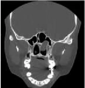
Figure 1
CT image
Coronal CT reconstruction showing hypodense soft tissue with regular edges and polypoid appearance extending from nasal cavity to nasopharinx and left sphenoid cavity.
Figure 2
Preoperative T2 Weighted MR image Figure 2a
Figure 2b
Figure 2c
Figure 2d
Figure 2e
Coronal T2-weighted MR image shows polypoid solid tissue extending from nasopharyngeal space to the left sphenoid sinus.
Coronal T1-weighted MR image after intravenous injection of paramagnetic contrast agent shows polypoid enhancing tissue extending from nasopharyngeal space to the left sphenoid sinus.
Sagittal T1-weighted MR image before intravenous injection of paramagnetic contrast agent shows polypoid solid tissue extending from nasopharyngeal space to the left sphenoid sinus.
Axial T1-weighted MR image after intravenous injection of paramagnetic contrast agent shows polypoid enhancing tissue extending from nasopharyngeal space to the left sphenoid sinus.
Axial T1-weighted MR image before intravenous injection of paramagnetic contrast agent shows
Figure 2f Figure 3 Endoscopic images Figure 3a Figure 3b Figure 3c Figure 3d Figure 4
polypoid solid tissue at left nasopharyngeal space.
Axial T1-weighted MR image after intravenous injection of paramagnetic contrast agent shows polypoid solid tissue at left nasopharyngeal space.
Endoscopic image showing expansive solid tissue growing into naso-pharyngeal space.
Endoscopic image showing expansive solid tissue growing into naso-pharyngeal space.
Endoscopic image of the surgery procedure showing complete excision of the mass.
Endoscopic image of the surgery procedure showing complete excision of the mass.
MRI follow-up Figure 4a
Figure 4b
Figure 4c
Figure 4d
Figure 5
At one year MRI follow-up axial T1-weighted image before intravenous injection of Gadolinium-DTPA shows the presence of solid tissue unremoved by the surgical approach.
At one year MRI follow-up sagittal T1-weighted image after intravenous injection of Gadolinium-DTPA shows the persistence of a huge enhancing tissue at the surgical site.
At one year MRI follow-up axial T1-weighted image after intravenous injection of Gadolinium-DTPA shows the persistence of a huge enhancing tissue that results unremoved from the surgical approach.
At one year MRI follow-up coronal T1-weighted image after intravenous injection of Gadolinium-DTPA shows the persistence of a huge enhancing tissue that results unremoved from the surgical approach.
H&E histopathology examination
High cell density tissue composed of compact arrangement of spindle cells (Antoni A Schwannoma) at H&E histopathology examination.