Università Degli Studi di Catania
Dottorato Internazionale di Ricerca in Neurobiologia.
XXIV CICLO
Coordinatore: Prof. Roberto Avola
Dott.ssa Giuseppina Vitale
“Aminergic systems in sleep disorders of neurological diseases”
PhD THESIS
Tutor: Prof. V. SOFIA
Co-Tutor: Prof. M. ZAPPIA
Index
1 INTRODUCTION 1
1.1 Dopamine and Sleep 1
1.2 Periodic Lower Limb Movements (PLMS) 2
1.2.a Definition and characteristics 2
1.2.b Role and pathogenesis of dopamine 4 1.2.c Impact of PLMS in different pathological conditions 6
1.2.d PLMS in epilepsy 7
1.3 Epilepsy and Dopamine 8
2 AIMS 9
3 MATERIALS AND METHODS 10
4 RESULTS 12 4.1 Clinical data 12 4.2 Daytime sleepiness 12 4.3 Hypnic structure 12 4.4 Characteristics of PLMS 14 4.5 Effects of Pramipexole 15 5 DISCUSSION 19 6 CONCLUSIONS 22 7 REFERENCES 23
1 1. INTRODUCTION
1.1. DOPAMINE and SLEEP
Dopamine is a catecholaminergic neurotransmitter. Of the four major dopaminergic pathways, three originate from the substantia nigra in the mesencephalon. One of these, known as the nigrostriatal pathway, plays a distinctive role in controlling motion, while the other two, the mesolimbic and the mesocortical pathways, regulate emotional and affective responses. The nigrostriatal pathway is affected in Parkinson’s disease and generally in motor disorders, while the mesolimbic and the mesocortical pathways are altered in patients with schizophrenia.
The role of dopamine (DA) on sleep in both physiological and pathological conditions remains an unsettled issue. Low dopamine doses are known to be sleep promoting by acting on D2 receptors, whereas high dopamine doses, usually effective in increasing motor activity, stimulate wakefulness and suppress both slow-wave and REM sleep through postsynaptic D1 receptors.
A variety of sleep disorders implicate a dysfunction in the dopaminergic system, particularly REM Behaviour Disorder (RBD), Restless Leg Syndrome (RLS) and Periodic Limb Movements during Sleep (PLMS). The latter were the object of this thesis and therefore the following paragraphs will focus exclusively on periodic movements involving the lower limbs.
2 1.2 PERIODIC LIMB MOVEMENTS DURING SLEEP (PLMS)
1.2.a Definition and characteristics:
Periodic Limb Movements during Sleep (PLMS) were first described by Symonds in 1953 [2]. PLMS involve stereotyped, rhythmic extension movements of the big toe and dorsiflexion of the foot, occasionally associated with flection of the leg and thigh during sleep (fig. 1).
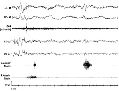
According to the criteria first set out by Coleman [3] and recently revised, PLMS can be defined as an activation of the tibialis anterior muscles lasting between 0.5 and 10 seconds, separated by an intermovement interval ranging from 5 to 90 seconds, and organised in series of 4 or more consecutive leg movements (fig. 2) [4]. They preferably appear during NREM light sleep stages 1 and 2 – although they may persist even in NREM stages 3 and 4 and in REM sleep – and tend to occur more frequently during the first half of the night (fig. 3). A PLMS index (number of PLMS per hour of sleep) of 5 or more is generally considered pathological in the young adult [5]. As mentioned above, PLMS are particularly common in Restless Legs Syndrome during NREM sleep, but they have also been described in association with various sleep disorders including Narcolepsy, REM Sleep Behaviour Disorder (RBD), Sleep Apnea Syndrome, Insomnia and Hypersomnia [6].
PLMS may be associated – in varying proportions – with cortical arousal, defined as an abrupt change in the EEG pattern causing the alpha or theta rhythm to be reactivated, with a duration ranging between 3 and 10 seconds. Additionally, autonomic activation is observed with each PLMS episode. Autonomic activation in PLMS is characterised by tachycardia (lasting about 10 seconds), followed by bradycardia, regardless of the presence of arousals [7,8].
3 Fig. 1: Semiology of PLMS
Fig. 2: Polygraphic characteristics of PLMS
4
1.2.b Role and pathogenesis of dopamine
The pathogenesis of PLM is still controversial. However, the most commonly accepted hypothesis is that PLM as well as RLS are caused by a dysfunction of both the central and the subcortical dopaminergic system. There is no shortage of evidences supporting this thesis, among which are: (a) an immediate, direct effect of low doses of dopamine agonists [17, 18]; (b) the role of dopaminergic drugs as PLM-inducing agents [19]; (c) the role of iron deficiency as a causative agent of both RLS and PLM (iron being a coenzyme for tyrosine hydroxylase, a key enzyme in the metabolism of dopamine) [20]; and (d) the higher RLS and PLM frequency in patients with Parkinson's disease [21]. The circadian distribution of PLMS is inversely related to dopamine values in the blood and cerebrospinal fluid, which verge on the low-side during evening and night-time hours and gradually increase throughout the day [22].
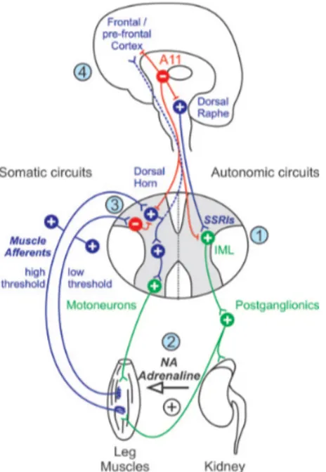
The role of the dopaminergic system in RLS has been recently clarified by a number of studies using functional magnetic resonance imaging and nuclear medicine techniques (namely, SPECT and PECT). Most of these studies – albeit with mixed results – have highlighted the following: (a) increased dopamine and dopamine turnover levels in the cerebrospinal fluid; (b) reduction in the striatal binding for all D2 receptors (using SPECT) in patients with PSMS [23]; (c) dysfunction of the nigro-striatal dopamine receptors particularly in the putamen with increased levels of endogenous dopamine (using PET); (d) a slight but significant decrease in FDOPA uptake (using PET) in both the putamen and the caudate [24]. From an anatomical standpoint, which dopaminergic system is implied in the pathogenesis of RLS and PLM is not entirely clear. Over the past few years, research has focused on a small dopaminergic diencephalic nucleus (approximately 300 neurons in rats and 130 in mice) located in the dorsal-posterior region of the hypothalamus, which is generally referred to as A11 (fig. 4). The dopaminergic diencephalic nucleus has projections to the neocortex, to the dorsal raphe nucleus and, most importantly, it has inhibitory projections to two of the spinal nuclei: the intermediolateral tracts of the spinal cord (i.e. the preganglionic orthosympathetic neurons) and the spinal dorsal horns (i.e. the sensitive neurons) [25]. A dysfunction in such descending pathway could generate a certain degree of hyperexcitability at the spinal level, which is thought to be the underlining cause of PLM and RLS symptoms. The dopamine receptors present on these areas of the spinal cord belong to the D3 family. D3 agonists are
5 currently the first-line treatment for RLS and PLMS. It should be stressed that various sympathetic neurons originate from the intermediolateral tracts of the spinal cord, all forming synaptic connections with other neurons that innervate muscle spindles. The alteration of this pathway could increase the sensitive influence from the spindles on the posterior horns through the sensitive peripheral pathway. The A11 area is also closely linked to the hypothalamic suprachiasmatic nucleus – the main endogenous pacemaker of circadian rhythms – and could therefore act as a modulator of the circadian progression of symptoms. An experimental lesion of the A11 in rats results in hypermotor activity, which is reduced by dopamine agonists [25].
6
1.2.c Impact of PLMS in different pathological conditions
Periodic nocturnal myoclonus is particularly frequent in Restless Legs Syndrome (RLS) and other sleep disorders, such as REM Sleep Behaviour Disorder (RBD), Narcolepsy, Obstructive Sleep Apnea Syndrome (OSAS), Insomnia and Hypersomnia, although it can also be found in healthy, asymptomatic subjects aged 65 years and over [26].
Polysomnographic studies, whose aim is to evaluate the differential characteristics of PLM in sleep disorders, have emphasized the highly variable nature of the phenomenon. In particular, the most important differences identified were related to the periodicity of PLM and to its distribution throughout the various sleep stages within a given pathological framework. As regards the myoclonus distribution through the night, two different patterns have been identified [27]. In the first pattern, PLM is particularly noticeable during the first half of the night (with a peak between 11 p.m. and 4 a.m.) [28] and then decreases. This is the typical PLM distribution found in patients with RLS or PLMD. In the second pattern, which is typical of narcoleptic and OSAS patients, PLMS is distributed relatively evenly throughout the night. [27].
Important differences have also been reported in the distribution of nocturnal myoclonus throughout the various stages of sleep: in RLS and PLMS patients, it typically occurs during NREM phases. In RBS patients, on the contrary, nocturnal myoclonus tends to occur during REM sleep and also shows a reduced frequency, probably due to a lower association between PLMS and arousal [29].
Finally, narcoleptic patients have a pathological PLMS index, although to a lesser extent compared to patients with RLS. The hypnic structure of these patients is characterised by a lower CAP rate, meaning that the cause of the reduced PLMS frequency is most likely to be identified with a reduced fluctuation of the arousals [30].
Data reported above suggest that the various pathological frameworks may contain different PLM pathogenic phenotypes. High PLMS rates have been reported in some neurodegenerative disorders (ND), such as Parkinson’s Disease and Multiple System Atrophy (MSA), in which the high coexistence rate of PLMS and RBD is considered revealing of an existing alteration in motor control during sleep.
Polysomnographic studies in patients with PD and MSA show a significant reduction in total sleep time and in sleep efficiency, as well as anomalies in the REM stages of sleep, such as
7 absence of REM atonia or typical RBD manifestations, and a pathological PLM index [31-34].
The reasons for the fragmentation of the hypnic profile are to be found in both the high frequency of the awakenings, which is in turn closely related to the motor effects of the conditions themselves, and in the equally high frequency of complex phenomena, such as PLMS and RBD, during sleep. The latter are due to an increased motor activity, during both REM and NREM stages, which is typical of such pathologies [32, 33]. In the above mentioned ND, as well as in RBD that often precedes them, some of the data indicate a correlation between the extent of the periodic nocturnal myoclonus and the severity of the neurodegenerative process, thus assigning a possible prognostic significance to PLMS.
1.2.d PLMS in Epilepsy
Few studies have documented a pathological PLMS index in patients with Epilepsy. A work published in 1994 investigated the sleeping patterns of 6 patients with Partial Epilepsy. Polysomnographic recordings showed a reduction of sleep efficiency and the presence of PLMS in 4 patients. In two cases these were associated with a high arousal index [35]. More recently, researchers have investigated the video-polysomnographic patterns of patients with Angelman Syndrome, highlighting the presence of PLMS, a phenomenon which they attributed to Epilepsy rather than to Mental Retardation [36]. A recent study, carried out in 2008, evaluated the spectrum of polysomnographic abnormalities in children with Epilepsy. The study revealed that in a sample of 40 subjects, 10% had periodic movements of the lower limbs [37]. The occurrence of PLMS in Epilepsy could be related to the fragmentation of the hypnic structure often found in affected patients. This hypothesis has been suggested by a study describing the association between PLMS, minor motor events and interictal discharges in Nocturnal Frontal Lobe Epilepsy (NFLE). EEG abnormalities, in fact, may lead to a fluctuant arousal, which in turn ‘allows’ the occurrence of PLMS episodes [38].
8 1.3 EPILEPSY AND DOPAMINE
Recent studies point to a role of dopamine in Epilepsy which, however, still remains fascinating, complex and largely unresolved. There is evidence that many dopamine agonists and antagonists modulate the brain network, exerting either anti- or pro-convulsant effects [39]. The potential role of the dopaminergic pathways in seizure expression or control patterns has been reported in both generalized and focal epilepsies. In particular, nuclear medicine studies have shown dysfunctions in the dopaminergic system in some epileptic syndromes, such as Autosomal Dominant Nocturnal Frontal Lobe Epilepsy (ADFNLE) [40], Juvenile Myoclonic Epilepsy (JME) [41], Ring 20 Syndrome [42] and Temporal Lobe Epilepsy [43, 44].
Considering the possibility of the dopaminergic pathways being involved in PLM genesis, these data may serve to explain the presence of PLM in patients with Epilepsy, thus providing valuable insights into the pathogenesis of both disorders.
9 2. AIMS
The aims of this study were to evaluate the possibility of an association between Epilepsy and Periodic Lower Limb Movements as evidenced by video-polisomnographic recordings, to define the polysomnographic characteristics of PLMS (i.e.: duration, distribution through the night and thorough the various stages of sleep, and inter-PLM interval), and to highlight any differences with PLMS in association with other neurological disorders.
Given the dopaminergic nature of PLMS, the association between PLMS and Epilepsy might have represented an additional, indirect evidence of the involvement of dopamine in the pathogenesis of Epilepsy.
Another aim was to evaluate the influence of some dopamine agonists commonly used in the treatment of PLMS on the PLMS pattern of some of the patients constituting our sample, as well as any impact these might have on epileptiform discharges.
10 3. MATERIALS AND METHODS
We selected all patients referred to our neurological clinic from 2008 to 2012, who had previously been diagnosed with epilepsy based on the ILAE criteria, who showed no signs of Snoring, Sleep Apnea, RLS, RBD and other degenerative neurologic conditions and who were not being treated with antidopaminergic and/or antidepressant drugs.
All patients underwent Epworth Sleepiness Scale measurements, EEG recordings during wakefulness, brain MRI and nocturnal video-polisomnographic recordings (carried out after a one-night adaptation period in our laboratory).
For video-polisomnographic recordings, the following parameters were taken into account: standard bipolar EEG derivations (according to the International System 10-20), electrooculogram (EOG), derivation from the mylohyoid muscle and from the anterior left and right tibialis muscles, ECG, oronasal flow, bands for detecting thoracic and abdominal movements, and oximetry.
Next, we reviewed each patient’s clinical history as well as the clinical history of his/her family members, together with their EEG, neuroradiological and video-polysomnographic data. We also analysed and scored any occurring PLMS event. PLMS events were scored according to the criteria set forth by AASM in “Manual for scoring” (2007) [4]. According the AASM criteria, each sequence should consist of 4 or more leg movements, separated by an intermovement interval ranging from 5 to 90 seconds. The activation of the tibialis anterior muscles should last between 0.5 and 10 seconds, with a burst amplitude of at least 25% recorded at the beginning of the polygraphic study during a spontaneous dorsiflexion of the foot. We therefore selected patients who had a PLM index considered as pathological (PLMI>5). For each of these patients we analysed the following PLM characteristics: (a) number; (b) index; (c) mean duration. Subsequently, 5 patients were randomly selected among those with Epilepsy who had a PLMS index > 5. All 5 patients underwent other two nocturnal video-polisomnographic recordings. A first basal recording was carried out after a one-night adaptation period in our laboratory, with patients being administered their current anti-epileptic treatment alone. The second recording was carried out after administration of a dopamine agonist drug. All patients were administered Pramipexole 0.18 mg half an hour before going to bed. Each of the various stages of sleep, as well as any occurring PLMS events, were separately scored and analysed. We analysed the following PLM characteristics:
11 (a) number; (b) index; (c) mean duration. In both recordings, epileptiform discharges were also counted and analysed.
12 4. RESULTS
Eighty-five patients (36 males, 49 females, mean age 43.5±7.7 years ) who had been previously diagnosed with Epilepsy met the inclusion criteria for this study. Of these, 17/85 (20%) patients (12 females, 5 males, mean age 42.4± 9.8 years, range: 31-58 years) had a pathological PLM index (> 5).
4.1 Clinical data:
Of the 85 patients in our sample, 12/17 (70.6%), were diagnosed with Temporal Lobe Epilepsy, 2/17 (11.8%) with Frontal Lobe Epilepsy and 3/17 (17.6%) with Juvenile Myoclonic Epilepsy.
Mean age at onset of symptoms was 24.7± 14.25, with a 6-52 year range.
In 7/17 patients (41.17%), the morphological analysis of the brain with MRI did not show any existing lesions. In 5/17 patients (29.4%), MRI evidenced the presence of encephalic lesions potentially related to clinical manifestations. Finally, in 5/7 patients (29.4%) nonspecific lesions of the ischemic vascular type were observed.
4.2. Daytime sleepiness
The averaged mean scores on the Epworth Sleepiness Scale were 8.0 ± 2.12, with a 1-12 range. Only one patient achieved a score indicative of daytime sleepiness (> 10).
4.3. Hypnic structure:
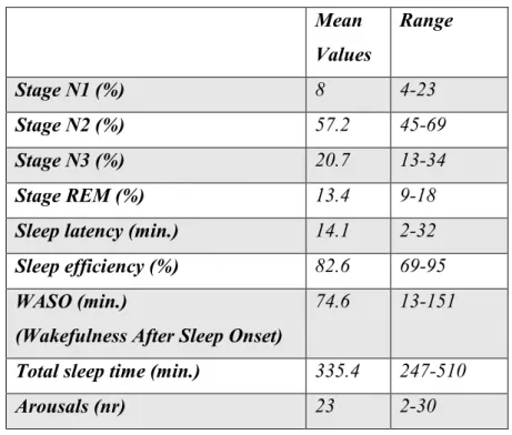
The hypnic profile exhibited a standard sleep latency, a standard representation of the various stages of NREM sleep, and a slight reduction in the percentage of REM sleep (tab. 1).
13 Mean Values Range Stage N1 (%) 8 4-23 Stage N2 (%) 57.2 45-69 Stage N3 (%) 20.7 13-34 Stage REM (%) 13.4 9-18
Sleep latency (min.) 14.1 2-32
Sleep efficiency (%) 82.6 69-95
WASO (min.)
(Wakefulness After Sleep Onset)
74.6 13-151
Total sleep time (min.) 335.4 247-510
Arousals (nr) 23 2-30
Table 1: Mean values and range of the main sleep assessment parameters.
However, it showed a high fragmentation due to frequent arousal episodes (mean arousal: 23), and a reduced sleep efficiency (mean: 82.6%).
14
4.4. Characteristics of PLMS:
Periodic lower limb movements registered in all 17 patients exhibited a mean PLMS index of 18.78 corresponding to the presence of a periodic nocturnal myoclonus of medium entity (tab. 2).
PLMS index
Distribution through the night Sleep stage
Case 1 15 1st half of the night N2
Case 2 12 1st half of the night N2
Case 3 6 1st half of the night N2
Case 4 7 1st half of the night N2
Case 5 12 2nd half of the night N2/REM
Case 6 6 2nd half of the night N2/N3
Case 7 17 1st half of the night N2
Case 8 19 1st half of the night N2
Case 9 30 All night N2/REM
Case 10 6 1st half of the night N2/N3
Case 11 20 2nd half of the night N2
Case12 10 All night N2
Case 13 66 1st half of the night N2/N3
Case 14 17 1st half of the night N2
Case 15 8 All night N2/N3
Case 16 51 All night N2
Case 17 18 1st half of the night N2
18.78 (Mean)
Table 2: Periodic lower limb movements during sleep.
In addition, in 10/17 (58.8%) patients, PLMS appeared to be mainly distributed during the first half of the night; in 3/17 (17.6%) patients, in the second half of the night; and in 4/17 (23.6 %) throughout the night.
15 In 11/17 (66.7%) PLMS occurred during N2, in 4/17 (22.2%) during both N2 and N3, and in 2/17 (11.1%) during both N2 and REM sleep.
4.5: Effects of Pramipexole:
Five patients (3 F, 2 M, mean age: 32.8, range: 25-40 years), underwent two video-PSGs, before and after being administered Pramipexole. All patients had previously been diagnosed with Temporal Lobe Epilepsy, 3 were being treated with carbamazepine, 1 with levetiracetam, and 1 with topiramate.
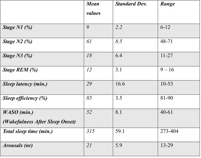
In Basal PSG, the hypnic profile exhibited a slight increase in sleep latency, a standard representation of the various phases of NREM sleep and a slight reduction in the percentage of REM sleep (tab. 3).
Mean values
Standard Dev. Range
Stage N1 (%) 9 2.2 6-12
Stage N2 (%) 61 8.5 48-71
Stage N3 (%) 18 6.4 11-27
Stage REM (%) 12 3.1 9 – 16
Sleep latency (min.) 29 16.6 10-53
Sleep efficiency (%) 85 3.5 81-90
WASO (min.)
(Wakefulness After Sleep Onset)
52 8.1 40-61
Total sleep time (min.) 315 59.1 273-404
Arousals (nr) 21 5.9 13-29
16 However, it showed a high fragmentation due to frequent arousals (mean arousal occurrences: 21), and a reduced sleep efficiency (mean sleep efficiency: 85%).
Fig. 6: Hypnogram case 3, Basal PSG
Periodic lower limb movements registered in all 5 patients exhibited a mean PLMS index of 11.4 corresponding to the presence of a periodic nocturnal myoclonus of medium-to-mild entity (tab. 4). PLMS index Distribution throughout the night Sleep stage
Case 1 15 1st half of the night N2
Case 2 13 1st half of the night N2
Case 3 6 1st half of the night N2
Case 4 9 1st half of the night N2
Case 5 14 2nd half of the night N2/N3
11.4 (mean)
Table 4: Periodic lower limb movements during sleep in Basal PSG
In addition, in 4/4 (80%) patients, PLMS appeared to be mainly distributed during the first half of the night; in 1/5 (20%) patients, in the second half of the night. In 4/5 (80%) PLMS occurred during N2, in 1/9 (20%) patients during both N2 and N3.
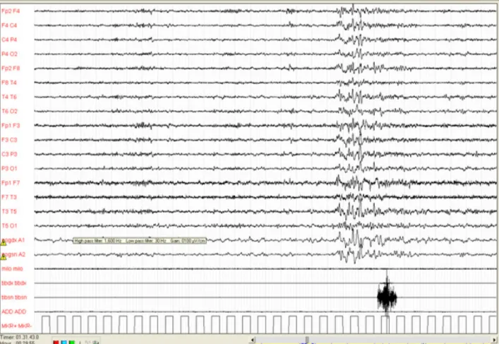
17 The mean epileptiform discharges (ED) number registered in all 5 patients was 130, with a mean index of discharges (ED index) per hour of sleep of 25. In one patient (case 3) the discharges were related to PLM and preceded it by about 1-2 sec.
Fig. 7: PLMS associated to epileptiform discharges during N2 (case 3).
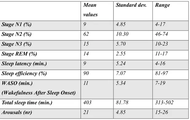
In the second PSG (carried out after administration of Pramipexole 0.18 mg), the hypnic profile exhibited a slight increase in sleep latency, a standard representation of the various phases of NREM sleep and a slight increase in the N2 stage (tab. 5).
18 Mean
values
Standard dev. Range
Stage N1 (%) 9 4.85 4-17
Stage N2 (%) 62 10.30 46-74
Stage N3 (%) 15 5.70 10-23
Stage REM (%) 14 2.55 11-17
Sleep latency (min.) 9 5.24 4-16
Sleep efficiency (%) 90 7.07 81-97
WASO (min.)
(Wakefulness After Sleep Onset)
11 5.34 7-19
Total sleep time (min.) 403 81.78 313-502
Arousals (nr) 21 4.85 15-26
Table 5 : Mean values and range of the main sleep assessing parameters of the second PSG.
None of the patients exhibited a PLMS index > 5 after administration of Pramipexole. The mean epileptiform discharges (ED) number registered in all 5 patients in the second PSG was 81, with a mean index of discharges (ED index) per hour of sleep of 12.06.
19 5. DISCUSSION
The study confirms the presence of Periodic Leg Movements during Sleep (PLMS) in patients with Epilepsy. In our sample, the frequency of PLMS episodes stood at 23%. These movements mainly occurred during the first half of the night, in connection with the N2 stage, with a severity ranging from mild to moderate. They therefore had characteristics and distributions which made them more similar to the PLMS described during RLS than to those associated to neurodegenerative conditions.
In addition, the PLMS in our sample were more frequent in patients with Temporal Lobe Epilepsy.
This association between PLMS and Epilepsy could be simply explained by the fragmentation of the hypnic structure which is often found in patients with Epilepsy, as well as being a secondary cause of seizures and of EEG anomalies. The fluctuant nature of the arousals is believed to be responsible for the phenomenon, ‘allowing’ the occurrence of phasic events such as PLMS [38]. It should not be ignored, on the other hand, that PLMS may also be found in patients in whom such fragmentation is less marked, which means other hypotheses need to be formulated.
In particular, considering the dopaminergic nature of PLMS, their occurrence in association with Epilepsy may serve to strengthen the role of dopamine in the pathophysiology of this disorder. This is still a matter of dispute in research [39]. Dopamine might have an inhibitory influence on epileptogenesis, interfering with the propagation of the discharge and contributing to its interruption. The dopaminergic system could therefore be part of an endogenous anticonvulsant mechanism capable of preventing seizures as well as their secondary generalization . Animal studies point to a neuroprotective role for dopamine through a form of control inhibiting the excitatory neurotransmission of glutamate and of excitotoxicity in Epilepsy. Nuclear medicine studies have shown a reduction in D2/D3 receptor uptake in patients with Temporal Lobe Epilepsy caused by Hippocampal Sclerosis, relative to the irritative region [43, 44]. This may serve to explain the high incidence of Temporal Lobe Epilepsy in our patient sample. [18F] fluoro-L-DOPA PET studies have demonstrated alterations of the dopaminergic network also in patients with Epilepsy with Ring Chromosome 20 [42]. In 2008, Fedi et al. studied 12 patients with Autosomal Dominant Nocturnal Frontal Lobe Epilepsy (ADNFLE) due to a mutation of the nicotinic receptor alfa-4 subunit. As shown by PET, these patients had a reduced D1 receptor uptake to the striatum
20 as compared to healthy controls. This uptake reduction appears to be secondary to a reduction of the receptors themselves, as regards receptor internalization phenomena, or to increased levels of extracellular dopamine. Given the importance of the control of the striatum on the thalamus-cortical projections to the frontal lobe, the authors suggest that the dopaminergic dysfunction observed in the striatum of the patients may lead to an aberrant modulation of these projections. This would mainly occur during NREM sleep, when the physiological concentration of dopamine is lower, thus contributing to the genesis of critical episodes with motor semiology similar to Paroxysmal Dystonia [40].
In another PET study, Ciumas et al. examined 12 patients with Juvenile Myoclonic Epilepsy (JME), demonstrating a reduction in DAT uptake to the substantia nigra. These patients showed reduced performances when undergoing tests assessing their psychomotor, executive and attentional functions [41].
Our study evaluated patients who underwent video-polysomnographic recordings. Video-polysomnography is a test often carried out in our clinic on patients with nocturnal seizures, or in drug-resistant patients with a long history of disease. In the first case, the aim is to clarify any diagnostic doubts that may subsist, while in the second case, video-polysomnographic recordings serve to define the best-effective therapeutic strategy. This may explain why only three patients in our sample showed signs of JME, an epileptic condition whose diagnosis does not normally require a PSG. A larger JME sample would be necessary to verify the actual incidence of PLMS on this typology of epilepsy. More recently, the presence of a dopaminergic dysfunction in JME was confirmed by Landvogt C. et al., who demonstrated a bilateral reduction in the D2/D3 receptor uptake to the posterior putamen in patients with JME [49]. In another paper published in 2010 on Neuroimage, Ciumas et al. revealed a dopaminergic dysfunction in both JME and Epilepsy with Grand Mal Seizures [50].
The analysis of the 5 patients who underwent two VPSG (before and after administration of a dopamine agonist drug) showed a close to 50% reduction of epileptiform discharges in patients with PLMS and Epilepsy after administration of a dopamine agonist, namely Pramipexole. The reduction in the frequency of epileptiform discharges after the administration of Pramipexole might be a secondary indirect effect due to the improvement (drug-induced) of hypnic structure. It is thought that this particular dopamine agonists may reduce the number of PLMS and improve sleep patterns and structure, thereby reducing the occurrence of epileptiform discharges. However, in a recent article published on Sleep in
21 June 2010, the authors (Ferri et al.) analysed the effects of acute treatment with dopamine agonists on sleep architecture in Restless Legs Syndrome, showing that Pramipexole does not modify the Cap rate [52]. This finding indeed supports the hypothesis that treatment with dopamine agonists does not reduce the epileptiform discharges owing to a secondary, indirect effect leading to a less stable sleep architecture, but appears to act directly on epileptogenesis. Moreover, a recent SPECT study by Del Sole et al (October 2010) demonstrated an involvement of the striatal neurons in Epilepsy with Ring Chromosome 20 Syndrome [51]. The relationship found between mosaicism and alteration in DAT expression could suggest that drugs acting on the dopamine system may indeed have a role in the treatment of this type of epilepsy.
22 6. CONCLUSIONS
The study confirmed the presence of PLMS in patients with Epilepsy, in line with previous – although scarce – findings in literature. This results, however, would need to be confirmed by additional prospective studies carried out on a larger patient population. The presence of PLMS in Epilepsy could indeed support the role of dopamine in epileptogenesis, corroborating the thesis – which has met with ever growing favour over the past few years – that Epilepsy is a disorder due to a disharmonic balance between cortical and subcortical functions. Critical episodes would be due to a lack of control by the cortex on the subcortical structures, leading to their inhibition [45, 46].
The involvement of the dopaminergic systems may be related to some motor manifestations typical of certain typologies of seizures. This could have significant repercussions even in the therapeutic field, opening up new possibilities through the employment of dopamine agonists which may well find use in the treatment of some forms of Epilepsy.
23 7. REFERENCES
1. Rye D. Parkinson’s disease and RLS: the dopaminergic bridge. Sleep Medicine 2004; 5: 317-328 2. Symonds CP. Nocturnal myoclonus. J Neurol Neurosurg Psychiatry 1953; 16: 166-171
3. Coleman RM, Pollack CP, Weitzman ED. Periodic movements in sleep (nocturnal myoclonus): relation to sleep disorders. Ann Neurol 1980; 8: 416-421
4. Iber C, Ancoli-Israel S, Chesson A Jr, Quan SF, eds, The AASM Manual for the Scoring of Sleep and Associated Events. Rules, Terminology and Technical Specifications. 1st Edn. Westchester, American Academy of Sleep Medicine, 2007.
5. American Academy of Sleep Medecine. International Classification of Sleep Disorders. Diagnostic ad Coding Manual. 2 nd ed., 2005.
6. Hornyak M, Feige B, Riemann D, Voderholzer U. Periodic leg movements in sleep and periodic limb movement disorder: prevalence, clinical significance and treatment. Sleep Med Rev 2006; 10: 169– 177.
7. Ferri R, Zucconi M, Rundo F, Spruyt K, Manconi M, Ferini-Strambi L. Heart rate and spectral EEG changes accompanying periodic and non-periodic leg movements during sleep. Clin Neurophysiol 2007;118:438-48.
8. Fantini ML, Michaud M, Gosselin N (2002) Periodic leg movements in REM sleep behavior disorder and related autonomicand EEG activation. Neurology 59:1889–1894
9. L. Ferini-Strambi. Insonnia e dopamina: aspetti clinici .Neurol Sci (2005) 26:S317–S321
10. Trenkwalder C, Bucher SF, Oertel WH, Proeckl D, Plendl H, Paulus W. Bereitschaftspotential in idiopathic and symptomatic restless legs syndrome Electroencephalogr Clin Neurophysiol. 1993 Apr;89(2):95-103.
11. Provini F, Vetrugno R, Meletti S, Plazzi G, Solieri L, Lugaresi E, Coccagna G,Montagna P. Motor pattern of periodic limb movements during sleep. Neurology. 2001 Jul 24;57(2):300-4.
12. Bara-Jimenez W, Aksu M, Graham B, Sato S, Hallett M.Periodic limb movements in sleep: state-dependent excitability of the spinal flexor reflex. Neurology. 2000 Apr 25;54(8):1609-16.
13. Smith RC. The Babinski response and periodic limb movements disorders. J Neuropsychiatry Clin. Neurosci. 1992; 4: 233-234.
14. Winkelmann J, Wetter TC, Trenkwalder C, Auer DP. Periodic limb movements in syringomyelia and syringobulbia. Mov Disord. 2000 Jul;15(4):752-3.
15. De Mello MT, Poyares DL, Tufik S. Treatment of periodic leg movements with a dopaminergic agonist in subjects with total spinal cord lesions. Spinal Cord. 1999 Sep;37(9):634-7
16. Quatrale R, Manconi M, Gastaldo E, Eleopra R, Tugnoli V, Tola MR, Granieri E. Neurophysiological study of corticomotor pathways in restless legs sindrome Clin Neurophysiol. 2003 Sep;114(9):1638-45
24
17. Saletu M, Anderer P, Saletu B, et al. Sleep laboratory studies in restless legs syndrome patients as compared with normals and acute effects of ropinirole. 2. Findings on periodic leg movements, arousals and respiratory variables. Neuropsychobiology 2000; 41: 190–199.
18. Manconi M, Ferri R, Zucconi M, et al. First night efficacy of pramipexole in restless legs syndrome and periodic leg movements. Sleep Med 2007; 8: 491–497.
19. Nishimatsu O, Horiguchi J, Inami Y, Sukegawa T, Sasaki A. Periodic limb movement disorder in neuroleptic-induced akathisia. Kobe J Med Sci. 1997 Oct;43(5):169-77.
20. Allen R. Dopamine and iron in the pathophysiology of restless legs syndrome (RLS). Sleep Med. 2004 Jul;5(4):385-91.
21. Wetter TC, Collado-Seidel V, Pollmacher T et al (2000) Sleep and periodic leg movement patterns in drug-free patients with Parkinson’s disease and multiple system atrophy. Sleep 23:361–367
22. Michaud M., Dumont M., Parquet J., et al. Circadian variation of the effect of immobility on symptoms of restless legs syndrome. Sleep 2005; 28: 843-846.
23. Michaud M, Soucy JP, Chabli A, Lavigne G, Montplaisir J. SPECT imaging of striatal pre- and postsynaptic dopaminergic status in restless legs syndrome with periodic leg movements in sleep. J Neurol. 2002 Feb;249(2):164-70.
24. H.M. Ruottinen; M. Partinen; C. Hublin; et al. An FDOPA PET study in patients with periodic limb movement disorder and restless legs syndrome Neurology 2000;54:502–504
25. William G. Ondo, Yi He, Shalini Rajasekaran and Wei-Dong Le. Clinical Correlates of 6-Hydroxydopamine Injections Into A11 Dopaminergic Neurons in Rats: A Possible Model for Restless Legs Syndrome? Movement Disorders Vol. 15, No. 1, 2000, pp. 154–158
26. Fantini ML, Manconi M, Zucconi M, Cappa S, L. Ferrini-Strambi. Il mioclono notturno nelle malattie neurodegenerative. Neurol Sci (2004) 25: S541-S544
27. Culpepper WJ, Badia P, Shaffer JI. Time-of-night patterns in PLMS activity. Sleep 1992; 15: 306-311
28. Hening WA, Walters AS, Wagner M, Rosen R, Chen V, Kim S, Shah M, Thai O. Circadian rhythm of motor restlessness and sensory symptoms in the idiopathic restless legs syndrome. Sleep 1999; 22: 901-912
29. M. Manconi, R. Ferri, M. zucconi e al. Time structure analysis of leg mevements during sleep in REM Sleep Behaviour Disorder. Sleep, vol. 30, No. 12, 2007
30. R. Ferri, M. Zucconi, M. Manconi e al. Different periodicity and time structure of Leg Movement During Sleep in Narcolepsy/Cataplexy and Restelss Legs Syndrome. Sleep 2006; 29 (12): 1587-1594 31. Wetter TC, Collado-Seidel V, Pollmächer T, Yassouridis A, Trenkwalder C. Sleep and periodic leg
movement patterns in drug-free patients with Parkinson's disease and multiple system atrophy. Sleep 2000 May 1; 23(3):361-7
32. R.Manni, M. Terzaghi, C. Pacchetti, G. Nappi. Sleep disorders in Parkinson’s disease: facts and new perspectives. Neuro sci (2007) 28: S1-S5
33. Alex Iranzo. Article review: exploring the relationship between Parkinson disease and resless legs syndrome. Sleep medicine 4 (2003) 161-162
25
34. R. Vetrugno, F. Provini, e al. Sleep disorders in multiple system atrophy: a correlative video-polisomnographic study. Sleep medicine 5 (2004) 21-30
35. Newell SA, Drake ME Jr. Sleep apnea and periodic leg movements in epilepsy. Clin Electroencephalogr. 1994 Oct;25(4):153-5.
36. S. Miano, O. Bruni, M. Elia., et al. Sleep breathing and periodic leg movement pattern in Angelman Syndrome: A polysomnographic study. Clinical Neurophysiology 116 (2005) 2685–2692
37. J. Kaleyias, M. Cruz, J. S. Goraya, et al. Spectrum of Polysomnographic Abnormalities in Children With Epilepsy. Pediatric Neurology Vol. 39 No. 3: 170-176
38. Nobili L, Sartori I, Terzaghi M, et al. Relationship of epileptic discharges to arousal instability and periodic leg movements in a case of nocturnal frontal lobe epilepsy: a stereo-EEG study. Sleep. 2006 May 1;29(5):701-4.
39. Sheryl R, et al. Dopamine and epilepsy. Neurology 2008; 71: 784-785
40. Fedi M, Berkovic SF, Scheffer IE, et al. Reduced striatal D1 receptor binding in autosomal dominant nocturnal frontal lobe epilepsy. Neurology 2008;71:795–798.
41. Ciumas C, Robins Wahlin T-B, Jucaite A, Lindstrom P, Halldin C, Savic I. Reduced dopamine transporter binding in patients with juvenile myoclonic epilepsy. Neurology 2008;71:788–794
42. Biraben A, Semah F, Ribeiro MJ, Douaud G, Remy P, Depaulis A. PET evidence for a role of the basal ganglia in patients with ring chromosome 20 epilepsy. Neurology 2004;63:73–7.
43. Werhahn KJ, Landvogt C, Klimpe S, et al. Decreased dopamine D2/D3-receptor binding in temporal lobe epilepsy: an [18F]fallypride PET study. Epilepsia 2006;47: 1392–1396.
44. Bouilleret V, Semah F, Chassoux F, et al. Basal ganglia involvement in temporal lobe epilepsy: a functional and morphologic study. Neurology 2008;70:177–184.
45. Tassinari CA, Meletti S, Gardella E, et al. (2005) Ethological approach to epileptic “automatisms” viewed as fixed action patterns induced by a release of central pattern generators. Epilepsia 46[Suppl 6]:124
46. Tassinari CA, Rubboli G, Gardella E et al. Central pattern generators for a common semiology in fronto-limbic seizures and in parasomnias. A neuroethologic approach. Neurol Sci 2005; 26: s225-s232 47. Karvonen MK, Kaasinen V, Korja M, Marttila RJ. Ropinirole diminishes myoclonus and improves
writing and postural balance in an ULD patient. Mov Disord. 2010 Mar 15;25(4):520-1. PubMed PMID: 20155865.
48. Martinez HR, Cantù- Martinez L. and al. Epilepsy, parkinsonism, and neuroleptic malignant syndrome in a child. 2006 Dec;21(12):1073-5.
49. Landvogt C, Buchholz HG, Bernedo V, Schreckenberger M, Werhahn KJ. Alteration of dopamine D2/D3 receptor binding in patients with juvenile myoclonic epilepsy. Epilepsia. 2010 Sep;51(9):1699-706.
50. Ciumas C, Wahlin TB, Espino C, Savic I.The dopamine system in idiopathic generalized epilepsies: identification of syndrome-related changes. Neuroimage. 2010 Jun;51(2):606-15. Epub 2010 Feb 24.
26
51. Del Sole A, Chiesa V, Lucignani G, Vignoli A, Giordano L, Lecchi M, Canevini MP. Exploring dopaminergic activity in ring chromosome 20 syndrome: a SPECT study. Q J Nucl Med Mol Imaging. 2010 Oct;54(5564-9):564-569.
52. Ferri R. Manconi M. Aricò D. et al. “ Acute dopamine-agonist treatment in restless legs syndrome: effects on sleep architecture and NREM sleep instability.” Sleep 2010 Jun 1; 33(6): 793-800

![Bibliografia [1] M. Sanjeev Arulampalam, S. Maskell, N. Gordon and T.Clapp A Tutorial on Particle Filter for Ondine Nonlinear/Non-Gaussian Bayesian Tracking.](data:image/gif;base64,R0lGODlhAQABAIAAAP///wAAACH5BAEAAAAALAAAAAABAAEAAAICRAEAOw==)