https://doi.org/10.1177/2058738419827745 International Journal of
Immunopathology and Pharmacology Volume 33: 1–6
© The Author(s) 2019 Article reuse guidelines: sagepub.com/journals-permissions DOI: 10.1177/2058738419827745 journals.sagepub.com/home/iji
Creative Commons Non Commercial CC BY-NC: This article is distributed under the terms of the Creative Commons Attribution-NonCommercial 4.0 License (http://www.creativecommons.org/licenses/by-nc/4.0/) which permits non-commercial use, reproduction and distribution of the work without further permission provided the original work is attributed as specified on the SAGE and Open Access pages (https://us.sagepub.com/en-us/nam/open-access-at-sage).
Introduction
Mucositis is not a disease that needs to be treated, but rather a disease that should be prevented. In order to prevent mucositis, everyone should know what causes this disease. The occurrence rate of mucositis is high,1 even with patients who pay attention to oral hygiene.
Peri-implantitis is an infection of both bone and soft peri-implant tissues. It is a pathology that can occur at a distance of 3–9 years and which leads to the progressive reabsorption of the bone surround-ing the implant, further leadsurround-ing to total loss of the implant.1 The implant loss is related to major bone
Reuterinos
®as adjuvant for
peri-implant treatment: A pilot study
Dorina Lauritano1 , Francesco Carinci2 , Annalisa Palmieri3,
Francesca Cura3, Silvia Caruso4 and Valentina Candotto5
Abstract
The objective of this study was to evaluate the effects of lozenges-containing Lactobacillus reuteri as an adjuvant treatment of peri-implant mucositis and to detect the level of L. reuteri colonization in the peri-implant tissues of treated patients. A total of 10 patients were selected. Subjects with at least one implant affected by peri-implant mucositis, with gingival index (GI) of ⩾2 in each quadrant, evaluated at the buccal aspect of all teeth. Patients included in the study were partially edentulous and had implants with mucositis or peri-implantitis. Implants with radiographic bone loss of ⩾5 mm and/ or ⩾50% of the implant length were excluded, and only one implant per patient was included. Each patient received
L. reuteri–containing lozenges. Microbiological sampling was performed at baseline and on day 28 and analysed by
polymerase chain reaction (PCR). Our results indicate that the use of the probiotic did not influence the peri-implant microbiota in a statistically significant way, although there was a reduction in the number of periodontal and peri-implant species. The lack of statistically significant microbiological changes could be explained either by the small sample population or by the short evaluation period. Therefore, the poor colonization of L. reuteri in the peri-implant pockets can be explained by the different anatomical and histological characteristics of the interface of the dental–gingival unit with respect to the periodontal sulcus. The administration of a daily lozenge of L. reuteri for 4 weeks had a limited effect on the microbiological analysis. Probiotics provide an alternative therapeutic approach to consider in the prevention and treatment of peri-implant diseases, but further long-term prospective studies with standardized variables are needed.
Keywords
gingivitis, L. reuteri, mucositis, oral microbiota, probiotic
Date received: 18 October 2018; accepted: 9 January 2019
1 Department of Medicine and Surgery, Milan Center for Neuroscience,
University of Milano-Bicocca, Milan, Italy
2 Department of Morphology, Surgery and Experimental Medicine,
University of Ferrara, Ferrara, Italy
3 Department of Experimental, Diagnostic and Specialty Medicine,
University of Bologna, Bologna, Italy
4 Paediatric Dentistry, Department of Life, Health and Environmental
Sciences, University of L’Aquila, L’Aquila, Italy
5 Department of Biomedical, Surgical and Dental Sciences, University of
Milan, Milan, Italy
Corresponding author:
Lauritano Dorina, Department of Medicine and Surgery, Milan Center for Neuroscience, University of Milano-Bicocca, Milan, Italy. Email: [email protected]
defects; it is difficult to position a new implant with a new tooth. Peri-implantitis is a disease very similar to periodontitis,1 always mediated by bac-teria, and therefore must be treated in a similar way, with a series of protocols that lead to the elim-ination of infection and bacteria within the implant.1 The main causes of peri-implantitis are patho-genic bacteria;1 hence, it is very important to remove or prevent these bacteria that tend to accu-mulate on the implant surface, especially if it is rough. Another important factor is the individual predisposition of the patient: there are patients who often fall sick, especially smokers, and patients who, instead, have a lower tendency.1 Thus, the individual response component of the host is important too.1 The implant surface is another pos-sible cause of peri-implantitis: a smoother surface is less susceptible to infection of the peri-implant tissues and therefore leads to peri-implantitis.1
Prevention of peri-implantitis is mandatory because once the disease has manifested, it is very difficult to eradicate and control. To prevent peri-implant mucositis, it is important to use peri-implants that do not have excessively rough surfaces that therefore could favour the accumulation of bacte-ria on the implant surface; in particular, it is impor-tant that the patient undergoing implant therapy is then followed with hygiene protocols every 3–6 months by the professional or even by dental hygienists in order to avoid excessive accumula-tion of plaque at the level of the implant surface and peri-implant tissues.
There are two phases of treatment for peri-implant mucositis: initially, through the action of a dental hygienist, it is necessary to remove the greatest amount of bacteria from the tissues and subgingival areas of the implant surface and thus maintain this health condition for as long as possi-ble (even forever). The patient must perform a scrupulous oral hygiene, and if this is not enough, it is necessary to perform surgical therapies, detaching the peri-implant tissues and soft tissues, smoothing the implant surface and remove all the bacteria. Then, it is necessary to close the gingival tissues and eventually, in some cases, regenerate the bone or part of the bone that has been lost. Probiotics for peri-implantitis
prevention
Probiotics are defined as living and viable microor-ganisms which, when administered in adequate
quantities, confer benefits to the organism. They can interact positively with the intestinal immune system and help prevent gastrointestinal disorders.
When our intestinal flora loses its balance, the bad bacteria take the upper hand over the good ones (dysbiosis): this is the moment when it becomes very important to introduce probiotics to restore the correct balance. To achieve this goal, it is necessary to take products based on probiotics that are able to survive the acidity of the gastric environment, reach the intestine and fight the harmful germs, restoring the balance of the intestinal flora, by adhering to the intestinal mucosa and carrying out beneficial actions. The use of probiotics has not been proposed for mucositis treatment and prevention.
To the best of our knowledge, probiotics effi-cacy on the oral microflora preventing the coloni-zation of periodontal pathogens, and thus preventing the microbiological shifts associated with mucositis, has not been investigated.
Probiotic tablets containing Lactobacillus reu-teri have been formulated with the aim of prevent-ing and treatprevent-ing gprevent-ingivitis, although to the investigator’s knowledge, there are no controlled studies evaluating its efficacy.2–4
Therefore, the purpose of this investigation is to study the clinical and microbiological effects of L. reuteri and to evaluate the patterns of colonization in peri-implant pockets.
Materials and methods Subjects
A total of 10 healthy volunteers were recruited among patients of a private practice from February to October 2017, provided they fulfilled the fol-lowing criteria:
•
• Subjects with at least one implant affected by peri-implant mucositis, with gingival index (GI) of ⩾2 in each quadrant, evaluated at the buccal aspect of all teeth. Patients included in the study were partially edentulous and had implants with mucositis or peri-implan-titis. Implants with radiographic bone loss of ⩾5 mm and/or ⩾50% of the implant length were excluded, and only one implant per patient was included.
•
• Subjects were excluded if they had used any systemic antibiotics in the previous 3 months, probiotic preparations or oral antiseptics in the previous month or if they had any
systemic disease or condition that could interfere with the study results (e.g. diabetes and immunological disorders, pregnancy, ongoing drug therapy that could affect the signs of mucositis).
Study design
All selected subjects signed an informed consent to participate in the study. This study was conducted in accordance with the Declaration of Helsinki, and the protocol was approved by the Ethics Committee of 06.09.2013 prot. n. 29579 University Study of L’Aquila. After non-surgical mechanical therapy, subjects were randomly assigned to take either one probiotic lozenge or one placebo loz-enge every day for 4 weeks. Participants were asked not to change their oral hygiene habits and to refrain from taking other probiotic products throughout the duration of the study. A total of 10 patients were selected. Clinical measurements were taken in the whole mouth (GI) and at the implant site (probing pocket depth) at baseline and after 4 weeks. Microbiological examination was performed at the same study time points that clini-cal measurements were made. Each selected site will be subjected to microbial analysis. For the col-lection of subgingival samples, the site was iso-lated using cotton rolls. Sterile absorbable paper points (size 60) were used for the collection of sub-gingival samples and were immediately transferred to microbiological laboratory for processing. They were instructed on the use of the tablet medications (Reuterinos®; Noos s.r.l., Rome, Italy) and were scheduled for new evaluations, after 4 weeks, with additional clinical and microbiological examina-tions. After 4 weeks, they were scheduled for a baseline examination. At this visit, the clinical and microbiological examinations were carried out.
Treatments
Subjects were randomly assigned following a com-puter-generated randomization list. Each subject identified by a unique study number was instructed to chew one tablet per day (Reuterinos; Noos s.r.l.), during 28 days.
Clinical examination
Clinical variables were evaluated at baseline and 4 weeks. The variable included the GI,5 as normally
assessed in studies evaluating oral hygiene prod-ucts. The same examiner evaluated this index by selecting randomly in each patient two quadrants: either upper right and lower left or upper left and lower right quadrants (half of the mouth scoring).5
Microbiological analysis
The microorganisms processed were the three bac-terial species, which were involved in most of the periodontitis cases, that constitute the red complex group: Porphyromonas gingivalis, Tannerella for-sythia and Treponema denticola, as described in previous studies.6–9
Both P. gingivalis and T. denticola occur con-comitantly with the clinical signs of periodontal destruction.1 They appear closely ‘linked’ topolog-ically in the developing biofilm, with an in vitro ability to produce a number of outer membrane– associated proteinases, and are considered the first pathogens involved in the clinical destruction of periodontal tissues. Moreover, P. gingivalis and T. denticola and T. forsythia show a higher preva-lence in disease than in health suggesting that these bacteria are associated with the local development of periodontitis.1
Real-time polymerase chain reaction. Primers and oligo-nucleotide probes will be designed based on 16S ribosomal RNA (rRNA) gene sequences of the Human Oral Microbiome Database (HOMD 16S rRNA RefSeq Version 10.1) counting 845 entries. All the sequences will be aligned in order to find either consensus sequence or less conservative spots. Two real-time polymerase chain reaction (PCR) runs will be performed for each sample. The first reaction will quantify the total amount of bac-teria using two degenerate primers and a single probe matching a highly conservative sequence of the 16S rRNA gene. The second reaction will detect and quantify the three red complex bacteria, that is, P. gingivalis, T. forsythia and T. denticola, in a multiplex PCR. This reaction will include a total of six primers and three probes that are highly specific for each species. Oligonucleotide concen-trations and PCR conditions will be optimized to ensure sensitivity, specificity and no inhibitions in case of unbalanced target amounts. Absolute quan-tification assays will be performed using the Applied Biosystems 7500 Sequence Detection System. The amplification profile will be initiated by a 10-min incubation period at 95°C to activate
polymerase, followed by a two-step amplification of 15 s at 95°C and 60 s at 57°C for 40 cycles. All these experiments will be performed including nontemplate controls to exclude contamination of reagents.
Plasmids containing synthetic DNA target sequences (Eurofin MWG Operon, Ebersberg, Germany) will be used as standard for the quantita-tive analysis. Standard curves for each target will be constructed in a triplex reaction, using a mix of the same amount of plasmids, in serial dilutions ranging from 101 to 107 copies. There is a linear relationship between the threshold cycle values plotted against the log of the copy number over the entire range of dilutions. The copy numbers for individual plasmid preparations will be estimated using the Thermo NanoDrop spectrophotometer.
The absolute quantification of total bacterial genome copies in samples allowed for the calcula-tion of relative amount of red complex species. To prevent contamination of samples and PCR, plas-mid purification and handling will be performed in a separate laboratory with dedicated pipettes.
Statistical analysis
Descriptive statistics were performed using Microsoft Excel spreadsheets. The Freeman– Halton extension of Fisher’s exact test was used to compute the (two-tailed) probability of obtaining a distribution of values in a 2 × 3 contingency table, given the number of observations in each cell. Odds ratio calculation was performed online at the OpenEpi website (www.openepi.com).
Results
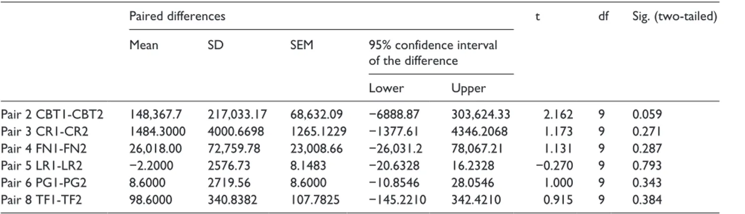
After treatment, there was a reduction in specific and total bacterial loading, although no statistical significant difference was detected (Tables 1 and 2). After 4 weeks, GI values were better to baseline as well as gingival inflammation.
Discussion
Lactobacillus reuteri is a species of bacterium belonging to the Lactobacillaceae family that natu-rally colonizes the gastrointestinal tract of humans and animals. Some clinical studies have shown that adequate administration of L. reuteri can bring ben-efits to human health.10,11 For this reason, L. reuteri
is currently considered a probiotic organism. Some strains of L. reuteri (mainly ATCC55730 and DSM17938) are currently used as therapeutic agents against various intestinal disorders.
Lactobacillus reuteri, belonging to the genus Lactobacillus, is a gram-positive bacterium, which, due to its unique metabolic properties, belongs to the group of lactic bacteria (also called ‘lactic fer-ments’) that colonize both men and animals. In humans, it has been isolated in the gastrointestinal tract and in samples of faecal and vaginal material. It is also present in breast milk, together with other lactic bacteria of the genera Lactobacillus and Bifidobacterium. Based on the study by Sinkiewicz,10 who considered more than 200 women from seven different countries in the world, L. reuteri was iso-lated in human milk in both urban and rural areas, with colonization rates of up to 50%.
One of the most studied Lactobacilli and having great effectiveness today is L. reuteri, described for the first time by Gerhard Reuter in 1980: commonly already present in the intestinal mucosa since the first hours of life, L. reuteri is part of that important immunity that is transmitted from the mother to the baby also through the mother’s milk.11
The experimental design of this clinical trial aimed to study the clinical and microbiological impact of the use of probiotic tablets containing L.
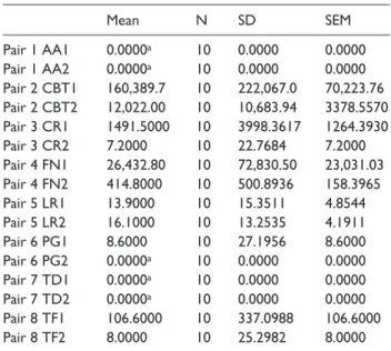
Table 1. Mean amounts of specific bacterial species before
and after Reuterinos® treatment.
Mean N SD SEM Pair 1 AA1 0.0000a 10 0.0000 0.0000 Pair 1 AA2 0.0000a 10 0.0000 0.0000 Pair 2 CBT1 160,389.7 10 222,067.0 70,223.76 Pair 2 CBT2 12,022.00 10 10,683.94 3378.5570 Pair 3 CR1 1491.5000 10 3998.3617 1264.3930 Pair 3 CR2 7.2000 10 22.7684 7.2000 Pair 4 FN1 26,432.80 10 72,830.50 23,031.03 Pair 4 FN2 414.8000 10 500.8936 158.3965 Pair 5 LR1 13.9000 10 15.3511 4.8544 Pair 5 LR2 16.1000 10 13.2535 4.1911 Pair 6 PG1 8.6000 10 27.1956 8.6000 Pair 6 PG2 0.0000a 10 0.0000 0.0000 Pair 7 TD1 0.0000a 10 0.0000 0.0000 Pair 7 TD2 0.0000a 10 0.0000 0.0000 Pair 8 TF1 106.6000 10 337.0988 106.6000 Pair 8 TF2 8.0000 10 25.2982 8.0000
SD: standard deviation; SEM: standard error of the mean.
aThe correlation and t cannot be computed because the standard error
reuteri and further to assess the patterns of L. reu-teri colonization in peri-implant pockets. Similar to another study,12 our results indicate that the use of the probiotic did not influence in a statistically significant way the peri-implant microbiota, although there was a reduction in the number of periodontal and peri-implant species. The lack of statistically significant microbiological changes could be explained either by the small sample pop-ulation or by the short evaluation period. Therefore, the poor colonization of L. reuteri in the peri-implant pockets can be explained by the different anatomical and histological characteristics of the interface of the dental–gingival unit with respect to the periodontal sulcus.
The administration of a daily lozenge of L. reu-teri for 4 weeks had a limited effect on the micro-biological analysis. Probiotics provide an alternative therapeutic approach to consider in the prevention and treatment of peri-implant diseases, but further long-term prospective studies with standardized variables are needed.
Declaration of conflicting interests
The author(s) declared no potential conflicts of interest with respect to the research, authorship and/or publication of this article.
Funding
The author(s) received no financial support for the research, authorship and/or publication of this article.
ORCID iDs
Dorina Lauritano https://orcid.org/0000-0002-3550-1812 Francesco Carinci https://orcid.org/0000-0001-9639-6676
References
1. Esposito M, Grusovin MG, Tzanetea E, et al. (2018) Interventions for replacing missing teeth: Treatment of peri-implantitis. Cochrane Database of Systematic
Reviews 6: CD004970.
2. Shimauchi H, Mayanagi G, Nakaya S, et al. (2008) Improvement of periodontal condition by probiotics with Lactobacillus salivarius WB21: A randomized, double-blind, placebo-controlled study. Journal of
Clinical Periodontology 35: 897–905.
3. Mayanagi G, Kimura M, Nakaya S, et al. (2009) Probiotic effects of orally administered Lactobacillus
salivarius WB21-containing tablets on
periodon-topathic bacteria: A double-blinded, placebo-con-trolled, randomized clinical trial. Journal of Clinical
Periodontology 36: 506–513.
4. Jacobsen CN, Rosenfeldt NV, Hayford AE, et al. (1999) Screening of probiotic activities of forty-seven strains of Lactobacillus spp. By in vitro techniques and evaluation of the colonization ability of five selected strains in humans. Applied and Environmental
Microbiology 65: 4949–4956.
5. Lobene RR, Weatherford T, Ross NM, et al. (1986) A modified gingival index for use in clinical trials.
Clinical Preventive Dentistry 8: 3–6.
6. Roncati M, Lauritano D, Cura F, et al. (2016) Evaluation of light-emitting diode (led-835 nm) application over human gingival fibroblast: An in vitro study. Journal of Biological Regulators and
Homeostatic Agents 30(2 Suppl. 1): 161–167.
7. Lauritano D, Bignozzi CA, Pazzi D, et al. (2016) Evaluation of the efficacy of a new oral gel as an adjunct to home oral hygiene in the management of chronic periodontitis. A microbiological study using PCR analysis. Journal of Biological Regulators and
Homeostatic Agents 30(2 Suppl. 1): 123–128.
8. Lauritano D, Candotto V, Bignozzi CA, et al. (2018) The role of zinc plus octenidine in the regulation of gene
expres-Table 2. Output of paired samples t test.
Paired differences t df Sig. (two-tailed) Mean SD SEM 95% confidence interval
of the difference Lower Upper Pair 2 CBT1-CBT2 148,367.7 217,033.17 68,632.09 −6888.87 303,624.33 2.162 9 0.059 Pair 3 CR1-CR2 1484.3000 4000.6698 1265.1229 −1377.61 4346.2068 1.173 9 0.271 Pair 4 FN1-FN2 26,018.00 72,759.78 23,008.66 −26,031.2 78,067.21 1.131 9 0.287 Pair 5 LR1-LR2 −2.2000 2576.73 8.1483 −20.6328 16.2328 −0.270 9 0.793 Pair 6 PG1-PG2 8.6000 2719.56 8.6000 −10.8546 28.0546 1.000 9 0.343 Pair 8 TF1-TF2 98.6000 340.8382 107.7825 −145.2210 342.4210 0.915 9 0.384
sion: An in vitro study. Journal of Biological Regulators
and Homeostatic Agents 32(2 Suppl. 1): 237–244.
9. Lauritano D, Candotto V, Bignozzi CA, et al. (2018) Zinc plus octenidine: A new formulation for treating periodontal pathogens. A single blind study. Journal
of Biological Regulators and Homeostatic Agents
32(2 Suppl. 1): 231–236.
10. Sinkiewicz G and Nordström EA (2005) Occurrence of Lactobacillus reuteri, lactobacilli and bifidobacte-ria in human breast milk. Pediatric Research 58: 415.
11. Reuter G (2001) The Lactobacillus and Bifidobacterium microflora of the human intestine: Composition and succession. Current Issues in Intestinal Microbiology 2: 43–53.
12. Galofre M, Palao D, Vicario M, et al. (2018) Clinical and microbiological evaluation of the effect of
Lactobacillus reuteri in the treatment of
mucosi-tis and peri-implantimucosi-tis: A triple-blind randomized clinical trial. Journal of Periodontal Research 53(3): 378–390.