a.a. 2007/2010
Università degli Studi di Catania
Scuola Superiore di Catania
International PhD
in
STEM CELLS
XXIII cycle
IDENTIFICATION OF NOVEL THERAPEUTIC
TARGETS FOR COLON ADENOCARCINOMA
Dr. EROS FABRIZI
Coordinator of PhD Tutor
2
Index
Abstract 4 Introduction 5 1. Colon cancer. 5 1.1. Risk factors. 5 1.2. Pathology. 6 1.3. Pathogenesis. 61.3.1. APC/β-catenin pathway and chromosomal instability. 7
1.3.2. Microsatellites instability. 11
1.4. Therapy. 11
1.4.1. Surgery, chemotherapy and radiotherapy. 12
1.4.2. New therapies. 13
2. Cancer stem cells. 15
2.1. Colon cancer stem cells. 19
2.2. Cancer stem cells therapeutic implication. 22
29
Methods 24
1. Colon cancer stem cells isolation and cultivation. 24
2. Kinase inhibitors library in vitro screening. 24
3 commercially available inhibitors.
4. Viability assay. 25
5. Mice models of xenotransplantation. 26
6. Western blotting. 26
7. Phosphoproteomic analysis. 27
8. Statistical analysis. 31
Results
1. Phosphoproteomic analysis of Colon-CSC 31
2. In vitro of a library of kinase inhibitors on Colon-CSC 32
3. Titration of positive targets on colon-CSC 40
4. Combination with chemiotherapeutic agents 42
5. Definition of signal transduction pathways involved in
survival/proliferation of colon-CSC 43
Discussion 63
4
Abstract
Colorectal cancer (CRC) is the third most common form of cancer in the Western world. Despite the emergence of new targeted agents and the use of various therapeutic combinations, none of the treatment options available is curative in patients with advanced cancer. A growing body of evidence is increasingly supporting the idea that malignancies originate from a small fraction of cancer cells, called Cancer Stem Cells (CSC), that show self-renewal and pluripotency and are capable of initiating and sustaining tumor growth.
Several studies have shown that, with respect to the bulk of tumor cells, CSC posses a higher degree of resistance to chemotherapy and radiotherapy that could explain the inefficacy of current therapies. The ability to isolate and study these tumor cells provided a powerful tool for the investigation of drug-and radio-resistance mechanisms thus paving the way for the development of novel targeted therapies aimed at the tumor complete eradication.
The aim of this PhD thesis was to use CSC lines, derived from CRC specimens, to individuate new potential molecular targets for the development of novel therapies. To this end four colon-CSC lines were subjected to phosphoproteomic analysis by RPPA (Reverse
Phase Protein Array) technology. Through this analysis phosphorylation levels of various
protein kinases and their substrates were evaluated in order to create an activation map of the main colon-CSC proliferation and cell survival pathways. In parallel colon-CSC lines have been screened in vitro to the action of 80 commercially available protein-kinase inhibitors. This screening has revealed a partial correlation between in vitro sensitivity and phosphoproteomic analysis, but in this study, was not possible to identify predictive factors to infer colon-CSC sensitivity to specific kinase inhibitors. Colon-CSC was sensitive to the inhibition of protein kinase C (PKC), known regulator of cell proliferation and survival. Among PKC inhibitors, the most interesting was the UCN-01, a staurosporine derivative that can also inhibit PDK1 and Chk1. This compound has been already used in clinical trials as antineoplastic agent in combination with conventional chemotherapy. The in vitro treatment of colon-CSC with UCN-01 has demonstrated its ability to enhance the irinotecan cytotoxicity by increasing the apoptotic response. The combined action of UCN-01 and irinotecan caused a marked reduction in the levels of antiapoptotic proteins such as Bcl-XL and Mcl-1 and the activation of caspase 3.
The in vivo administration of UCN-01/irinotecan combination, in a mouse model of subcutaneous xenograft, confirmed the observations obtained in vitro, leading to a significant reduction in tumor growth compared to the single treatments. UCN-01 has also
5
shown efficacy in the inhibition of Chk1, as demonstrated by the reduction of the phosphorylation of its target protein cdc25. Inhibition of Chk1, an important regulator of cell cycle, in combination with chemotherapy, could help in reducing the viability of colon-CSC, thus preventing cell cycle arrest and repair DNA damage induced by irinotecan. Although UCN-01 exerts its effect by inhibiting the activity of various protein kinases, this reduced selectivity could be the basis of its effectiveness. The present study demonstrated that it is possible to identify, among the commercially available compounds, those that interfere with processes that regulate colon-CSC survival or proliferation and therefore are potentially able to interfere with tumor growth. The use of newly developed inhibitors, combined with the analysis of genetic alterations or phosphoproteomic, will identify factors predictive of response to therapy and lead to the possibility of developing individualized therapeutic strategies, increasing the likelihood of success of targeted therapy.
Introduction
1. Colorectal CancerColorectal cancer (CRC) is the third most common form of tumor, after lung and breast cancer. Each year more than a million people worldwide develop this type of neoplasm and in Europe about 2.5 x10⁵ people are diagnosed every year, covering approximately 9% of new cases [1]. In Italy, the annual incidence is about 50 x105 for men, 38 x105 for women. The incidence of this tumor follows an age-dependent distribution: under 45 years there is a low incidence (2 x10⁵), and then it progressively raises in the 45-54 years age group (20 x105), reaching 55 x105 cases between 54 and 64 years, 150 x105 in the range 65-74 years and more than 250 x105 in patients over 75 years. Similarly the 5-year survival rate changes in an age dependent manner, from 63% in the younger age groups (15-45 years) to 49% in older patients (over 75 years) [1].
1.1 Risk factors
The majority of CRC cases occurs sporadically and only 5% of cases can be attributed to inherited mutations. Epidemiological studies have identified an association between several environmental factors and CRC onset risk. In addition to the male sex and age these include, a high fat diet, red meat consumption, inadequate fiber intake, a sedentary lifestyle and smoking. Together with the environmental risk factors, a genetic
6
predisposition has been found and it was estimated that at least 20% of CRC cases have a family history. Inherited syndromes that may lead to CRC include familial adenomatous polyposis (FAP) and hereditary non-polyposis colorectal cancer (HNPCC or Lynch syndrome). These diseases occur in individuals who have inherited heterozygous mutations in specific genes in which the acquisition of a second mutation, in the normal allele, leads to the loss of the corresponding protein. In FAP cases, homozygous mutations lead to excessive proliferation of intestinal epithelial cells with a subsequent polyp formation. Lynch syndrome is, on the other hand, characterized by the lack of DNA mismatch repair system components that leads to the acquisition of a "mutator phenotype". This is in turn associated with an increased risk of neoplastic transformation [1].
1.2 Pathology
One of the most important CRC progression feature is the "adenoma to carcinoma sequence", a widely used term to describe the multistep pathogenesis processes that characterize this tumor. This involves the progressive acquisition of genetic alterations in intestinal epithelial cells which is reflected in the histological progression from normal epithelium to a more malignant phenotype passing through several degrees of dysplasia with increasing severity and carcinoma in-situ (CIS) [3]. In particular, the majority of colorectal cancers arise from malignant transformation of polyps. A polypoid lesion may develop from a normal mucosa in about five years and it will require at least two more years to turn into CIS stage. Three more years are finally needed to reach the stage of invasive cancer.
The main colorectal cancer histotype is the adenocarcinoma which accounts for 90-95% of all large intestine cancers, while 17% is represented by mucinous adenocarcinomas [1]. The ability to identify adenomatous polyps or pre-neoplastic lesions, together with the high survival rate associated with earlier stages of the disease, make colorectal cancer an ideal target for screening programs, whose main purpose is to prevent its sporadic forms (more than 90% of cases) [1, 2].
1.3 Pathogenesis
In 1990 Fearon and Vogelstein proposed a genetic model to describe colorectal tumorigenesis [6]. According to this model colon cancer development is driven by mutations in oncogenes and tumor suppressor genes, whose total accumulation, rather than the order of appearance, is considered important in tumor development, even though their
7
Figure 1. Fearon and Vogelstein model for colorectal carcinogenesis [6]. Patients with FAP inherit a
mutation in APC and develop numerous dysplastic foci in colon crypts. Progression towards a more malignant phenotype is then driven by the acquisition of other mutations (e.g. K-Ras, SMAD4/2 and p53) and increased genomic instability.
acquisition sequence often reflect tumor clinical progression (Figure 1). Genetic mutations responsible for sporadic colorectal cancer are the same that characterize the hereditary forms and it is possible to distinguish two major pathogenetic pathways: the APC/β-catenin pathway and the microsatellites instability pathway.
1.3.1 APC/β-catenin pathway and chromosomic instability
APC/β-catenin pathway is altered in 85% of sporadic CRC cases. APC (adenomatous polyposis coli) gene mutation is one of the earliest events in the adenoma-carcinoma sequence. Heterozygous mutations in this gene are responsible for FAP and, if untreated, this disease inevitably evolves in CRC [3]. An APC key function is -catenin intracellular level control. Indeed, APC takes part in Wnt signal transduction pathway involved in regulation of cell proliferation and survival (Figure 2). This pathway is activated by Wnt binding to its receptor Frizzled (FZD) that interacts, at a cytoplasmatic level, with a protein complex formed by Axin, APC and GSK-3β (destruction complex). In the absence of Wnt signal, this complex is responsible for -catenin degradation via the ubiquitin-proteasome pathway. APC mutatations impair protein ability to interact with the destruction complex, leading to -catenin stabilization and its accumulation in the nucleus. The nuclear functions of this protein lie in its ability to act as a co-activator of the TCF family of transcription factors activating the transcription of specific genes, including c-MYC and
8
Cyclin D1 (CCND1). Mutations in the APC gene were observed in 40-80% of colorectal cancers reinforcing the belief that this mutation is one of the earliest events in adenoma-carcinoma sequence. Finally about half of tumors bearing a wild type APC gene show mutations in the -catenin gene, thus suggesting that these latter mutations can replace those in APC [3].
Although APC mutation is an early event this alteration seems not to be self-sufficient for adenomas progression [9]. Each new mutation leads additional control genes lost (chromosomal instability) and is correlated with a greater tendency for the acquisition of new malignant aberrations. Another frequent genetic alteration that occurs early in the
Figure 2. Wnt signal transduction pathway. (A) In the absence of Wnt the cytosolic β-catenin is
degradated through the APC/Axin/GSK-3β complex. (B) When Wnt binds to the receptor, DSH (Dishevelled) is activated by preventing the binding of β-catenin to the complex, the β-catenin is stabilized and, moving into the nucleus, can associate with transcription factors fostering the expression of target genes [8].
9
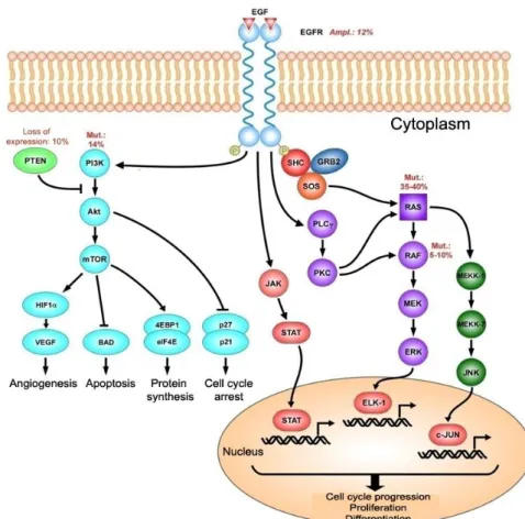
adenoma-carcinoma sequence is the KRAS activating mutation [3]. This gene, belonging to the RAS family (KRAS, HRAS and NRAS), encodes for a small GTPase localized on cell membrane cytoplasmic side and is involved in proliferation and differentiation signals coming from receptor tyrosine kinases, such as the epidermal growth factor receptor (EGFR) [9]. These receptors are coupled to the RAS family proteins through a series of adaptor proteins known as GRB2/SOS and the downstream signal transduction pathway elements consist in the serine-threonine kinase Raf and the MAP (mitogen activate protein) kinase cascade (Figure 3).
RAS proteins are activated by GTP binding and inactivated by their intrinsic GTPase activity, through the action of GAP (GTPase Activating Protein) proteins, called p120, RasGAP and neurofibromin (NF1 gene product). All of the KRAS oncogene mutations
Figure 3. Signal transduction pathways activated by EGF binding to its receptor (EGFR).
10
associated with cancer are located in the GTP binding domain and are responsible for a reduction in GTPase activity, causing constitutive protein activation [3]. Activating KRAS mutations characterize the 35-42% of CRCs and adenomas and the percentage tends to increase in advanced lesions. The high genetic alteration frequency found in adenomas suggests that this is a quite early event in tumor development. However, the low occurrence frequency of this mutation in small adenomas suggests that RAS alterated function could not be a tumor initiating factor but rather has a role in conferring a proliferative advantage to cells that have already acquired a mutation in the APC gene [9]. The deletion of a chromosome 18 region is another important genetic alteration in the CRC pathogenesis, found in about 70% of cases. Its frequency shows an increase proportional to tumor staging: 10-30% of early adenomas and 60% of the most advanced ones. Initially, the DCC gene (Deleted in Colorectal Cancer), contained within this region, was considered a tumor suppressor associated with tumorigenesis. Experiments on mutant mice bearing a homozygous deletion, however, did not confirm this hypothesis [3]. Other tumor suppressor genes have been further identified in this region. Among these SMAD2 and SMAD4 (Small Mother Against Decapentaplegic 2-4), whose protein products act as mediators of intracellular TGF- (transforming growth factor ) signal. TGF- transduction pathway regulates various processes such as cell growth, differentiation, extracellular matrix production and apoptosis in various cell types. Mutations in SMAD2/4 and TGF- were found in several human neoplasms, including colorectal cancer, in which the deletion of SMAD4 is known to abolish the antiproliferative TGF- signals [3]. The short arm region of chromosome 17 is frequently subject to deletion in CRC, an event that involves TP53 gene lost. TP53 gene mutation or deletion is one of the most common events found in human cancers. This gene alterations or 17p region lost are observed in 4-26% of adenomas, in approximately 50% of adenomatous polyps and in 50-75% of adenocarcinomas [3]. p53 protein plays a major function as a transcription factor regulating physiological processes such as stress response, DNA damage repair, cell cycle regulation and apoptosis. In particular, genotoxic stresses, trigger cell cycle arrest mediated by p53, that for this function, has been described as the "the genome guardian" [10].
11 1.3.2 Microsatellites instability
About 15% of CRC cases show high level of microsatellites instability (MIN) that results in frame shift mutations and base substitutions. This phenomenon is indicative of a reduced efficiency of DNA mismatch repair (MMR). MIN has been found in almost all HNPCC patients, and only in 10-15% of sporadic cases.
To date, there are five known genes involved in MMR: MSH2, MLH1, PMS1, PMS2, and MSH6. The majority of HNPCC patients bear a heterozygous somatic mutation in at least one of these genes, whereas in sporadic cancers somatic mutations in MMR genes are much less frequent. Noteworthing is the high mutation frequency in the TGF- RII (TGF- receptor type II) gene associated with MIN-positive CRC. The gene encoding this receptor contains sequence similar to microsatellite repeats and therefore particularly sensitive to defects in MMR. Inactivating mutations of the TGF- RII were observed in 90% of MIN-positive CRC [3].
1.4 Terapy
1.4.1 Surgery, chemotherapy and radiotherapy
The most important therapeutic treatment for CRC is surgery. This procedure aims to remove the affected bowel section and its lymphatic system. The currently used screening programs, allows an early diagnosis (adenomatous polyps), ensuring, after surgery, disease resolution in about 50% of cases [1]. Systemically administered chemotherapy is used as adjuvant therapy for tumors that have reached the third stage of progression (invasion of serosa and lymph node involvement), with the aim of reducing recurrence risk. Moreover, it represents the first-line treatment in metastatic patients and aims to prolong survival and improve quality of life. Unfortunately, drug treatments generally produce only a partial and short-termed clinical response [11].
The most widely used anticancer drugs in CRC treatment are 5-fluorouracil (5-FU), oxaliplatin, irinotecan and their possible combinations. 5-FU is a pro-drug that is converted by the cell in fluorodeoxiuridin-5-monophosphate (FdUMP), which can interfere with the activity of the enzyme thymidylate synthase, responsible for the conversion of dUMP to dTMP, a precursor of dTTP, thus resulting in a dTTP production impairment and consequently in DNA synthesis inhibition [12]. As a single agent, 5-FU shows little activity against the most advanced forms of cancer [11, 13]. Although the initial response rate is improved by combining 5-FU with folinic acid (leucovorin), thanks to its ability to
12
inhibit thymidylate synthase [14], there is not a significant increase in survival rate [15]. This led to the development of new drugs with an analogous mechanism of action such as capecitabine, which used alone or in combination with leucovorin, induces a better response rate with a lower toxicity profile [16].
Oxaliplatin was developed as an analogue of cisplatin in order to achieve greater therapeutic efficacy. Like all alkylating compounds, oxaliplatin is able to form guanine-guanine or adenine-guanine-guanine adducts between DNA complementary strands. These adducts hinder DNA polymerase progression during replication, thus interfering with normal cell division processes. Clinical studies have proven oxaliplatin effectiveness either as CRC first-line therapy or as a secondary treatment of 5-FU refractory cancers [17, 18].
Irinotecan is a camptothecin derivative that acts by introducing double-stranded breaks in DNA stabilizing topoisomerase I-DNA complex and preventing replication fork progress during DNA synthesis [21, 22]. Clinical studies have shown that in patients insensible to 5-FU treatment, irinotecan produced a response rate of 13.5% and tumor stabilization in 44% of cases with a median survival of 45 weeks [23]. This has led to irinotecan acquisition as secondary treatment of 5-FU insensible CRC patients.
Studies investigating oxaliplatin/5-FU/leucovorin (FOLFOX) combination benefits started after the observation of a synergistic effect of these drugs in vitro and in mice models [24]. In a recent clinical trial (MOSAIC) patients have shown a significant increase in 3 years disease-free survival, with a 23% reduction in recurrence risk compared to control and a moderate toxicity profile [25].
Some studies on metastatic patients [32, 33] evaluated the irinotecan/5-FU/leucovorin combination effectiveness (FOLFIRI). Compared to irinotecan alone, the results have shown a 21% to 39% increase in response rate, a 4.3 to 7 months increase in progression free survival and a 12.6 to 14.8 months increase of median survival. In cases of unresponsiveness to first-line therapy it is possible to proceed with palliative chemo/radiotherapy treatment to reduce bleeding and pain. For radiotherapy, there is no standard protocol and the system is determined by the patient's general condition and severity of symptoms.
Liver is the most common CRC metastatization site. Liver metastases tend to appear within two years after surgical removal of primary tumor in 70-80% of cases [26, 27]. The main therapeutic approach in the treatment of liver metastases involves the surgical removal of liver affected tissue, followed by chemotherapy. The 5-year survival does not exceed 40-50% [28-31].
13 1.4.2 New therapies for CRC
Over the past 30 years many signal transduction pathways regulating tumor progression processes such as cell division, growth, survival, programmed death and angiogenesis have been described. For this reason in the last decade we have seen an overwhelming development of new therapeutic strategies aimed at the selective inhibition of cancer cells deregulated pathways.
Human genome sequencing has revealed that 20% of the approximately 32,000 human genes encoding proteins are involved in signal transduction. Some of these proteins are frequently mutated in cancer and, among these, protein kinases and phosphatases play a central role [34]. In 1.3.1 paragraph a classical example of signal transduction pathway (EGFR) has already been described representing how its deregulation is involved in colorectal carcinogenesis. Signal transduction involved protein kinases are classified in tyrosine and serine/threonine kinases depending on their specific enzymatic activity. Generally the former are receptors with intrinsic tyrosine kinase activity and are essential for plasma membrane originating signal triggering. Serine/threonine kinases are instead generally responsible for signal amplification via intracellular phosphorylation cascade events. These signaling cascades usually converge in transcription factor activation with consequent specific gene expression. Activating mutations of one or more kinases result in deregulated proliferation, growth or cell survival signals.
Cancer research ultimate goal is the selective eradication of the tumor cell compartment preserving normal cell counterpart. Selective deregulated pathway inhibition is therefore, the basis for a large proportion of both clinical and basic research studies. There are two main mechanisms for kinases activity inhibition: monoclonal antibodies directed against receptor tyrosine kinases or highly specific small molecules able to block the catalytic activity. Once bound to its specific receptor, monoclonal antibodies can either prevent receptor-ligand interaction or cause receptor degradation. Monoclonal antibodies can also sequester the receptor ligand avoiding their interaction [35]. Small molecules of natural or synthetic origin act by competing with ATP (adenosine triphosphate) for binding to kinases catalytic site. Other small molecules block kinases catalytic activity by interacting with allosteric regulation sites [35].
Targeted therapy protocols have also been considered in CRC treatment. Since in about 80% of colon cancers there is an EGFR overexpression [35] blocking its increased signals has recently been an investigational therapeutic approach. The chimeric monoclonal
14
antibody cetuximab can specifically bind to EGFR extracellular domain, thereby preventing its ligand binding. In a phase III clinical trial Cetuximab has demonstrated efficacy leading to an increase in both overall and disease free survival with respect to active comparators [36]. In addition, cetuximab is able to increase oxaliplatin and irinotecan sensitivity to chemotherapy in otherwise refractory tumors, probably by abolishing EGFR antiapoptotic functions [37, 38]. In a second Phase III clinical study based on cetuximab/irinotecan combination it has been shown a significant increase in objective response rate from 4.2% to 16.4% and progression-free survival from 2.6 to 4 months compared with irinotecan alone [39]. In the CRYSTAL trial Cetuximab was administered as first-line treatment to metastatic patients in combination with FOLFIRI protocol. This resulted in a significant increase in both the objective response and progression free survival [40]. However, Cetuximab effectiveness has however been questioned in tumors bearing a constitutively active RAS. In these contexts the antibody antiproliferative effect is missing and this was clinically confirmed [41]. The explanation lies in RAS downstream position that guarantees a constitutive proliferation signal even in the absence of receptor function thus, eliminating Cetuximab effects. KRAS activating mutations have therefore been proposed as an anti-EGFR therapy predictive marker of resistance. PI3K/Akt represents a second signal transduction pathway which may be the target of treatment (Figure 3). PI3K (phosphatidylinositol 3-kinase) can be activated either directly by receptor tyrosine kinases such as EGFR or upon interaction with the RAS family proteins. The main role of PI3K is the phosphorylation of the lipid membrane by creating sites for recruitment and activation of various proteins including protein kinase B/Akt, an important regulator of survival, growth and cell proliferation. PI3K mutations occur in 15-30% of CRC cases in which the kinase acquires the ability to constitutively activate PKB/Akt in absence of growth factors. Similarly, PTEN (phosphatase and tensin homologue) tumor suppressor gene is frequently mutated or deleted in CRC (approximately 30% of cases) [42]. PTEN is a phosphatase that antagonizes PI3K activity and whose mutation or no function causes Akt hyperactivation. Several small molecules inhibitors that target PI3K or mTOR (mammalian target of rapamycin, Akt activation main target) are currently in clinical trials for CRC as a single treatment or in combination with chemotherapy [42].
Tumor growth and development can also be blocked by intervening on cancer cells ability to stimulate angiogenesis. Angiogenesis primary regulator is Vascular Endothelial Growth Factor (VEGF) produced by tumor cells, which can bind to its specific tyrosine kinase
15
receptor (VEGFR) expressed by endothelial cells. VEGF binding promotes the VEGFR phosphorilation triggering a signal transduction cascade that promotes endothelial cells proliferation, survival and migration [43]. Bevacizumab, a humanized monoclonal antibody directed against VEGF, is one of the clinically proven therapies in CRC. Several clinical studies have evaluated the effectiveness of bevacizumab combined with conventional chemotherapy protocols such as FOLFIRI and FOLFOX leading to a significant increase in overall and progression-free survival [44, 45]. VEGFR block by using anti-angiogenic small molecules is an alternative approach for CRC treatment. Sorafenib is a VEGFR, PDGFR (platelet derived receptor growth factor) and RAF inhibitor whose use has been approved for hepatocellular carcinoma treatment [46]. The combination of sorafenib/cetuximab/irinotecan in patients with metastatic CRC is currently under clinical evaluation [42]. Sunitinib is another VEGFR, PDGFR and c-kit (stem cell factor tyrosine kinase receptor) inhibitor approved for renal cell carcinoma treatment [47] and its combination in FOLFIRI and FOLFOX regimens is now under study for CRC treatment [42].
In recent years the development of new treatment protocols has improved patients prognosis in malignancies such as CRC, but we are still looking for a definitive strategy. Many tumors, indeed, after an initial positive response, tend to recur with a frequency that varies according their histological type and progression staging [48].
2. Cancer stem cells
Normal adult stem cells have been described in small quantities in most of the body tissues, where they carry out functions of tissue maintenance and regeneration [49]. There are two properties that help to define a stem cell:
•self-renewal. Stem cells have unlimited replicative potential.
• multipotency. Stem cells have the ability to differentiate into the different cell types that compose the organ of origin.
These properties are due to the ability of stem cells to perform both symmetrical and asymmetrical mitotic divisions. The former produces two identical daughter stem cells, while the latter give rise to a stem and to a more differentiated cell. This allows stem cells to provide cell replacement for the whole lifespan.
The idea that a subpopulation of stem cells may guide the development of the tumor dates back from the early twentieth century [50-52]. This hypothesis arises from studies that
16
assessed cancer cells heterogeneity in terms of both morphological and proliferative potential [53]. Indeed, in cancer cell population only a small fraction is able to form colonies in semisolid culture. It is also known that xenotransplantation experiments (into immunocompromised mice) requires a large number of cells, indicating that only a small fraction of cells is equipped with tumorigenic capacity [54]. Two models have been proposed to explain this heterogeneity (Figure 4). The "stochastic" model proposes that all the cells within a tumor have tumorigenic capacity. The explanation why only a small part of them is able to regenerate the tumor in vitro and in vivo is therefore due to extrinsic factors such as experimental or microenvironmental conditions.
According to "cancer stem cells" (CSC) model, instead, there is a specific subpopulation of cells capable of expressing tumorigenic properties. CSC in fact shares with normal tissue stem cells the ability to self-renew and generate differentiated progeny responsible for tumor formation and maintenance.
Figure 4. Two general models to explain cellular heterogeneity in solid tumors. According
to the stochastic model there is a degree of heterogeneity among the tumor cells and most of them have the ability to form new tumors. According to the CSC model only a small population of cells (represented in yellow) can give rise to new tumors [55].
17
CSC existence was confirmed in 1997 by John Dick and colleagues who first isolated human leukemic CSC [59]. The experimental procedure adopted is analogous to that used for hematopoietic stem cells (HSC) isolation [60, 61]. CD34+/CD38- cells were isolated by FACS (Fluorescence Activated Cell Sorting) from patients with AML (acute myeloid leukemia) and inoculated in immunodeficient mice (NOD/SCID) to test their tumorigenicity. The authors found that CD34+/CD38- were able to regenerate the leukemic compartment whereas, the CD34+/CD38+ more differentiated population, lack this ability. This study was followed by other leading to the possibility to identify CSC populations also in solid tumors including breast, central nervous system, prostate, lung, liver and colon cancer (Table 3).
Once having identified cancer cells with stem-like properties several assumptions about their possible role in cancer development have been made. One possibility is that mutations occur that alter self-renewing regulation in normal stem cells given the ability of stem cells to self-renew indefinitely while maintaining replicative potential. A second
18
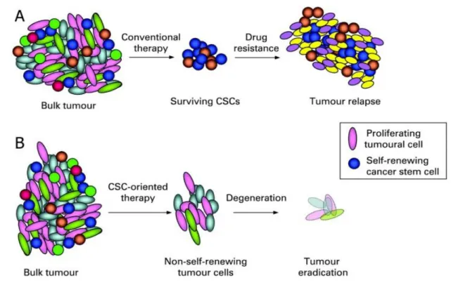
hypothesis is that these cells are more likely to accumulate mutations over time increasing chances of neoplastic transformation [83]. CSC model identification has, however, led to new approaches in cancer experimental research. This is true especially in light of recent works that have been demonstrated CSC resistance to chemotherapy and radiotherapy. One of the most commonly used methods for CSC enrichment from primary cultures of tumor cells, is the Hoechst 33342 dye exclusion [88-90]. Goodell and colleagues demonstrated that this phenomenon involves the multidrug resistance transporter (MDR1), a member of the ABC (ATP Binding Cassette) family transporters [91]. Several studies [92, 93] showed that the CSC associated chemoresistance is due to increased expression or activity of MDR1. Other gene expression studies have identified a correlation between CSC chemotherapy resistance and high expression levels of BCRP1 (Breast Cancer Resistance Protein) multidrug resistance gene. Similarly, genes involved in mismatch repair system in DNA as MGMT (methyl-guanine methyl transferase) and genes encoding proteins such as antiapoptotic Bcl-2, Bcl-XL and FLIP have been associated with chemoresistance [94]. As with chemotherapy, radiotherapy resistance has been linked to the CSC. In a recent paper [95] has been shown that the stem cell subpopulation contributes to tumor radioresistance through more efficient checkpoint activation by DNA damage, compared to the remaining tumor population. For the aforementioned reasons, tumor CSC subpopulation can be considered responsible for tumor initiation, progression and spreading to other organs. Thus, CSC must be considered as a new target for therapy aimed at complete tumor healing (Figure 5).
19
Figure 5. (A) Conventional therapy was mainly addressed towards the more
mitotically active cells, thus resulting less effective on resting cells such as CSC. After an initial shrinkage the tumor tends to recur because of the small stem cell population remained alive. (B) A targeted therapy can selectively kill CSC thus eliminating tumor regenerative capacity [96].
2.1 Colon cancer stem cells
Colon epithelium is formed by a columnar cell monolayer organized into functional units called crypts of Lieberkühn. Crypt based resident multipotent stem cells are able to generate actively proliferating progenitors which, in turns, gives rise to three main terminally differentiated colonic cell types: enterocytes, goblet cells and enteroendocrine cells. Differentiated cells are continuously produced and migrate to the apical end of the crypts, which are shed into the intestinal lumen (Figure 6a) [97].
The stem cells compartment existence in the intestine has been inferred through DNA label retention experiments (e.g. tritiated thymidine or bromodeoxyuridine), by virtue of reduced mitotic activity [98, 99].
20
Such studies conducted over the past 30 years to identify intestine normal stem cells have led to a first model, known as "+4 position" model [101, 102]. In this model the intestinal stem cells are localized in position +4 from the base of the crypt, just above the Paneth cells. These stem cells divide at a relatively slow rate for the whole lifespan, giving rise to cells able to divide at a higher rate ("transit amplifying-cells”). These in turn can differentiate into enterocytes, goblet cells and enteroendocrine cells, which migrate to the apex of the crypt, or Paneth cells that are located below the +4 position at the base of the crypt.
Recently, the characterization of a rare undifferentiated cell population, interspersed between the Paneth cells named Crypt Base Columnar Cells, CBC, [103, 104], lead to the definition of an alternative model, the "stem cell compartment” model (Figure 6b). According to this model the CBC are the normal intestinal stem cells [105, 106]. Stem cell compartment maintenance at the crypt base is dependent on a variety of signals from surrounding mesenchymal cells, which control self-renewal, migration, proliferation, differentiation and apoptosis. Among these signal transduction pathways Wnt plays a major role [107-109]. In a recent study [111] it has been demonstrated in mice that the expression of the LGR5 (Leucine Rich Repeats Receptor 5 G protein-coupled) gene, is Wnt-dependent and is limited to the stem cell population at the base of the crypts. In particular, the authors demonstrated that the expression of Lgr5+ is restricted to CBCs. Lgr5+ cells were then proposed as a stem cell population of normal colon in a murine system. Despite these evidences the true colon stem cells have not yet been uniquely identified due to lack of specific biomarkers.
Figure 6. (A) Schematic representation of the crypt of Lieberkühn. (B) CBC Lgr5+ cells can differentiate and
give rise to all differentiated colon epithelial cells [100]. B A
21
In a recent study related to humans, Lgr5 overexpression has been placed in relation to colorectal carcinogenesis and correlated with Wnt/ -catenin aberrant pathway activation [112]. Lgr5 expression was observed in rare cells at the crypt base in normal mucosa. Significantly higher expression was observed in adenomas and in CRC cases. Another study [113] showed, in a murine tumor model system, that stem cell regulation operated by Wnt signal, is a dynamic property also influenced by extrinsic factors such as the microenvironment. In particular, authors demonstrated that myofibroblasts present in the crypt surrounding stroma, are capable of regulating the Wnt activation through hepatocyte growth factor (HGF) secretion. HGF binding to its receptor (c-Met) present on crypt base cell membrane is responsible for the increased nuclear levels of -catenin, through a molecular mechanism not yet clear. Wnt pathway activation cause increased expression of Lgr5 and maintenance of the tumorigenic potential of colon-CSC.
In recent years several studies have evaluated, by flow cytometry, the functionality of some biomarkers in the identification of colon-CSC (Table 3).
In a first study conducted in our laboratory [75] we proposed CD133 as a colon-CSC marker. This protein, also known as Prominin-1, is a glycoprotein expressed in neural, hematopoietic, epithelial and endothelial cells. Using surgically removed samples from primary CRC, we identified, in a large number of cases, a small population of cells (about 2.5%) expressing CD133 marker. CD133+ cells do not express cytokeratin 20 (CK20), a component of intermediate filaments expressed in differentiated intestinal epithelial cells. The next step was the isolation, from primary tumors, of a population enriched in CD133+ cells, to test the tumorigenicity by subcutaneous injection in NOD/SCID mice. Differently from CD133-, CD133+ cells gave rise to tumors that faithfully reproduced parental tumor. CD133+ cells can be maintained in culture, in an appropriate serum free medium enriched with growth factors. In these condition, cells proliferate indefinitely maintaining their tumorigenic ability. Colon-CSC can be induced to differentiate in vitro by removing growth factors and supplementing the culture medium with serum.:Differentiation involves a morphological change and increased expression of CK20, with a concomitant decrease of CD133. To assess the long term tumorigenic ability of the colon-CSCs, we submitted the CD133+ cells to serial transplantation in mice, showing that they were able to maintain tumorigenicity even after several in vivo passages. Following these observations, the CD133+ population has been associated with the initiation and maintenance of CBC and was therefore proposed as a preferential target of antineoplastic therapy.
22
A second working group has parallely came to the same conclusion through a similar experimental procedure [74]. Cells obtained from human primary CRC specimens were inoculated into the renal capsule of NOD/SCID mice resulting in the formation of tumors that replicate the parental tumor in terms of morphology and expression of CRC marker such as CEA and p53. Using the CD133 membrane marker, the tumor cell population was split into two subsets by FACS CD133+ and CD133-, which are respectively 2% and 98% of the total. These two subpopulations were inoculated into immunodeficient mice to test the possible enrichment of the tumor initiating cells. Only the CD133+ population was able to generate a tumor xenografts. Both papers identified in the CD133+ sub-population colon cancer initiating cells but do not provide any information about their contribution to metastases formation. More recently it has been shown that the expression of CD133 marker is unable to distinguish unambiguously the population of CSC in metastatic CRC, since both CD133+ and CD133- subpopulations, contained cells with tumorigenic ability [114].
Dalerba and coworkers [76] have proposed an alternative protocol for colon-CSC isolation, by means of CD44 and EpCAM (Epithelial cell adhesion molecule) surface markers. The injection in NOD/SCID mice of CD44+/EpCAMhigh cells, purified by FACS, produced tumor formation with a high frequency, whereas the CD44-/EpCAMlow population did not show any tumorigenic ability.
2.2 Cancer stem cells therapeutic implication
CSC ability to maintain multipotency and tumorigenicity and their ability to faithfully reproduce the parental tumor in mice models of xenotransplantation, makes them an excellent model to study tumorigenesis and to evaluate the effectiveness of new cancer therapies. The development of new targeted therapy strategies will require a better CSC characterization in terms of their genomic and proteomic characteristics. The era of large scale genetic analysis has led to valuable new information on the molecular aberrations that contribute to tumorigenesis and has increased the sample size that is possible to analyze [115]. However, even though this approach could allow a better patient stratification [116, 117] and provide potential prognostic or predictive factors, genome analysis was not able to overcome the limitations of traditional cancer therapy.
Emerging technologies are not only able to qualitatively infer protein translation but also to quantify protein expression levels and measure the amount of proteins in an active state. Proteins activity is indeed regulated through post-translational modifications such as
23
phosphorylation and cleavage. Phosphorylation, in particular, plays a major role in the regulation of signal transduction pathways that control proliferation, survival and cell death processes.
RPPA (Reverse Phase Protein Array) represents a novel approach designed to conduct large-scale phosphoproteomic quantitative analysis. [119, 120] (Figure 7).This technology is a larger scale extension of western or dot blotting. Nano-amounts of protein lysates or laser capture microdissected biological sections are automatically spotted on nitrocellulose coated slides and then proteins of interest are detected by using specific antibodies. The high detection sensitivity permits to print on each spot less than one microgram of material, much less than that required in a Western blotting experiment. In addition this technology allows users to analyze hundreds of samples simultaneously on a single slide in a relatively short time. RPPA great potential lies in the possibility of molecularly characterize a large number of tumor samples, allowing the identification of prognostic/diagnostic factors as well as potential targets for novel therapies. In recent years, research interest has focused on the possibility to asses personalized therapy calibrating treatments on the basis of anticancer drugs response predictive biomarkers. Tumors of different patients are in fact considered as distinct molecular entities, whose development could be guided by several deregulated signal transduction pathways leading to a different responsiveness to the same treatment. RPPA analysis offers the opportunity to analyze individual signal transduction activation profile providing new molecular targets and opening new therapeutic ways.
24
An RPPA technology important application is its association with in vitro and in vivo screening of potential new drugs for cancer therapy. In our laboratory we analyzed several samples of CSC, including those derived from CRC, in order to create an activation profile of different signal transduction pathways. The purpose of these studies is to correlate pathways signal transduction activation levels with the response to in vitro and in vivo treatments. A colon-CSC RPPA preliminary profile analysis of susceptibility to inhibitors of protein kinase, gave positive indications on the association between molecular activity and sensitivity to inhibitors. Even though this analysis must be confirmed on a larger patients number, the great potential of this technology for novel anticancer therapies finding is already emerged.
25
Methods
1. Colon cancer stem cells isolation and cultivation
In this study we used four different colon-CSC lines isolated from colorectal carcinoma tissue specimens. Tissues were mechanically dissociated with sterile scissors and forceps in DMEM (Dulbecco's Modified Eagle Medium, Gibco Invitrogen Inc., BRL, Rockville, MD) containing Streptomycin 500 g/mL, Penicillin 500 U/mL (PAA Laboratories Inc.) and Amphotericin-B 5 g/mL (Fungizone, Gibco). Then tissues were dissociated enzymatically at 37°C using type II collagenase 1.5 mg/mL (Gibco Invitrogen Inc.) DNase I 20 g/mL (Roche, Mannheim, Germany) for a time depending on piece size. Cell suspension was then passed through a sterile 100 m filter. Following a further series of washes in PBS cells were then placed in culture in a DMEM-F12 (Hepes15mM L-glutamine, Invitrogen Life Technologies Inc., Grand Island, NY) containing medium and supplemented with b-FGF (Fibroblastic Growth Factor type II) 10 ng/mL and EGF (Epidermal Growth Factor) 20 ng/mL (PeproTech, Rocky Hill, NJ). Culture medium was replaced twice a week until the formation of spheroids in suspension was observed. Spheroids were passaged by mechanical dissociation followed by replating of individual cells and small aggregates in fresh medium.
CD133+ cells isolation were performed from freshly dissociated samples or from established cultures cell suspension. CD133 staining was performed by incubating cells for 45 minutes at 4 ° C with an anti-CD133/1-PE antibody (Miltenyi Biotec Inc., Bergisch Gladbach, Germany). After a series of washes in PBS CD133+ population was separated using fluorescence activated cell sorter FACSAriaIII ™ (Becton Dickinson, San Jose, CA).
2. Kinase inhibitors library in vitro screening
For in vitro screening experiments colon-CSC spheroids were subjected to enzymatic dissociation with recombinant trypsin (TrypLE Express ™, Gibco Invitrogen Inc.) for 3 minutes at 37 ° C. After being counted with Trypan Blue dye in a Burker chamber to exclude death cells, 5000 cells per well were plated onto 96-well microplates in 80 L of culture medium. Each different treatment were performed 24 hours after plating by adding 20 L of culture medium.
Inhibitors included in the Biomol Library (Enzo Life Sciences/Biomol http://www.enzolifesciences.com/BmL -2832/kinase-inhibitor-library) were initially tested
26
at a 5 concentration. All inhibitors were resuspended in DMSO (dimethyl sulfoxide, Sigma-Aldrich Inc., Saint Louis, MO). Some samples were therefore treated with DMSO 0.1% as a vehicle control. Staurosporine was used as positive control.
3. Positive hits titration and combination with chemotherapeutics or commercially available inhibitors
Compounds that resulted effective in the first screening were then titrated using three scalar concentrations (1 M, 500 nM, 250 nM), in addition to the vehicle control (0.1% DMSO).
Selected compounds were tested in vitro in combination with irinotecan (Sigma-Aldrich Inc.) at 250 nM concentration: ERK inhibitor and its commercial negative control (ERK inhibitor FR180204 II, ERK inhibitor II-Negative control, Calbiochem Inc., Merck, Germany) were both used at 1 M, 500 nM, 250 nM and 125 nM; Akt and PKC commercially available inhibitors and the multiple inhibitor (Akt inhibitor II and X, PKC inhibitor Set, PDK1/Akt/Flt Dual Pathway Inhibitor, Merck Calbiochem Inc.) were used at 10 M, 5 M, 2,5 M, 1 M and 500 nM; PDK1 commercially available inhibitor (OSU-03012, Alexis Biochemicals, San Diego, CA) was used at 5 M. In the same experiment, cells were treated individually and in combination with Akt inhibitor X at 5 μM and PDK1/Akt/Flt Dual Pathway Inhibitor at 125 μM. DMSO 0.1% was used as a vehicle control.
UCN-01 (Sigma-Aldrich Inc.) dose-response treatment was conducted for the four lines of colon-CSC at 1 μM, 500 nM, 250 nM, 125 nM and 62.5 nM. In a second step of experiments we performed the following treatments (single and in combination): irinotecan 50 μM, oxaliplatin (Sigma-Aldrich Inc.) 10 μM, UCN-01 1 μM. DMSO 0.1% was used as a vehicle control.
4. Viability assay
In vitro viability was assayed measuring cellular ATP content by luminometry. To this end
we used the chemiluminescence assay CellTiter-Glo™ (Promega Inc., Madison, WI) following manufacturer instructions. This method takes advantage of cells ATP content to convert luciferin, the substrate of the enzyme luciferase, in an unstable compound. Before undergoing spontaneous oxidation the compound is able to emit photons in the 510-650 nm range of wavelength.
27
Viability was tested at different times of treatment. The intensity of the luminescence, proportional to cell viability, was measured by Victor 2™ (Wallac, Perkin Elmer Inc., Norwalk, CT) microplate reader. Vehicle control (DMSO 0.1%) luminescence values were averaged and arbitrarily set to 100%. The absolute values of luminescence for each treatment were then normalized with respect to vehicle control and then expressed as a percentage.
5. Mice models of xenotransplantation
In vivo experiments were conducted with immunodeficient NOD/SCID mice. Before
injection in both flanks, colon-CSC spheroids were mixed in Matrigel (Becton Dickinson) in a 1:1 ratio. In two independent experiments, mice were injected with a suspension of 2x10⁶ CSCs. Established tumors were regularly monitored over a four weeks period. When the tumors reached a 2-5 mm diameter, mice were divided into three groups and treated respectively with DMSO (vehicle), irinotecan 10 mg/kg (irinotecan group), administered only on the first day of treatment, UCN-01 5 mg/kg (UCN-01 group), administered from the first to the sixth day and the combination irinotecan/UCN-01 (combo group). Tumor growth was monitored regularly by measuring the major and minor axes during treatment and for seven days following the end of treatment. Tumor volume was then extrapolated using the modified ellipsoid volume formula [121]. Growth ratio with respect to the control was then calculated.
6. Western blotting
To determine caspase 3, phospho-Akt (S473), phospho-PKCα/βII (T638/641), Bcl-XL, Mcl-1, phospho-PDK1 (S241) and phospho-cdc25 expression levels, colon-CSC spheroids were subjected to enzymatic dissociation, after being counted 5x10⁵ cells were plated 6-well in 2 mL of culture medium. Samples were subsequently treated with 0.1% DMSO (Sigma-Aldrich), UCN-01 (Sigma-Aldrich) 1 μM, irinotecan (Sigma-Aldrich) 50 , oxaliplatin (Sigma-Aldrich) 10 μM and their combinations. Protein lysates were prepared by resuspending cells in a T-PER lysis buffer (Pierce) with 300 mM NaCl and Protease Inhibitor Cocktail and Phosphatase Inhibitor Cocktails I and II (Sigma-Aldrich) according to manufacturer instruction. After a 30 minutes of-ice incubation, cells were centrifuged
28
for 10 minutes at 13000 RPM at +4°C. The recovered supernatantwas then directly used for Western blotting experiments or stored at -80°C.
Protein concentration has been assessed using Bradford protein assay (Bio-Rad Laboratories, Richmond, CA), which is based on Coomassie Blue G-250 dye ability to change its maximum absorption wavelength from 465 nm to 595 nm in response to protein binding.
Proteins were subjected to electrophoresis on acrylamide gel containing SDS (Sodium Dodecyl Sulfate). NuPage Novex Bis-Tris gel 1.0 mm x 15 well 4-12% (Gibco Invitrogen Inc) were used. An equivalent of 20 μg of protein, supplemented with loading buffer NuPage LDS sample buffer (25mM Tris-HCl, pH 6.8, SDS 10%, 50% glycerol, 5% β-mercaptoethanol, 0.01% bromophenol blue, (Gibco Invitrogen Inc.) was incubated at 95°C for 3 minutes and loaded. SeeBlue Plus 2 (Gibco Invitrogen Inc.) was used as molecular weight marker. Electrophoresis were performed in MOPS (3-[N-morpholino]-propansulfonico) buffer at 120 V for about 2 hours.
Protein transfer was made by using an elettroblot (Bio-Rad Laboratories) filled with transfer buffer (25mM Tris-HCl pH 8, 200 mM glycine and 20% methanol) at 80 V for 2 hours at 4°C. Membrane was saturated using TBST (25mM Tris-HCl pH 8, 150 mM NaCl, 0.2% Tween-20) containing 5% powder milk (Blottin-Grade Blocker non- fat dry milk, Bio-Rad). Primary antibodies incubation was performed overnight at 4°C. After the
29
appropriate secondary-HRP conjugated antibodies incubations, protein signals were visualized by chemiluminescence (Pierce Super Signal West Pico).
7. Phosphoproteomic analysis
A RPPA (Reverse Phase Protein Array) module consists of a solid support that can quantify proteins of interest previously immobilized onto nitrocellulose coated slides through the use of specific antibodies. Sample proteins immobilization was obtained through the use of an "Arrayer" tool that deposit (printing) microspot (about 30 nanoliters of sample) onto nitrocellulose slides. The immunostaining procedure, (staining) is automatically performed by an Autostainer. A portion of the printed slides are processed to detect the amount of total protein (used to normalize the antibody signal) by a fluorescence method. Once generated, signal, is detected by a high resolution scanner. The images obtained are analyzed by a software that can automatically identify the spots and quantify their relative intensity.
Printing
This procedure is performed by an Aushon 2470 Arrayer that takes advantage from a 20 "pin" head (5x4) that permits samples spotting onto nitrocellulose coated slides. Samples are diluted 1:2 with print loading buffer (Tris-glycine 2x SDS, Invitrogen) containing 2.5% β-mercaptoethanol (Sigma-Aldrich). After being heated at 100°C for 5 minutes and loaded in four different dilutions (1:1, 1:2, 1:4 and 1:8) on a 384-well plate (Genetix), samples are then spotted on the appropriate slide (FAST slides, Whatman, Fisher) by the instrument. Protein concentration is one of the most important parameter, in fact, too small amounts of protein per-spot can give rise to false negative results, while to high amounts can cause signal saturation.
Protein quantification
All protein signals were normalized on the basis of total protein values. This parameter has been estimated by SYPRO Ruby Protein Blot Stain (Invitrogen). After slides printing some of them were selected for total protein determination as described below:
• Fixing solution incubation (7% acetic acid and 10%methanol).
30 • One minute wash.
Slides are then dried at room temperature and then scanned with a fluorescence scanner (Vidar Systems Comporation, Revolution 4550).
Staining
Staining was performed using the TSA and DAB (Tyramide Signal Amplification, Diaminobenzidine, DAKO, Denmark) system of signal amplification. In this procedure, a primary antibody is detected with a biotinylated secondary antibody. This process allows a significant signal amplification useful for the detection of femtomolar antigen amount. Before the immunostaining, slides are incubated for 15 minutes with a stripping solution (Reblot mild antibody stripping solution, Chemicon) to promote antigenic sites exposure. After two PBS washes slides are treated for two hours with PBS containing powder I-block 0.2% (Applied Biosystems/Tropix) and Tween-20 0.1%, in order to saturate nitrocellulose aspecific binding sites. Before primary antibody staining, slides are subjected to a streptavidin pretreatment in order to saturate endogenous biotin eventually present in the sample. The entire staining procedure is automatically carried out using a DAKO Autostainer.
Data analysis
After staining, slides are scanned with a flatbed scanner (UMAX PowerLook, UMAX, Dallas, TX) at a 1800 dpi resolution and saved as *.TIFF image file using Photoshop 6.0 (Adobe , San Jose, CA). Images are then analyzed with the MicroVigene software 2.9.9.9 (Vigen Tech, North Bedford, MA).
Expression levels of each protein in the four lines of colon-CSC were "standardized" as follows: (xn-μ)/σ, where xn represents the intensity of the single protein in the sample n, μ is the mean value and σ the standard deviation of the individual proteins intensity calculated for all samples analyzed. In this way data are represented as relative expression values between the four lines in a range between -1.5 and 1.5 standard deviations. Data hierarchical clustering was performed through the T-MEV (http://www.tm4.org) open-source program.
32 8. Statistical analysis
All statistical analyses were conducted using GraphPad Prism 4 program (GraphPad Software Inc., www.graphpad.com).
In vitro experiments statistical significance was calculated by ANOVA test while for in vivo experiments we used Student t test. In figures 17 and 19, the significance is indicated
by asterisks: one asterisk represented a p-value ≤ 0.05, two asterisks corresponded to p ≤ 0.01, and three asterisks corresponded to p ≤ 0.001.
Results
1. Phosphpoproteomic analysis of Colon-CSC
We performed an RPPA analysis on a group of representative protein kinases, tested on four lines of colon-CSC (Figure 8). This analysis revealed a complex panel of kinase activities and was not possible to identify a single target protein common to all colon-CSC lines. These data reflect problems in identifying molecular targets for multifactorial diseases such as cancer, in which various individuals with different genetic abnormalities share similar histology and clinical course. To better understand the different characteristics of each of our colon-CSC line in terms of kinase activation and phosphorylation state we decided to directly test their in-vitro sensitivity to a panel of kinase inhibitors, to functionally test the importance of various molecular targets belonging to different signal transduction pathways for colon-CSC.
33
Figure 8: Hierarchical clustering of a panel of phosphoproteins expression in the four lines of colon-CSC.
Color scale represents the normalized intensity of expression (values from -1.5 ≤ to ≥ 1.5 standard deviations). The signal transduction pathways which are known to be involved in the phosphoproteins investigated are listed below.
2. In vitro screening of a library of kinase inhibitors on colon-CSC
Libraries of synthetic compounds with known specificity have been screened in-vitro with the aim to study the sensitivity of tumor cells to the inhibition of a specific signal transduction pathway and the consequent development of targeted drug therapies. The ability to test in-vitro such a particular cell model, as the colon-CSC, with a library of kinase inhibitors, provides additional benefit to the study of aberrations in signal transduction pathways. This experimental approach allowed to assess and discover the importance of pathways involved in tumor progression and maintenance, in a cellular system that is the ideal target for cancer therapy.
In this study we tested the in-vitro sensitivity of colon-CSC to the action of a series of 80 kinase inhibitors that belong to a commercially available chemical library (Enzo Life Sciences/Biomol http://www.enzolifesciences.com/BML-2832/kinase-inhibitor-library). This library specifically contains inhibitors of BTK, CaM Kinase, CDK, CKI/II, EGFR, GSK, IKK, Insulin Receptor, JAK, JNK, MAPK, MEK, MLCK, PI 3-kinase, PDGFR, PKA, PKC, RAF, SAPK, Src-family, and VEGFR. Table 4 shows the names of the compounds in the library with their corresponding molecular target.
34
The wide coverage of most studied signal transduction pathways, mainly implicated in tumorigenesis, provides a good chance to identify possible relations between sensitivity to the compounds and molecular aberrations directly involved with neoplastic processes.
35
36
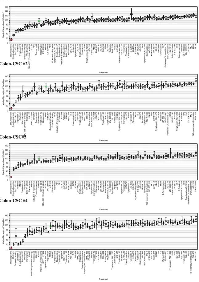
Colon-CSC spheroids were subjected to enzymatic dissociation in order to obtain a single-cell suspension ensuring reproducibility in the number of single-cells plated in each well (coefficients of variation < 10%). Colon-CSC were then plated in quadruplicate for each treatment and the 80 Biomol inhibitors were added the following day. Colon-CSC viability has been assessed after 48h of treatment through a method based on the quantification of cellular ATP. Figure 9 shows the viability for each of the four colon-CSC line tested. Following a supervised analysis, different inhibitors were found to significantly influence
37
the viability of colon-CSC 48h after treatment in-vitro. Inhibitors that showed a significant effect on colon-CSC, led to either a reduction or an increase in viability. Even though both interesting biological phenomena, we have chosen to continue with the study of inhibitors that led to a viability reduction. This choice was dictated by our experimental design, since the objective of this study was to identify possible new therapies aimed at eradicating colorectal cancer.
38
Colon-CSC #1
Colon-CSC #2
Colon-CSC#3
Colon-CSC #4
Figure 9. Viability chart after 48h of treatment with the compounds included in the library of kinase
inhibitors ( M concentration). Each symbol represents the average of at least three independent experiments and each error bars showed one standard deviation. Positive and negative controls are respectively in red and in green.
39
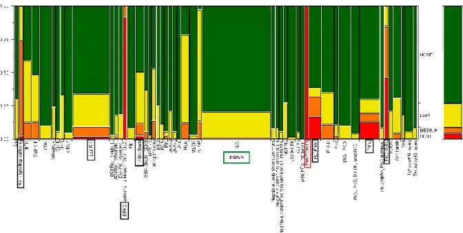
In order to summarize the data and obtaining a clear overview of inhibitors effects on colon-CSC, we created a scale of sensitivity and results of all experiments have been plotted in a two-dimensional map as showed in the Figure 10.
Figure 10. Synthetic map indicating the cumulative sensitivity of 4 colon-CSCs lines to the library of kinase
inhibitors. The scale has been calculated comparing the viability data as follows: NO EFFECT (green)> 90%, 70% <LOW (yellow) <90%, 50% <Medium (orange) <70%, HI (red) <50%.
As shown in the boxes in Figure 10 the most important signal transduction pathways responsible for colon-CSC viability include EGFR, AKT, ERK, PKC, GSK3 and PDGFR. It is clearly evident that most of compounds included in the library had no significant effect in reducing the viability of colon-CSC. Such a result was expected: in fact the number of inhibitors that cause an effect, on large scale studies, is related to the concentration used.
40 3. Titration of positive target on colon-CSC
The specificity of kinase inhibitors is not only due to their tridimensional structure, but also to the concentration at which they are used. The vast majority of synthetic inhibitors interacts directly with the kinase ATP-binding pocket (ATP-competitive), or avoids the conversion from the inactive to the active state. The ATP-binding pocket is an enzymatic site that posses highly conserved amino acid modules among the hundreds of kinases expressed by eukaryotic cells. Therefore the concentration used in each treatment may be crucial in influencing the spectrum of specificity of each inhibitor. The aim of this study is to identify selective inhibitors of kinases responsible for survival and proliferation of colon-CSC, therefore we screened lower doses of the inhibitors previously seen to be effective at a 5 M concentration. Figure 11 shows a viability chart of the four lines of colon-CSC treated with three different concentrations of inhibitors.
41
Figure 11. Viability chart after 48h of treatment with the library of kinase inhibitors (concentrations 1 M,
0,5 M and 0,25 M). Each symbol represents the average of at least three independent experiments and each error bar is calculated using one standard deviation. Vehicle control is represented by 0.1% DMSO.
The chart shows that the 4 lines of colon-CSC responded similarly, even with different degrees of sensitivity, to the lower doses of the inhibitors. The concentration decrease
42
brought to the disappearance of growth inhibition effects or death due to some inhibitors thus narrowing the pool of effective compounds and indicating that effect observed at higher concentrations could be due to involvement of pathways different from the one they were specific for. Signal transduction pathways that remained critical at lower concentrations were: PDGFR, ERK, GSK3 , Akt and PKC.
4. Combination with chemotherapeutic agents
Since the actual potential of new compounds in the therapy of colon cancer is likely the association with established chemotherapeutic regimens, we assessed these compounds
in-vitro ability in potentiating chemotherapy effect on colon-CSC. The rationale behind these
regimens implicates that is possible to achieve better effects while maintaining doses tolerated by the patients. With this in mind we evaluated whether low concentrations of kinase inhibitors, although not effective alone, could enhance the Irinotecan cytotoxic or cytostatic effects.
Unfortunately after 72h of treatment none of the combinations used resulted in a significant impairment of cell viability compared with administration of single agents alone or with Irinotecan (Figure 12).
43
Figure 12. Viability chart after 72h of treatment with a set of inhibitors used alone or in combination with
Irinotecan. The working concentration was 250nM for both inhibitors and Irinotecan. Vehicle control is represented by 0.1% DMSO. The thickened bar has been included as reference values of a single treatment with chemotherapy.
5. Definition of signal transduction pathways involved in survival/proliferation of colon-CSC
In order to assess the actual significance of a signaling pathway in CSC survival and proliferation we decided to use different inhibitors acting at different levels of the same pathway thus producing a more complete block. We were particularly interested in approaches with a relatively straight possibility of translation to clinical settings and therefore we performed a literature search, considering separately each of these molecular targets, to find molecules already in clinical use that could be used to confirm our data. For the PDGFR, some inhibitors were already approved by the FDA (Food and Drug Administration) such as Imatinib and Sunitinib. These are known to inhibit the tyrosine
![Figure 1. Fearon and Vogelstein model for colorectal carcinogenesis [6]. Patients with FAP inherit a](https://thumb-eu.123doks.com/thumbv2/123dokorg/4506599.34304/7.892.141.797.253.440/figure-fearon-vogelstein-model-colorectal-carcinogenesis-patients-inherit.webp)