Corresponding author Alessio Mancini
E-mail: [email protected]
n INTRODUCTION
C
lostridium difficile is the leading cause of infec-tious diarrhoea in the industrialized world. This spore-forming anaerobe bacteria may cause a spectrum of diseases, ranging from pseudomem-branous colitis to uncomplicated mild diarrhoea[1]. It predominantly affects elderly and frail hos-pital and nursing home patients [2].
C. difficile is known to express up to three toxins: toxin A (TcdA), toxin B (TcdB) and less commonly a third toxin called binary toxin (CDT) [1]. Symp-toms are caused by toxins A and B encoded by the tcdA and tcdB genes located within the path-ogenicity locus (PaLoc) [3]. These toxins cause extensive colonic inflammation, epithelial tissue damage, and cell death [1]. By contrast it is not clear if the binary toxin genes increase the viru-lence of C. difficile or if they are simply
epidemio-A three-year study entailing molecular
characterization and epidemiology
of Clostridium difficile in an Italian
tertiary care hospital
Alessio Mancini1,3, Giorgio La Vigna1, Sandra Pucciarelli1,
Francesca Elena Lombardi2, Simone Barocci3
1School of Biosciences and Veterinary Medicine, University of Camerino, Camerino, Italy; 2School of Medicine and Surgery, Marche Politecnica University, Ancona, Italy;
3ASUR MARCHE AV2, O.U. Clinical Pathology, Senigallia (AN), Italy
In Italy, there are limited studies on the molecular epi-demiology of Clostridium difficile, possibly due to insuf-ficient laboratory diagnostic capacity, low awareness and lack of high-quality surveillance systems. The aim of this study was to evaluate the diffusion of C. difficile in a tertiary care hospital and to genotype all the col-lected strains in order for hospital staff to take correc-tive action. All specimens were subjected to a CDI di-agnostic algorithm. This included highly specific toxin PCRs and multilocus sequence typing (MLST) to obtain clear, unequivocal genotypization. During a three-year study period, as part of routine C. difficile testing, 711 stool samples were collected from 522 patients to de-tect the presence of toxigenic genes. After testing, 106 different samples were identified as toxigenic. The pro-portions of non-toxigenic and toxigenic isolates were
SUMMARY
respectively 8.7% (62/711) and 14.9% (106/711). The most infection findings in wards for toxigenic strains were in Internal Medicine (56), followed by Neurology (11) and Gastroenterology (11). Three novel sequence types (STs) were found. The two most prevalent STs in wards were clade 1 ST-378 (40) and clade 1 ST-379 (33). Other healthcare-acquired strains were clade 4 ST-37 (11) and clade 5 ST-11 (7). Two STs, namely clade 3 ST-5 (10) and clade 1 ST-380 (5), were isolated among exter-nal patients. To prevent an increase in outbreak prob-ability, an active surveillance programme combined with proper hand hygiene, environmental cleaning and contact precautions should be implemented.
Keywords: C. difficile, MLST, prevalence, sequence type, nosocomial infections.
logic markers of strains with increased virulence. The development of genotypic methods since the late 1990s has facilitated the understanding of C. difficile epidemiology. Genotypic methods are divided into band-based and sequence-based ap-proaches. The most commonly used band-based approach is the PCR ribotyping [4].
The most frequently used sequence-based meth-od is the multilocus sequence typing (MLST). MLST is a microbial genotyping method with a high discrimination potential, using nucleotide sequences of housekeeping gene fragments [5]. Each unique combination of alleles is assigned to a Sequence Type (ST) number that can be used to group MLST results by evolutionary relationships into clades. An Internet-accessible MLST database (http://pubmlst.org/) allows the comparison of MLST results from different laboratories main-taining the ownership of the data.
The knowledge of C. difficile distribution and genotyping is the first step in monitoring and understanding the epidemiology inside hospi-tals. In Italy there are limited studies on the C. difficile molecular epidemiology, possibly due to insufficient laboratory diagnostic capacity, low awareness, and lack of high-quality surveillance systems [6, 7].
C. difficile infection represents a significant bur-den on the health care system. Although this in-fection is sporadic, nosocomial transmission is still important and outbreaks are a constant threat in hospitals [8]. The first objective of this study is to characterize C. difficile populations inside a tertiary care hospital in Central Italy, to estimate strain diversity in a variety of wards. The sec-ond objective is the genotyping of all toxinogen-ic strains and the comparison of our results with other previously reported.
n PATIENTS AND METHODS
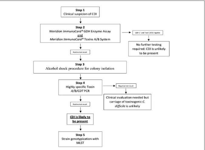
This study was carried in a hospital located in Central Italy with 288 beds divided in wards and a mean of 31,000 inpatient days per semester. A ward is a spatial unit provided with rooms where a unique staff of health-care and co-workers are active. All patients admitted from January 2015 to January 2018 were included in the study. In order to optimize the diagnosis of CDI we combined dif-ferent tests as suggested by Crobach et al. [9]
(Fig-ure 1). The advantage of an algorithm is that tests can be combined in such a way that the percent-age of false-positive and false-negative results can be decreased. CDI cases were defined as subject with at least one toxin-positive stool and we con-sidered the total number of patients and analyses, including the analyses repeated for single subjects and changes of result concerning single patients during time. All specimens were obtained from a source cohort of patients aged >18 years who ex-perienced clinically significant diarrhoea, abdom-inal pain and cramps, lower quadrant tenderness, fever, leucocytosis and hypoalbuminemia accord-ing to the current guidelines [10]. In step 2 of the algorithm, all the stool samples were tested for the presence of the glutamate dehydrogenase en-zyme and the toxins. ImmunoCard® GDH Enzyme
Assay (Meridian Bioscience inc., United States) detect glutamate dehydrogenase, an enzyme that is produced by both toxigenic and nontoxigenic strains of C. difficile. All the strains were also test-ed with the ImmunoCard® toxins A/B System
(Me-ridian Bioscience inc., United States). This assay is able to detect both toxin A and B produced by the pathogenicity locus (PaLoc) of toxinogenic C. difficile witch encodes both the toxin A gene (tcdA) and the toxin B gene (tcdB). Toxin A/B EIAs tend to be the most specific assays, while GDH EIAs is the more sensitive test [9]. The GDH positive strains but negative for toxins were discarded as suggested by the European Centre for Disease Prevention and Control (ECDC) guidelines and according with the Hospital standards.
In step 3, only positive C. difficile stool samples to GDH and one of the toxin (A or B) or both, were subjected to alcohol shock procedure to isolate the pathogenic strains. Of the first stool dilution (1:2), 0.5 mL was added and mixed by vortexing to an equal volume of absolute sterile ethanol. After incubation at room temperature for 1h, se-rial tenfold dilutions were prepared and samples of 1 mL of the serial dilutions were plated on to BHIA medium. Plates were incubated at 37° C under anaerobiosis conditions using a “gas gen-erating kit” system (Oxoid, UK). The plates were placed in an incubator for at least 48 h in 10% C02 atmosphere. Isolated bacteria were stored in Bac-terial Freezing Kit tubes (Ops Diagnostics, USA) for further analysis.
In step 4, all the isolates were subjected to a highly specific toxin-PCRs. These PCRs can
con-firm the production of TcdA and⁄or TcdB and, in addition to these toxins, several strains isolated from outbreaks and severe infections have been shown to harbour the genes encoding the bina-ry toxin CDT. For these reasons tcdA, tcdB, cdtA and cdtB primers were used, as shown by Pers-son et al. [11].
Eventually, in step 5, MultiLocus Sequence Typ-ing were performed to have a clear and unique genotypization. All samples positive in the step 2 (GDH+ and Toxin A or B positive or both) and confirmed in step 3 with the molecular assay were subjected to MLST and seven house-keeping gene were sequenced as previously described [5]. ST and clades of C. difficile isolates were determined by querying an official website (http://pubmlst. org/). The allele sequences of every strain was concatenated into a super-gene alignment and compared each other to construct the
phylogenet-ic tree [12]. MEGA 6 software (http://www. me-gasoftware.net/) was used to build the tree with a maximum likelihood method.
n RESULTS
During the 3-year study period, 711 stool samples were collected from 522 patients. The gender dis-tribution was 367 (51.7%) males and 344 (48.3%) females. The mean age was 74.3±20.5 years. Only following clinical suspicion, 168 samples out of 711 (23.6%) were positive to Meridian Immuno-Card® GDH Enzyme Assay and, 82.1% (n.584) of
these were adults over the age of 65.
After GDH screening, a patient was considered to suffer from infection only if the sample was positive to Meridian ImmunoCard® toxins A/B
System (step 2) and eventually positive at least
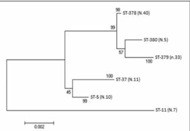
Figure 2 - Phylogenetic analysis (Maximum likelihood)
based on the alignment of seven housekeeping genes. The number of isolations is reported in round brackets.
for one toxin (A or B) after toxin-PCRs (step 4). With this approach, a total of 106 (14.9%) pa-tients were diagnosed with C. difficile infection (CDI) (Table 1). The results from the step 2 toxins assay and those confirmed through molecular techniques (step 4) were identical, according to the high specificity of EIAs methods as denot-ed in literature. The 77.3% of the patients (n = 82) were males and the 22.7% (n = 24) were fe-male with a mean age of 83.3±17.2 years. The highest number of toxinogenic strains in wards was found in Internal Medicine Unit (n = 56), followed by Neurology (n = 11) and Gastroen-terology (n = 11). Among all the CDI, the 85.9% were inpatients, while outpatients counted for 15 toxinogenic-positive strains representing the 14.1% of all toxinogenic isolations.
After the toxin-PCRs, 78 strains were toxin A/B positive and CDT negative. Seventeen strains were toxin A/B positive and CDT positive and 11 strains toxin A negative, toxin B positive and CDT negative. In order to obtain a sequence-based genotypi-zation of these six unique banding profiles, we performed MLST by sequencing adk1, atpA1, dxr3, glyA1, recA2, sodA5 and tpi2 genes. Af-ter matching the six housekeeping genes with MLST public database (http://pubmlst.org/), six different STs were detected. We found three known and three unknown sequence types. All the new loci combinations were sent to the data-base curator and three novel ST were assigned, ST-378, ST-379 and ST-380. These three new STs, according the classification provided by the soft-ware database, belonged to clade 1. The three sequence types, already present in the public database, were clade 4 ST-37, clade 3 ST-5 and clade 5 ST-11 frequently associated with animals. All the CDIs-causative toxinogenic strains and
Table 1 - Total toxinogenic C.difficile isolations in wards. All positive strains (GDH+ and toxin A/B or both positive)
were subjected to MLST. Sequence Type GDH test PaLoc Toxin A Toxin B CDT MLST
clade Cardiology Dyalisys
Internal Medicine Neurology Gastro-enterology Outpatients Total CDIs ST-378 + + + + - 1 0 6 23 0 11 0 40 ST-379 + + + + - 1 0 0 33 0 0 0 33 ST-380 + + + + - 1 0 0 0 0 0 5 5 ST-11 + + + + + 5 7 0 0 0 0 0 7 ST-5 + + + + + 3 0 0 0 0 0 10 10 ST-37 + + - + - 4 0 0 0 11 0 0 11
the matching ward of origin are listed in Table 1. Some strains were isolated from hospital wards like Gastroenterology, Internal Medicine, Dial-ysis, Cardiology, Neurology, while others from outpatients.
In order to understand the evolutionary relation-ship of these strains, we constructed a maximum likelihood phylogenetic tree (Figure 2), after the concatenation of alleles sequences from every ST. The phylogenetic tree demonstrated a heter-ogeneous genetic characteristics of C. difficile iso-lates in this collection. The STs clustered into two groups with one outlier (ST-11). According to the classification provided by the software database (http://pubmlst.org/), the closest related strains were the ST-378, ST-379 and ST-380 corresponding to the clade 1. ST-5 and ST-37 apparently grouped together but they were less related, because they belonged to clade 3 and clade 4 respectively. A single clade 5 ST-11 outlier was found.
n DISCUSSION
C. difficile continues to be the most common cause of healthcare-associated infection in the devel-oped world. A previous European C. difficile hos-pital-based survey has shown that the incidence of CDI and the distribution of causative types differs greatly from hospital to hospital [13]. This may represent a possible limitation, making diffi-cult the comparison of our data with other publi-cations.
The most common STs (ST-378, ST-379) were found in Internal Medicine and in Gastroenterol-ogy wards. Both these types carried toxin A and B but were negative to the binary toxin. The high C. difficile rate in these wards was probably due to systemic antibiotics exposure, advanced age, followed by gastric acid-suppressive medications which are the three most notable risk factors for developing CDI [14].
Our results show the presence of ST-378 also in Dialysis, due to frequent transfer of patients from Internal Medicine and other wards. These transfer patients could be a reservoir for C. difficile spores and it helps to explain the reason why there was a presence of the two most common ST in just three wards.
ST-380, one of the community-acquired strains, has resulted phylogenetically close to the most common strains isolated in wards. As ST-378 and ST-379 belong to MLST Clade 1 (>100 STs) and carried the same toxins.
Another healthcare-acquired strain was ST-11 (toxin A+B+, CDT+) found in Cardiology ward.
This strain, from clade 5, was most distant from the other lineages. Although clade 5 strains are currently present at low frequency, prospective surveillance demonstrates the continued expan-sion found it in Australia, USA and Europe [15]. The recent trends in epidemiological data show that it is an important type found in the Dutch healthcare system, Belgian provinces and among patients in Holland [16, 17].
In Neurology ward, we found eleven ST-37 strains belonging to Clade 4, also known as the toxin A-B+
clade and this is in line with our results [5]. Some studies showed that all ST-37 isolates exhibited multi-drug resistance and also indicated that the recurrence rate has increased in recent years fol-lowing the use of metronidazole and/or vanco-mycin [18]. This ST has caused widespread disease
in East Asia and recently has been reported as the predominant type in China [19]. Finally, from 12 outpatients, we isolated twelve ST-5 carrying the two main toxins A+B+ and the binary toxin CDT+.
This community-acquired strain is a binary-tox-in-positive and it is among the 15 most prevalent STs in Europe. This is the most common type in clade 3/HA2 and in contrast with our results, most of literature reports that it is much more prevalent inside hospitals than in community. In a Czech study, samples were collected from 32 healthcare facilities (7 tertiary care hospitals, 24 secondary care hospitals and 1 specialized care hospital) but ST-5 was found in only 1 hospital [20].
Nowadays, ST-17 has been reported as one of the most prevalent genotypes circulating in hos-pital settings in Italy and it has been accounted for >20% of all Italian isolates [7]. This sequence type is highly transmissible and generally shows a multidrug-resistant phenotype that seem to contribute in conferring an adaptive advantage to these strains, allowing their successful spread in our country [21]. In our study, there is no sign of infection caused by this sequence type. Maybe to start a longer period surveillance could help us to better investigate its presence in our hospital and to understand its spread.
To avoid the increase of outbreak probability, an active surveillance program combined with prop-er hand hygiene, environmental cleaning and contact precautions, should be improved. Con-tact precautions are crucial while patients transfer between wards. Healthcare workers, particularly when understaffed, may unintentionally contrib-ute to transmission of infectious diseases through poor infection control practices. Daily cleaning of all rooms with bleach wipes may reduce the in-cidence of hospital-acquired C. difficile infection and routine use of gloves may also be an effective mean to reduce nosocomial transmission of C. dif-ficile spores. In addition to reducing the burden of spores in the environment, a key aspect of pre-venting C. difficile infection in older adults is to minimize their vulnerability by avoiding unnec-essary antibiotic exposure through antimicrobial stewardship [22].
Conflict of interest
The authors certify that they have no affiliations with or involvement in any organization or enti-ty with any financial interest (such as honoraria;
educational grants; participation in speakers’ bu-reaus; membership, employment, consultancies, stock ownership, or other equity interest; and ex-pert testimony or patent-licensing arrangements), or non-financial interest (such as personal or pro-fessional relationships, affiliations, knowledge or beliefs) in the subject matter or materials dis-cussed in this manuscript.
ACKNOWLEDGMENTS
The authors would like to thank Fausta Carbini for her critical review of the manuscript.
n REFERENCES
[1] Jones A.M., Kuijper E.J., Wilcox M.H. Clostridium dif-ficile: A European perspective. J. Infect. 66, 115-128, 2013. [2] Kelly C.P., LaMont J.T. Clostridium difficile - More difficult than ever. N. Engl. J. Med. 359, 1932-1940, 2008. [3] Denève C., Janoir C., Poilane I., Fantinato C., Col-lignon A. New trends in Clostridium difficile virulence and pathogenesis. Int. J. Antimicrob. Agents. 33, Suppl 1, S24-S28, 2009.
[4] Bidet P., Barbut F., Lalande V., Burghoffer B., Petit J.-C.: Development of a new PCR-ribotyping method for Clostridium difficile based on ribosomal RNA gene sequencing. FEMS Microbiol. Lett. 175, 261-266, 1999. [5] Griffiths D., Fawley, W. Kachrimanidou M., et al. Multilocus sequence typing of Clostridium difficile. J. Clin. Microbiol. 48, 770-778, 2010.
[6] Aschbacher R., Indra A., Wiedermann C.J. et al. Pre-dominance of Clostridium difficile 027 during a five-year period in Bolzano, Northern Italy. Infez. Med. 25, 1, 13-20, 2017.
[7] Riccobono E., Di Pilato V., Della Malva N. et al. Draft genome sequence of Clostridium difficile belonging to ri-botype 018 and sequence type 17. Genome Announc. 4, e00907-e00916, 2016.
[8] Janežič S., Štrumbelj I., Rupnik M. Use of modified PCR ribotyping for direct detection of Clostridium dif-ficile ribotypes in stool samples. J. Clin. Microbiol. 49, 3024-3025, 2011.
[9] Crobach M.J.T., Planche T., Eckert C., et al. Europe-an Society of Clinical Microbiology Europe-and Infectious Dis-eases: update of the diagnostic guidance document for Clostridium difficile infection. Clin. Microbiol. Infect. 20 Suppl 2, 1-26, 2016.
[10] Fehér C., Mensa J. A comparison of current guide-lines of five international Societies on Clostridium difficile infection management. Infect. Dis. Ther. 5, 207-230, 2016. [11] Persson S., Torpdahl M., Olsen K.E.P. New multi-plex PCR method for the detection of Clostridium dif-ficile toxin. A (tcdA) and toxin. B (tcdB) and the binary toxin (cdtA/cdtB) genes applied to a Danish strain col-lection. Clin. Microbiol. Infect. 14, 1057-1064, 2008. [12] Gadagkar S.R., Rosenberg M.S., Kumar S. Inferring species phylogenies from multiple genes: Concatenat-ed sequence tree versus consensus gene tree. J. Exp. Zool. Part B Mol. Dev. Evol. 304, 64-74. 2005.
[13] Bauer M.P., Notermans D.W., Van Benthem B.H., et al. Clostridium difficile infection in Europe: A hospi-tal-based survey. Lancet 377, 63-73, 2011.
[14] Jump R.L.: Clostridium difficile infection in older adults. Aging Health 9, 403-414, 2013.
[15] Stabler R.A., Dawson L.F., Valiente E., et al. Macro and micro diversity of Clostridium difficile isolates from diverse sources and geographical locations. PLoS One. 7, 1-12, 2012.
[16] Knetsch C.W., Connor T.R., Mutreja A., et al. Whole genome sequencing reveals potential spread of Clostrid-ium difficile between humans and farm animals in the Netherlands, 2002 to 2011. Eurosurveillance 19, 1-12, 2014.
[17] Rodriguez C., Fernandez J., Van Broeck J., et al. Clostridium difficile presence in Spanish and Belgian hospitals. Microb. Pathog. 100, 141-148, 2016.
[18] Tang C., Cui L., Xu Y., et al. The incidence and drug resistance of Clostridium difficile infection in Mainland China: A systematic review and meta-analysis. Sci. Rep. 6, 37865, 2016.
[19] Du P., Cao B., Wang J., et al. Sequence variation in tcdA and tcdB of Clostridium difficile: ST37 with truncat-ed tcda is a potential epidemic strain in China. J. Clin. Microbiol. 52, 3264-3270, 2014.
[20] Krutova M., Nyc O., Matejkova J., Allerberger F., Wilcox M.H., Kuijper E.J. Molecular characterisation of Czech Clostridium difficile isolates collected in 2013-2015. Int. J. Med. Microbiol. 306, 479-485, 2016.
[21] Spigaglia P., Barbanti F. Emergence of Clostridium difficile infection caused by multi-resistant and highly virulent strains: the Italian experience in the last dec-ade. ECCMID. Proceedings 2015 Poster: P0601
[22] Mancini A., Verdini D., La Vigna G., Recanatini C., Lombardi F.E., Barocci S. Retrospective analysis of no-socomial infections in an Italian tertiary care hospital. New Microbiol. 39(3), 197-205, 2016.