Artificial Organs
29(3):203–206, Blackwell Publishing, Inc.
© 2005 International Center for Artificial Organs and Transplantation
203
Blackwell Science, LtdOxford, UKAORArtificial Organs0160-564X2005 International Society for Artificial OrgansMarch 2005293203206Original ArticleFES OF DENERVATED MUSCLESM. MÖDLIN ET AL.
Received November 2004.
Presented in part at the 8th Vienna International Workshop on
Functional Electrical Stimulation, held September 10–13, 2004, in Vienna, Austria.
Address correspondence and reprint requests to Dr. Helmut Kern, Ludwig Boltzmann Institute of Electrical Stimulation and Physical Rehabilitation, Department of Physical Medicine and Rehabilitation, Wilhelminenspital Montleartstr. 37, A-1171 Vienna, Austria. E-mail: [email protected]
Electrical Stimulation of Denervated Muscles:
First Results of a Clinical Study
*Michaela Mödlin, *Claudia Forstner, *Christian Hofer, †Winfried Mayr, ‡Wolfgang Richter,
§Ugo Carraro, #Feliciano Protasi, and *Helmut Kern
*Ludwig Boltzmann Institute of Electrical Stimulation and Physical Rehabilitation, Department of Physical Medicine and Rehabilitation, Wilhelminenspital; †Department of Biomedical Engeneering and Physics, General Hospital Vienna; ‡Department of Radiology, Wilhelminenspital, Vienna, Austria; §Laboratory of Applied Myology, Department of Biomedical
Sciences, University of Padua; and #Center for Research on Ageing, University G D’Annunzio, Chieti, Italy
Abstract: To evaluate the effects of electrical stimulation
on denervated muscles in spinal cord injured humans, the EU Project RISE was started in 2001. The aims of this project are: to design and build sufficient stimulators; to develop stimulation protocols by means of mathematical models, animal experiments, and practice in humans with denervated lower limbs; to develop examination methods and devices for evaluation of electrical stimulation training effects; and to acquire basic scientific knowledge on den-ervated and stimulated denden-ervated muscle. In the clinical study 27 spinal cord injured individuals were included,
fur-thermore 13 pilot patients participated. After a series of initial examinations they underwent an electrical stimula-tion program for their denervated lower limb muscles. Some of the patients have already follow up examinations. A marked increase of muscle mass and quality was observed, the trophic situation of the denervated lower
limbs had improved obviously. Key Words: Denervated
muscle—Spinal cord injury—Electrical stimulation— Computerized tomography scan—Force measurement— Human.
Spinal cord injured individuals with conus–cauda lesions and as a consequence denervation of their lower extremities suffer from severe atrophy of their lower limb muscles. After some years a lot of the muscle tissue is replaced by fat and connective tissue. The trophic situation of the paralyzed limbs worsens rapidly. Secondary problems like decubital ulcers, dysfunction of wound healing or osteoporosis occur. These changes can be reversed by a high intensity electrical stimulation therapy of the muscles (1,2).
For this kind of electrical stimulation a new proto-col had to be set up. Special stimulation devices had to be developed, since there were no commercially available stimulators which could generate the high
current intensities needed to stimulate denervated muscle (3).
In the EU project RISE, started in 2001, scientists and physicians from Austria, Germany, Italy, Great Britain, Slovenia, and Iceland are working together to develop a rehabilitation method for this particular kind of paraplegia, to construct a stimulation device and to acquire basic scientific knowledge concerning denervated and denervated stimulated muscle
PATIENTS AND METHODS
Forty spinal cord injured individuals were exam-ined and started electrical stimulation training (5 women, 35 men). Twenty-seven of them took part in the EU project RISE, 13 were pilot study patients.
They had to have a conus or cauda lesion with complete denervation of the m. quadriceps femoris for at least 6–12 months. Only patients without implants, hazardous infections or disease were included, pregnant women were not allowed to participate.
Paraplegia was caused by traumatic fracture of TH 12 in 57.5%, of L1 in 12.5% and of TH 11 in 7.5%.
204 M. MÖDLIN ET AL.
Artif Organs, Vol. 29, No. 3, 2005
In 22.5% of the patients, problems of regional blood circulation caused damage of the spinal cord over longer distances.
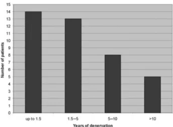
At the beginning of electrical stimulation the mean age was 43 years in female, 36 years in male partici-pants. The mean time span of denervation was 5.2 years. In 14 individuals it was 0.7–1.5 years, in 13 between 1.5 and 5 years, in 8 between 5 and 10 years and in 5 cases more than 10 years (Fig. 1).
Before starting the electrical stimulation program the patients had to undergo a series of examinations to prove that they met the inclusion criteria (com-plete denervation of m. quadriceps femoris with absent voluntary movement, sensation, and reflexes) and to describe the status of the denervated muscles at the onset.
Test stimulation, clinical and neurological exami-nations, neurophysiological assessment, biopsies of skin and quadriceps muscles, computerized tomogra-phy scans (CT scans) of the thighs, bone density measurement, skin examinations, and mechanical evaluations were carried out.
For the test stimulation the patient was sitting with extended lower limbs, two pairs of large electrodes
(~200 cm2), each inside a wet sponge bag, positioned
above his thighs. The quadriceps muscle was stimu-lated with biphasic rectangular impulses of defined durations (145, 42, 5, 2.6, and 1.3 ms) and a maximum intensity of 160 V peak to peak (Vpp). By palpating the muscle belly and the patella it was decided if a contraction of the stimulated muscle could be elic-ited. Only patients whose quadriceps muscles con-tracted by applying 5 ms or longer lasting impulses were included.
The clinical and neurological examination com-prised different tests of motor and nerve function,
sensibility, and of the range of movement of the joints.
The components of the neurophysiological ment were: BMCA (brain motor control assess-ment), LSEP (lumbosacral somatosensory evoked potentials), SSR (sympathetic skin response), transc-ranial and lumbar magnetic stimulation, and needle EMG (electromyography) (4).
Biopsies were taken from the vastus lateralis of the m. quadriceps femoris and analyzed histochemically and morphologically under light and electron micro-scopy (5).
Transverse CT scans of both thighs were taken every 10 cm beginning at the top of trochanter major. The cross-sectional areas of m. quadriceps femoris
and the hamstrings were measured in cm2, the
density of these areas was measured in Houndsfield units (2).
Force measurements were performed as soon as tetanic contractions of the thigh muscles could be elicited. Knee extension torque was measured on an isometric knee dynamometer stimulating the quadri-ceps muscle with a standardized program (1,2).
After passing this initial examinations, patients started their electrical stimulation training. The train-ing was carried out at home after appropriate instruc-tion in stimulating not only the quadriceps muscle, but also the gluteal muscle and calf bilaterally.
Electrical stimulation was applied by a specially developed stimulation device (3) with large
elec-trodes (~200 cm2) in sponge bags which were placed
over the muscles. After 4–6 months, when the skin had adapted to electrical stimulation, the electrodes on the thighs were applied to the skin directly with gel.
Every four to eight weeks accurate check ups and appropriate adaptations of the stimulation protocol were made.
Depending on the results of the test stimulation (impulse duration necessary to elicit a muscle con-traction) the electrical stimulation program was set up. Sometimes it had to be started with very long biphasic rectangular impulses lasting up to 120– 150 ms. These impulses were applied with a fre-quency of 2 Hz in bursts of 5 s duration, 2 s pause with an intensity up to 160 Vpp. Training was per-formed once a day for about 15 min (in intervals) in the beginning and was than extended to 20–30 min.
After some months of regular electrical stimula-tion it was possible to reduce impulse durastimula-tion to 70 ms (5 Hz) and subsequently to 40 ms. With 40 ms impulse duration and an impulse pause of 10 ms (20 Hz) and bursts of 2 s (2 s pause) tetanic contrac-tions could be elicited.
FES OF DENERVATED MUSCLES 205
Artif Organs, Vol. 29, No. 3, 2005
With increasing force of quadriceps muscle as a result of training, knee extension in a sitting position could be performed. As soon as knee extension
reached 45–60∞ (measured from sitting position
toward knee extension) additional weight around the ankle was used to increase training intensity. This resistance training was performed twice a week with 3–6 series of 12–15 repetitions of leg extension with maximum stimulation intensity, 60% of maximum possible additional weight, and pauses of 2–3 min between series (2).
Apart from the frequent assessment of the effects of the electrical stimulation training, patients were checked to assess if they still met the inclusion crite-ria once a year.
RESULTS
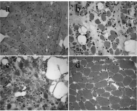
Denervation of skeletal muscle causes rapid loss of both mass as well as contractile force which is fol-lowed by severe structural changes. Figure 2 shows images of muscle biopsies at different stages of denervation after H&E (hematoxylin and eosin) staining.
In a human quadriceps muscle denervated for 0.7 years (Fig. 2a), the analyzed sections consist mainly of small, multiangular myofibers with a mean
diameter of 18.6 mm (±7.4 mm SD). In the quadriceps
muscle of a patient with 4 years of denervation (Fig. 2b) atrophic and severely atrophic myofibers
(mean diameter 9.0 mm, ±10.5 mm SD) have been
substituted by adipocytes and collagen. After 8.7 years of denervation the biopsy (Fig. 2c) demon-strates the characteristics of a typical long-term den-ervated and degenerated human muscle with very few muscle fibers which are severely atrophic and multiangular. The mean diameter of these myofibers
is 7.9 mm (±3.7 mm SD).
In a human thigh muscle denervated 2 years and subsequently stimulated 8 years (Fig. 2d), the ana-lyzed sections mainly consisted of large round, but still multiangular myofibers with a mean diameter of
48.2 mm (±14.8 mm SD). Adipocytes were absent.
The CT scans of the thighs 20 cm below the top of the trochanter major demonstrated the severe atro-phy of the denervated muscles at the onset of stimu-lation. After 1–2 years of denervation the cross-sectional area of the quadriceps muscle was reduced to approximately 40% of normal values. The longer the duration of denervation was, the more pro-nounced was the atrophy.
Furthermore, an increase of fat occurred in the denervated muscles and subcutaneously.
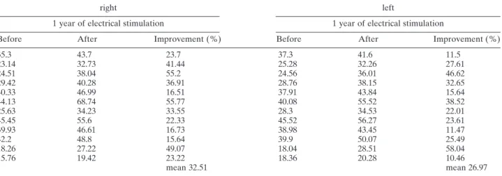
After about one year of electrical stimulation a distinct increase of the cross-sectional area of the
quadriceps muscle (mean: +29.74%, range from
10.45% to 58.04%) but also of the hamstrings could be observed (Fig. 3). Values of individual subjects are listed in Table 1.
At the beginning of the electrical stimulation pro-gram only single twitches could be elicited in the denervated quadriceps muscle. With the increase of
FIG. 2. Biopsies of m. quadriceps femoris, H&E stained, light microscopy: (a) 0.7 year of denervation; (b) 4 years of denervation; (c),8.7 years of denervation; (d) 2 years of denervation and 8 years of stimulation.
206 M. MÖDLIN ET AL.
Artif Organs, Vol. 29, No. 3, 2005
excitability of the stimulated denervated muscles the impulse duration could be reduced and the elicitation of tetanic contractions was possible. The tetanic con-tractions were very weak in the beginning, but after some months a force output up to 5–20 Nm could be measured.
CONCLUSIONS
This work shows that structurally and functionally denervated muscles deteriorate with increasing
dura-tion of denervadura-tion. With an adequate stimuladura-tion protocol which can only be carried out with a spe-cially adapted stimulation device, these changes can be reversed. It is possible to increase muscle mass and to enhance the trophic situation and thereby decrease the secondary problems. Furthermore training of long-term denervated muscles is also pos-sible, even if it takes more time to achieve similar properties.
Acknowledgments: This work was supported
by EU Commission Shared Cost Project RISE (Contract No. QLG5-CT-2001-02191).
REFERENCES
1. Kern H. Funktionelle Elektrostimulation paraplegischer Patienten. Österr Z Phys Medical 1995;5(Suppl. 1): 1–79. 2. Kern H, Hofer C, Mödlin M, Forstner C, Mayr W, Richter W.
Functional electrical stimulation (FES) of long-term dener-vated muscles in humans: clinical observations and laboratory findings. Basic Appl Myol 2002;12:291–9.
3. Hofer C, Mayr W, Stöhr H, Unger E, Kern H. A stimulator for functional activation of denervated muscles. Artif Organs
2002;26:276–9.
4. Dimitrijevic MR, Hsu CY, McKay WB. Neurophysiological assessment of spinal cord and head injury. J Neurotrauma
1992;9(Suppl. 1):293–300.
5. Kern H, Boncompagni S, Rossini K, et al. Long-term dener-vation in humans causes degeneration of both contractile and excitation-contraction coupling apparatus, which is reversible by functional electrical stimulation (FES): a role for myofiber regeneration? J Neuropathol Exp Neurol 2004; 63:919–31.
TABLE 1. Cross-sectional area (cm2) of m. quadriceps fem. in the CT scan 20 cm below the top of trochanter major
right left
1 year of electrical stimulation 1 year of electrical stimulation
Before After Improvement (%) Before After Improvement (%)
35.3 43.7 23.7 37.3 41.6 11.5 23.14 32.73 41.44 25.28 32.26 27.61 24.51 38.04 55.2 24.56 36.01 46.62 29.42 40.28 36.91 28.76 38.15 32.65 40.33 46.99 16.51 37.91 43.84 15.64 44.13 68.74 55.77 40.08 55.52 38.52 25.63 34.23 33.55 28.3 34.53 22.01 45.45 55.6 22.33 45.52 56.27 23.61 39.93 46.61 16.73 38.98 43.45 11.47 42.2 48.8 15.64 39.9 50.07 25.49 18.26 27.22 49.07 18.04 28.51 58.04 15.76 19.42 23.22 18.36 20.28 10.46 mean 32.51 mean 26.97
FIG. 3. CT scans of the right thighs 20 cm below trochanter major: (a) >10 years denervated; (b) same subject as (a), 1 year stimulated; (c) 1.7 years denervated; (d) same subject as (c), 1 year stimulated.