ARTICLE
Clinical characteristics and outcomes of
inpatients with neurologic disease and
COVID-19 in Brescia, Lombardy, Italy
Alberto Benussi, MD, Andrea Pilotto, MD, Enrico Premi, MD, Ilenia Libri, MD, Marcello Giunta, MD,Chiara Agosti, MD, Antonella Alberici, MD, Enrico Baldelli, MD, Matteo Benini, MD, Sonia Bonacina, MD, Laura Brambilla, MD, Salvatore Caratozzolo, MD, Matteo Cortinovis, MD, Angelo Costa, MD,
Stefano Cotti Piccinelli, MD, Elisabetta Cottini, MD, Viviana Cristillo, MD, Ilenia Delrio, MD, Massimiliano Filosto, MD, Massimo Gamba, MD, Stefano Gazzina, MD, Nicola Gilberti, MD,
Stefano Gipponi, MD, Alberto Imarisio, MD, Paolo Invernizzi, MD, Ugo Leggio, MD, Matilde Leonardi, MD, Paolo Liberini, MD, Martina Locatelli, MD, Stefano Masciocchi, MD, Loris Poli, MD, Renata Rao, MD, Barbara Risi, MD, Luca Rozzini, MD, Andrea Scalvini, MD, Francesca Schiano di Cola, MD, Raffaella Spezi, MD, Veronica Vergani, MD, Irene Volonghi, MD, Nicola Zoppi, MD, Barbara Borroni, MD, Mauro Magoni, MD, Alessandro Pezzini, MD, and Alessandro Padovani, MD, PhD
Neurology
®
2020;95:e910-e920. doi:10.1212/WNL.0000000000009848Correspondence Dr. Padovani alessandro.padovani@ unibs.it
Abstract
ObjectiveTo report clinical and laboratory characteristics, treatment, and clinical outcomes of patients admitted for neurologic diseases with and without coronavirus disease 2019 (COVID-19). Methods
In this retrospective, single-center cohort study, we included all adult inpatients with confirmed COVID-19 admitted to a neuro-COVID unit beginning February 21, 2020, who had been discharged or died by April 5, 2020. Demographic, clinical, treatment, and laboratory data were extracted from medical records and compared (false discovery rate corrected) to those of neurologic patients without COVID-19 admitted in the same period.
Results
One hundred seventy-three patients were included in this study, of whom 56 were positive and 117 were negative for COVID-19. Patients with COVID-19 were older (77.0 years, interquartile range [IQR] 67.0–83.8 years vs 70.1 years, IQR 52.9–78.6 years, p = 0.006), had a different distribution regarding admission diagnoses, including cerebrovascular disorders (n = 43, 76.8% vs n = 68, 58.1%), and had a higher quick Sequential Organ Failure Assessment (qSOFA) score on admission (0.9, IQR 0.7–1.1 vs 0.5, IQR 0.4–0.6, p = 0.006). In-hospital mortality rates (n = 21, 37.5% vs n = 5, 4.3%, p < 0.001) and incident delirium (n = 15, 26.8% vs n = 9, 7.7%, p = 0.003) were significantly higher in the COVID-19 group. Patients with COVID-19 and without COVID with stroke had similar baseline characteristics, but patients with COVID-19 had higher modified Rankin Scale scores at discharge (5.0, IQR 2.0–6.0 vs 2.0, IQR 1.0–3.0, p < 0.001), with a significantly lower number of patients with a good outcome (n = 11, 25.6% vs n = 48, 70.6%, p < 0.001). In patients with COVID-19, multivariable regressions showed increasing odds of in-hospital death associated with higher qSOFA scores (odds ratio [OR] 4.47, 95% confidence interval [CI] 1.21–16.5, p = 0.025), lower platelet count (OR 0.98, 95% CI 0.97–0.99, p = 0.005), and higher lactate dehydrogenase (OR 1.01, 95% CI 1.00–1.03, p = 0.009) on admission. Conclusions
Patients with COVID-19 admitted with neurologic disease, including stroke, have a significantly higher in-hospital mortality and incident delirium and higher disability than patients without COVID-19.
MORE ONLINE
COVID-19 Resources For the latest articles, invited commentaries, and blogs from physicians around the world
NPub.org/COVID19
From the Neurology Unit (A.B., A. Pilotto, I.L., M.G., E.B., S.B., M.C., S.C.P., V.C., A.I., M. Locatelli, S.M., B.R., L.R., A.S., F.S.d.C., N.Z., B.B., A. Pezzini, A. Padovani), Department of Clinical and Experimental Sciences, University of Brescia; Neurology Unit (A.B., A. Pilotto, C.A., A.A., S.C., E.C., M.F., S. Gipponi, P.L., L.P., R.R., L.R., I.V., B.B., A. Pezzini, A. Padovani), Vascular Neurology Unit (E.P., A.C., I.D., M.G., N.G., R.S., V.V., M.M.), and Neurophysiology Unit (S. Gazzina, U.L.), Department of Neurological and Vision Sciences, ASST Spedali Civili, Brescia; Neurology Unit (M.B.), University of Bologna; Department of Neuroimmunology and Neuromuscular Diseases (L.B.) and Neurology (M. Leonardi), Public Health and Disability Unit, Foundation IRCCS Neurological Institute Carlo Besta, Milan; and Neurology Unit (P.I.), Fondazione Poliambulanza Hospital, Brescia, Italy.
Since February 20, 2020, Lombardy, Italy, has experienced a major outbreak of coronavirus disease 2019 (COVID-19), caused by the severe acute respiratory syndrome coronavirus 2 (SARS–CoV-2), with >50,000 cases and 9,000 deaths in the region as of April 5, 2020.1
The clinical spectrum of SARS–CoV-2 appears to be wide, encompassing asymptomatic infections, mild upper respiratory tract illness, and severe pneumonia with respiratory failure and death.2 Several factors have been associated with increased mortality, including older age, high Sequential Organ Failure Assessment (SOFA) score, and increased d-dimer levels.3 To date, only 2 retrospective case series from convenience samples from 3 hospitals in Wuhan, China, have been pub-lished4or posted on preprint servers without peer review.5 The most common neurologic manifestations were dizziness (16.8%), headache (13.1%), and encephalopathy (2.8%). Stroke complicated COVID-19 infection in 5.9% of cases, with patients being older with more cardiovascular comorbid conditions and more severe pneumonia.5,6
It is still unclear whether patients with neurologic disease and COVID-19 have a different neurologic outcome compared to patients without COVID-19 and if this is achieved at the cost of days of hospitalization or increased mortality. Moreover, it is not known if stroke severity at admission and stroke severity at discharge are similar in the 2 populations and if acute-phase treatments such as endovascular therapy and IVfibrinolysis have similar outcomes. We aimed to describe the clinical and laboratory characteristics, treatment, and clinical outcomes of patients with neurologic disease with and without COVID-19.
Methods
Standard protocol approvals, registrations, and patient consents
This study received approval from the ethics standards committee on human experimentation (local ethics com-mittee of the ASST Spedali Civili Hospital, Brescia: NP 4051, approved April 6, 2020). The requirement for in-formed consent was waived by the ethics commission for patients who were no longer alive or reachable at the time of approval, while full written informed consent was re-quired for all other participants.
Study design and participants
This retrospective cohort study included adult inpatients (≥18 years old) admitted primarily for neurologic disease
from the General Neurology Unit and Vascular Neurology Unit, Department of Neurologic and Vision Sciences, ASST Spedali Civili Hospital, Brescia, Italy, from February 21 to April 5, 2020. This hospital in Brescia was designated as a hub for acute cerebrovascular diseases during the COVID-19 outbreak in a metropolitan area of >1,200,000 people.7The original units were divided into 2 separate sections for patients affected (neurological neuro-COVID unit) and not affected (non-COVID unit) by COVID-19, and staff neurologists were equally divided between the 2 units.8
Our study included all adult inpatients who were hospitalized for neurologic diseases and had a definite outcome (discharge home or to a rehabilitation facility or death).
The criteria for discharge for patients with COVID-19 were absence of fever for at least 24 hours, a respiratory rate <22 breaths/min, and substantial improvement at chest x-ray or CT scan.
Data collection
Epidemiologic, demographic, clinical, laboratory, treatment, and outcome data were extracted from both printed and electronic medical records with standardized anonymized data collection forms. All data were imputed and checked by 4 physicians (A.B., A. Pilotto., M.G., and I.L.). The admission data of included patients ranged from February 21 to April 5, 2020.
Demographical and clinical data
The following demographic and clinical data were acquired for all patients, which were present on admission: age, sex, smoking habits, comorbid conditions (diabetes mellitus, hypercholesterolemia, hypertension, coronary heart dis-ease, malignancy, chronic kidney disdis-ease, immunodefi-ciency), quick SOFA (qSOFA) score, premorbid modified Rankin Scale (mRS) score, and NIH Stroke Scale (NIHSS) score (for cerebrovascular disease only); during hospitalization: antibiotic, antiviral, or high-flow oxygen therapy, in-hospital mortality, incident delirium, fever during hospitalization, number of diagnostic tests, acute-phase therapies such as IV fibrinolysis, endovascular therapy, or bridging therapy (IVfibrinolysis followed by endovascular therapy) (for ischemic stroke only); or at discharge: days of hospitalization, mRS score, and NIHSS score (for cerebrovascular disease only). The qSOFA score uses 3 criteria, assigning 1 point for low systolic blood pressure (≤100 mm Hg), high respiratory rate (≥22 breaths/min), or altered mentation (Glasgow Coma Scale score <15), with a range from 0 (least impairment) to 3 (greatest impairment).
Glossary
CI= confidence interval; COVID-19 = coronavirus disease 2019; IQR = interquartile range; mRS = modified Rankin Scale; NIHSS= NIH Stroke Scale; qSOFA = quick SOFA; SARS–CoV-2 = severe acute respiratory syndrome coronavirus 2; SOFA = Sequential Organ Failure Assessment.
Laboratory procedures
SARS–CoV-2 detection in respiratory specimens was per-formed by real-time reverse-transcriptase PCR methods, as described elsewhere.9Both nasopharyngeal and oropharyn-geal swabs were performed in all patients. If 2 consecutive tests obtained at least 24 hours apart were negative and there was high suspicion of COVID-19 (i.e., interstitial pneumonia at chest x-ray, low arterial partial pressure of oxygen), a bronchoalveolar lavage was performed.
Routine blood examinations comprised complete blood count, erythrocyte sedimentation rate, serum biochemical tests including C-reactive protein, liver and renal function, lactate dehydrogenase, creatine kinase, high-sensitivity tro-ponin T, serum ferritin, and coagulation profile. High-sensitivity troponin T, ferritin, and d-dimer were performed in only a subset of patients (≈20%).
Definitions
Fever was defined as axillary temperature of at least 37.5°C. The diagnosis of delirium was defined by the presence of features 1 and 2 and either 3 or 4 of the Confusion Assessment Method.10 Antiviral treatment was defined as lopinavir/ritonavir 400/ 100 mg twice daily, darunavir 800 mg once daily + ritonavir 100 mg once daily, or darunavir/cobicistat 800/150 mg once daily. In patients with stroke, a good outcome was defined as an mRS score≤2. Diagnostics tests were defined as MRI (head), CT (head/thorax/abdomen), x-ray (thorax/abdomen), EEG, echography (heart/neck), and Holter monitor.
Statistical analysis
Continuous and categorical variables are reported as median (interquartile range [IQR]) and number (percent), re-spectively. Differences between patients with and without COVID-19 were compared by the Mann-Whitney U test,χ2 test, or Fisher exact test as appropriate.
To explore the risk factors associated with in-hospital death, univariable and multivariable logistic regression models were implemented. For multivariate analysis, to avoid overfitting in the model, variables were chosen on the basis of previous findings and clinical constraints.3Previous studies have shown age, qSOFA scores, and several laboratoryfindings to be asso-ciated with in-hospital mortality. Therefore, we chose age, qSOFA scores, platelet count, C-reactive protein, and lactate dehydrogenase for our multivariable logistic regression model. A 2-sided value of p < 0.05 was considered significant and cor-rected for multiple comparisons with the Benjamini-Hochberg false discovery rate.11Data analyses were carried out with SPSS software (version 21.0; Armonk, NY) and GraphPad Prism (version 8.0; GraphPad Software, La Jolla, CA).
Data availability
All study data, including raw and analyzed data, and materials will be available from the corresponding author on reasonable request.
Results
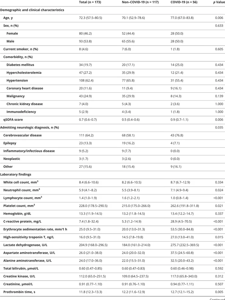
Two hundred fourteen adult patients were hospitalized in the Neurology and Vascular Neurology Unit of the ASST Spedali Civili di Brescia Hospital from February 21 to April 5, 2020. After the exclusion of 41 patients who were still hospitalized as of April 5, 2020, we included 173 inpatients in the final analysis. Of these, 56 (32.4%) resulted positive for COVID-19 and were admitted to the neuro-COVID unit (figure). Demographic, clinical, and laboratory characteristics of in-cluded patients are reported in table 1 (between-group dif-ferences with 95% confidence intervals [CIs] are reported in table e-1, links.lww.com/WNL/B124).
Results are reported as median (IQR) or number (percent), while false discovery rate–adjusted p values for multiple com-parisons are reported for each test. Compared to patients without COVID-19, patients with COVID-19 were older (77.0 years, IQR 67.0–83.8 years vs 70.1 years, IQR 52.9–78.6 years, p = 0.006), had a different distribution regarding admission diagnoses, particularly for cerebrovascular disorders (n = 43, 76.8% vs n = 68, 58.1%, p = 0.035), and had a higher qSOFA score on admission (0.9, IQR 0.7–1.1 vs 0.5, IQR 0.4–0.6, p = 0.006). No significant differences were observed for comorbid conditions, including diabetes mellitus, hypercho-lesterolemia, hypertension, coronary heart disease, chronic kidney disease, immunodeficiency, or malignancy (all p > 0.050). Patients with COVID-19 had higher in-hospital mor-tality (n = 21, 37.5% vs n = 5, 4.3%, p < 0.001) and higher incidence of delirium (n = 15, 26.8% vs n = 9, 7.7%, p = 0.003) and fever (n = 27, 48.2% vs n = 14, 12.0%, p < 0.001), while days of hospitalization were similar (n = 6.0 IQR 3.3–10.0 vs 5.0, IQR 4.0–8.0, p = 0.424). Of patients who were discharged (excluding in-hospital deaths), days of hospitalization were increased in patients with COVID-19 (8.0, IQR 5.0–11.0 vs 5.0, IQR 4.0–8.0, p = 0.005).
Treatments were different between the 2 groups, with a wider use of high-flow oxygenation (n = 43, 76.8% vs n = 11, 9.4%, p < 0.001), antibiotic therapy (n = 36, 67.9% vs n = 19, 16.2%, p < 0.001), and antiviral treatments (n = 38, 67.9% vs n = 2, 1.7%, p < 0.001) in the COVID-19 group.
Laboratory analysis showed an increased neutrophil and platelet count; reduced lymphocyte count; and increased C-reactive protein, erythrocyte sedimentation rate, high-sensitivity troponin T, lactate dehydrogenase, aspartate and alanine aminotransferase, prothrombin time, andfibrinogen (all p < 0.05) in patients with COVID-19 compared to patients without COVID-19. No differences were observed for whole white blood cell count, hemoglobin, bilirubin, creatine kinase, creatinine, activated thromboplastin time, d-dimer, or serum ferritin (all p > 0.05) (table 1).
Patients with COVID-19 had worse functional outcomes as measured by the mRS (5.0, IQR 2.3–6.0 vs 2.0, IQR 1.0–3.0,
p < 0.001), with similar premorbid mRS scores (1.0, IQR 1.0–2.0 vs 1.0, IQR 0.0–2.0, p = 0.903).
We observed a significant increase in cerebrovascular disease rates in the COVID-19 group (n = 43, 76.8% vs n = 68, 58.1%, p = 0.018), with a similar distribution among TIA (n = 5, 11.6% vs n = 8, 11.9%), ischemic stroke (n = 35, 81.4% vs n = 50, 74.6%), and hemorrhagic stroke (n = 3, 7.0% vs n = 9, 13.4%) within groups, p = 0.560 (table 2; between-group differences with 95% CIs are reported in table e-2, links.lww. com/WNL/B124).
Patients admitted for ischemic stroke had similar baseline characteristics, including sex, comorbid conditions, pre-morbid mRS score, and NIHSS score on admission. There were no differences in access to acute-phase therapies such as endovascular treatment (n = 2, 5.0% vs n = 8, 13.8%, p = 0.785), IVfibrinolysis (n = 4, 10.0% vs n = 2, 3.4%, p = 0.384), or bridging therapy (n = 3, 8.6% vs n = 3, 6.0%, p = 0.785). Patients with COVID-19 had higher mRS scores at discharge (5.0, IQR 2.0–6.0 vs 2.0, IQR 1.0–3.0, p < 0.001), with a sig-nificantly lower number of patients with a good outcome (n = 11, 25.6% vs n = 48, 70.6%, p < 0.001). This difference in outcomes was also confirmed considering only patients who underwent acute-phase therapies (mRS score at discharge p = 0.009, good outcome p = 0.047).
Moreover, patients with cerebrovascular disease (including TIA and ischemic and hemorrhagic stroke) and COVID-19 had an increased platelet count, reduced lymphocyte count, and higher C-reactive protein, erythrocyte sedimentation rate, lactate dehydrogenase, aspartate and alanine aminotransferase, prothrombin time, andfibrinogen levels compared to patients with cerebrovascular disease but without COVID-19 (table 2).
In univariable analysis, increased age, higher qSOFA scores, thrombocytopenia, and elevated C-reactive protein and lac-tate dehydrogenase were associated with in-hospital death in the COVID-19 group (table 3). In the multivariable logistic regression model, we found that higher qSOFA scores (odds ratio 4.47, 95% CI 1.21–16.50, p = 0.025), lower platelets (odds ratio 0.98, 95% CI 0.97–0.99, p = 0.005), and higher lactate dehydrogenase (odds ratio 1.01, 95% CI 1.00–1.03, p = 0.009) on admission were all associated with increased odds of death in patients with COVID-19 (table 3).
Discussion
This retrospective cohort study identified several differences between patients with and without COVID-19 who were hospitalized for a neurologic disease. In particular, patients with COVID-19 were older, had higher qSOFA scores on admission, and had a higher rate of cerebrovascular disorders compared to patients without COVID-19. During hospitali-zation, patients with COVID-19 had a higher incidence of delirium and fever, with prolonged hospital length of stay and increased in-hospital mortality rates. In addition, patients with COVID-19 had significant differences in laboratory values on admission, including blood count analysis, acute-phase pro-teins, and coagulation profiles.
We identified potential risk factors for a poor prognosis at an early stage such as high qSOFA score, thrombocytopenia, and increased lactate dehydrogenase levels. Previous reports have also found that SOFA and qSOFA scores were associated with in-hospital mortality, as well as d-dimer and older age, in adult inpatients with COVID-19.3The qSOFA is a bedside prompt that may identify patients with suspected infection who are at greater risk for a poor outcome outside the intensive care unit. FigureWeekly admissions of patients with neurologic diseases with and without coronavirus disease 2019 (COVID-19)
Table 1Demographic, clinical, laboratory characteristics, treatment, and clinical outcomes of all included patients Total (n = 173) Non–COVID-19 (n = 117) COVID-19 (n = 56) p Value Demographic and clinical characteristics
Age, y 72.3 (57.5–80.5) 70.1 (52.9–78.6) 77.0 (67.0–83.8) 0.006 Sex, n (%) 0.633 Female 80 (46.2) 52 (44.4) 28 (50.0) Male 93 (53.8) 65 (55.6) 28 (50.0) Current smoker, n (%) 8 (4.6) 7 (6.0) 1 (1.8) 0.605 Comorbidity, n (%) Diabetes mellitus 34 (19.7) 20 (17.1) 14 (25.0) 0.434 Hypercholesterolemia 47 (27.2) 35 (29.9) 12 (21.4) 0.434 Hypertension 108 (62.4) 77 (65.8) 31 (55.4) 0.434
Coronary heart disease 20 (11.6) 11 (9.4) 9 (16.1) 0.434
Malignancy 43 (24.9) 35 (29.9) 8 (14.3) 0.139
Chronic kidney disease 7 (4.0) 5 (4.3) 2 (3.6) 1.000
Immunodeficiency 5 (2.9) 4 (3.4) 1 (1.8) 1.000
qSOFA score 0.7 (0.6–0.7) 0.5 (0.4–0.6) 0.9 (0.7–1.1) 0.006
Admitting neurologic diagnosis, n (%) 0.035
Cerebrovascular disease 111 (64.2) 68 (58.1) 43 (76.8) Epilepsy 23 (13.3) 19 (16.2) 4 (7.1) Inflammatory/infectious disease 9 (5.2) 9 (7.7) 0 (0.0) Neoplastic 3 (1.7) 3 (2.6) 0 (0.0) Other 27 (15.6) 18 (15.4) 9 (16.1) Laboratory findings
White cell count, mm3 8.4 (6.6–10.6) 8.2 (6.6–10.5) 8.7 (6.7–12.9) 0.334
Neutrophil count, mm3 5.9 (4.1–8.2) 5.5 (3.9–8.1) 7.1 (4.9–9.4) 0.024
Lymphocyte count, mm3 1.4 (1.0–1.9) 1.6 (1.2–2.1) 1.0 (0.8–1.4) <0.001
Platelet count, mm3 228.0 (178.5–290.5) 215.0 (175.0–266.0) 262.6 (191.8–311.8) 0.021
Hemoglobin, g/dL 13.3 (11.9–14.5) 13.2 (11.8–14.5) 13.4 (12.2–14.7) 0.337
C-reactive protein, mg/L 7.4 (1.8–32.4) 5.3 (1.2–14.9) 28.9 (4.5–70.5) <0.001 Erythrocyte sedimentation rate, mm/1 h 25.0 (9.5–31.0) 20.0 (13.0–31.3) 53.5 (30.0–84.8) <0.001 High-sensitivity troponin T, ng/L 16.0 (9.5–31.0) 14.5 (7.8–19.8) 27.0 (13.0–41.0) 0.015 Lactate dehydrogenase, U/L 204.9 (168.0–296.5) 184.0 (161.0–214.0) 275.7 (232.5–369.5) <0.001 Aspartate aminotransferase, U/L 26.0 (21.0–38.0) 24.0 (20.0–32.0) 37.5 (24.5–60.8) <0.001 Alanine aminotransferase, U/L 24.0 (17.0–36.0) 22.0 (15.5–31.0) 32.5 (20.0–43.2) <0.001 Total bilirubin,μmol/L 0.60 (0.47–0.85) 0.60 (0.47–0.83) 0.60 (0.46–0.98) 0.592 Creatine kinase, U/L 112.0 (65.0–251.5) 109.0 (64.5–237.5) 117.0 (65.8–343.0) 0.312
Creatinine,μmol/L 0.91 (0.77–1.10) 0.91 (0.76–1.10) 0.94 (0.77–1.11) 0.507
Prothrombin time, s 11.8 (12.3–13.3) 12.2 (11.6–12.9) 12.7 (12.1–15.2) 0.005
qSOFA can be performed rapidly by the clinician without the need for laboratory analysis.12
We observed a significant increase of stroke rates in patients with COVID-19, with worse outcomes compared to the group without COVID-19, including higher mRS scores at discharge and a significantly lower number of patients with a good outcome, at par with access to acute-phase therapies. As recently suggested in a statement by the American Heart Association/American Stroke Association Stroke Council Leadership, stroke mechanisms in COVID-19 could include different processes, including the release of proinflammatory cytokines with a direct effect on plaque rupture through local inflammation and activation of co-agulation factors or cardioembolism from virus-related cardiac injury.13–17
Moreover, a direct effect of the virus on endothelial cells or on heart tissue could be hypothesized, considering that the receptor for SARS–CoV-2, the angiotensin-converting en-zyme 2,18–20 is expressed on vascular endothelial cells and myocytes.21–23
In our cohort, we observed several indexes of an altered coagulability in patients with COVID-19 compared to patients without COVID-19. Prothrombin time and d-dimer
were increased in the former group, as well as inflammatory indexes such as C-reactive protein and erythrocyte sedi-mentation rate. This marker profile is consistent with what has been observed in disseminated intravascular coagulation and may play an important role in stroke incidence and severity in patients with COVID-19.24 Abnormal co-agulation parameters have also been shown to be associated with poor prognosis in patients with COVID-19–associated pneumonia.25
From the present analysis, patients who underwent endovascular therapy or IVfibrinolysis in the COVID-19 group had more severe outcomes, including increases in NIHSS and mRS scores at discharge. This raises a critical issue of whether patients with COVID-19 should be equally treated with acute-phase therapies; additional studies on broader populations should try to shed further light on this matter.
What also emerges from this study is that differences in laboratory and most clinical features between neurology admissions of patients with and without COVID-19 mostly reflect the features of COVID-19 infection, including fever, thrombocytopenia, elevated lactate dehydrogenase, and high morbidity and mortality.3,9,26,27 The concomitant neuro-logic comorbidity such as stroke or other major neuroneuro-logic Table 1 Demographic, clinical, laboratory characteristics, treatment, and clinical outcomes of all included patients
(continued)
Total (n = 173) Non–COVID-19 (n = 117) COVID-19 (n = 56) p Value Activated thromboplastin time, s 29.3 (27.3–31.2) 29.3 (27.5–31.3) 29.4 (27.3–31.1) 0.959 D-dimer, mg/L 680.5 (313.3–2,239.5) 317.0 (200.0–913.0) 735.5 (329.8–2,710.5) 0.285 Fibrinogen, mg/dL 348.4 (298.5–461.1) 321.0 (288.5–376.1) 484.0 (376.3–600.3) <0.001 Serum ferritin,μg/L 377.0 (173.5–706.5) 155.0 (66.5–340.25) 392.0 (194.5–827.0) 0.119 Treatments and examinations
Antibiotic treatment, n (%) 55 (31.8) 19 (16.2) 36 (64.3) <0.001
Antiviral treatment, n (%) 40 (23.1) 2 (1.7) 38 (67.9) <0.001
High-flow oxygen therapy, n (%) 54 (31.2) 11 (9.4) 43 (76.8) <0.001
Diagnostic tests, n 5.0 (4.0–7.0) 5.0 (3.0–7.0) 5.0 (4.0–8.0) 0.553
Outcomes
In hospital mortality, n (%) 26 (15.0) 5 (4.3) 21 (37.5) <0.001
Incident delirium, n (%) 24 (13.9) 9 (7.7) 15 (26.8) 0.003
Fever during hospitalization, n (%) 41 (23.7) 14 (12.0) 27 (48.2) <0.001
Hospital length of stay, d 5.0 (4.0–8.0) 5.0 (4.0–8.0) 6.0 (3.3–10.0) 0.424
mRS score premorbid 1.0 (0.0–2.0) 1.0 (0.0–2.0) 1.0 (1.0–2.0) 0.903
mRS score discharge 2.0 (1.0–4.0) 2.0 (1.0–3.0) 5.0 (2.3–6.0) <0.001
Abbreviations: COVID-19 = coronavirus disease 2019; mRS = modified Rankin Scale; qSOFA = quick Sequential Organ Failure Assessment.
Data are median (interquartile range), or number (percent). The p values were calculated by the Mann-Whitney U test, χ2test or Fisher exact test as
Table 2Demographic, clinical, laboratory characteristics, treatment, and clinical outcomes of patients with cerebrovascular disease
Total (n = 111) Non–COVID-19 (n = 68) COVID-19 (n = 43) p Value
Cerebrovascular event, n (%) 0.560
TIA 13 (11.7) 8 (11.8) 5 (11.6)
Ischemic stroke 85 (76.6) 50 (73.5) 35 (81.4)
Hemorrhagic stroke 13 (11.7) 10 (14.7) 3 (7.0)
Demographic and clinical characteristics
Age, y 76.3 (61.4–82.5) 74.8 (58.3–80.4) 76.9 (66.8–85.2) 0.204 Sex, n (%) 0.765 Female 49 (44.1) 28 (41.2) 21 (48.8) Male 62 (55.9) 40 (58.8) 22 (51.2) Current smoker, n (%) 7 (6.3) 6 (8.8) 1 (2.3) 0.526 Comorbidity, n (%) Diabetes mellitus 24 (21.6) 14 (20.6) 10 (23.3) 0.781 Hypercholesterolemia 33 (29.7) 23 (33.8) 10 (23.3) 0.526 Hypertension 77 (69.4) 51 (75.0) 26 (60.5) 0.388
Coronary heart disease 16 (14.4) 8 (11.8) 8 (18.6) 0.643
Malignancy 24 (21.6) 19 (27.9) 5 (11.6) 0.220
Chronic kidney disease 5 (4.5) 3 (4.4) 2 (4.7) 1.000
Immunodeficiency 4 (3.6) 3 (4.4) 1 (2.3) 1.000
qSOFA score 0.7 (0.6–0.8) 0.5 (0.4–0.7) 0.9 (0.7–1.1) 0.077
Laboratory findings
White cell count, mm3 8.5 (6.6–10.6) 8.3 (6.6–10.5) 8.9 (6.7–11.7) 0.424
Neutrophil count, mm3 6.1 (4.5–8.5) 5.5 (4.1–8.1) 7.3 (5.0–9.6) 0.067
Lymphocyte count, mm3 1.3 (0.9–1.8) 1.5 (1.2–2.1) 1.0 (0.8–1.4) <0.001
Platelet count, mm3 231.0 (181.0–294.0) 211.5 (174.5–264.3) 276.0 (197.0–315.0) 0.036
Hemoglobin, g/dL 13.2 (11.5–14.6) 12.7 (11.5–14.4) 13.4 (12.1–14.9) 0.133
C-reactive protein, mg/L 8.1 (2.0–35.7) 5.1 (1.5–12.9) 28.0 (4.4–71.1) <0.001 Erythrocyte sedimentation rate, mm/1 h 25.0 (14.0–50.0) 19.5 (12.3–30.8) 49.0 (29.3–85.0) <0.001 High-sensitivity troponin T, ng/L 16.0 (10.0–29.0) 16.0 (9.3–21.3) 27.0 (10.0–41.0) 0.067 Lactate dehydrogenase, U/L 220.0 (168.0–277.5) 177.0 (156.8–221.5) 277.7 (239.0–365.0) <0.001 Aspartate aminotransferase, U/L 27.7 (21.0–38.0) 24.0 (21.0–30.5) 36.0 (28.0–52.0) <0.001 Alanine aminotransferase, U/L 25.0 (16.0–35.0) 20.5 (15.0–27.8) 31.0 (20.0–40.7) <0.001 Total bilirubin,μmol/L 0.64 (0.48–0.94) 0.64 (0.48–0.91) 0.66 (0.48–1.0) 0.424 Creatine kinase, U/L 109.0 (66.0–241.0) 104.5 (65.3–221.5) 114.0 (69.0–337.0) 0.424
Creatinine,μmol/L 0.95 (0.77–1.14) 0.95 (0.77–1.14) 0.95 (0.77–1.11) 0.864
Prothrombin time, s 12.5 (12.0–14.1) 12.4 (11.8–13.0) 12.8 (12.2–15.6) 0.029
Activated thromboplastin time, s 29.0 (26.8–31.0) 29.1 (26.7–31.4) 28.6 (27.3–30.7) 0.770 D-dimer, mg/L 693.0 (325.0–2,868.0) 317.0 (197.0–437.0) 735.5 (364.3–2,910.8) 0.626 Continued
disorder could possibly increase even further the high mortality observed in this patient group. Moreover, it is still not clear whether COVID-19 may increase the incidence of stroke28and other neurologic diseases such as encephalitis29 or immune-mediated neurologic disorders.30–32
With the surge of intensive care unit admissions during the pandemic2 and as underlined by the American Heart Association/American Stroke Association Stroke Council Leadership, it has become necessary to prioritize intensive care unit resources, at the possible expenses of patients with stroke,15,33 withholding ventilation when necessary and raising issues on potential legal liabilities.34 These aspects should be addressed in neurology units that deal with acute-phase diseases and patients with COVID-19.
We acknowledge that this study has some limitations. First, due to the retrospective study design, not all laboratory
tests were performed in all patients, including high-sensitivity troponin T, ferritin, and d-dimer. Therefore, their role could have not been thoroughly assessed in this study. Second, results on stroke do not take into account several factors due to the retrospective design, including stroke subtypes, infarct volume, and recanalization rates. Third, interpretation of our findings could be limited by sample size and by the single-center design. Fourth, there could be a selection bias due to the unwillingness of patients with COVID-related symptoms or infection to come for hospital neurologic evaluation unless ex-tremely necessary such as stroke, epilepsy, or other major neurologic disorders. Additional larger, multicenter, pro-spective studies should be performed to confirm these findings.
To the best of our knowledge, this is the largest retro-spective cohort study among patients with neurologic Table 2 Demographic, clinical, laboratory characteristics, treatment, and clinical outcomes of patients with
cerebrovascular disease(continued)
Total (n = 111) Non–COVID-19 (n = 68) COVID-19 (n = 43) p Value Fibrinogen, mg/dL 351.0 (291.0–466.0) 312.0 (261.1–386.4) 471.0 (368.0–545.0) <0.001 Serum ferritin,μg/L 411.0 (244.0–739.0) 152.0 (38.0–266.0) 452.5 (268.0–871.0) 0.130 Treatments and examinations
Antibiotic treatment, n (%) 39 (35.1) 11 (16.2) 28 (65.1) <0.001
Antiviral treatment, n (%) 27 (24.3) 0 (0.0) 27 (62.8) <0.001
High-flow oxygen therapy, n (%) 40 (36.0) 7 (10.3) 33 (76.7) <0.001
Diagnostic tests, n 6.0 (4.0–8.0) 6.0 (5.0–8.0) 6.0 (4.0–8.0)
Acute-phase therapies (ischemic stroke only), n (%) 22 (25.9) 13 (26.0) 9 (25.7) 1.000
IV fibrinolysis 6 (6.1) 2 (3.4) 4 (10.0) 0.384 Endovascular therapy 10 (10.2) 8 (13.8) 2 (5.0) 0.785 Bridging therapy 6 (7.1) 3 (6.0) 3 (8.6) 0.785 Outcomes In hospital mortality, n (%) 19 (17.1) 4 (5.9) 15 (34.9) <0.001 Incident delirium, n (%) 13 (11.7) 4 (5.9) 9 (20.9) 0.047
Fever during hospitalization, n (%) 29 (26.1) 8 (11.8) 21 (48.8) <0.001
Hospital length of stay, d 5.0 (4.0–8.0) 5.0 (4.0–8.0) 6.0 (4.0–9.0) 0.425
mRS score premorbid 1.0 (0.0–2.0) 1.0 (0.0–2.0) 1.0 (0.0–1.0) 0.727
mRS score discharge 2.0 (1.0–5.0) 2.0 (1.0–3.0) 5.0 (2.0–6.0) <0.001
Good outcome 59 (53.2) 48 (70.6) 11 (25.6) <0.001
NIHSS admission 5.0 (2.0–15.0) 4.0 (2.0–14.8) 10.0 (3.0–15.0) 0.147
NIHSS discharge 3.0 (0.0–10.0) 2.0 (0.0–6.8) 9.0 (1.0–19.0) 0.005
Abbreviations: mRS = modified Rankin Scale; NIHSS = NIH Stroke Scale; qSOFA = quick Sequential Organ Failure Assessment.
Bridging therapy is IV fibrinolysis followed by endovascular therapy. Data are median (interquartile range) or number (percent). The p values were calculated by the Mann-Whitney U test, χ2test, or Fisher exact test as appropriate and corrected for multiple comparisons with the Benjamini-Hochberg false discovery
disorders and COVID-19 with a definite outcome. Patients with COVID-19 and neurologic disease have worse clinical and neurologic outcomes with higher in-hospital mortality rates compared to patients without COVID-19.
Acknowledgment
The authors express their gratitude to the nurses, auxiliary staff, technicians, and all the colleagues who collaborated for the management of patients during the COVID-19 outbreak. Table 3Risk factors on admission associated with in-hospital mortality in patients with COVID-19
Univariable OR (95% CI) p Value Multivariable OR (95% CI) p Value Demographic and clinical characteristics
Age, y 1.06 (1.00–1.12) 0.043 1.08 (0.99–1.18) 0.090
Female sex (vs male) 0.11 (0.11–1.03) 0.057
Current smoker (vs nonsmoker) 0.000 1.000
Comorbidity present (vs not present)
Diabetes mellitus 0.36 (0.09–1.50) 0.161
Hypercholesterolemia 0.48 (0.11–2.03) 0.319
Hypertension 0.32 (0.10–0.99) 0.048
Coronary heart disease 0.81 (0.18–3.63) 0.778
Malignancy 1.00 (0.21–4.69) 1.000
Chronic kidney disease 1.70 (0.10–28.70) 0.713
Immunodeficiency 0.000 1.000
qSOFA score 5.12 (1.77–14.83) 0.003 4.47 (1.21–16.50) 0.025
Laboratory findings
White cell count 1.02 (0.93–1.11) 0.730
Neutrophil count 1.02 (0.93–1.13) 0.651
Lymphocyte count 0.45 (0.13–1.50) 0.192
Platelet count per 1 mm3 0.99 (0.98–0.99) 0.012 0.98 (0.97–0.99) 0.005
Hemoglobin 1.18 (0.86–1.61) 0.303
C-reactive protein 1.01 (1.00–1.02) 0.031 1.00 (0.99–1.01) 0.981
Erythrocyte sedimentation rate 1.01 (0.99–1.03) 0.460 High-sensitivity troponin T 1.03 (1.00–1.07) 0.078 Lactate dehydrogenase 1.01 (1.00–1.02) 0.004 1.01 (1.00–1.03) 0.009 Aspartate aminotransferase 1.02 (1.00–1.03) 0.112 Alanine aminotransferase 1.00 (0.99–1.02) 0.599 Total bilirubin 0.79 (0.31–2.03) 0.630 Creatine kinase 1.00 (1.00–1.00) 0.080 Creatinine 4.71 (0.88–25.3) 0.071 Prothrombin time 1.07 (0.96–1.20) 0.210
Activated thromboplastin time 1.02 (0.91–1.15) 0.682
D-dimer 1.00 (1.00–1.00) 0.058
Fibrinogen 1.00 (1.00–1.01) 0.108
Serum ferritin 1.00 (1.00–1.00) 0.773
Moreover, they thank Dr. Marco Trivelli, Dr. Camilo Rossi, and Dr. Loretta Jacquot for their support in setting up a neuro-COVID unit.
Study funding
No targeted funding reported. Disclosure
A. Benussi, A. Pilotto, E. Premi, I. Libri, M. Giunta, C. Agosti, A. Alberici, E. Baldelli, M. Benini, S. Bonacina, L. Brambilla, S. Caratozzolo, M. Cortinovis, A. Costa, S. Cotti Piccinelli, E. Cottini, V. Cristillo, I. Delrio, M. Filosto, M. Gamba, S. Gazzina, N. Gilberti, S. Gipponi, A. Imarisio, P. Invernizzi, U. Leggio, M. Leonardi, P. Liberini, M. Locatelli, S. Masciocchi, L. Poli, R. Rao, B. Risi, L. Rozzini, A. Scalvini, F. Schiano di Cola, R. Spezi, V. Vergani, I. Volonghi, N. Zoppi, B. Borroni, M. Magoni, and A. Pezzini report no disclosures relevant to the manuscript. A. Padovani is consultant and served on the scientific advisory board of GE Healthcare, Eli-Lilly, and Actelion Ltd Pharmaceuticals and received speaker honoraria from Nutricia, PIAM, Lansgstone Technology, GE Health-care, Lilly, UCB Pharma, and Chiesi Pharmaceuticals. Go to Neurology.org/N for full disclosures.
Publication history
Received by Neurology April 18, 2020. Accepted in final form May 13, 2020.
AppendixAuthors
Name Location Contribution
Alberto Benussi, MD
University of Brescia, Italy
Designed and
conceptualized study; major role in the acquisition of data; analyzed the data; drafted the manuscript for intellectual content
Andrea Pilotto, MD
University of Brescia, Italy
Major role in the acquisition of data; analyzed the data; revised the manuscript for intellectual content
Enrico Premi, MD
ASST Spedali Civili, Brescia, Italy
Interpreted the data; analyzed the data; revised the manuscript for intellectual content
Ilenia Libri, MD
University of Brescia, Italy
Major role in the acquisition of data; revised the manuscript for intellectual content
Marcello Giunta, MD
University of Brescia, Italy
Major role in the acquisition of data; revised the manuscript for intellectual content
Chiara Agosti, MD
ASST Spedali Civili, Brescia, Italy
Interpreted the data; revised the manuscript for intellectual content
Antonella Alberici, MD
ASST Spedali Civili, Brescia, Italy
Interpreted the data; revised the manuscript for intellectual content
Appendix (continued)
Name Location Contribution
Enrico Baldelli, MD
University of Brescia, Italy
Interpreted the data; revised the manuscript for intellectual content
Matteo Benini, MD
University of Bologna, Italy
Interpreted the data; revised the manuscript for intellectual content
Sonia Bonacina, MD
University of Brescia, Italy
Interpreted the data; revised the manuscript for intellectual content Laura Brambilla, MD Foundation IRCCS Neurologic Institute Carlo Besta, Milan, Italy
Interpreted the data; revised the manuscript for intellectual content
Salvatore Caratozzolo, MD
ASST Spedali Civili, Brescia, Italy
Interpreted the data; revised the manuscript for intellectual content Matteo Cortinovis, MD University of Brescia, Italy
Interpreted the data; revised the manuscript for intellectual content
Angelo Costa, MD
ASST Spedali Civili, Brescia, Italy
Interpreted the data; revised the manuscript for intellectual content
Stefano Cotti Piccinelli, MD
University of Brescia, Italy
Interpreted the data; revised the manuscript for intellectual content
Elisabetta Cottini, MD
ASST Spedali Civili, Brescia, Italy
Interpreted the data; revised the manuscript for intellectual content
Viviana Cristillo, MD
University of Brescia, Italy
Interpreted the data; revised the manuscript for intellectual content
Ilenia Delrio, MD
ASST Spedali Civili, Brescia, Italy
Interpreted the data; revised the manuscript for intellectual content
Massimiliano Filosto, MD
ASST Spedali Civili, Brescia, Italy
Interpreted the data; revised the manuscript for intellectual content
Massimo Gamba, MD
ASST Spedali Civili, Brescia, Italy
Interpreted the data; revised the manuscript for intellectual content
Stefano Gazzina, MD
ASST Spedali Civili, Brescia, Italy
Interpreted the data; revised the manuscript for intellectual content
Nicola Gilberti, MD
ASST Spedali Civili, Brescia, Italy
Interpreted the data; revised the manuscript for intellectual content
Stefano Gipponi, MD
ASST Spedali Civili, Brescia, Italy
Interpreted the data; revised the manuscript for intellectual content
Alberto Imarisio, MD
University of Brescia, Italy
Interpreted the data; revised the manuscript for intellectual content Paolo Invernizzi, MD Fondazione Poliambulanza Hospital, Brescia, Italy
Interpreted the data; revised the manuscript for intellectual content
Ugo Leggio, MD
ASST Spedali Civili, Brescia, Italy
Interpreted the data; revised the manuscript for intellectual content
References
1. Protezione Civile. Coronavirus. Available at: protezionecivile.gov.it/attivita-rischi/ rischio-sanitario/emergenze/coronavirus. Accessed April 5, 2020.
2. Grasselli G, Pesenti A, Cecconi M. Critical care utilization for the COVID-19 out-break in Lombardy, Italy: early experience and forecast during an emergency re-sponse. JAMA 2020;19:2–3.
3. Zhou F, Yu T, Du R, et al. Clinical course and risk factors for mortality of adult inpatients with COVID-19 in Wuhan, China: a retrospective cohort study. Lancet 2020;395:1054–1062. 4. Mao L, Jin H, Wang M, et al. Neurologic manifestations of hospitalized patients with
coronavirus disease 2019 in Wuhan, China. JAMA Neurol Epub 2020 Apr 10. 5. Li Y, Wang M, Zhou Y, et al. Acute cerebrovascular disease following COVID-19:
a single center, retrospective, observational study. SSRN 2020; Available at SSRN: https://ssrn.com/abstract=3550025.
6. Mao L, Wang M, Chen S, et al. Neurological manifestations of hospitalized patients with COVID-19 in Wuhan, China: a retrospective case series study. JAMA Neurol Epub 2020 April 10.
7. Dato Istat. Popolazione residente al 1o gennaio 2019 nella provincia di Brescia. Available at: demo.istat.it/bilmens2019gen/index.html. Accessed April 5, 2020. 8. COVID-19: neurologists in Italy to colleagues in US: look for poorly-defined neurologic
conditions in patients with the coronavirus. Available at: journals.lww.com/neuro-todayonline/blog/breakingnews/pages/post.aspx?PostID=920. Accessed April 5, 2020. 9. Huang C, Wang Y, Li X, et al. Clinical features of patients infected with 2019 novel
coronavirus in Wuhan, China. Lancet 2020;395:497–506.
10. Inouye SK, van Dyck CH, Alessi CA, Balkin S, Siegal AP, Horwitz RI. Clarifying confusion: the Confusion Assessment Method: a new method for detection of de-lirium. Ann Intern Med 1990;113:941–948.
11. Pike N. Using false discovery rates for multiple comparisons in ecology and evolution. Methods Ecol Evol 2011;2:278–282.
12. Seymour CW, Liu VX, Iwashyna TJ, et al. Assessment of clinical criteria for sepsis for the third international consensus definitions for sepsis and septic shock (sepsis-3). JAMA 2016;315:762–774.
13. Guo T, Fan Y, Chen M, et al. Cardiovascular implications of fatal outcomes of patients with coronavirus disease 2019 (COVID-19). JAMA Cardiol Epub 2020 Mar 27. 14. Inciardi RM, Lupi L, Zaccone G, et al. Cardiac involvement in a patient with
coro-navirus disease 2019 (COVID-19). JAMA Cardiol 2020;2019:4–9.
15. AHA/ASA Stroke Council Leadership. Temporary Emergency Guidance to US Stroke Centers During the Coronavirus Disease 2019 (COVID-19) Pandemic: On Behalf of the American Heart Association/American Stroke Association Stroke Council Leadership. Stroke 2020;51:1910–1912.
16. Davidson JA, Warren-Gash C. Cardiovascular complications of acute respiratory infections: current research and future directions. Expert Rev Anti Infect Ther 2019;17:939–942. 17. Smeeth L, Thomas SL, Hall AJ, Hubbard R, Farrington P, Vallance P. Risk of myocardial
infarction and stroke after acute infection or vaccination. N Engl J Med 2004;351:2611–2618. 18. Yan R, Zhang Y, Li Y, Xia L, Guo Y, Zhou Q. Structural basis for the recognition of the
SARS-CoV-2 by full-length human ACE2. Science 2020;2:1444–1448.
19. Shang J, Ye G, Shi K, et al. Structural basis of receptor recognition by SARS-CoV-2. Nature 2020;581:221–224.
20. Lan J, Ge J, Yu J, et al. Structure of the SARS-CoV-2 spike receptor-binding domain bound to the ACE2 receptor. Nature 2020;581:215–220.
21. Mendoza Torres E, Oyarz´un A, Mondaca Ruff D, et al. ACE2 and vasoactive peptides: novel players in cardiovascular/renal remodeling and hypertension. Ther Adv Car-diovasc Dis 2015;9:217–237.
22. Hamming I, Timens W, Bulthuis MLC, Lely AT, Navis GJ, van Goor H. Tissue distribution of ACE2 protein, the functional receptor for SARS coronavirus: afirst step in understanding SARS pathogenesis. J Pathol 2004;203:631–637.
23. Gallagher PE, Ferrario CM, Tallant EA. Regulation of ACE2 in cardiac myocytes and fibroblasts. Am J Physiol Heart Circ Physiol 2008;295:2373–2379.
24. Levi M, Toh CH, Thachil J, Watson HG. Guidelines for the diagnosis and manage-ment of disseminated intravascular coagulation. Br J Haematol 2009;145:24–33. 25. Tang N, Li D, Wang X, Sun Z. Abnormal coagulation parameters are associated with
poor prognosis in patients with novel coronavirus pneumonia. J Thromb Haemost 2020:18:844–847.
26. Grasselli G, Zangrillo A, Zanella A, et al. Baseline characteristics and outcomes of 1591 patients infected with SARS-CoV-2 admitted to ICUs of the Lombardy region, Italy. JAMA 2020:323:1574–1581.
27. Guan WJ, Ni ZY, Hu Y, et al. Clinical characteristics of coronavirus disease 2019 in China. N Engl J Med 2020;19:3–4.
28. Oxley TJ, Mocco J, Majidi S, et al. Large-vessel stroke as a presenting feature of covid-19 in the young. N Engl J Med 2020;375:e60.
29. Pilotto A, Odolini Si, Masciocchi S, et al. Steroid-responsive severe encephalopathy in SARS-CoV-2 infection. Ann Neurol Epub 2020 May 17.
30. Dinkin M, Gao V, Kahan J, et al. COVID-19 presenting with ophthalmoparesis from cranial nerve palsy. Neurology Epub 2020 May 1.
31. Alberti P, Beretta S, Piatti M, et al. Guillain-Barr´e syndrome related to COVID-19 infection. Neurol Neuroimmunol Neuroinflammation 2020;7:1–4.
32. Guti´errez-Ortiz C, M´endez A, Rodrigo-Rey S, et al. Miller Fisher syndrome and polyneuritis cranialis in COVID-19. Neurology Epub 2020 Apr 17.
33. White DB, Lo B. A framework for rationing ventilators and critical care beds during the COVID-19 pandemic. JAMA 2020;146:e61S–e74S.
34. Cohen IG, Crespo AM, White DB. Potential legal liability for withdrawing or with-holding ventilators during COVID-19: assessing the risks and identifying needed reforms. JAMA Epub 2020 Apr 1.
Appendix (continued)
Name Location Contribution
Matilde Leonardi, MD
Foundation IRCCS Neurologic Institute Carlo Besta, Milan, Italy
Interpreted the data; revised the manuscript for intellectual content
Paolo Liberini, MD
ASST Spedali Civili, Brescia, Italy
Interpreted the data; revised the manuscript for intellectual content
Martina Locatelli, MD
University of Brescia, Italy
Interpreted the data; revised the manuscript for intellectual content Stefano Masciocchi, MD University of Brescia, Italy
Interpreted the data; revised the manuscript for intellectual content
Loris Poli, MD ASST Spedali Civili, Brescia, Italy
Interpreted the data; revised the manuscript for intellectual content
Renata Rao, MD
ASST Spedali Civili, Brescia, Italy
Interpreted the data; revised the manuscript for intellectual content
Barbara Risi, MD
University of Brescia, Italy
Interpreted the data; revised the manuscript for intellectual content
Luca Rozzini, MD
University of Brescia, Italy
Interpreted the data; revised the manuscript for intellectual content
Andrea Scalvini, MD
University of Brescia, Italy
Interpreted the data; revised the manuscript for intellectual content Francesca Schiano di Cola, MD University of Brescia, Italy
Interpreted the data; revised the manuscript for intellectual content
Raffaella Spezi, MD
ASST Spedali Civili, Brescia, Italy
Interpreted the data; revised the manuscript for intellectual content
Veronica Vergani, MD
ASST Spedali Civili, Brescia, Italy
Interpreted the data; revised the manuscript for intellectual content
Irene Volonghi, MD
ASST Spedali Civili, Brescia, Italy
Interpreted the data; revised the manuscript for intellectual content
Nicola Zoppi, MD
University of Brescia, Italy
Interpreted the data; revised the manuscript for intellectual content Barbara Borroni, MD University of Brescia, Italy Designed and conceptualized study; revised the manuscript for intellectual content
Mauro Magoni, MD
ASST Spedali Civili, Brescia, Italy
Interpreted the data; revised the manuscript for intellectual content
Alessandro Pezzini, MD
University of Brescia, Italy
Interpreted the data; revised the manuscript for intellectual content Alessandro Padovani, MD, PhD University of Brescia, Italy Designed and conceptualized study; drafted the manuscript for intellectual content; revised the manuscript for intellectual content
DOI 10.1212/WNL.0000000000009848
2020;95;e910-e920 Published Online before print May 22, 2020
Neurology
Alberto Benussi, Andrea Pilotto, Enrico Premi, et al.
COVID-19 in Brescia, Lombardy, Italy
Clinical characteristics and outcomes of inpatients with neurologic disease and
This information is current as of May 22, 2020
Services
Updated Information &
http://n.neurology.org/content/95/7/e910.full
including high resolution figures, can be found at:
References
http://n.neurology.org/content/95/7/e910.full#ref-list-1
This article cites 22 articles, 0 of which you can access for free at:
Subspecialty Collections http://n.neurology.org/cgi/collection/prognosis Prognosis http://n.neurology.org/cgi/collection/covid_19 COVID-19 e http://n.neurology.org/cgi/collection/all_cerebrovascular_disease_strok
All Cerebrovascular disease/Stroke
following collection(s):
This article, along with others on similar topics, appears in the
Permissions & Licensing
http://www.neurology.org/about/about_the_journal#permissions
its entirety can be found online at:
Information about reproducing this article in parts (figures,tables) or in
Reprints
http://n.neurology.org/subscribers/advertise
Information about ordering reprints can be found online:
rights reserved. Print ISSN: 0028-3878. Online ISSN: 1526-632X.
1951, it is now a weekly with 48 issues per year. Copyright © 2020 American Academy of Neurology. All ® is the official journal of the American Academy of Neurology. Published continuously since