UNIVERSITÀ DEGLI STUDI DI PISA
DIPARTIMENTO CARDIO - TORACICO
DOTTORATO DI RICERCA
IN FISIOPATOLOGIA E CLINICA
DELL’APPARATO CARDIOVASCOLARE E RESPIRATORIO
Tesi di Dottorato
THE ROLE OF BIOMARKERS IN CARDIOVASCULAR EMERGENCY.
A CLINICAL STUDY
ON A NOVEL BIOCHEMICAL METHOD
FOR THE DIAGNOSIS OF AORTIC DISSECTION.
Relatore: Candidata:
Chiar.mo
Prof. Alessandro DISTANTE Dott.ssa Mariarosaria GALDI
INDEX INDEX……….2 ABBREVIATIONS……….3 ABSTRACT………4 INTRODUCTION……….….5 1. BIOMARKERS……….…….6
1.1 Biomarkers of Cardiovascular Disease……….…7
2. AORTIC DISSECTION..………..8
2.1 Biomarkers of Aortic Dissection………..11
3. AIM OF THE STUDY……….15
4. METHODS………17
5. STATISTICAL ANALYSIS………20
6. RESULTS………..21
6.1 Results of Patients Characteristics..……….…………21
6.2 Results of preliminary study with calponin...………..22
7. DISCUSSION………24 8. CONCLUSIONS………...26 REFERENCES……….27 TABLES………34 FIGURES………..48 ACKNOWLEDGMENTS...……….58
ABBREVIATIONS
CVD = cardiovascular disease MI = myocardial infarction ECG = electrocardiogram cTnT = cardiac troponin T cTnI = cardiac troponin I CK-MB = creatine kinase-MB ACS = acute coronary syndrome AD = aortic dissection
AAS = acute aortic syndrome CT = computerized tomography MRI = magnetic resonance imaging TEE = transesophageal echocardiography TTE = transthoracic echocardiography
SMMHC = smooth muscle myosin heavy chain DD = D-dimer
sELAF = solubleelastin fragments MMPs = matrix metalloproteinases
ABSTRACT
Background: Aortic dissection (AD) is a life-threatening medical emergency of the aorta, characterizedby disruption of the aortic media by blood entering througha laceration of the luminal vascular wall. Therefore, rapid diagnosisand timely management play an essential role in patient survival. Although newer diagnostic methods have greatly improved the diagnosis of AD, the diagnosis is still frequently missed today because the clinical manifestations of AD are often diverse and the clinical presentation may mimic signs and symptoms of other diseases. A reliable biochemical diagnostic method for AD would be beneficial.
Objectives: to analyze the main demographic characteristics (sex, age) and a common symptom of
cardiovascular emergency, as chest pain, in patients with suspected or confirmed AD; to enstablish the utility of the troponin- like protein of smooth muscle, calponin, as a diagnostic biomarker of AD.
Methods: From April 2004 to June 2007, the patients with suspected AD were enrolled in the
multicenter study. Clinical data forms were completed for each of the patients. Blood plasma was drawn on admission and used for measurements. Finally, an immunoassay against circulating calponins was generated by Biosite Incorporated.
Results: In Italy, 412 patients (62,8 ± 13,4 years) have been enrolled including 151 (36,7%) with AD (60,9 ± 13,4 years) and 261 (63,3%) with a different final diagnosis (63,8 ± 12,7 years). The chest pain was the most common symptom (77,4%): the half of patients (50,5%) had severe pain and referred that the chest pain had a abrupt onset (54,2%).
From all enrolled patients into the international study, the plasma specimens of 217 patients have been analysed including 59 cases of AD and 158 cases with an initial suspicion of AD but a different final diagnosis. Basic and acidic calponins, respectively, showed greater than two-fold and three-fold elevations in patients with AD.
Conclusions: The descriptive analysis of data shows that chest pain was the most common
symptom in cases of AD but, given the relative frequency of chest pain in patients presenting to emergency room and the relative infrequency of AD, the availability of biomarkers could be of great assistance in carrying out the differential diagnosis between the diseases that are accompanied by chest pain.
Acid and basic calponins have the potential for use as an early diagnostic biomarker for AD but the results of this preliminary experience using an initial assay show moderate sensitivities and specificities with negative predictive values which should be further improved upon.
INTRODUCTION
Cardiovascular disease (CVD) is the leading cause of death in most western societies and is increasing steadily in many developing countries (1). Thus, primary prevention and secondary prevention of CVD are public health priorities (2).
Greatprogress has been made in the past quarter century in the diagnostic biochemical testing of CVD. Beginning withthe introduction of the serum transaminase assays (3), followed by clinical applicationof enzyme activity assays (e.g., lactate dehydrogenase, creatininekinase) which greatly improved the diagnosis of acute myocardial infarction (4-5), and recently assays of structural proteins (e.g., cardiac myosin light chain and troponin)(6-7) have established their roles in clinical medicine.
Clinicians have used additional tools to facilitate clinical assessment and to enhance their ability to identify the patient at risk for CVD.
Biomarkers are one such tool to better identify high-risk individuals, to diagnose disease conditions promptly and accurately, and to effectively prognosticate and treat patients with disease (8).
Biomarkers, defined as alterations in the constituents of tissues or body fluids, provide a powerful approach to understanding the spectrum of CVD with applications in at least 5 areas: screening, diagnosis, prognostic, prediction of disease recurrence, and therapeutic monitoring (9).
With expansion of the number and types of existing biomarkers, the opportunity to improve diagnosis, risk stratification, and selection of therapy using these non-invasive, affordable tools continues to grow. Congruent with this evolution, the practicing clinician will benefit from a thorough understanding of the clinical, biology and technology evidence underlying the use of established and emerging biomarkers in CVD (10).
The research doctorate in physiopathology of cardiovascular apparatus has represented an important opportunity not only to acquire a better knowledge in the field of cardiology, but also to propose an innovative experience in the use of biological markers for the early diagnosis of CVD and in particular of the aortic dissection (AD).
This study has been funded by Biosite Incorporated, a pharmaceutical firm involved in biomarkers discovery, and it has been carried out together with a research team of ISBEM ScpA (Scientific Biomedical Euro Mediterraneam Institute) from Brindisi.
1. THE BIOMARKERS
The term biomarker (biological marker) was introduced in 1989 as a Medical Subject Heading (MeSH) term: “measurable and quantifiable biological parameters which serve as indices for health- and physiology-related assessments”.
In 2001, an NIH (National Institutes of Health) working group standardized the definition of a biomarker as “a characteristic that is objectively measured and evaluated as an indicator of normal biological processes, pathogenic processes, or pharmacologic responses to a therapeutic intervention” (11).
A biomarker may be measured on a biosample (as a blood, urine, or tissue test), it may be a recording obtained from a person (blood pressure, electrocardiogram, or Holter), or it may be an imaging test (echocardiogram or computerized tomography scan).
A simplistic way to think of biomarkers is as indicators of disease trait (risk factor or risk marker), disease state (preclinical or clinical), or disease rate (progression) (12).
Accordingly, biomarkers can be classified as antecedent biomarkers (identifying the risk of developing an illness), screening biomarkers (screening for subclinical disease), diagnostic biomarkers (recognizing overt disease), staging biomarkers (categorizing disease severity), or prognostic biomarkers (predicting future disease course, including recurrence and response to therapy, and monitoring efficacy of therapy) (11).
Regardless the purpose for its use, a new biomarker will be clinical valuable only if it is accurate, it is reproducibly, obtained in a standardized fashion, it is acceptable to the patient, it is easy to interpret by clinicians, it has high sensitivity and high specificity for the outcome which is expected to identify (13).
Table 1 displays the desirable properties of biomarkers overall and of biomarkers of screening, diagnosis, and prognosis (14).
Although several biomarkers satisfy one or more of these criteria, no single marker has been identified yet that satisfies all of them.
Today, in the whole world, there are investigators researching new biomarkers. The entire process, from the discovery of a biomarker in a laboratory, to the development of an assay and finally to its delivery, requires many years (15) [Figure 1]. Briefly, the process begins with the identification of target biomarkers with the use of standardized technology platforms, followed by validation of the assays (16, 17), statistical evaluation of biomarker distributions in reference samples and in those with disease, and assessment of the correlation between biomarker levels (or expression patterns of biomarkers) and clinical measurements that define disease status (15).
1.1 Biomarkers of Cardiovascular Disease
Biochemical markers play a crucial role in accurate diagnosis of CVD and, more importantly, in assessing risk and directing appropriate therapy that improves clinical outcome. Development and utilization of biomarkers has evolved substantially over the past three decades. The earliest biomarkers, such as alanine aminotransferase and lactate dehydrogenase, have fallen out in use with the development of more sensitive and specific assays for creatine kinase isoenzyme MB (CK-MB) and particularly cardiac troponin. Cardiac troponin T or I (cTnT o cTnI) measurements are now considered surrogates for necrosis and myocardial infarction (MI) when elevated in the setting of acute cardiac ischemia.
Biomarkers have provided important information for the clinical assessment of patients with suspected MI patients since the early 1950s. As displayed [Figure 2], utilization of biomarkers has evolved substantially over the past 30–40 yrs.
Biomarkers were previously considered to be one of the three important variables, along with changes on the electrocardiogram (ECG) and clinical signs and symptoms, necessary for the diagnosis of MI as defined by the World Health Organization (WHO) in 1979 (18). The biomarkers cTnT and cTnI are now designated as surrogates for necrosis and MI when elevated in the setting of acute cardiac ischemia, according to the consensus document of the European Society of Cardiology (ESC) and the American College of Cardiology (ACC) (19).
The overall expectation of a CVD biomarker is to enhance the ability of the clinician to optimally manage the patient. For instance, in a person with chronic or atypical chest pain, a biomarker may be expected to facilitate the identification of patients with chest pain of an ischemic etiology (angina). In a patient presenting to the emergency room with acute severe chest pain (suspected acute coronary syndrome, ACS), a biomarker may help to differentiate patients with an acute MI from those with unstable angina (e.g., troponin I or T), acute pulmonary embolism (e.g., D-dimer or ventilation perfusion scan), or an AD (e.g., transesophageal echocardiogram) in a timely fashion to facilitate targeted management.
In addition to their diagnostic utility, the ability of cardiac biomarkers to facilitate risk assessment in chest pain patients has allowed emergency physicians and cardiologists to rapidly identify and treat higher-risk patients with suspected ACS.
However, the sensitivities of these cardiac biomarkers obtained on initial presentation may be poor and are dependent on the time from the onset of symptoms to presentation, the duration of ischemia, and the amount of myocardial tissue involved (20).
2. AORTIC DISSECTION
Aortic dissection (AD) is an acute CVD associated with high mortality and morbidity (21) and is the most frequent and serious form of acute aortic syndrome (AAS).
Knowledge regarding the incidence of AD in the general population is limited. Studies suggest an incidence of 2.6 to 3.5 cases per 100 000 person-years (22-24).
AAS is an acute process in the aortic wall involving a weakening of the medial layer with the risk of aortic rupture and other complications. It consists of three disease entities: aortic dissection, intramural hematoma, and penetrating atherosclerotic ulcer (25). It has an incidence of around 30 cases per million per year, of which 80% are aortic dissections, 15% intramural hematomas, and 5% penetrating atherosclerotic ulcers (26, 27). The ascending aorta is affected in 60% of cases (type A) and unaffected in 40% (type B). It mainly affects men (70%), with a mean age of 60 years as proved by the International Registry of Acute Aortic Dissection (IRAD) (28).
IRAD is an ongoing multi-nationalmulti-centre registry started in 1996 that includes consecutive patients with acute AD at 22 aortic centers of 11 countries. The main objective of IRAD wasto assess the etiology, mode of presentation, clinical features,management, and outcomes of patients with AD.
The dissection is characterized by separation of the layers within the aortic wall. Blood passes through the tear separating the intima from the media or adventitia, creating a false lumen. Propagation of the dissection can proceed in anterograde or retrograde fashion from the initial tear involving side branches and causing complications such as malperfusion syndromes, tamponade, or aortic valve insufficiency (29-32). Both acquired and genetic conditions share a common pathway leading to the breakdown in the integrity of the intima. The most common risk condition for AD is hypertension, with chronic exposure of the aorta to high pressures leading to intimal thickening, fibrosis, calcification, and extracellular fatty acid deposition (33, 34-36). Marfan’s syndrome, vascular Ehlers-Danlos syndrome, annuloaortic ectasia, bicuspid aortic valve are genetic conditions that often cause acute AD. As displayed [Table 2], other risk conditions exist for AD (37).
The Stanford classification of AD distinguishes between type A and type B (38,39) [Figure 3]. In type A, the dissection involves the ascending aorta. In type B, only the descending aorta is involved. The DeBakey classification subdivides the dissection into 3 types, with type I dissection involving the entire aorta, type II dissection involving only the ascending aorta, and type III dissection sparing the ascending aorta and arch.
Acute AD of the ascending aorta is highly lethal, with a mortality of 1% to 2% per hour early after symptom onset (40,41).
Instantaneous onset of severe chest (85%) and/or back (46%) pain are characteristic presenting symptoms; however, abdominal pain (22%), syncope (13%), and stroke (6%) are common (28,42-44,45,46). Acute type A dissection is a surgical emergency. Medical management alone is associated with a mortality rate of nearly 20% by 24 hours after presentation, 30% by 48 hours, 40% at day 7, and 50% at 1 month. Even with surgical repair, mortality rates are 10% at 24 hours, 13% at 7 days, and nearly 20% at 30 days, as recently documented in the largest registry of AD, although randomized data are not available [Figure 4].
AD affecting the descending aorta is less lethal than type A dissection but not strikingly different regarding the clinical presentation. Instantaneous onset of severe back (64%) and/or chest (63%) pain are frequently reported symptoms, as is sudden abdominal pain (43%). Stroke is less common (21%), and presentation with an ischemic leg or peripheral ischemic neuropathy is encountered on occasion (1). Patients with uncomplicated type B dissection have a 30-day mortality rate of 10% [Figure 4]. Conversely, those who develop an ischemic leg, renal failure, visceral ischemia, or contained rupture often require urgent aortic repair; their mortality rate is 20% at day 2 and 25% at day 30 (44). Acute AD is a life-threatening medical emergency of the aorta; therefore, rapid
diagnosis and timely management play an essential role in patient survival. The typical
manifestation of acute AD is an acute onset of severechest pain. However, symptoms may mimic more common disorders such as myocardial ischemia or stroke, and physical findings may be absent or suggestive of a diverse range of other conditions (47,48). The diagnosis of AD begins with clinical suspicion, which is the most crucial step in diagnosing this disease that should be confirmed rapidlyand accurately, preferably with an easily available non-invasivemodality.
The most frequently used modalities to identify AD are computerized tomography (CT), magnetic resonance imaging (MRI), transesophageal echocardiography (TEE), and angiography.In various studies, each of these imaging techniques has beenreported to have high sensitivity, specificity and diagnostic accuracy.
A recently published metaanalysis (49) showed that diagnostic accuracy is practically the same (95%-100%) for CT, TEE, and MRI. Most patients require multiple imaging studies to diagnose and characterize AD.
The best combination for correctly diagnosing acute AD and its complications is CT and transthoracic echocardiography (TTE) (50, 51) [Table 3].
The choice of initial imaging modality may reflect availability rather than preference. Although TEE is accurate and can be performed quickly at the bedside with minimal risk, CT was the most common initial assessment performed [Table 4].
Despite recent reports of high sensitivity and specificity of MRI, it was rarely used as a first diagnostic imaging method (52, 53).
Availability, time delay, restricted ability to monitor patients during imaging, and incompatibility with implanted metal devices are likely explanations for its limited use. Aortography, previously the criterion standard, was used infrequently, and rarely as the initial study method. Despite improved diagnostic and therapeutic techniques, overall in hospital mortality for acute AD was 27.4% (25). Because of the limitations of the various diagnostic modalities and the fact that many medical facilities do not have the equipment and expertise necessary to perform some or all of these test, many studies evaluated the role of biomarkers in diagnosis of AD. In suspected cases, diagnostic speed isof utmost importance. Bedside tests like the ECG, chest radiograph,and TTE cannot rule out the diagnosis of AD (41).More advanced imaging modalities can refute the diagnosis, butthey are either semi-invasive (i.e., TEE) or time-consuming and are not available in the emergency departement setting (i.e., CT scanning, MRI, and aortography). Moreover, the accuracy of these tests is dependent on their performance and interpretation byskilled individuals. So, a simple and quick laboratory testto rule out AD would be of great value.
Until now, laboratory testinghas played only a minor role in the assessment of acute AD,and tests are performed only for the exclusionof other diseases. Bedside specific biomarkers are not yet in clinical use, although biochemical diagnosis of AD may become feasible according to studies that have been done. In suspected AD, swift non invasive diagnostic imaging is advised to differentiate conditions requiring immediate action (involvement of the ascending aorta) from less dramatic scenarios (48, 53, 54).
2.1 Biomarkers of Aortic Dissection
Although newer diagnostic methods have greatly improved the diagnosis of AD, the diagnosis is still frequently missed today.
Because the clinical manifestations of AD are often diverse, the clinical presentation may mimic signs and symptoms of other diseases. A high level of suspicion maintained by the physician is therefore vital todefinite diagnosis. The newer and preferred diagnostic methods(e.g., MRI, CT,
TTE and TEE) are still limited by availability and technique. Also, the patients may be
hemodynamically unstable,with diagnostic methods difficult to perform, and thus the diagnosisof the disease, even with available diagnostic equipment, mayremain obscure (55).
A reliable biochemical diagnostic method for AD would bebeneficial. Various biomarkers that can facilitate the diagnosis of AD have been studied in recent years [Table 5].
Research on the behavior of these and other new biomarkers may modify diagnostic strategies regarding AD in the near future and be of great assistance in carrying out the differential diagnosis between the diseases that are accompanied by chest pain.
Smooth muscle myosin heavy chain (SMMHC) was the initial pioneering discovery to showed that detection of circulating smooth muscle protein released from the aortic medial layer could be used to diagnose the disease (56-59).
AD is characterizedby disruption of the aortic media by blood entering througha laceration of the luminal vascular wall. The insult causes extensivedamage to smooth muscle cells of the media, leading to release of structural proteins of the smooth muscle cells, including SMMHC, into the circulation.
A rapid 30-minute immunoassay of SMMHC has been developed (58), showing high sensitivityand specificity, but the test is not widely used today.Patients with acute AD who presented within 3 hours after onset had elevated levels of circulating SMMHC protein. The temporal course of circulating SMMHC levels showed peak levels at onset with rapid normalization of level within the initial 24 hours [Figure 5]. In these patients, the assay had a sensitivity of 90.9%, within thefirst 12 hours after onset of AD, a specificity of 98% compared with healthy volunteers, and a specificity of 83% compared with patients who had acute MI (59).
The sensitivity and specificity of this assay in the first 3 hours after onset are similar if not superior to those of TTE (sensitivity, 59% to 85%; specificity, 63% to 96%), conventional CT (sensitivity, 83% to 94%; specificity, 87% to 100%), or aortography (sensitivity, 88%; specificity, 94%). However, the assay’s sensitivity and specificity were lower than those of TEE (sensitivity, 98% to
99%; specificity, 77% to 97%), helical CT (both almost 100%), or MRI (both 98%) (52,53,61) [Figure 6].
It is important to note that because this assay is the first available biochemical diagnostic tool for AD, comparison with these established diagnostic methods (all of which are imaging procedures) provides only an estimation of its performance. Another important point is that biochemical testing can be done at a fraction of the cost of CT or MRI (approximately 10%) and is similar in cost to measuring cardiac enzymes (for example, myoglobin or troponin). The cost of a relatively inexpensive blood test is likely to outweigh the small risk for overlooking or failing to exclude the diagnosis of AD. In addition, manual or automated measurements can be performed easily in a similar manner to that of other conventional enzyme immunoassays.
The assay shows tremendous clinical possibilities, providingan easy, fast, and accurate method for screening of AD.The biochemical method shows promise in the differential diagnosisas well, as exemplified by acute MI. In the cardiovascularinstitution, as parameter of the clinical assessment, the method may play an assisting role in the diagnosis, along with other available diagnostic methods. In the facilities that do not have available diagnostic instruments (e.g., CT, TEE), the biochemical method may provide a highlyuseful tool for screening of AD in patients presenting with chest pain and to aid in the clinical judgment of the assessmentand management of such cases. To further develop additional markers, investigators have examined the role of creatine kinase BB-isozyme, which is selectively expressed in smooth muscle and is also elevated in this disease (60). In particular, the BB-isozyme is preferentially expressed in smooth muscle and in brain in contrast to the MB-isozyme which is restricted to cardiac muscle and used in the diagnosis of acute MI and the MM-isozyme which is limited to skeletal muscle. A study has shown that creatine kinase BB-isozyme is elevated in patients with AD (61). The analysis showed that peak level for creatine kinase BB-isozyme may be delayed as compared to SMMHC which may allow for use of differential diagnostic temporal profiles similar to use of multiple cardiac marker in acute MI.
The combination of SMMHC and creatine kinase BB-isozyme showed promise for biochemical diagnosis of acute AD by circulating smooth muscle proteins [Figure 7].
Additional biochemical markers have been studied more into depth, including acute-phase reactants such as the white blood cell count, C-reactive protein, fibrinogen, and D-dimer.
D-dimer (DD) is a typical degradation product of cross-linked fibrin. Elevated DD levels generally can be seen with intravascularactivation of the coagulation system and secondary fibrinolysis,in particular in patients with malignancies (62), disseminatedintravascular coagulation (63), severe infections, complicatedrenal disease, recent trauma or surgery, and following fibrinolytictherapy (64).
Following an incidental observation, the relationship between elevated DD levelsand acute AD has been systematicallyinvestigated (65).
The result of the DD test was positive in all patients with AD. The degree of the elevationwas correlated to the delay from the onset of symptoms to laboratorytesting and showed a trendto the extent of the dissection, but not to the outcome.
The DD test has demonstrated its usefulness in diagnosing AD, especially after the first 6 h (66). This suggest that testingfor DD should be part of the initial assessment of patientswith chest pain, especially if AD is suspected.A negative test result makes the presence of the disease unlikely. More recently, soluble elastin fragments (sELAF) have been measured in the serum of patients with acute AD (67).
Elastin is one of the major structural matrix proteins of thearterial wall. Mature elastin is composed of soluble elastin subunits, which are intermolecularly cross-linked into a fibrous network (desmosine and isodesmosine formation) andthus construct a highly polymerized insoluble protein. The main pathological feature of the aortic media in acute AD is a higher grade of elastin degradation (68-71). Once an initial tearis formed, the dissection tends to expand to the degraded elastinlayers, along with an inflammatory infiltrate, a major sourceof proteolytic enzymes such as elastases and metalloproteinases,which thus dramatically promote the fragmentation process ofthe elastin network in the media (70-72). As a result, sELAF are supposed to be released into the circulating blood. Therefore, sELAF in the serum might be anew and potentially useful variable for aid in the diagnosisof acute AD.
For this reason, it was developed an ELISA system for measuringsELAF in the serum that was reliable and reproducible. Usingthis system, it was demonstrated that acute AD patients within 48 hours after theonset showed an increase in the sELAF levels in serum.
In the last years, many studies have demonstrated that the elevation of matrix metalloproteinases (MMPs) might represent an opportunity to diagnostically characterize acute or chronic aortic processes not only in aterosclerotic aneurysms but also in AD (73).
AD is characterized by an acute phase of medial dissection and a subacute-chronic phase of vessel wall repair. MMPs, through degradation of extracellular matrix, may play an important role in these processes.
Recently, the potential diagnostic role of MMP-9 and MMP-2 in different phases of AD has been examined (74). MMP-9 plasma levels were increased in patients affected by type A and type B AD presenting within 1 h from onset of symptoms compared to controls. No differences were detected in MMP-2 plasma levels compared to controls. In type B AD, mean MMP-9 plasma levels increased significantly from hospital admission to 2-month follow-up. Conversely, no difference in
MMP-2 plasma levels was evident during follow-up. The expression of MMP-9 was evident at immunohistochemistry in the acute phase, whereas a marked expression was detected in the subacute phase. This pilot study suggests that the acute and subacute phase of both type A and type B aortic dissection is characterized by an increase of MMP-9 plasma levels. A marked increase is also evident in the subacute phase of medically treated type B AD as an expression of aortic wall remodelling. An increase of proteolytic activity could accompany attempts of the dissected aorta to heal itself but such a phenomena might further weaken the aortic wall, predisposing it to dilation and/or rupture.
The cardiac markers are an important parameter but are most potent when are used in combination with other diagnostic measures such as the imaging techniques, so each contributes to different diagnostic information. For AD, biochemical testing will likely show an optimal effect when is used in combination with imaging according to the ideal diagnostic algorithm [Figure 8].
3. AIM OF STUDY
The purpose of study is:
- to analyze the main demographic characteristics (sex, age) and a common symptom of CVD, as chest pain, in patients with suspected or confirmed AD and, thus, to assess the timeliness of a early biomarker for differential diagnosis;
- to enstablish the utility of the troponin-like protein of smooth muscle, calponin, as a potentially reliable biomarker for AD. In paticular, this study was conducted based on previous studies which showed that smooth muscle proteins released from the aortic medial smooth muscle cells at time of aortic insult can allow for biochemical detection of the disease as shown for the smooth muscle proteins, SMMHC and CK BB-isozyme.
Calponin was first isolated from chicken gizzard smooth muscle as a 34 kDa actin- and calmodulin-binding protein (75) and later shown to exist as multiple isoforms: h1- ( or basic), h2- (neutral) and acidic calponins; -calponin is an alternatively spliced variant of h1-calponin and it has been detected at the protein level only in smooth muscle cells of the urogenital tract.
The domain organization of the three genetic isoforms of calponins is highly similar: all molecules
contain an N-terminal calponin homology (CH) domain, followed by a short linker sequence
connecting the high affinity binding site (ABS1), the adjacent triple CLIK repeats harboring the ABS2, and the C-terminal tail [Figure 9].
The tissue-specific h1 variant is involved in the regulation of the contraction/relaxation cycle in smooth muscle, probably by blocking the weak bindingsite for myosin on actin (76) andplays a key role in stabilizing the structural integrity ofblood vessels (77). In adult vertebrates, basic calponin expression is restricted to differentiated smooth muscle cells where it is localized to the contractile and cytoskeletal actin filaments.
Basic calponin expression is down-regulated when vascular smooth muscle cells re-enter the cell cycle and proliferate, changing from a contractile to a synthetic phenotype (as occurs in response to vascular injury), and basic calponin is a useful marker of the contractile phenotype of smooth muscle cells. During embryonic development, this isoform is expressed in other tissues, including the heart, but disappears during late fetal development.
The observations that loss of the smooth-muscle-specific calponin variant resulted in increased fragility of the blood vesselsaccompanied by decreased cell adhesion and causing frequentleakage of the vessels significantly underscore the hypothesis that calponin's function as a structural component of the actin cytoskeletonis of considerable biological significance for smooth muscle functionand regulation.
Neutral (h2-) and acidic isoforms of calponin are expressed in smooth muscle and non-muscle cells, but at much lower levels.
The biological functionof h2 calponin is less clear but based on localization studies thisvariant has been implicated in the organization of the actincytoskeleton (78-79). Acidiccalponin is thought to control neurite outgrowth and branching (80), and neuronal regeneration (81). Although calponins display a high degree of sequence similarity withinthe N-terminal two thirds of the molecule, they differ significantlyin their C-terminal regions.
The role of the N-terminal type 3 calponin homology (CH) domain in calponin is not well
understood (82) and a number of different functions have beenassigned to this region including the binding to phospholipidsand extracellular regulated Ser/Thr kinases(ERK) involved in MAP kinase signaling pathways. In general, however, the CH domain is dispensablefor actin binding in vitro and in vivo and probably plays a role in targeting calponin to the cell cortex or in recruiting additionalcalponin-binding partners.
The three genetic isoforms of calponin, h1, h2 and acidic, are distinguished mostly by their individual C-terminal tail sequences. Deletion of these sequences beyond the last homologous residue Cys273 increases actin filament association for all three isoforms, indicating a negative regulatory role for the unique tail regions (83).
One common biological property of all three calponin isoforms is binding to actin filaments; however, the subtle differences in biological functions, which can be predicted from the tissue-specific expression patterns of the individual isoforms,have not been determined.
4. METHODS
The clinical study "A novel biochemical method for the diagnosis of aortic dissection", has been carried out with Biosite Incorporated, a pharmaceutical firm from San Diego (USA) which, through combined expertise in diagnostic discovery and commercialization, it is able to identify potential markers of disease and proteins with high diagnostic utility, apply validated disease markers to advanced testing platforms.
ISBEM ScpA, an advanced research centre in the biomedical and healthcare field, set up a multidisciplinary group (biologists, clinicians, biothecnologist and sociologist) whose main activity was to organize, carry out and manage the clinical study according to Good Clinical Practice (GCP). GCP is an international ethical and scientific quality standard for designing, conducting, recording, and reporting trials that involve the participation of human subjects. Compliance with this standard provides public assurance that the rights, safety, and well being of trial subjects are protected, consistent with the principles that have their origin in the Declaration of Helsinki, and that the clinical trial data are credible.
It has been create a Italian network among the divisions of the cardiac surgery, cardiology, vascular surgery and emergency room; forty-six major centres, from all over Italy, were selected to take part in the study.
The requirements for selection of clinical centres were the following:
- the health areas had to be accessible and near to big aggregations in order to register the epidemiological phenomenon of AD of a given territory and to obtain homogeneous study populations with regard to environmental factors , life styles and culture;
- there had to be at least one doctor per each clinical centre with a proven technical-scientific background interested in the study protocol and oriented to scientific cooperation.
The study started after the institutional review board approval was obtained and start up site visits took place for a correct and detailed information on the protocol and on the specific operating procedures to investigators.
Only the patients, who had expressed their consent, were included in the study; in fact, the Informed Consent is fundamental in compliance with the regulations and laws in force in the field of clinical trials and with the rules of GCP, and in accordance with ethical requirements; thus, a form for the informed consent was drafted with a relevant informative file for the patient.
The patients have been enrolled into the study based on the following criteria: • Age 18 and older;
• Patients suspected of having acute or chronic AD who were referred for one or more of the following imaging tests:
Evaluation for AD based on computed tomography (CT) scan;
Evaluation for AD based on trans-esophageal echocardiogram (TEE); Evaluation for AD based on aortography/angiography;
Evaluation for AD based on magnetic resonance imaging (MRI); • Patients who presented within 24hrs of symptom onset.
Plasma specimens, treated with anticoagulant EDTA, have been taken during hospitalisation using standard blood draw procedures. Samples have been collected at the following time points:
• Presentation (at the time of enrollment) • 3 hours after enrollment
• 6 hours after enrollment • 12 hours after enrollment • 24 hours after enrollment • 48 hours after enrollment • 72 hours after enrolment
As the stability of the analytes was not known, extra precautions were necessary to ensure the stability of the analyte in plasma and whole blood specimens.
The following protocol has been applied:
1. Within 45 minutes of collecting the blood, the samples should be centrifuged at 2000xg for ten minutes at 4 ºC to separate the plasma from the cells.
2. Using a single sheet of 20 barcode labels, affix one barcode label to the collection tube and one barcode label to the appropriate area on the Case Report Form.
3. Label five 2 ml cryotubes with the barcode number that matches the same number on the collection tube and Case Report Form.
4. Aliquot the plasma into the 2 ml cryotubes that are labelled with the barcode label.
5. Store the labeled sample in a –20 ºC or colder freezer within one hour of collecting the blood. If samples are to be stored longer than 5 days, transfer the samples to a –80 ºC or colder freezer.
6. Transfer the samples to Biosite Inc. on dry ice.
All patient samples and patient information forms have been collected, processed, and sent to Biosite Inc. used for measurements in the present study.
Clinical data forms were completed for each of the patients with parameters including demographics, history, physical findings, management, imaging studies, and outcomes, as developed by the IRAD [Figure 10], and have been entered in an electronic database specifically created. All forms have been reviewed for clinical face validity and analytical internal validity. Moreover, in February 2007, external validation was performed by an audit group, which was set up by Biosite Inc.
Finally, it has been developed an immunoassay against circulating calponins for an initial studies to address their role in biochemical diagnosis of AD.
Monoclonal antibodies against full length recombinant acidic calponin, peptide fragments of basic calponin (peptides included amino acids 274-281 and 289-297 of basic calponin), and full length recombinant neutral calponin were derived, and sandwich-type enzyme immunoassays were generated by Biosite Inc. according to standard procedures and protocols.
The normal range was 2.04 ng/ml for acidic calponin, 124.31 ng/ml for basic calponin, and 14.08 ng/ml for neutral calponin (84) [Table 6].
Note that neutral calponin was not further pursued after initial analysis demonstrated a lack of correlation with AD.
5. STATISTICAL ANALYSIS
Continuous variables were reported as mean and their standard deviations. Categorical variables were described using frequency tables.
Associations between categorical variables were examined by using Chi –square.
Statistical analysis was performed using the SAS statistical software (SAS Institute Inc, Cary, NC) versions 8.2 per Microsoft Windows.
6. RESULTS
6.1 Results of Patients Characteristics
Consenting patients with suspected AD who presented to participating Italian centres between April 2004 and June 2007 have been enrolled in the study.
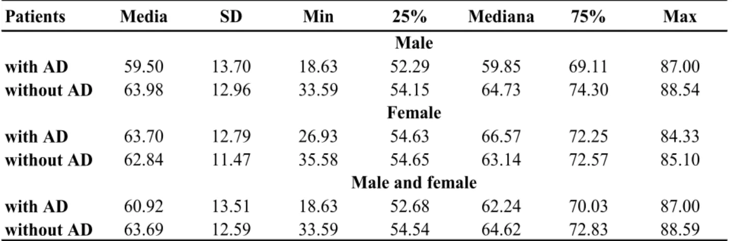
Four hundred and twelve patients (62,8 ± 13,4 years) have been enrolled including 151 (36,7%) with AD (60,9 ± 13,4 years) and 261 (63,3%) with an initial suspicion of AD but a different final diagnoses (63,8 ± 12,7 years) [Tables 7-9].
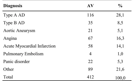
The other main diagnoses were: aortic aneurysm 21 (5,1%), angina 67 (16,3%), acute MI 58 (14,1%), pulmonary embolism 4 (1,0%), panic disorder 22 (5,3%) and other 89 (21,6%) [Table 10]. 71,1% of enrolled patients were males (61,7 ± 12,9 years), of whom 100 (66,2%) with AD (59,5 ± 13,6 years) and 193 (73,9%) without AD (62,8± 12,4 years); the females were 28,9% with higher mean age (65,4 ± 12,9 years), of whom 51 (33,8%) with AD (63,7 ± 12,7 years) and 68 (26,1%) without AD (66,7± 12,9years) [Tables 8-9]. Therefore, the number of males with AD was twice as many females, but the females had mainly AD.
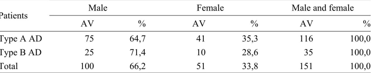
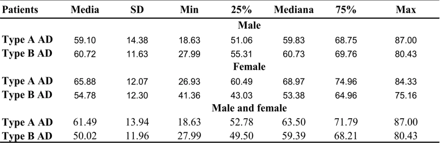
AD type A was identified in 76.8% of patients (61,5 ± 13,8 years) that they were mostly males (64,7%) but, also in this case, the prevalence of disease was bigger in females (35,3%). 23,2% of patients presented AD type B (59,0 ± 11,6 years): the majority, 71,4%, were males (60,7 ± 11,4 years), while the females (28,6%) had a mean age more lowland (54,8 ± 11,7 years of age) [Tables 11-12-13].
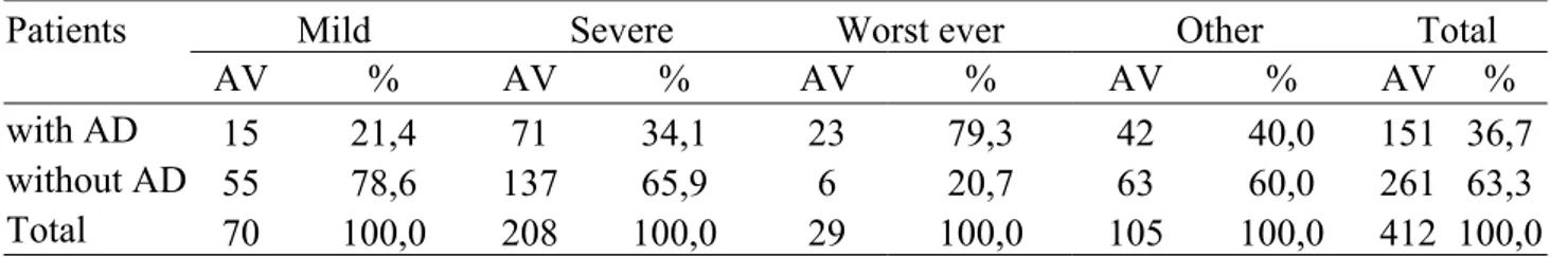
The chest pain was the most common presenting symptom; of 412 patients enrolled 319 (77,4%) complained of chest pain, including 111 (34,8%) with AD and 208 (65,2%) with other final diagnoses. In particular, of the 151 patients with AD 111 (73,5%) had pain, whilst 40 (26,5%) didn’t; of the 261 non AD cases 208 (79,7%) presented pain, whilst 53 (20,3%) didn’t [Tables 14-15].
Of note, pain was described as severe in 50,5% of patients of whom 71 (34,1%) with AD and 137 (65,9%) with other pathologies [Tables 16-17].
Furthermore, 173 (54,2%) patients referred that the chest pain had abrupt onset: 77 (44,5 %) patients had AD, whilst 96 (55,5%) did not have AD; in particular, the pain was characterized by a abrupt onset in 77 (69,4%) of the 111 patients with AD [Tables 18-19].
6.2 Results of preliminary study with calponin
Of all enrolled patients into the international study, by today, the plasma specimens of subgroup of 217 patients including 59 cases of acute AD and 158 cases with an initial suspicion of AD but a different final diagnosis have been analysed [Table 20].
Of the 59 AD cases (59 ± 14.5 years of age), 34 were males (58%). The non-AD cases (63 ± 14.8 years of age) included 116 males (73%). The other final diagnosis included MI (n=37), angina pectoris (n=34), pulmonary embolism (n=3), non-dissecting thoracic aortic aneurysm (n=17) or uncertain but not AD (n=67).
Acidic calponin showed a greater than two-fold increase for all dissections presenting within the first 6 hrs of symptom onset (4.10 ng/ml, n=16; normal reference, 2.04 ng/ml) which was particularly notable for type A (4.70 g/ml, n=14) as compared to type B patients (2.84 ng/ml, n=2). Type A patients in the 6-12 hr range also showed elevations (5.08 ng/ml, n=16) but not type B patients (2.43 ng/ml, n=4). Levels began to drop-off in the 12-24 hr range for type A (3.23 ng/ml, n=13) and were not significantly elevated in type B patients (2.64 ng/ml, n=9). Patients without AD did not show elevations at any of the examined time points (0-6 hr, 2.29 ng/ml, n=52; 6-12 hr, 2.65 ng/ml, n=34; 12-24 hr, 2.62 ng/ml, n=72) [Figure 11].
Basic calponin showed a more than three-fold increase at 377.56 ng/ml (normal reference, 123.31 ng/ml) for all dissections when sampled within the first 6 hrs of symptom onset (n=16) which was similar for type A (379.04 ng/ml, n=14) and type B patients (316.24 ng/ml, n=2). The 6-12 hr time-window showed similar, greater than three-fold, elevations in type A patients (348.79 ng/ml, n=16) but with a drop-off for type B patients (171.96 ng/ml). Levels in both type A and type B patients had fallen in the later 12-24 hr group (all patients, 169.24 ng/ml, n=22; type A patients, 172.05 ng/ml, n=13; type B patients, 171.96 ng/ml, n=9). Patients without AD did not show elevations at any of the examined time points (0-6 hr, 166.70 ng/ml, n=52; 6-12 hr, 179.41 ng/ml, n=34; 12-24 hr, 159.98 ng/ml, n=72) [Figure 12].
Neutral calponin did not show elevations in any AD patient regardless of type or time from onset of symptoms (0-6 hr, 5.11 ng/ml, n=16; 6-12 hr, 18.17 ng/ml, n=21; 12-24 hr, 13.19 ng/ml, n=22; normal reference, 14.08 ng/ml). As expected, neutral calponin did not show elevations in non-AD controls (0-6 hr, 15.03 ng/ml, n=52; 6-12 hr, 8.19 ng/ml, n=34; 12-24 hr, 12.30 ng/ml, n=72).
Thus, acidic and basic calponins showed greater than two-fold and three-fold elevations respectively during the initial 6 hrs with type A and remained elevated through to 12 hrs. For type B dissection, acidic and basic calponin levels were elevated in the very early presenters (0-6 hrs)
but not afterward. Neutral calponin did not show disease-associated changes and was not further pursued.
Further analysis according to final diagnosis was done for acidic and basic calponin [Figure 13]. Sensitivity and specificity of detection of acute AD were also analysed by receiver operating characteristics (ROC) curves.
The optimal clinical decision limit value was determined from these ROC curve analyses which showed that the optimal value for acidic calponin was 2.8 ng/ml which resulted in a sensitivity of 50% and specificity of 87% for the initial 6 hrs, and 2.3 ng/ml which resulted in a sensitivity of 58% and specificity of 72% for the initial 24 hr period.
Similarly, the optimal value for basic calponin was 159 ng/ml which resulted in a sensitivity of 63% and specificity of 73% for the initial 6 hrs, and 139 ng/ml which resulted in a sensitivity of 50% and specificity of 66% for the initial 24 hr period. According to type, type A showed an optimal value for acidic calponin at 2.8 ng/ml which resulted in a sensitivity of 50% and specificity of 87% for the initial 6 hrs, and 2.3 ng/ml which resulted in a sensitivity of 58% and specificity of 72% for the initial 24 hr period. Similarly, the optimal value for basic calponin was 159 ng/ml which resulted in a sensitivity of 64% and specificity of 73% for the initial 6 hrs, and 141 ng/ml which resulted in a sensitivity of 50% and specificity of 67% for the initial 24 hr period.
The predictive values (negative and positive) as calculated with a prevalence of 1 in 10,000 were 0.84 and 0.56 in the initial 6 hrs and 0.84 and 0.41 in the initial 24 hrs, respectively, for acidic calponin, and 0.86 and 0.44 in the initial 6 hrs and 0.80 and 0.33 in the initial 24 hrs, respectively, for basic calponin. [Table 21] Importantly, both acidic and basic calponin had higher negative predictive values.
7. DISCUSSION
Although AD is a rare pathology, the creation of a big national network among the divisions of cardiac surgery, cardiology, vascular surgery and emergency room has been very useful as, not only has it enabled to enroll a significant number of patients with suspected AD, but it could also represent a valid tool to guarantee the uniform treatment of all patients suffering of this pathology. From the descriptive analysis of data it is inferred that the majority of enrolled patients presented chest pain as the main symptom, and half of patients reported a severe pain with a abrupt onset. The chest pain characterizes the onset of various and significant CVD, thus it is evident how important it is to have/do a timely differential diagnosis for those pathologies with a similar symptomatology, in many cases necessary for the patient’s own survival; in particular, the majority of patients with AD showed chest pain thus the availability of a reliable biochemical test would have been useful, in association with imaging test, for an early diagnosis which is a prerequisite for improved treatment and survival.
There currently is no readily available, reliable, bedside biomarker assay for AD. Thus, this experience is promising for identification of a future serum biomarker or panel of markers that may aid in the more rapid diagnosis of AD.
The immunoassay developed against basic calponin showed the greatest elevations. Acidic calponin also showed diagnostic elevations, but disease-associated changes in neutral calponin levels was non-diagnostic. Analysis by type and time after onset showed that acidic calponin reliably detects AD within the first 12 hrs with superior performance in type A patients. Basic calponin showed superior performance for the first 6 hrs.
Importantly, calponin measurements allowed for detection of the disease in patients with a more delayed presentation (out to 12 hrs) which should be a welcome addition for diagnostic use in comparison with SMMHC which previously has been shown to possess superior accuracy for patients presenting within six hours after onset.
Combined use of calponin and SMMHC assays might allow for improved detection of acute AD by biochemical means as well as to potentially determine the time of onset.
This would be analogous to use of multiple diagnostic biomarkers for diagnosis of acute MI with the initial peak in myoglobin being followed by later elevations in CK MB-isozyme and troponins. One of the strengths of the present study was that diagnostic performance of the assays was determined in patients who were enrolled on the basis of a clinical suspicion of AD and not by comparison with healthy controls. Thus, the test's accuracy reflects 'real-world' conditions.
limited in number and therefore did not allow for unequivocal results in certain cohorts (e.g. high AUC values in type B dissection for basic calponin based on two diseased cases).
8. CONCLUSIONS
The increasing ageing of the western population and the consequent increase of the aortic diseases make it more necessity to develop sensitive and specific instruments for a differential diagnosis. The use of an efficient and rapid biochemical method, which can address toward specific diagnostic examination and relevant therapeutic choices in the shortest possible time, represents for the patient the fundamental “life-saving” selection.
The descriptive analysis of data shows that chest pain was the most common symptom in cases of AD but, given the relative frequency of chest pain in patients presenting to emergency department and the relative infrequency of AD, the availability of a reliable biochemical method could be of great assistance in carrying out the differential diagnosis between the diseases that are accompanied by chest pain.
Patients who present with abrupt onset of severe chest pain are straightforward candidates for imaging; a biochemical test to screen for dissection offers great potential to improve case finding and at the same time potentially reduce the expense of unnecessary imaging for patients who don't have AD.
Acid and basic calponins have the potential for use as an early diagnostic biomarker for AD but the results of this preliminary experience using an initial assay show moderate sensitivities and specificities with negative predictive values which should be further improved upon.
Future studies will be aimed at further improving the assay and determining the optimal combination of this and other biomarkers for diagnosis of AD
REFERENCES
1. Christoph A. Nienaber and Kim A. Eagle. Aortic Dissection: New Frontiers in Diagnosis and Management: Part I: From Etiology to Diagnostic Strategies. Circulation 2003;108;628-635.
2. Pearson TA, Blair SN, Daniels SR, Eckel RH, Fair JM, Fortmann SP, Franklin BA, Goldstein LB, Greenland P, Grundy SM, Hong Y, Houston Miller N, Lauer RM, Ockene IS, Sacco RL, Sallis JF Jr, Smith SC Jr, Stone NJ, Taubert KA. AHA guidelines for primary prevention of cardiovascular disease and stroke: 2002 update: consensus panel guide to comprehensive risk reduction for adult patients without coronary or other atherosclerotic vascular diseases. Circulation. 2002;106:388 –391.
3. LaDue JS, Wroblewski F, Karmen A. Serum glutamic oxaloacetic transaminase activity in human acute transmural myocardial infarction. Science. 1954;120:497-499.
4. Sorensen NS. Creatine phosphokinase in the diagnosis of myocardial infarction. Acta Med
Scand. 1963;174:725-734.
5. Kibe O, Nilsson NJ. Observations on the diagnostic and prognostic value of some enzyme tests in myocardial infarction. Acta Med Scand. 1967;182:597-610.
6. Katoh H, Sugi M, Chino S, Ishige M, Kuroda M, Fujimoto M, Nagai R, Yazaki Y. Development of an immunoradiometric assay kit for ventricular myosin light chain I with monoclonal antibodies. Clin Chem. 1992;38:170-171.
7. Cummins B, Auckland ML, Cummins P. Cardiac-specific troponin-I radioimmunoassay in the diagnosis of acute myocardial infarction. Am HeartJ.1987;113:133-144.
8. Ramachandran S. Vasan.Biomarkers of Cardiovascular Disease Molecular Basis and Practical Considerations Circulation 2006;113;2335-2362.
9. Tardif JC, Heinonen T, Orloff D, Libby P. Vascular Biomarkers and Surrogates in Cardiovascular Disease. Circulation 2006;113;2936-2942.
10. DAVID A. MORROW. CARDIOVASCULAR BIOMARKERS Pathophysiology and Disease Management. 2006 Humana Press Inc.
11. Biomarkers Definitions Working Group. Biomarkers and surrogate endpoints: preferred definitions and conceptual framework. Clin Pharmacol Ther. 2001;69:89 –95.
12. Fox N, Growdon JH. Biomarkers and surrogates. Neuro Rx. 2004;1:181.
14. Sackett DL, Haynes RB, Guyatt GH, Tugwell P. The Interpretation of Diagnostic Data: Clinical Epidemiology, a Basic Science for Clinical Medicine. Boston, Mass: Little, Brown; 1991:69 –152.
15. Pepe MS, Etzioni R, Feng Z, Potter JD, Thompson ML, Thornquist M, Winget M, Yasui Y. Phases of biomarker development for early detection of cancer. J Natl Cancer Inst. 2001;93:1054 –1061.
16. Barker PE. Cancer biomarker validation: standards and process: roles for the National Institute of Standards and Technology (NIST). Ann N Y Acad Sci. 2003;983:142–150. 17. Schulte PA, Perera FP. Validation. In: Schulte PA, Perera FP, eds. Molecular Epidemiology:
Principles and Practice. San Diego, Calif: Academic Press; 1993:79 –107.
18. Nomenclature and Criteria for Diagnosis of Ischemic Heart Disease. Report of the Joint International Society and Federation of Cardiology/World Health Organization Task Force on Standardization of Clinical Nomenclature. Circulaton 1979;59:607–609.
19. Alpert JS, Thygesen K, Antman E, Bassand JP. Myocardial infarction redefined—a consensus document of The Joint European Society of Cardiology/American College of Cardiology Committee for the redefinition of myocardial infarction. J Am Coll Cardiol 2000;36:959–969.
20. Balk EM, Ioannidis JP, Salem D, Chew PW, Lau J. Accuracy of biomarkers to diagnose acute cardiac ischemia in the emergency department: a meta-analysis. Ann Emerg Med 2001;37(5):478–494.
21. Miller DC. Acute dissection of the aorta: continuing need for earlier diagnosis and treatment. Mod Concepts Cardiovasc Dis. 1985;54:51-55.
22. Meszaros I, Morocz J, Szlavi J, Schmidt J, Tornoci L, Nagy L, Szep L. Epidemiology and clinicopathology of aortic dissection. Chest. 2000; 117: 1271–1278.
23. Bickerstaff LK, Pairolero PC, Hollier LH, Melton LJ, Van Peenen HJ, Cherry KJ, Joyce JW, Lie JT. Thoracic aortic aneurysms: a population-based study. Surgery. 1982; 92: 1103– 1108.
24. Clouse WD, Hallett JW Jr, Schaff HV, Spittell PC, Rowland CM, Ilstrup DM, Melton LJ 3rd. Acute aortic dissection: population-based incidence compared with degenerative aortic aneurysm rupture. Mayo Clin Proc. 2004; 79: 176–180.
25. Vilacosta I, San Roman JA. Acute aortic syndrome. Heart. 2001;85:365-8.
26. Nienaber Ch, Von Kodolitsch Y, Petersen B, Loose R, Helmchen U, Haverich A, et al. Intramural hemorrhage of the thoracic aorta. Circulation. 1995;92:1465-72.
27. Maraj R, Rerkpattanapipat P, Jacobs L, Makornwattana P, Kother M. Metaanalysis of 143 reported cases of aortic intramural hematoma. Am J Cardiol. 2000;86:664-8.
28. Hagan PG, Nienaber CA, Isselbacher EM, Bruckman D, Karavite DJ, Russman PL, et al. The International Registry of Acute Aortic Dissection (IRAD): new insights into an old disease. JAMA. 2000;283:897-903.
29. Meszaros I, Morocz J, Szlavi J, Schmidt J, Tornoci L, Nagy L, Szep L. Epidemiology and clinicopathology of aortic dissection. Chest. 2000; 117: 1271–1278.
30. Roberts CS, Roberts WC. Aortic dissection with the entrance tear in the descending thoracic aorta: analysis of 40 necropsy patients. Ann Surg. 1991; 213: 356–368.
31. Masuda Y, Takanashi K, Takasu J, Watanabe S. Natural history and prognosis of medical treatment for the patients with aortic dissections [in Japanese]. Nippon Geka Gakkai Zasshi. 1996; 97: 890–893.
32. Bogaert J, Meyns B, Rademakers FE, Bosmans H, Verschakelen J, Flameng W, Marchal G, Baert AL. Follow-up of aortic dissection: contribution of MR angiography for evaluation of the abdominal aorta and its branches. Eur Radiol. 1997; 7: 695–702.
33. Larson EW, Edwards WD. Risk factors for aortic dissection: a necropsy study of 161 cases. Am J Cardiol. 1984; 53: 849–855.
34. Reed D, Reed C, Stemmermann G, Hayashi T. Are aortic aneurysms caused by atherosclerosis? Circulation. 1992; 85: 205–211.
35. Stefanadis CI, Karayannacos PE, Boudoulas HK, Stratos CG, Vlachopoulos CV, Dontas IA, Toutouzas PK. Medial necrosis and acute alterations in aortic distensibility following removal of the vasa vasorum of canine ascending aorta. Cardiovasc Res. 1993; 27: 951–956. 36. Von Kodolitsch Y, Aydin MA, Koschyk DH, Loose R, Schalwat I, Karck M, Cremer J, Haverich A, Berger J, Meinertz T, Nienaber CA. Predictors of aneurysmal formation after surgical correction of aortic coarctation. J Am Coll Cardiol. 2002; 39: 617–624.
37. Thomas T. Tsai, MD; Christoph A. Nienaber, MD; Kim A. Eagle, MD Acute Aortic Syndromes. Circulation 2005;112;3802-3813.
38. Daily PO, Trueblood HW, Stinson EB, Wuerflein RD, Shumway NE. Management of acute aortic dissections. Ann Thorac Surg. 1970; 10: 237–247.
39. DeBakey ME, Beall AC Jr, Cooley DA, Crawford ES, Morris GC Jr, Garrett HE, Howell JF. Dissecting aneurysms of the aorta. Surg Clin North Am. 1966; 46: 1045–1055.
40. DeSanctis RW, Doroghazi RM, Austen WG, Buckley MJ. Aortic dissection. N Engl J Med. 1987;317:1060 –1067.
41. Erbel R, Alfonso F, Boileau C, Dirsch O, Eber B, Haverich A, Rakowski H, Struyven J, Radegran K, Sechtem U, Taylor J, Zollikofer C, Klein WW, Mulder B, Providencia LA. Diagnosis and management of aortic dissection. Eur Heart J. 2001;22:1642–1681.
42. Mehta RH, O’Gara PT, Bossone E, et al. Acute type A aortic dissection in the elderly: clinical characteristics, management, and outcomes in the current era.J Am Coll Cardiol. 2002;40:685–692.
43. Januzzi JL, Isselbacher EM, Fattori R, et al. Characterizing the young patient with aortic dissection: results from the International Registry of Aortic Dissection (IRAD). J Am Coll Cardiol. In press.
44. Suzuki T, Mehta RH, Ince H, et al. Clinical profiles and outcomes of acute type B aortic dissection in the current era: lessons learned from the International Registry of Aortic Dissection (IRAD). Circulation. In press.
45. Mehta RH, Suzuki T, Hagan PG, et al. Predicting death in patients with acute type A aortic dissection. Circulation. 2002;105:200–206.
46. Nallamothu BK, Mehta RH, Saint S, et al. Syncope in acute aortic dissection: diagnostic, prognostic and clinical implications. Am J Med. 2002;113:468–471.
47. Hirst A, Johns VJ, Krime SJ. Dissecting aneurysm of the aorta: a review of 505 cases.
Medicine. 1958; 37:217-279.
48. Eagle KA, Quertermous T, Kritzer GA, et al. Spectrum of conditions initially suggesting acute aortic dissection but with negative aortograms. Am J Cardiol. 1986;57:322-326.
49. Shiga T, Wajima Z, Apfel CC, Inoue T, Ohe Y. Diagnostic accuracy of transesohageal echocardiography, helical computed tomography, and magnetic resonance imaging for suspected thoracic aortic dissection. Arch Intern Med. 2006;166:1350-6.
50. Evangelista A. Progress in the Acute Aortic Syndrome.
51. Von Kodolitsch Y, Krause N, Spielmann R, Nienaber CA. Diagnostic potential of combined transthoracic echocardiography and x-ray computed tomography in suspected aortic dissection. Clin Cardiol. 1999;22:345-52.
52. Nienaber CA, Spielmann RP, Von Kodolitsch Y, et al. Diagnosis of thoracic aortic dissection: magnetic resonance imaging versus transesophageal echocardiography.
Circulation. 1992;85:434-447.
53. Nienaber CA, von Kodolitsch Y, Nicolas V, et al. The diagnosis of thoracic aortic dissection by non-invasive imaging procedures. N Engl J Med. 1993;328:1-9.
54. Cigarroa JE, Isselbacher FM, De Sanetis RW, et al. Diagnostic imaging in the evaluation of suspected aortic dissection: old standards and new directions. N Engl J Med. 1993;328:35– 43.
55. Spittell PC, Spittell JA Jr, Joyce JW, Tajik AJ, Edwards WD, Schaff HV, Stanson AW. Clinical features and differential diagnosis of aortic dissection: experience with 236 cases (1980 through 1990). Mayo Clin Proc. 1993;68:642-651.
56. Katoh H, Suzuki T, Hiroi Y, Ohtaki E, Suzuki S, Yazaki Y, Nagai R. Diagnosis of aortic dissection by immunoassay for circulating smooth muscle myosin. Lancet 1995;345:191-2. 57. Katoh H, Suzuki T, Yokomori K, Suzuki S, Ootaki E, Watanabe M, Yazaki Y, Nagai R. A
novel immunoassay of smooth muscle myosin heavy chain in serum. J Immunol Methods 1995;185:57-63.
58. Suzuki T, Katoh H, Watanabe M, Kurabayashi M, Hiramori K, Hori S, Nobuyoshi M, Tanaka H, Kodama K, Sato H, Suzuki S, Yazaki Y, Nagai R. A novel biochemical method for aortic dissection -- the results of a prospective study using an immunoassay of smooth muscle myosin heavy chain. Circulation 1996;93:1244-9.
59. Suzuki T, Katoh H, Tsuchio Y, Hasegawa A, Kurabayashi M, Ohira A, Hiramori K, Sakomura Y, Kasanuki H, Hori S, Aikawa N, Abe S, Tei C, Nakagawa Y, Nobuyoshi M, Misu T, Sumiyoshi T, Nagai R. Diagnostic implications of raised smooth muscle myosin heavy chain levels in acute aortic dissection: the smooth muscle myosin heavy chain (SMH) study. Annals Int Med 2000;133:537-541.
60. Suzuki T, Katoh H, Kurabayashi M, Yazaki Y, Nagai R. Biochemical diagnosis of aortic dissection by raised concentrations of creatine kinase-BB isozyme. Lancet 1997;350:784-5. 61. Suzuki T, Katoh H, Nagai R. Biochemical diagnosis of aortic dissection ~from bench to
bedside~. Jpn Heart J 1999;40:527-34.
62. Costantini, V, Zacharski, LR Fibrin and cancer. Thromb Haemost 1993;69,406-414.
63. Bick, RL Disseminated intravascular coagulation: objective laboratory diagnostic criteria and guidelines for management. Clin Lab Med 1994;14,729-768.
64. Kraus, M Fibrin(ogen)-spaltprodukte, D-Dimere. Thomas, L eds. Labor und diagnose 5th ed. 1998,648-651 Books Verlagsgesellschaft mbH. Frankfurt/Main, Germany.
65. Thomas Weber, MD; Sonja Högler, MD; Johann Auer, MD; Robert Berent, MD; Elisabeth Lassnig, MD; Erich Kvas, ScD and Bernd Eber, MD D-dimer in Acute Aortic Dissection
Chest. 2003;123:1375-1378.
66. Eggebrecht H, Naber CK, Bruch C, Kroger K, Von Birgelen C, Schmermund A, et al. Value of plasma fibrin D-dimers for detection of acute aortic dissection. J Am Coll
Cardiol. 2004;44:804-9.
67. Tadashi Shinohara; Kimihiro Suzuki; Makoto Okada; Masaru Shiigai; Masashi Shimizu; Tadaaki Maehara; Fumitaka Ohsuzu. Soluble Elastin Fragments in Serum Are Elevated in Acute Aortic Dissection. Arteriosclerosis, Thrombosis, and Vascular Biology. 2003;23:1839.
68. Nakashima Y, Shiokawa Y, Sueishi K. Alterations of elastic architecture in human aortic dissecting aneurysm. Lab Invest. 1990; 62: 751–760.
69. Akashima Y, Sueishi K. Alteration of elastic architecture in the lathyritic rat aorta implies the pathogenesis of aortic dissecting aneurysm. Am J Pathol. 1992; 140: 959–969.
70. Schlatmann TJ, Becker AE. Pathogenesis of dissecting aneurysm of aorta: comparative histopathologic study of significance of medial changes. Am J Cardiol. 1977; 39: 21–26. 71. Schlatmann TJ, Becker AE. Histologic changes in the normal aging aorta: implications for
dissecting aortic aneurysm. Am J Cardiol. 1977; 39: 13–20.
72. Murray CA, Edwards JE. Spontaneous laceration of ascending aorta. Circulation. 1973; 47: 848–858.
73. Ishii T, Asuwa N. Collagen and elastin degradation by matrix metalloproteinases and tissue inhibitors of matrix metalloproteinase in aortic dissection. Hum Pathol 2000;31:640-646. 74. Sangiorgi G, Trimarchi S, Carbone GL, et al. Increased plasma levels of matrix
metalloproteinases-2 and -9 in acute and subacute aortic dissection. Circulation 2002;106 (Suppl 19):218 (Abstract).
75. Takahashi K, Hiwada, K. & Kokubu. Biochemical and Biophysical Research Communications 1986; 141: 20-26.
76. EL-Mezgueldi M. and Marston S. B. The effect of smooth muscle calponin on the strong and weak binding sites of F-actin. J. Biol. Chem. 1996; 271,28161 –28167.
77. Taniguchi S., Takeoka M., Ehara T., Hashimoto S., Shibuki H., Yoshimura N., Shigematsu H., Takahashi K. and Katsuki M. Structural fragility of blood vessels and peritoneum in calponin h1-deficient mice, resulting in an increase in hematogenous metastasis and peritoneal dissemination of malignant tumor cells. Cancer Res. 2001; 61:7627 -7634.
78. Danninger C. and Gimona M. Live dynamics of GFP-calponin: isoform-specific modulation of the actin cytoskeleton and autoregulation by C-terminal sequences. J. Cell Sci. 2000; 113: 3725 –3736.
79. Fukui Y., Masuda H., Takagi M., Takahashi K. and Kiyokane, K. The presence of h2-calponin in human keratinocyte. J. Dermatol. Sci. 1997; 14: 29 –36.
80. Ferhat L., Rami G., Medina I., Ben-Ari Y. and Represa A. Process formation results from the imbalance between motor-mediated forces. J. Cell Sci. 2001; 114: 3899 –3904.
81. Ferhat L., Charton G., Represa A., Ben-Ari Y., der Terossian E. and Khrestchatski M. Acidic calponin cloned from neural cells is differentially expressed during rat brain development. Eur. J. Neurosci. 1996; 8:1501 -1509.
82. Gimona M., Djinovic-Carugo K., Kranewitter W. J. and Winder S. J. Functional plasticity of CH domains. FEBS Lett. 2002; 513: 98 –106.
83. Burgstaller G., Kranewitter W. J. and Gimona M. The molecular basis for the autoregulation of calponin by isoform-specific C-terminal tail sequences Journal of Cell Science 2002; 115: 2021-2029.
84. Suzuki T, Distante A, Zizza A, Trimarchi S, Villani M, Salerno JA Tupputi De Luca L, Renzulli A, Sabino F, Nowak R, Birkhahn R, Hollander JE, Counselman F, Bossone E, Eagle K. on behalf of the International Registry of Acute Aortic Dissection Substudy on Biomarkers (IRAD-Bio) Investigators. Preliminary experience with the smooth muscle troponin-like protein, calponin, as a novel biomarker for diagnosing acute aortic dissection. European Heart Journal, april 2008.
TABLES
Table 1. Measures of biomarker test performance. Adapted from reference 3
Sensitivity
is defined as the ability of a test to detect disease (condition of interest) when it is truly present, i.e., it is the probability of a positive test result given that the patient has the disease.
Specificity
is the ability of a test to exclude the disease (condition of interest) in patients who do not have disease, i.e., it is the probability of a negative test result given that the patient does not have the disease.
Predictive value
tells us how good the test is at predicting the true positives or true negatives, i.e., the probability that the test will give the correct diagnosis.
Positive Predictive Value
is the probability that a patient has the disease given that the test results are positive.
Negative Predictive Value
is the probability that a patient does not have the disease or condition given that the test results are indeed negative.
ROC curve
is a plot of the sensitivity versus specificity of a diagnostic test, in which the different points on the curve correspond to different cut points used to determine whether the test results are positive.
Table 2. Risk Conditions for Aortic Dissection
Long-standing arterial hypertension ● Smoking, dyslipidemia, cocaine/crack Connective tissue disorders
● Hereditary vascular disease ● Marfan syndrome
● Vascular Ehlers-Danlos syndrome (type 4) ● Bicuspid aortic valve
● Coarctation of the aorta
● Hereditary thoracic aortic aneurysm/dissection Vascular inflammation
● Giant cell arteritis ● Takayasu arteritis ● Behcet’s disease ● Syphilis ● Ormond’s disease Deceleration trauma ● Car accident ● Fall from height Iatrogenic factors ● Catheter/instrument intervention ● Valvular/aortic surgery ● Side or cross-clamping/aortotomy ● Graft anastomosis ● Patch aortoplasty ● Aortic wall fragility
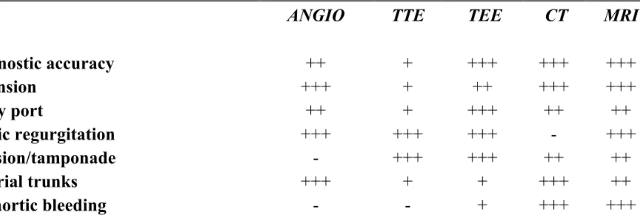
Table 3. Usefulness of imaging techniques in the diagnosis of acute aortic syndrome*(50)
ANGIO TTE TEE CT MRI
Diagnostic accuracy ++ + +++ +++ +++ Extension +++ + ++ +++ +++ Entry port ++ + +++ ++ ++ Aortic regurgitation +++ +++ +++ - +++ Effusion/tamponade - +++ +++ ++ ++ Arterial trunks +++ + + +++ ++ Periaortic bleeding - - + +++ +++
*Angio indicates catheter angiography; TEE, transesophageal echocardiography; TTE, transthoracic echocardiography; MRI, magnetic resonance imaging; CT, computerized tomography. +++ Excellent; ++ Good; + Sufficient; - Insufficient
Table 4. Advantages of imaging during the acute phase study (50)
ANGIO TTE TEE CT MRI
Speed + +++ +++ ++ +
Portability - +++ +++ - -
Monitoring +++ +++ +++ + -
Availability + +++ ++ +++ +
Tolerance + +++ ++ +++ +
*Angio indicates catheter angiography; TEE, transesophageal echocardiography; TTE, transthoracic echocardiography; MRI, magnetic resonance imaging; CT, computerized tomography. +++ Excellent; ++ Good; + Sufficient; - Insufficient
Table 5. Biomarkers for diagnosis of AD. Endothelial Marker
(von Willebrand Factor, Thrombomodulin, etc.)
Markers of the Smooth Muscle
(Smooth Muscle Myosin Heavy Chain, Creatine kinase, etc.)
Markers of the Adventitia/Extracellular Matrix
(Collagen, Elastin, Matrix Metalloproteinases, etc.)
Coagulation Markers
(D-dimer, etc.)
Inflammation Marker
(C- Reactive Protein, etc.)
Table 6. Normal reference range for assay (76).
n Average Median SD percentile 95th percentile 99th Acidic calponin (ng/ml) 218 1.19 1.01 0.92 2.04 5.73
Basic calponin (ng/ml) 282 44.09 31.74 41.81 124.31 162.15