Copyright Ó 2016 Cognizant, LLC. E-ISSN 1555-3892 www.cognizantcommunication.com
Received March 17, 2015; final acceptance December 18, 2015. Online prepub date: September 21, 2015.
Address correspondence to Michela Bosetti, Ph.D., Dipartimento di Scienze del Farmaco, Università del Piemonte Orientale, Laboratorio Anatomia Umana, Via Solaroli 17, 28100 Novara, Italia. Tel: +39-0321-660557; Fax: +39-0321-660633; E-mail: [email protected]
1043
Human Lipoaspirate as Autologous Injectable Active Scaffold for
One-Step Repair of Cartilage Defects
Michela Bosetti,* Alessia Borrone,† Antonia Follenzi,† Fanuel Messaggio,‡ Carlo Tremolada,§ and Mario Cannas†
*Dipartimento di Scienze del Farmaco, Università del Piemonte Orientale, Novara, Italy †Dipartimento di Scienze della Salute, Università del Piemonte Orientale, Novara, Italy
‡Diabetes Research Institute, University of Miami, Miami, FL, USA §IMAGE Institute, Milan, Italy
Research on mesenchymal stem cells from adipose tissue shows promising results for cell-based therapy in cartilage lesions. In these studies, cells have been isolated, expanded, and differentiated in vitro before trans-plantation into the damaged cartilage or onto materials used as scaffolds to deliver cells to the impaired area. The present study employed in vitro assays to investigate the potential of intra-articular injection of micro-fragmented lipoaspirate as a one-step repair strategy; it aimed to determine whether adipose tissue can act as a scaffold for cells naturally present at their anatomical site. Cultured clusters of lipoaspirate showed a spontaneous outgrowth of cells with a mesenchymal phenotype and with multilineage differentiation poten-tial. Transduction of lipoaspirate clusters by lentiviral vectors expressing GFP evidenced the propensity of the outgrown cells to repopulate fragments of damaged cartilage. On the basis of the results, which showed an induction of proliferation and ECM production of human primary chondrocytes, it was hypothesized that lipoaspirate may play a paracrine role. Moreover, the structure of a floating culture of lipoaspirate, treated for 3 weeks with chondrogenic growth factors, changed: tissue with a high fat component was replaced by a tissue with a lower fat component and connective tissue rich in GAG and in collagen type I, increasing the mechani-cal strength of the tissue. From these promising in vitro results, it may be speculated that an injectable autolo-gous biologically active scaffold (lipoaspirate), employed intra-articularly, may 1) become a fibrous tissue that provides mechanical support for the load on the damaged cartilage; 2) induce host chondrocytes to proliferate and produce ECM; and 3) provide cells at the site of injury, which could regenerate or repair the damaged or missing cartilage.
Key words: Lipoaspirate; Cartilage defects; Injectable autologous active scaffold; In vitro
INTRODUCTION
The treatment of degenerative joints is becoming a very frequent necessity. Increased life expectancy and inappropriate medical treatments (e.g., removal of the meniscus) are leading to an increase in chondropathies and osteoarthritis, which also occur at a relatively young age. Chondrocytes are the parenchymal cells of carti-lage and have limited regenerative capacity; thus, after traumatic injury, the cartilage undergoes degenerative changes, which may be irreversible (18). The more super-ficial lesions tend to progress toward degeneration (29); those that penetrate the subchondral bone progress toward the formation of fibrocartilage tissue. This differs from normal hyaline cartilage in both biomechanical and bio-chemical properties and frequently undergoes subsequent
degeneration (30). Any cartilage lesion should therefore be considered as the beginning of a chronic degenerative disease with little chance of cure due to the poor regen-erative capacity of this type of cell (7).
Modern treatments for damaged cartilage and for the prevention of osteoarthritis include the intra-articular administration of hyaluronan (15), treatment with platelet-rich plasma (PRP) (3), bone marrow stimulation techniques (subchondral drilling, abrasion, microfracture) (24), osteo-chondral grafting (mosaicplasty) (23), autologous chondro-cyte implantation (ACI), and matrix-assisted autologous chondrocyte implantation (MACI) with autologous chon-drocytes cultured on collagen membranes prior to reimplan-tation (11). Chondrocyte-based therapy has demonstrated promising clinical results, but the procedure requires an
invasive protocol, in which autologous cartilage is har-vested from the patient at a healthy anatomical site. This produces damage to the normal cartilage at that site lead-ing to pathological changes. It is therefore evident that to avoid the disadvantages arising from the use of autologous chondrocytes, alternative cell sources must be investigated. Mesenchymal stem cells (MSCs) obtained from adult tis-sues of different origins (umbilical cord blood, adipose tissue, bone marrow, synovial membranes, periosteum, and muscle) have recently been investigated and their chondrogenic potential assessed and compared (14,28,34). Bone marrow and adipose tissue are the most readily avail-able sources of MSCs. Moreover, adipose tissue is readily available in large quantities and can be obtained through less invasive procedures; of the many cell types contained in adipose tissue, MSCs (ASCs) comprise up to 2%, whereas only 0.02% of cells in bone marrow are MSCs (BM-MSCs) (20). MSCs have shown promising results for cartilage repair (31); however, in the studies published to date, cells were isolated from the originating tissue, then expanded and differentiated in vitro, prior to transplanta-tion into the damaged cartilage or seeding in materials used as scaffolds to deliver cells to the injured area. No optimal stimulation regimen has yet been proposed to differenti-ate stem cells into chondrocytes prior to implantation; fur-ther difficulty lies in the large number of processing steps (enzymatic digestion, cell expansion, and differentiation), which increases the translational barriers.
With the aim of avoiding excessive handling of stem cells and side effects derived from the scaffolds used for cell-based cartilage repair, this in vitro study investi-gated whether the intra-articular injection of autologous lipoaspirate might be an alternative cell-based therapy in treating cartilage diseases and in preventing osteoarthri-tis. Lipoaspirate is a natural scaffold rich in stem cells, which, when injected into the intra-articular space, might give rise to different biological responses. To determine whether lipoaspirate can lead to the outgrowth of cells to the damaged cartilage, thus repopulating and repair-ing it, and whether lipoaspirate may have a paracrine effect on resident chondrocytes, we studied in vitro 1) the ability of resident cells in lipoaspirate to grow out from adipose tissue, 2) the phenotype of these out-growing cells, together with their differentiation poten-tial and their capability of repopulating an organ culture of human cartilage, and 3) the effect of the lipoaspirate on the proliferation rate of human chondrocytes and on their production of cartilaginous matrix. Furthermore, with the aim of determining whether lipoaspirate clus-ters might differentiate into a different tissue at the intra-articular site, thus providing intra-intra-articular mechanical reinforcement, the chondrogenic differentiation of cells not extracted from lipoaspirate, but simply at their own natural anatomical site, was examined.
MATERIALS AND METHODS
Materials
Lipoaspirates were obtained from five healthy female patients (age range 30–45) undergoing an elective lipo-suction from a single anatomical site (abdominal sub-cutaneous fat tissue) at the IMAGE Institute (Milan, Italy). The ethical committee of the University of Milan approved the design of the study. Exclusion criteria were body mass index (BMI) >30, diabetes, hypertension, and nicotine or alcoholic abuse. Written informed consent, specifying that residual material destined to be disposed of could be used for research, was signed by each par-ticipant before the biological materials were removed, in agreement with Rec(2006)4 of the Committee of Ministers Council of Europe on research on biological materials of human origin. After a preliminary infiltration of 400 ml of
saline solution, with adrenaline 2 mg/ml (S.A.L.F. Spa.,
Bergamo, Italy) as a vasoconstrictor and lidocaine 0.02% (AstraZeneca, Luton, UK) as an anesthetic, the aspiration
was performed using a 10-cc syringe with a Luer-Lok® tip
(BD Medical, VWR Int., Milano, Italy) connected to a dis-posable 19-cm blunt cannula (3 mm OD), with five oval holes (1 × 2 mm). From each of five patients, 210 ml of lipoaspirate was obtained and processed as follows, giving four batches of lipoaspirate: 10 ml was washed with 25 ml of phosphate-buffered saline (PBS; Sigma-Aldrich, Milan, Italy) on a 250-mm Nitex filter (Dutscher Scientific, Essex, UK) and used as control (CTR); 50 ml was centrifuged for 3 min at 1,500 × g (22) and 10 ml used as Coleman (Col); 50 ml was processed with a commercial device
(PureGraft®; Cytori Therapeutics, San Diego, CA, USA)
according to the manufacturer’s instructions (25) and 10 ml used (PurGr); 100 ml was processed with a commercial
device (Lipogems®; Lipogems Int. S.p.A., Milano, Italy)
that washes and micronizes lipoaspirate (4) following the manufacturer’s instructions, and 10 ml was used (LipoG). Chondrogenic Organ Culture Model
Each batch (500 ml) of lipoaspirate was cultured as
free-floating aggregates in a conical polypropylene cen-trifuge tube (EuroClone, Milan, Italy) in 4 ml of chon-drogenic medium containing 1 ml Dulbecco’s modified Eagle medium (DMEM-high glucose; Sigma-Aldrich), ascorbate-2-phosphate (100 mM; Sigma-Aldrich),
dexa-methasone (10−7 M; Sigma-Aldrich), and 1% insulin–
transferrin–selenite (ITS+1 media supplement; Sigma-Aldrich) for 3 weeks at 37°C in a humidified atmosphere
containing 95% air and 5% CO2. The tubes were gently
shaken daily to allow the culture media to cover the sam-ple, which otherwise tended to float. Half of the culture medium was changed twice a week with the addition of fibroblast growth factor 2 (FGF2) and transforming growth factor-b2 (TGF-b2) found in previous research
to be chondrogenic on bone marrow stem cells (6). Replicate aggregates from each batch of lipoaspirate were processed for the following determinations: histological and immunohistological evaluation, mechanical evalua-tions, biochemical studies, RNA extraction, and RT-PCR analysis. Before processing, digital images were taken of each lipoaspirate organ culture using a digital camera, on samples treated or not for 3 weeks with the chGF. Mechanical Evaluations
The compression properties of untreated and chGF-treated lipoaspirates were tested using an Electro Force BioDynamic Test Instrument (Bose; TA Instruments,
Eden Prairie, MN, USA). Samples of 2 ± 0.5 mm3 were
positioned inside the instrument’s bioreactor chamber between two pistons. Tests were performed with a constant crosshead speed of 0.05 mm/s. The instrument showed the real-time displacement of the piston and the force act-ing on the sample, producact-ing a force/displacement curve. Compressive strength and Young modulus were then cal-culated, using the surface area of the sample, initial length of the sample, compressed length, and force at breaking point. All samples were tested in triplicate, and data are presented as mean ± standard deviation (SD).
Histology and Immunohistochemistry
After 21 days of culture, neoformed tissues were rinsed in PBS and fixed overnight in 10% buffered for-malin (Diapath S.r.l., Martinengo, Italy) at 4°C. The fixed tissues were dehydrated by treatment with alcohol, embedded in paraffin, and sectioned to a thickness of
7 mm. For histological evaluation, sections were
depar-affinized using xylene, rehydrated, and stained with hematoxylin and eosin (H&E; Sigma-Aldrich) or tolui-dine blue (Sigma-Aldrich) to detect proteoglycan or with Sirius red F3BA (Chroma, Stuttgart, Germany) to detect collagen. Alternate sections were used for immunohis-tochemistry to detect vessels: rehydrated sections were
treated with 0.5% H2O2 (Sigma-Aldrich) to inactivate
endogenous peroxidases, incubated in 1 mg/ml pepsin (Sigma-Aldrich) in 0.5 M acetic acid (Sigma-Aldrich) for 2 h at 37°C, blocked with 1.5% goat serum (Vector Laboratories, Burlingame, CA, USA) in PBS for 1 h at room temperature, and incubated for 1 h at room
temper-ature with anti-Von Willebrand factor 25 mg/ml (Dako,
Glostrup, Denmark) and FITC-conjugated secondary antibody 10 mg/ml (Vector Laboratories).
Glycosaminoglycan and Collagen Quantification Weighed tissue (2 mg) was taken after 21 days of cul-ture, washed with PBS, and digested with 200 ml papain
(1 mg/ml in 50 mM NaH2PO4, pH 6.5, containing 2 mM
N-acetyl cysteine and 2 mM EDTA; all from
Sigma-Aldrich) for 16 h at 65°C. Glycosaminoglycans were
quantified by reaction with 1.9-dimethylmethylene blue (16 mg/ml DMMB; Sigma-Aldrich) (12) using chondroitin sulfate (Sigma-Aldrich) as a standard. Measurements were made with a microplate reader (BS 1000 Spectracount; Packard BioScience Company, Meriden, CT, USA) at a wavelength of 530 nm. The results are reported as mean ± SD (n = 3) mg sGAG content/mg of the tissue and as percent increase versus untreated sample (21 days of culture in basal medium).
Collagen was quantified according to Tullberg-Renert and Jundt (32). Briefly, 2 mg of tissue was extracted overnight at 4°C in 0.5 ml of acetic acid 0.2%, air dried, and stained with 1 ml of Sirius red dye (Sigma-Aldrich) reagent (1 mg/ml in saturated aqueous picric acid; Sigma-Aldrich) for 1 h. The dye solution was then removed, and the tissue was washed with 0.01 N hydrochloric acid (Sigma-Aldrich) to remove all nonbound dye; the stained material was dissolved in 0.2–0.3 ml 0.1 N of sodium hydroxide for 30 min at room temperature. The optical density (OD) was measured with a Bio-Rad microplate reader 3550 (Bio-Rad, Milan, Italy) at 550 nm against 0.1 N of sodium hydroxide as blank. The results are
reported as means ± SD (n = 3) mg collagen/ml and as
percent increase versus the untreated sample (21 days of culture in basal medium).
Real-Time PCR
Total cellular RNA was extracted using a Qiagen QIAsshredder kit (Qiagen, Dusseldorf, Germany) and quantified using a RiboGreen RNA quantification assay (Molecular Probes, Invitrogen, Milan, Italy). Then 200 ng was reverse transcribed (RT) using a High-Capacity cDNA Archive Kit (Applied Biosystems, Foster City, CA, USA). The RT thermal profile was 25°C for 10 min, 37°C for 120 min, 85°C for 5 min, and kept at 4°C. Primers and probe were provided by TaqMan gene expression assay kit (Applied Biosystems) and were added to the reaction mixture according to the manufac-turer’s directions: SOX-9 (Hs01001343_g1), COL2A1 (Hs00264051_m1), COL1A1 (Hs00164004_m1), and GADPH (Hs02758991_g1). Amplification reactions were performed with SSOFast Probes Supermix with ROX (Bio-Rad). mRNA levels were measured by real-time RT-PCR based on TaqMan methodology, using CFX96 Real-Time System C 1000 Thermal Cycler (Bio-Rad). Real-time data analysis was run with Bio-Rad CFX Manager 2.1 software. Target gene expression was nor-malized to GADPH mRNA expression. Relative differ-ential gene expression was calculated (27).
Isolation and Culture of Human Chondrocytes (hChs) Normal human articular cartilage was obtained from the femoral or radial heads of three male subjects with dis-placed fractures. The mean age of the group was 33 years
(range: 20–41). Written consent was signed by each par-ticipant before biological materials were removed accord-ing to Rec (2006)4 of the Committee of Ministers Council of Europe and used after local ethics committee approval. Immediately after surgery was performed at the traumatol-ogy division of the Major Hospital of Novara, healthy car-tilage was taken under sterile conditions, washed with PBS supplemented with 50 U/ml penicillin, 50 mg/ml streptomy-cin, and 250 ng/ml Fungizone (PSF; all reagents from Sigma-Aldrich) and cut into pieces of approximately 2 × 2 mm, before enzymatic digestion, as follows and according to Archer et al. (1). Pieces were placed for 30 min at 37°C,
5% CO2 in DMEM/F12 (EuroClone) containing trypsin
(0.25% w/v; Sigma-Aldrich) and PSF (100–120 rpm shaking); the supernatant was discarded, and the cartilage fragments were further digested in DMEM/F12 with PSF containing 0.8 mg/ml collagenase II (Sigma-Aldrich) for
4 h at 37°C, 5% CO2 (100–120 rpm shaking). The digested
tissue was then allowed to settle, and the supernatant con-taining cells was centrifuged at 1,500 × g for 5 min. The cell pellet was washed with PBS-PSF and resuspended in growth medium: DMEM/F12 supplemented with PSF,
glutamine 200 mM (EuroClone), ascorbic acid 50 mg/ml
(Sigma-Aldrich), FBS 2% (EuroClone), 1 ng/ml TGF-b (Sigma-Aldrich), 1 ng/ml FGF2 (Sigma-Aldrich), 10 mg/ ml ITS+1 (Sigma-Aldrich), seeded in monolayer in culture
plates at low density (4,000 cell/cm2) and incubated in a
CO2 incubator at 5% CO2, 37°C.
Effect of Lipoaspirate on hCh Proliferation and Matrix Production
Chondrocytes were used for the experiments within the third expansion treatment. They were plated at a
density of 103 cells/well in a 96-multiwell plate, and
hChs were cultured in basal culture medium (DMEM/ F12 supplemented with antibiotics, glutamine ascorbic acid, 2% FBS, 1 ng/ml TGF-b, 1 ng/ml FGF2, 10 mg/ ml ITS+1 as described for hChs culture and abbreviated CTR) and compared to cells cultured in lipoconditioned culture medium made up of basal culture medium supple-mented with 15% of lipoaspirate culture medium that was obtained as follows. Lipoaspirate (1 ml) corresponding to 850 ± 20 mg of tissue was cultured in 10 ml of basal
medium in a CO2 incubator at 5% CO2, 37°C for 4 days;
upon this treatment, extracellular proteins were released into the medium. Chondrocytes were cultured in basal or in lipoconditioned culture medium for 1, 3, 6, and 12 days; after each incubation time, cell proliferation and cartilaginous matrix production were assayed.
For the proliferation assay, an ATP quantification Kit (ViaLight, Cambrex Profarmaco, Milan, Italy) was used following the manufacturer’s protocol. Briefly, cells were lysed with cell lysis reagent, supplied with the kit, and treated with ATP monitoring reagent, which utilizes
luciferase and was supplied with the kit. The light pro-duced was measured by a luminometer (Victorä X4; PerkinElmer, Milan, Italy) and expressed as relative luminescence units (RLUs) (9). For the cartilaginous matrix production assay 0.1 ml of each standard or cul-ture medium to be tested was added to 0.1 ml of DMMB dye in 96-microplate wells; they were read in a micoplate reader at A525.
Cell Outgrowth From Lipoaspirate
The outgrowth of cells from Col and LipoG clusters was studied by quantifying, on the bottom of the well, the number of cells that had grown out from 50 mg of tissue/ well, when the lipoaspirate was cultured in floating condi-tions in a 24-multiwell dish, from time 0 to 18 days. The outgrowth study was done also in a 3D matrix, as fol-lows: To 50 ml of collagen solution, obtained from rat tail tendon (10), a cluster of lipoaspirate was added, prior to gel formation, which was achieved within 1 h of incuba-tion at 37°C in culture medium (DMEM with 10% FBS, 100 U/ml penicillin, 100 μg/ml streptomycin, and 2 mM l-glutamine; all from Sigma-Aldrich). The kinetics of cell outgrowth and the number of cells/mg tissue were evalu-ated by quantifying 4¢,6-diamidino-2-phenylindole (DAPI; Sigma-Aldrich)-positive cells. Briefly, 3D collagen gels were fixed in formalin 4% for 1 h at room temperature and stained for 10 min with 300 nM DAPI (Molecular Probes), before counting nuclear positivity by fluorescence micros-copy (Leica Microsystems, Milan, Italy).
Cell Phenotype Characterization With Flow Cytometry Analysis
To determine whether the mechanical and enzymatic treatments, normally used to isolate stem cells from adi-pose tissue for regenerative medicine applications, may be avoided, the phenotype of cells obtained by outgrowth from the clusters was compared to that of cells obtained as follows. After digestion of lipoaspirate with type I col-lagenase 1 mg/ml in PBS (Sigma-Aldrich) at 37°C for 30–60 min, the stromal-vascular fraction (SVF) contain-ing ASCs was obtained through centrifugation for 10 min at 450 × g. Cell pellets were resuspended in red blood cell lysis buffer (2.06 g/L Tris base, pH 7.2, and 7.49 g/L
NH4Cl; Sigma-Aldrich) and incubated at room
tempera-ture for 10 min, to remove any remaining erythrocytes. Pellets were collected and filtered sequentially through 100- and 40-mm cell strainers (VWR Int.) to remove undi-gested tissue. The pellets were then washed and the cells resuspended in DMEM/Ham’s F12 medium (v/v 1:1) sup-plemented with 10% FBS, 100 U/ml penicillin, 100 μg/ ml streptomycin, and 2 mM l-glutamine, and plated in a
75-cm2 tissue culture flask coated with collagen (Thermo
Scientific, Waltham, MA, USA). ASCs were self-selected out of the SVF, based on their adherent phenotype, during
subsequent tissue culture passages, after which they were
maintained in a humidified atmosphere with 5% CO2 in
an incubator at 37°C. Cells were incubated with 1 mg/106
cells of fluorescent antibodies for 40 min at 4°C in the dark. The antibodies used were anti-CD146 (10 μg/ml), anti-CD105 (10 μg/ml), anti-CD90 (10 μg/ml), anti-CD73 (5 μg/ml), anti-CD45 (5 μg/ml), anti-CD44 (3.5 μg/ml), anti-CD34 (7.5 μg/ml), and anti-CD31 (2.5 μg/ml) (all from BioLegend, San Diego, CA, USA). After washing, cells were analyzed on a flow cytometer (FACSAria; BD Biosciences, San Jose, CA, USA) by collecting 10,000 events, and the data were analyzed using the FACSDiva Software (BD Biosciences).
Cell Differentiation Capacity
To confirm the mesenchymal activity of those cells that grew out naturally from the lipoaspirate clusters, while under culture conditions, their in vitro differentiation capacity was
studied according to Noël et al. (26). Briefly, 10 × 103 cells/
cm2 were cultured in DMEM-low glucose supplemented
with 10% FBS, 0.5 mM isobutyl-methyl xanthine (IBMX; Sigma-Aldrich), 200 μM indomethacin, 1 μM dexametha-sone, and 10 μg/ml insulin (all from Sigma-Aldrich); after 2 weeks they were stained with fresh Oil red O solution (Sigma-Aldrich) for adipogenesis. After 3 weeks of cul-ture in DMEM-low glucose supplemented with 10% FBS,
10 mM b-glycerophosphate, 0.2 mM ascorbic acid, and
10 nM dexamethasone (all from Sigma-Aldrich), for osteo-genic differentiation, cells were stained with calcein solu-tion (Sigma-Aldrich) to evidence mineralizasolu-tion (16) and with 5-bromo-4-chloro-3-indolyl phosphate (BCIP; Sigma-Aldrich) to evidence alkaline phosphatase activity (8). To
demonstrate chondrogenic differentiation, pellets of 5 × 10⁵
cells were cultured for 3 weeks in chondrogenic medium (Lonza, Cologne, Germany), formalin-fixed, embedded in paraffin, and immunostained for type II collagen by incuba-tion with rabbit polyclonal collagen type II primary anti-body 1 mg/ml (Biogenesis, Oxford, UK) and with secondary antibody conjugated to peroxidase (Vector Laboratories). Transduction and Examination of Lenti-GFP
Lipoaspirates
High-titer VSV-pseudotyped lentiviral vectors (LV) were produced in human embryonic kidney 293T cells
(ATCC® CRL-3216Ô) by transient transfection with the
lentiviral transfer vector construct pCCLsimPPT.PGK. GFP.WPRE (Lenti-GFP) and purified by ultracentrifu-gation, as described by Follenzi et al. (13). Expression titers for green fluorescent protein (GFP)-expressing vectors were determined on 293T cells by fluorescence-activated cell sorting (FACS) analysis (FACSCalibur; BD
Biosciences) and was between 5 × 10⁹ and 1010
transduc-ing units (TU)/ml. Clusters of lipoaspirate were trans-duced by Lenti-GFP into serum-free DMEM for 24 h at
37°C at several concentrations of LV particles (108–106
TU/ml), and the most efficient concentration was selected after 48 h by scoring GFP-positive cells using fluores-cence microscopy. The transduction efficiency was deter-mined by counting GFP-positive and -negative cells in at least 10 representative high-power fields against DAPI (D-1306; Molecular Probes).
Evaluation of the Repair of Damaged Cartilage
GFP-LipoG clusters were cocultured with an organ culture of healthy or mechanically damaged cartilage, obtained from the head of the femur of one of the three fracture cases, and cultured in a 24-well plate in DMEM/ F12 supplemented with PSF, 200 mM glutamine, 50 mg/ ml ascorbic acid, FBS 2%, 1 ng/ml TGF-b, 1 ng/ml FGF2, and 10 mg/ml ITS+1. At 1, 3, 6, 12, and 24 days after the beginning of the culture, fluorescence microscopy was used to detect whether GFP-positive cells had migrated out from the lipoaspirate, to populate the cultured car-tilage. To determine whether cells originating from the lipoaspirate (GFP-positive cells), which had repopulated the damaged cartilage, had differentiated into chondro-cytes, SOX-9-Texas red (Santa Cruz Biotechnology, Heidelberg, Germany) immunostaining was applied. The repopulated cartilage was formalin fixed, and after 1 h of incubation in 5% goat serum and 0.2% Triton X-100 (Sigma-Aldrich) in PBS it was incubated for 1 h at room temperature with a rabbit polyclonal antibody for SOX-9 2 mg/ml (Santa Cruz Biotechnology, Heidelberg, Germany) and a Texas red-conjugated secondary anti-body 10 mg/ml (Vector Laboratories).
Statistical Analysis
The data were analyzed statistically using the SPSS for Windows software package (Statistical Package for Social Science; IBM, Armonk, NY, USA). Multiple com-parisons of data were performed using one-way analy-sis of variance (ANOVA), and Bonferroni post hoc tests were applied to evaluate differences in mechanical prop-erties, biochemical results, real-time PCR, and FACS.
The value of p £ 0.05 level was considered to reflect
sta-tistical significance.
RESULTS
Chondrogenicity of Lipoaspirate
Morphological, mechanical, biochemical, and molecu-lar characterization showed that treatment of the lipoaspi-rate organ culture with the combined chondrogenic growth factors induced the formation of a tissue completely dif-ferent from the starting material and from that treated for 3 weeks in basal medium. Comparing the four lipoaspirates obtained with the four techniques described above, the washed lipoaspirate and that obtained with Col technol-ogy showed similar behavior, while that obtained with the
LipoG technique produced the most significant changes in cellular organization, in mechanical properties of the newly formed tissue and also in its microscopic and molecular aspects. From the macroscopic image (Fig. 1A, B) it can be seen that the structure of all lipoaspirates changed after 3 weeks of treatment with chondrogenic factors; the com-pactness of the tissue had increased in all cases. However, the Col and PurGr lipoaspirates started from a tissue with a more compact texture than that of the LipoG batch; before treatment, the latter was characterized by individual
iso-lated micronized fat globules of 0.4 ± 0.1 mm2, whereas
lipoaspirate, Col, and PurGr batches contained clusters
of 1.8 ± 0.9 mm2. The newly formed tissues also showed
statistically relevant differences in mechanical properties, measured under compression, using a Bose Electoforce instrument. The aggregation of the starting tissue was particularly evident in the case of the LipoG, which gen-erated a compact structure, with about 270% increase compared to the starting value in both tensile strength (Fig. 1C) and Young modulus (Fig. 1D). Figure 1C and D shows samples before the 3 weeks of culture in the chon-drogenic model and samples after the 3 weeks of culture in the chondrogenic model.
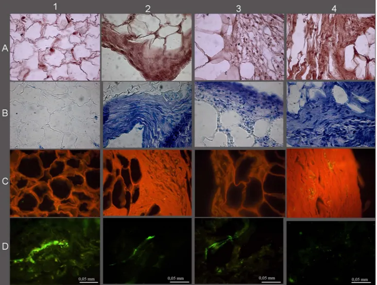
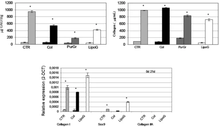
The histological and immunohistological analysis also showed considerable tissue modification (Fig. 2) with a reduction of the fatty component in favor of a connec-tive tissue rich in GAGs (Fig. 2B), in collagen (Fig. 2C), and with a reduced presence of blood vessels (Fig. 2D). Comparing the histology of the lipoaspirates obtained with the four techniques tested, the LipoG batch (Fig. 2, panel 4) underwent the most significant changes in cellu-lar organization, with only a few areas of adipose tissue, the remainder being almost completely replaced by col-lagen and GAGs. These data were confirmed by the bio-chemical quantifications, shown in Figure 3A for GAG and Figure 3B for collagen.
Since the PurGr batch of lipoaspirate gave the poor-est performance in the preliminary experiments for the remainder of the study only the CTR, Col, and LipoG batches were employed. Real-time PCR showed that, before treatment, all three lipoaspirates expressed simi-larly low levels of collagen type I, whereas none expressed mRNA for collagen type II, nor for SOX-9, at 40 cycles of PCR reaction. After 3 weeks of treatment in chondro-genic culture medium, the mRNA of lipoaspirate clusters was altered in a similar direction, with an increase in gene expression of collagen type I, and a small increase in gene expression of SOX-9 (Fig. 3C). The increase in SOX-9 gene expression was particularly evident in the LipoG batch. No collagen type II was detected.
Effect of Lipoaspirate on Chondrocytes
Culture medium from both Col and LipoG lipoaspirates was found to induce chondrocyte proliferation and ECM
production to the same extent. As shown in Figure 4A, already 3 days after the addition of 15% lipoaspirate cul-ture medium, a statistically significant increase in ATP levels occurred in both cases compared to the CTR. This increase was more evident at longer incubation times, reaching a plateau at 18 days of culture, with 80% more ATP in chondrocytes treated with 15% culture medium of the lipoaspirate and LipoG compared to CTR that were chondrocytes treated for the same period of time with 15% of the same culture medium used for lipoaspi-rate culture. Moreover, as shown in Figure 4B, culture medium from the Col and LipoG batches activated ECM synthesis of chondrocytes, as shown by increased levels of GAGs.
Cell Outgrowth From Lipoaspirates: Quantification,Phenotype, Differentiation Capacity, and Cartilage Repopulation
The next stage of the research showed that the LipoG batch of lipoaspirate, when transferred into tissue culture, without any processing began to leave cells out from the tissue clusters after 2–3 days. Cells attached to the plastic of the tissue culture wells and reached 70–80% conflu-ence in 7–12 days. Figure 5 shows representative images of cells grown out from the clusters: Figure 5A shows cells at the bottom of the plastic wells that had grown out from the clusters when cultured in floating condi-tions, and Figure 5B shows cell outgrowth in the 3D col-lagen culture model. Both lipoaspirates (Col and LipoG) showed good cell outgrowth from the clusters; the only difference was that, from the LipoG batch, outgrowth was faster than from the Col batch; further from the same weight of lipoaspirate, the number of cells from LipoG was higher than that from the Col batch. Of the two cul-ture models studied, cell outgrowth was faster in the 3D collagen matrix than it was from the floating culture. Cell outgrowth was already visible within 2 days from time 0 in the 3D culture model, whereas no cells were visible before 4–5 days in the floating culture.
FACS analyses (Fig. 5G) showed that cells obtained by outgrowth from the clusters expressed typical mesenchy-mal markers (CD90, CD73, CD105, and CD44) at high
percentages, close to 100%, and also CD146+ cell index
of pericyte population (Fig. 5, bar graph). No statistically significant difference was found versus cells obtained by enzymatic digestion of the lipoaspirate. Furthermore, cells outgrown from the lipoaspirates were found to have the developmental potential of hMSCs (Fig. 5C–F). Adipogenic differentiation showed a progressive loss of the fibroblastoid-like shape and the production of cyto-plasm lipid vacuoles (Fig. 5C). Osteogenic differentiation was revealed at the first week of induction by morpholog-ical changes (from fibroblastoid-like to trapezoidal cells) and, at the end of the induction period, by the formation
Figur e 1. Macroscopic results (A) before and (B ) after 3 weeks of culture with chondrogenic factors. (C, D) Mechanical results as (C) compressive strength and (D) Y oung modu -lus. The upper bar graph shows the results of samples before treatment (A columns) and after 3 weeks of culture in the chondrogenic model (B columns); the lower graph shows the percent increase following treatment. Results are expressed as mean ± SD of two experiments for each patient (n = 10). *p < 0.05 sample B compared to sample A. °p < 0.05 compared
of mineralized matrix (Fig. 5D) and of ALP-positive cells (Fig. 5E). Chondrogenic induction revealed abundant extracellular matrix positive to human type II collagen antibody (Fig. 5F).
LVs were used to transduce cultured clusters of lipoaspi-rate. Transduction was visualized by GFP expression, and a very high transduction efficiency was demonstrated as shown by direct fluorescence (Fig. 6A). Transduction was not limited to the surface of the clusters, but was also evi-dent in the inner part. Cell counts, performed with nuclear counterstaining, revealed that a mean of 62 ± 4% of cells of the cluster were GFP positive, and no cell loss was detected (p = 0.94 for comparison with control lipoaspi-rates, n = 3). Moreover, 49 ± 20% of cells outgrown from the lipoaspirate clusters were GFP positive.
When GFP-transduced clusters were placed in an organ culture of healthy or mechanically damaged carti-lage, it was found that, within a few days, positive cells
grew out from the clusters; moreover, they only repopu-lated the damaged cartilage (Fig. 6C) and not its healthy counterpart (Fig. 6B). Among GFP-positive cells, only 10% showed SOX-9 colocalization, indicating that, in the organ culture model used, only 10% of cells outgrow-ing from the lipoaspirate differentiated into chondrocytes (Fig. 6D).
DISCUSSION
The use of ASCs in regenerative medicine is a rapidly growing area of research. There is some in vitro evidence of success using these cells in osteochondral defect repair (17), and they have also recently been used successfully as a therapeutic tool in treating OA (5). ASCs have been available commercially for veterinary use since 2003, although little has been published documenting their clinical efficacy; such reports might minimize the gap between human and veterinary markets, accelerating the
Figure 2. Histological and histochemical results. (A) Hematoxylin and eosin, (B) toluidine blue, (C) Sirius red, (D) Von Willebrand.
CTR lipoaspirate before chondrogenic treatment (1), Col (2), PurGr (3), and LipoG (4) batches after 3 weeks of culture with chondro-genic factors.
introduction of stem cell-based cartilage repair for human diseases. Beneficial effects of ASCs have been reported in treating OA in some experimental animal models (19,33). However, these studies used a tissue-engineering approach, and a wide variety of biodegradable scaffolds were used to assist chondrogenic differentiation.
To our knowledge, use of an autologous biological tis-sue, naturally rich in stem cells, implanted in a different anatomical site from where it was harvested (i.e., autolo-gous transplantation of subcutaneous fat to intra-articular sites) has not been previously attempted. The present study has shown that it is possible to avoid stem cell isolation, expansion, and differentiation for several days in vitro, before autologous implantation with scaffold or scaffold-free approaches. We have demonstrated that microfragmented lipoaspirate clusters give rise to sponta-neous cell outgrowth both when cultured in floating con-ditions and in the 3D collagen matrix. Although adipose tissue contains adipose-derived stem cells, fibroblasts, endothelial cells, hematogenous cells, pericytes, adipo-cytes, and preadipoadipo-cytes, it was found that cells obtained naturally exhibit a mesenchymal phenotype with stem cell surface expression markers similar to those obtained through enzymatic extraction; they also have the capacity
for multilineage differentiation, showing the classical commitment to osteogenic, chondrogenic, and adipogenic lineages. These data show that micronized lipoaspirate, acting as a natural scaffold for mesenchymal cells that are simply trapped in the stromal vascular portion, may be considered a source of autologous multipotent undiffer-entiated cells. Since it requires no processing steps before use, the risk of infection or that of retaining enzymatic residues from tissue digestion during cell isolation can be avoided. This also reduces costs associated with a long period of cell expansion and differentiation (12–21 days) with residual of growth supplements, as well as the side effects associated with the natural or synthetic scaffolds used for cell-based cartilage repair. Despite these prom-ising results, however, the physiological role of native adipose-derived stromal/stem cells in vivo is not fully understood, and more histological studies must be per-formed before it may be deemed safe for clinical use. It will be necessary to identify in situ the expression of a wide range of markers; these may well include more than those studied to analyze culture-expanded preparations (2). The use of GFP-transduced lipoaspirates demon-strated, in vitro, that cells outgrown from lipoaspirate can repopulate an organ culture of damaged cartilage. It may
Figure 3. Biochemical quantification of GAG expressed as mg GAG per mg tissue (top left) and of total collagen expressed as
absolute value in μg/ml (top right). Expression of chondrocytic markers in lipoaspirate tissue extracts normalized with GAPDH and reported as relative differential gene expression (bottom). For each sample, the reported values are from samples before (first column) and after (second column) chondrogenic treatment (21 days of culture in chGF-enriched chondrogenic medium). *p < 0.05 compared to the sample before chondrogenic treatment. The results are given as means ± SD (n = 3).
therefore be hypothesized that, when intra-articular injec-tion of human lipoaspirate enters clinical trials, it will give rise to cells having mesenchymal potential to repopulate the lesion. Moreover, there appears to be a possibility that native stem-like cells arising from lipoaspirate may have a better physiological phenotype and higher regenerative potential than cells obtained with the isolation technique and culture conditions generally used for ASC prepara-tion prior to transplantaprepara-tion.
In addition to these data, the finding that lipoaspirate culture medium induced human primary chondrocyte pro-liferation and ECM synthesis reinforces the promise of such preparations, comprising micronized fat, becoming a therapeutic tool for cartilage regeneration. The paracrine and endocrine secretions of adipose tissue have recently been described (19); fat might not be considered only as an energy storage tissue, but rather as an active tissue involved in the metabolism (21), in immunomodulatory
Figure 4. Proliferation (A) and GAG production (B) of human primary chondrocytes cultured with 15% of culture medium from
lipoaspirates. Results in (A) are expressed as mean ± SD relative to control cells (n = 3); results in (B) are reported as mean ± SD (n = 3) in μg/ml of sulfated glycosaminoglycans quantified in culture medium of chondrocytes cultured for 2 days and 8 days in basal medium (CTR) and in culture media supplemented with 15% medium from lipoaspirates (Col and LipoG). *p < 0.05 compared to CTR.
FACING PAGE
Figure 5. Cell outgrowth from clusters. Representative images at phase contrast microscopy of cells outgrown from clusters in
the monolayer culture model (A) and in the 3D collagen matrix (B). (C–F) Representative microscopic images of four separate experiments of the multilineage differentiation capacity of the cells outgrowing from lipoaspirate. Adipogenic differentiation (C) was revealed by Oil red O staining for neutral lipids. Osteogenic differentiation was evidenced by the formation of mineralized matrix as shown in green by calcein staining (D) and ALP activity (E). Chondrogenic differentiation was revealed by immunohistochemical stain for collagen II (F). The bar graph (G) shows the immunophenotyping flow cytometry analysis of cells expanded from the enzymati-cally digested lipoaspirate, and of cells outgrowing from cultured lipoaspirate. Data are expressed as means ± SD (n = 3) of the percent of positive cells for the indicated markers.
activities, and in providing an extensive perivascular res-ervoir of multipotent, undifferentiated cell populations, involved in homeostasis and regeneration. As a large microvascular bed, fat appears to provide an ideal pool of undifferentiated cells in close proximity to perivas-cular access routes for the mobilization and relocation demands of injured or diseased structures. It is character-ized by different cell types (including adipocytes, preadi-pocytes, vascular cells, fibroblasts, pericytes, stem cells, and immune cells) that, by secreting a range of cytokines, growth factors, chemokines, and adipokines, might inter-act with the resident cells of damaged tissue, inducing repair and regeneration. As for ASC preparations, which are already in clinical trials, many questions remain to be clarified concerning lipoaspirate differentiation potential in vivo, and the mechanisms involved in repair or regen-eration (paracrine effects, differentiation, immunomodu-lation), together with any donor-specific variability in cell quality and activity.
In an attempt to determine the fate of lipoaspirate when injected into a joint, it was induced to differentiate in vitro to form cartilage, as has generally been done using MSCs in a 3D culture model (6). Micronized fat (LipoG) responds better to differentiating stimuli than did the other batches, which contained clusters of larger size; this may
be because the more micronized clusters have a greater surface area in contact with the culture medium containing chGF, giving the cells a greater response capacity. Based on these findings, it might be hypothesized that, when injected intra-articularly, lipoaspirate might become a fibrous tissue, having favorable mechanical, morphologi-cal, biochemimorphologi-cal, and molecular characteristics for its use at intra-articular sites. At in vitro examination, an abnor-mal change in the nature of the adipose tissue was seen to have occurred, with residual fat globules surrounded by thick fibrous tissue rich in collagen and GAG that was reminiscent of metaplasia. Although the newly formed tissue was not hyaline cartilage, as demonstrated by the lack of expression of type II collagen, it showed histolog-ical features and mechanhistolog-ical properties that in the joints, where the cartilage is damaged, would undoubtedly be superior to the classic viscosupplements used to alleviate the load acting on damaged cartilage. The fibrous intra-articular sleeve produced by the lipoaspirate preparation, by helping to support the load upon the injured articular heads, could provide a protective action on resident chon-drocytes, which being less stressed would be stimulated to regenerate, leading to repair of the damaged tissue.
In conclusion, intra-articular injection of lipoaspirate may be considered a possible strategy to resolve lesions
Figure 6. Lentiviral GFP transduction of the clusters (A). No GFP-positive cells growing out from GFP-transduced clusters are
vis-ible upon a healthy cartilage fragment (B). A mechanically damaged cartilage fragment was well repopulated (C). Few of the cells that repopulated the damaged cartilage differentiated into chondrocytes (GFP positive and SOX-9 positive, yellow highlighted with arrows) (D).
to the cartilage and osteoarthritis. This possibility shows promise for the production of a new generation of active injectable scaffolds (made of autologous tissue requiring no preparation steps), which could help clinicians to treat focal cartilage defects, osteochondral defects, and osteoar-thritis, using lipoaspirate from the patient to be treated. The use of human materials for transplantation (cells, tissues, and organs) is not without risks that may be related to the donor, the process of relocating the tissue from donor to the patient, or the condition of the receiver. In the application proposed in this study, since lipoaspirate was autologous, there would be no risk of rejection, or of transmitting dis-ease from donor to recipient; further, there would be no need for procurement, processing, testing, storage, or qual-ity control. Some limitations must be overcome before this procedure could enter widespread use, such as short- and long-term unpredictability, in vivo intra-articular perfor-mance, and the fate of cells outgrown from lipoaspirate and of the lipoaspirate itself (biodistribution).
Lipoaspirate and Lipogems® are both minimally
manipulated autologous materials, not combined with other products, and intended for homologous use; they are therefore classified as HCT/Ps in section 361 of the Public Health Service Act and FDA approved for clini-cal trials. For these reasons, the therapeutic approach is possible, but only clinical results will demonstrate its safety (ruling out inflammatory reactions, fibrosis, mal-differentiation, and any tumorigenic activity), and further studies are needed to demonstrate its efficacy that might be dependent on age, BMI, gender, ethnicity, existing dis-eases, or other factors.
ACKNOWLEDGMENTS: The authors would like to thank Dr. Massimiliano Leigheb from the Major Hospital of Novara for providing the cartilage fragments used in the study and Dr. Maurizio Sabbatini for preparing the figures. This work was supported by grants from the Italian Ministry of Education, University and Research (MIUR) PRIN 201288JKYY/2012 and from the research contract No. 6/2014. The study sponsors had no involvement in the study design, in the collection analysis and interpretation of data, in the writing of the manuscript, and in the decision to submit the manuscript for publication. Carlo Tremolada invented and has patented the Lipogems device; however, his only role in the present study was that of providing the biological material (excess of lipoaspirate destined to be disposed of) from his patients, who gave their informed consent. The other authors declare no conflicts of interest.
REFERENCES
Archer, C. W.; McDowell, J.; Bayliss, M. T.; Stephens, 1.
M. D.; Bentley, G. Phenotypic modulation in sub-popula-tions of human articular chondrocytes in vitro. J. Cell Sci. 97:361–371; 1997.
Baer, P. C.; Geiger, H. Adipose-derived mesenchymal 2.
stromal/stem cells: Tissue localization, characterization, and heterogeneity. Stem Cells Int. 2012:812693; 2012. Bendinelli, P.; Matteucci, E.; Dogliotti, G.; Corsi, M. M.; 3.
Banfi, G.; Maroni, P.; Desiderio, M. A. Molecular basis of
anti-inflammatory action of platelet-rich plasma on human chondrocytes: Mechanisms of NF-kB inhibition via HGF. J. Cell. Physiol. 225(3):757–766; 2010.
Bianchi, F.; Maioli, M.; Leonardi, E.; Olivi, E.; Pasquinelli, 4.
G.; Valente, S.; Mendez, A. J.; Ricordi, C.; Raffaini, M.; Tremolada, C.; Ventura, C. A new nonenzymatic method and device to obtain a fat tissue derivate highly enriched in peri-cyte-like elements by mild mechanical forces from human lipoaspirates. Cell Transplant. 22(11):2063–2077; 2013. Black, L. L.; Gaynor, J.; Adams, C.; Dhupa, S.; Sams, A. E.; 5.
Taylor, R.; Harman, S.; Gingerich, D. A.; Harman, R. Effect of intrarticular injection of autologous adipose-derived mes-enchymal stem and regenerative cells on clinical signs of chronic osteoarthritis of the elbow joint in dogs. Vet. Ther. 9(3):192–200; 2008.
Bosetti, M.; Boccafoschi, F.; Leigheb, M.; Bianchi, A. E.; 6.
Cannas, M. Chondrogenic induction of human mesenchy-mal stem cells using combined growth factors for cartilage tissue engineering. J. Tissue Eng. Regen. Med. 6:205–213; 2012.
Breyner, N. M.; Hell, R. C.; Carvalho, L. R.; Machado, C. 7.
B.; Peixoto, I. N.; Valerio, P.; Pereira, M. M.; Goes, A. M. Effect of a three-dimensional chitosan porous scaffold on the differentiation of mesenchymal stem cells into chondro-cytes. Cells Tissues Organs 191(2):119–128; 2010. Cox, W. G.; Singer, V. L. A high-resolution, 8.
based method for localization of endogenous alkaline phos phatase activity. J. Histochem. Cytochem. 47(11): 1443–1456; 1999.
Crouch, S. P.; Kozlowski, R.; Slater, K. J.; Fletcher J. The use 9.
of ATP bioluminescence as a measure of cell proliferation and cytotoxicity. J. Immunol. Methods 160(1):81–88; 1993. Elsdale, T.; Bard, J. Collagen substrata for studies on cell 10.
behaviour. J. Cell Biol. 54:626–637; 1972.
Emans, P. J.; Jansen, E. J.; van Iersel, D.; Welting, T. J.; 11.
Woodfield, T. B.; Bulstra, S. K.; Riesle, J.; van Rhijn, L. W.; Kuijer, R. Tissue-engineered constructs: The effect of scaffold architecture in osteochondral repair. J. Tissue Eng. Regen. Med. 7(9):751–756; 2013.
Farndale, R. W.; Sayers, C. A.; Barrett, A. J. A direct spectro-12.
photometric microassay for sulphated glycosaminoglycans in cartilage cultures. Connect. Tissue Res. 9(4):247–248; 1982.
Follenzi, A.; Sabatino, G.; Lombardo, A.; Boccaccio, C.; 13.
Naldini, L. Efficient gene delivery and targeted expression to hepatocytes in vivo by improved lentiviral vectors. Hum. Gene Ther. 13:243–260; 2002.
Freyria, A. M.; Mallein-Gerin, F. Chondrocytes or adult 14.
stem cells for cartilage repair: The indisputable role of growth factors. Injury 43(3):259–265; 2012.
Grace, H. L.; LaValley, M.; McAlindon, T.; Felson, D. T. 15.
Intra-articular hyaluronic acid in treatment of knee osteoar-thritis. JAMA 290(23):3115–3121; 2003.
Hale, L. V.; Ma, Y. F.; Santerre, R. F. Semi-quantitative fluo-16.
rescence analysis of calcein binding as a measurement of in vitro mineralization. Calcif. Tissue Int. 67(1):80–84; 2000. Hamid, A. A.; Idrus, R. B.; Saim, A. B.; Sathappan, S.; 17.
Chua, K. H. Characterization of human adipose-derived stem cells and expression of chondrogenic genes during induction of cartilage differentiation. Clinics 67(2):99–106; 2012.
Hunziker, E. B. Articular cartilage repair: Basic science and 18.
clinical progress. A review of the current status and pros-pects. Osteoarthritis Cartilage 10:432–463; 2002.
Jurgens, W. J.; Kroeze, R. J.; Zandieh-Doulabi, B.; van Dijk, 19.
A.; Renders, G. A.; Smit, T. H.; van Milligen, F. J.; Ritt, M. J.; Helder, M. N. One-step surgical procedure for the treat-ment of osteochondral defects with adipose-derived stem cells in a caprine knee defect: A pilot study. Biores. Open Access 2(4):315–325; 2013.
Kastrinaki, M. C.; Andreakou, I.; Charbord, P.; Papadaki, 20.
H. A. Isolation of human bone marrow mesenchymal stem cells using different membrane markers: Comparison of colony/cloning efficiency, differentiation potential, and molecular profile. Tissue Eng. Part C Methods 14:333– 339; 2008.
Kershaw, E. E.; Flier, J. S. Adipose tissue as an endocrine 21.
organ. J. Clin. Endocrinol. Metab. 89(6):2548–2556, 2004. Kurita, M.; Matsumoto, D.; Shigeura, T.; Sato, K.; Gonda, 22.
K.; Harii, K.; Yoshimura, K. Influences of centrifugation on cells and tissues in liposuction aspirates: Optimized centrif-ugation for lipotransfer and cell isolation. Plast. Reconstr. Surg. 121(3):1033–1041; 2008.
Kuroda, T.; Matsumoto, T.; Mifune, Y.; Fukui, T.; Kubo, 23.
S.; Matsushita, T.; Asahara, T.; Kurosaka, M; Kuroda, R. Therapeutic strategy of third-generation autologous chon-drocyte implantation for osteoarthritis. Ups. J. Med. Sci. 116(2):107–114; 2011.
Leunig, M.; Tibor, L. M.; Naal, F. D.; Ganz, R.; Steinwachs, 24.
M. R. Surgical technique: Second generation bone marrow stimulation via surgical dislocation to treat hip cartilage lesions. Clin. Orthop. Relat. Res. 470(12):3421–3431; 2012.
Mestak, O.; Sukop, A.; Hsueh, Y. S.; Molitor, M.; Mestak, 25.
J.; Matejovska, J.; Zarubova, L. Centrifugation versus PureGraft for fat grafting to the breast after breast-conserv-ing therapy. World J. Surg. Oncol. 12:178; 2014.
Noël, D.; Caton, D.; Roche, S.; Bony, C.; Lehmann, S.; 26.
Casteilla, L.; Jorgensen, C.; Cousin, B. Cell specific differences
between human adipose-derived and mesenchymal–stromal cells despite similar differentiation potentials. Exp. Cell Res. 314:1575–1584; 2008.
Pfaffl, M. W. A new mathematical model for relative 27.
quantification in real-time RT-PCR. Nucleic Acids Res. 29(9):2002–2007; 2001.
Sakaguchi, Y.; Sekiya, I.; Yagishita, K.; Muneta, T. 28.
Comparison of human stem cells derived from various mesenchymal tissues: Superiority of synovium as a cell source. Arthritis Rheum. 52:2521–2529; 2005.
Samuels, J.; Kransnokutsky, S.; Abramson, S. B. Osteo-29.
arthritis: A tale of three tissues. Bull. NYU Hosp. Jt. Dis. 66(3):244–250; 2008.
Sharkey, P. F.; Cohen, S. B.; Leinberry, C. F.; Parvizi, J. 30.
Subchondral bone marrow lesions associated with knee osteoarthritis. Am. J. Orthop. 41(9):413–417; 2012. Shenaq, D. S.; Rastegar, F.; Petkovic, D.; Zhang, B. Q.; He, 31.
B. C.; Chen, L.; Zuo, G. W.; Luo, Q.; Shi, Q.; Wagner, E. R.; Huang, E.; Gao, Y.; Gao, J. L.; Kim, S. H.; Yang, K.; Bi, Y.; Su, Y.; Zhu, G.; Luo, J.; Luo, X.; Qin, J.; Reid, R. R.; Luu, H. H.; Haydon, R. C.; He, T. C. Mesenchymal progen-itor cells and their orthopaedic applications: Forging a path towards clinical trials. Stem Cells Int. 16:519028; 2010. Tullberg-Reinert, H.; Jundt, G. In situ measurement of 32.
collagen synthesis by human bone cells with a Sirius Red-based colorimetric microassay: Effects of TGFb2 and ascorbic acid. Histochem. Cell Biol. 112:271–276; 1999. Veronesi, F.; Maglio, M.; Tschon, M.; Aldini, N. N.; Fini, 33.
M. Adipose-derived mesenchymal stem cells for cartilage tissue engineering: State-of-the-art in in vivo studies. J. Biomed. Mater. Res. A. 102(7):2448–2466; 2014.
Xue, J. X.; Gong, Y. Y.; Zhou, G. D.; Liu, W.; Cao, Y.; 34.
Zhang, W. J. Chondrogenic differentiation of bone marrow-derived mesenchymal stem cells induced by acellular carti-lage sheets. Biomaterials 33:5832–5840; 2012.