A. Catalano
a,⁎, D. Chilà
a, F. Bellone
a, G. Nicocia
a, G. Martino
b, I. Loddo
c, N. Morabito
a,
S. Benvenga
a,d,e, S. Loddo
aaDepartment of Clinical and Experimental Medicine, University Hospital of Messina, Messina, Italy
bDepartment of Cognitive Sciences, Psychology, Education and Cultural Studies, University of Messina, Messina, Italy
cDepartment of Laboratory Medicine and Advanced Biotechnologies, Mediterranean Institute for Transplantation and Advanced Specialized Therapies– ISMETT – IRCCS,
Palermo, Italy
dMaster Program on Childhood, Adolescent and Women’s Endocrine Health, University of Messina, Italy
eInterdepartmental Program of Molecular & Clinical Endocrinology and Women’s Endocrine Health, University Hospital Policlinico G. Martino, Messina, Italy
A R T I C L E I N F O Keywords: Electrolytes Hypercalcemia Hypocalcemia Inpatients Elderly Pediatrics A B S T R A C T
Disorders of calcium metabolism are frequently encountered in routine clinical practice. However limited data are available on the epidemiology of hypocalcemia and hypercalcemia in hospitalized patients. Our aim was to evaluate the frequency of hypocalcemia and hypercalcemia in hospitalized patients.
This is a retrospective study based on the laboratory results of all hospitalized subjects (n = 12,334) whose calcemia was determined between January 1st, 2011 and December 31st, 2014. Measurements of serum calcium were carried out by a single centralized laboratory. Hypocalcemia was defined as serum calcium levels < 8.2 mg/dl and hypercalcemia as serum calcium levels > 10.4 mg/dl. Albumin correction was applied to adjust serum calcium values.
Overall, hypocalcemia accounted for 27.72% (n = 3420) and hypercalcemia for 4.74% (n = 585) of the 12,334 inpatients. The highest prevalence of hypocalcemia was found in patients over 65 yr. (n = 2097, 61.31%) vs. younger subjects, while the highest prevalence of hypercalcemia was observed in patients aged 0–18 yr. (n = 380, 64.95%). Hypocalcemia was more often encountered in males (n = 1952, 57.07%) while no gender differences were found regarding hypercalcemia. Incidence of hypocalcemia changed over time varying from 35.42% (n = 1061) in 2011 to 21.93% (n = 672) in 2014 (r =−0.98; p = 0.01). Differently, incidence of hypercalcemia did not significantly increase significantly from 3.47% (n = 104) in 2011 to 6.92% (n = 211) in 2014 (r = 0.94; p = 0.052).
Despite increased awareness about electrolytes disturbance, physicians should consider calcium levels be-cause of life-threatening consequences associated to hypo- and hypercalcemia. Patient’s gender and age could be associated to a different risk of calcium disturbance in hospitalized patients.
Introduction
Calcium is the most abundant mineral in the body and participates with phosphorus to form calcium phosphate in bones and teeth. It is involved in many biological processes since it is essential for the normal functioning of nerves and muscles and plays a role in blood coagulation and in several enzymatic processes[1,2].
As expected perturbation of calcium homeostasis may have a deep impact on human pathology [2]. Serum calcium levels are usually maintained within a normal range and ionized calcium is tightly regulated by the actions of parathyroid hormone (PTH) and
1,25-dihydroxyvitamin D (1,25[OH]2D) on the kidney, bone and
gastro-intestinal tract[3,4].
PTH stimulates calcium release from bone and calcium resorption in the kidney, moreover it stimulates 1α-hydroxylation of 25-hydro-xyvitamin D leading to the production of active 1,25-dihydro25-hydro-xyvitamin D (calcitriol) which modulates gastrointestinal calcium absorption[3]. In a clinical setting, either elevation (hypercalcemia) or reduction (hypocalcemia) of serum calcium concentrations could depend by several pathologies and could be associated with life-threatening con-sequences[2].
Increasing life expectancy and improvement of standard medical
https://doi.org/10.1016/j.jcte.2018.05.004
Received 19 April 2018; Received in revised form 15 May 2018; Accepted 27 May 2018
⁎Corresponding author at: Department of Clinical and Experimental Medicine, University Hospital of Messina, Via C. Valeria, 98125 Messina, Italy.
E-mail address:[email protected](A. Catalano).
2214-6237/ © 2018 The Authors. Published by Elsevier Inc. This is an open access article under the CC BY-NC-ND license (http://creativecommons.org/licenses/BY-NC-ND/4.0/).
care have modified the epidemiology of some human diseases (e.g. renal failure, cancer). Thus, the incidence of calcium related disorders could be challenging over time[5–8].
However, the prevalence of hypo- and hypercalcemia is dependent by the specific setting of patients[6–8].
Persistent hypercalcemia has been reported to occur in up to 1% of individuals in general population [9,10]whereas the prevalence of hypocalcemia in hospital setting has been observed up to 3% [10]. Differently, hypocalcemia has been described to reach a prevalence of 18% in hospitalized patients and up to 85% in the intensive care units [5].
The main aim of our study was to update the incidence of hypo-calcemia and hyperhypo-calcemia in a large cohort of inpatients subjects. The secondary aim was to explore whether age and gender could be asso-ciated with serum calcium alterations.
Materials and methods
This is a retrospective study considering all the patients whose calcium concentrations were determined during hospital recovery at the University Hospital of Messina, Messina, Italy, over the period from January 2011 to December 2014. Any age and gender were considered. Total calcium levels were detected by a Centralized Laboratory of our hospital through an automated analyzer (Roche Modular Analytics P 800). All the measurements were corrected with serum albumin levels in accordance with the formula: corrected serum calcium = measured calcium + [(4.1-albumin) × 0.8] if albumin is < 4.1 g/dl, or measured calcium− [(4.1-albumin) × 0.8] if albumin is > 4.1 g/dl[11,12].
Hypercalcemia was defined for calcium levels above 10.4 mg/dl, whereas hypocalcemia was defined for calcium levels under 8.2 mg/dl in accordance with the reference range of our Laboratory.
Recruited subjects came from multiple medical and surgical de-partment clinics within our hospital.
The study was conducted in accordance with the ethical standards of our institutional research committee and with the 1964 Declaration of Helsinki and its later amendments. Written informed consent was not required. Statistical analyses were performed using MedCalc software (version 10.2.0.0; Mariakerke, 173 Belgium).
Statistical analyses were performed using MedCalc software (ver-sion 10.2.0.0; Mariakerke, 173 Belgium). Theχ2test was performed to calculate differences in the proportion of categorical variables. Linear regression was used to describe changes of incidences over time. Values of p < 0.05 were considered to indicate statistical significance. Results
In the period from January 1st, 2011 to December 31st, 2014 we
evaluated a sample of 12,334 internal patients whose calcium level was measured. We found 27.72% (n = 3420) of patients showing hypo-calcemia and 4.74% (n = 585) showing hyperhypo-calcemia. Concerning patients with hypocalcemia we observed 42.93% of cases (n = 1468) in females and 57.07% (n = 1952) in males (χ2
= 136, p < 0.0001). Adult subjects were more likely to exhibit low calcium levels: in fact, hypocalcemia was found in 61.31% (n = 2097) of subjects over 65 yr., in 33.3% (n = 1139) of subjects aged 19–65 yr., finally in 5.38% (n = 184) of subjects aged 0–18 yr. (χ2= 2407, p < 0.0001). At the
same time, we detected 585 cases of hypercalcemia of whom 50.95% (n = 298) were observed in female and 49.05% (n = 287) in male patients (χ2= 0.34, p = 0.55). The higher incidence of hypercalcemia
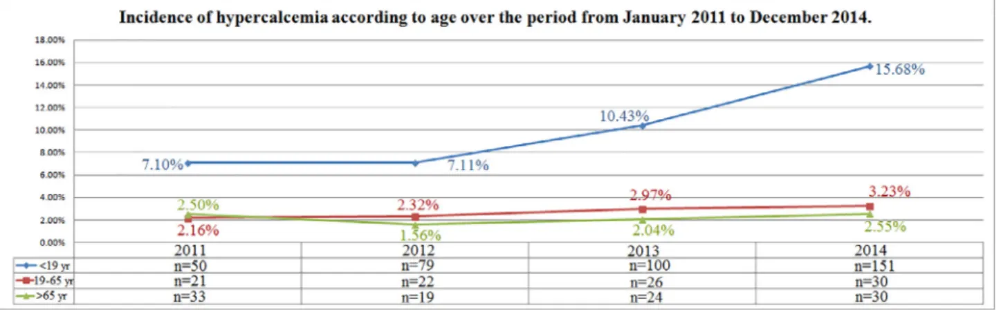
was found in subjects aged 0–18 (64.95%, n = 380), whereas in the age range19–65 yr. was 16.92% (n = 99) and in patients over 65 yr. (n = 106) was 18.11% (χ2= 395, p < 0.0001).
Incidence of hypocalcemia changed over time varying from 35.42% (n = 1061) in 2011 to 21.93% (n = 672) in 2014 (r =−0.98; p = 0.01); differently, incidence of hypercalcemia increased from 3.47% (n = 104) in 2011 to 6.42% (n = 211) in 2014 (r = 0.94; p = 0.052); normocalcemia cases increased from 61.10% (n = 1830) in 2011 to 71.13% (n = 2180) in 2014 (r = 0.93; p = 0.06) (Fig. 1). In-cidence of hypocalcemia according to gender was reduced during the observation period in both genders (r =−0.96, p = 0.03 and r =−0.99, p = 0.006 in females and males respectively) (Fig. 2). Dif-ferently, incidence of hypercalcemia increased over time in males (r = 0.99, p = 0.006) but not in females (r = 0.67, p = 0.32) (Fig. 3). Distribution of cases of hypocalcemia according to age remained unchanged over time in the group of subjects in the age range 0–18 yr. (r =−0.52, p = 0.47) while in subjects between 19 and 65 yr. and in those ones over 65 yr. a reduction of cases was observed (r =−0.96, p = 0.04 and r =−0.99, p = 0.007, respectively) (Fig. 4). A tendency of increasing incidence of hypercalcemia was observed in the age ranges 0–18 yr. (r = 0.92, p = 0.07) and 19–65 yr. (r = 0.97, p = 0.02), while incidence of hypercalcemia remained unchanged in the subjects aged over 65 yr. (r = 0.17, p = 0.82) (Fig. 5).
Discussion
Our research study shows the temporal trend of serum calcium abnormalities observed in hospitalized patients. As known total calcium includes ionized calcium, protein-bound calcium and calcium com-plexed with inorganic and organic anions. Since about 80% of the protein-bound calcium is associated with albumin, albumin levels could influence calcemia. Thus we considered only patients whose calcium serum concentration was corrected taking in account albumin levels [11–13].
Hypo- and hypercalcemia are calcium disorders commonly observed
in hospitalized patients, due to several causes as summarized inTables 1 and 2 [14–26].
Prevalence of hypocalcemia was previously reported to rank to 18% of all hospitalized patients and to be represented in 85% of patients in the intensive care units[5].
In our population the prevalence of hypocalcemia was 27.72%. A common cause of hypocalcemia in primary care is vitamin D deficiency which is, irrespective of latitude, a frequent condition often resulting in secondary increased levels of PTH [3]. Mild and slowly developing hypocalcemia could be an asymptomatic laboratoryfinding, but hypocalcemia could be also a life-threatening metabolic dis-turbance and acute hypocalcemia can provoke severe symptoms which requires hospitalization[4,5]. Hypocalcemia is frequently encountered
in hospitalized patients and could be also empathized by low dietary calcium intake.
We noticed a tendency to a decreasing incidence of hypocalcemia over the observation period, with a reduction of cases of 13.49% from 2011 to 2014. Ourfindings are consistent with the wide attention re-cently attributed to vitamin D deficiency, especially in the elderly. As vitamin D is a key regulator of calcium absorption, adequate vitamin D status could contribute to maintain eucalcemia[27–29]. Incidence of hypocalcemia decreased of 14.35% in particular in females. This is at least in part due to more frequent prescriptions of vitamin D and/or calcium in accordance with the prevention of osteoporosis[30]. How-ever a significant decreasing incidence of hypocalcemia was also ob-served in males as shown inFig. 2.
Fig. 2. Incidence of hypocalcemia according to gender over the period from January 2011 to December 2014.
Fig. 3. Incidence of hypercalcemia according to gender over the period from January 2011 to December 2014.
The prevalence of hypercalcemia in our hospital was 4.74%. Our findings are consistent with previous data by Aishah et al. who found a smaller prevalence of 2.4% in hospitalized patients[31]. No relevant gender differences was found about prevalence of hypercalcemia, but an increased incidence of hypercalcemia in males and younger subjects was detected over the observation period. Although we did not in-vestigate the causes of calcium abnormalities, we speculated that in the younger subjects an iatrogenic effect of multivitamins or calcium sup-plements could not be ruled out, in addition to the other causes of hypercalcemia reported inTable 2. Moreover, the high prevalence of hypercalcemia in young individuals is of concern and it might be im-portant to warn about the consequences that this alteration may lead to: hypercalcemia could be associated with hypercalciuria and be re-sponsible for the development of certain types of kidney stones (cal-cium oxalate dihydrate), the incidence of which is currently increasing in young individuals[32].
Despite increased awareness about electrolytes disturbances[33], physicians should consider calcium levels because of life-threatening consequences associated to hypo and hypercalcemia[2].
Gender and age of patient could be associated to a different risk of this electrolyte disorder in hospitalized patients.
A limitation of the present study is the impossibility of ruling out false values of calcemia due to some pre-analytical factors (e.g. venous occlusion, posture, alterations in protein binding, abnormal proteins, heparin, pH, drugs, temperature), but the large sample size is at the same time the main strength of this research. Although we did not
investigated causes of calcium disturbances, we recognize as a limit of the study also the lack of data on PTH concentrations. PTH should be considered in fact as a“couple” along with calcium concentrations and should be included in the initial diagnostic work-up investigating cal-cium disturbances.
In conclusion our study focused on the incidence of serum calcium abnormalities in hospitalized patients, highlighting age and gender differences. Hypo- and hypercalcemia are common electrolyte disorders in hospitalized patients. Despite increased awareness about electrolytes disturbances, physicians should consider calcium levels because of life-threatening consequences associated to hypo- and hypercalcemia. Gender and age of patient could be associated to a different risk of this electrolyte disorder in hospitalized patients.
Declarations of interest None.
Appendix A. Supplementary data
Supplementary data associated with this article can be found, in the online version, athttp://dx.doi.org/10.1016/j.jcte.2018.05.004. References
[1] Espay AJ. Neurologic complications of electrolyte disturbances and acid-base bal-ance. Handb Clin Neurol 2014;119:365–82.
[2] Akirov A, Gorshtein A, Shraga-Slutzky I, Shimon I. Calcium levels on admission and before discharge are associated with mortality risk in hospitalized patients. Endocrine 2017 Aug;57(2):344–51. http://dx.doi.org/10.1007/s12020-017-1353-y. Epub 2017 Jun 30.
[3] Holick MF. Vitamin D deficiency. N Engl J Med 2007;357(3):266–81.
[4] Body JJ, Bouillon R. Emergencies of calcium homeostasis. Rev Endocr Metab Disord 2003;4(2):167–75.
[5] Cooper MS, Gittoes NJ. Diagnosis and management of hypocalcemia. BMJ 2008;336(7656):1298–302.
[6] Ralston SH, Gallacher SJ, Patel U, Campbell J, Boyle IT. Cancer associated hy-percalcemia: morbidity and mortality. Clinical experience in 126 treated patients. Ann Intern Med 1990;112:499–504.
[7] Egbuna OI, Brown EM. Hypercalcemia and hypocalcemia conditions due to calcium-sensing receptor mutations. Best Pract Res Clin Rheumatol 2008;22(1):129–48. [8] Liamis G, Milionis HJ, Elisaf M. A review of drug-induced hypocalcemia. J Bone
Miner Metab 2009;27(6):635–42.
[9] Palmér M, Jakobsson S, Akerström G, Ljunghall S. Prevalence of hypercalcaemia in a health survey: a 14-year follow-up study of serum calcium values. Eur J Clin Invest 1988;18(1):39–46.
[10] Frolich A. Prevalence of hypercalcemia in normal and in hospital populations. Dan Med Bull 1998;45:436–9.
[11] Catalano A, Basile G, Lasco A. Hypocalcemia: a sometimes overlooked cause of heart failure in the elderly. Aging Clin Exp Res 2012;24(4):400–3.
[12] Labriola L, Wallemacq P, Gulbis B, Jadoul M. The impact of the assay for measuring albumin on corrected calcium concentrations. Nephrol Dial Transplant 2009;24:1834–8.
[13] Riancho JA, Arjona R, Sanz J, Olmos JM, Valle R, Barcello JR. Is the routine measurement of ionized calcium worthwhile in patients with cancer? Postgrad Med
Fig. 5. Incidence of hypercalcemia according to age over the period from January 2011 to December 2014.
Table 1
Main causes of hypocalcemia.
•
Absent-PTH 1. Primary Hypo-PTH 2. Post-surgical Hypo-PTH 3. Autoimmune Hypo-PTH 4. Hypomagnesemia•
Ineffective-PTH 1. Chronic renal failure 2. Hypo-vitamin D 3. Pseudo Hypo-PTH•
Abolished-PTH 1. Acute hyperphosphatemia a) Tumor lysis b) Rhabdomyolysis c) Acute renal failureTable 2
Main causes of hypercalcemia.
•
PTH-Dependent 1. Primary Hyper-PTH a) MEN b) Adenoma 2. Secondary Hyper-PTH a) Renal failure b) Aluminum intoxication 1. PTH-Independent a. Paraneoplastic syndrome b. Iatrogenic (thiazide diuretics, lithium, theophylline) c. Vitamin D intoxication•
Other 1. Immobilization 2. Hyperthyroidism 3. Acromegaly 4. Milk-alkali syndrome[21] Eftekhari F, Yousefzadeh D. Primary infantile hyperparathyroidism: clinical, la-boratory and radiographic features in 21 cases. Skeletal Radiol 1982;8:201–8. [22] Sargent JTS, Smith OP. Haematological emergencies managing hypercalcemia in
adults and children with haematological disorders. Br J Haematol 2010;149:465–77.
[23] Endelson GW, Kleerekoper M. Hypercalcemic crisis. Med Clin North Am 1995;79:79–92.
[24] Nussbaum SR, Gaz RD, Arnold A. Hypercalcemia and ectopic secretion of para-thyroid hormone by an ovarian carcinoma with rearrangement of the gene for
2013;10(3):191–4.
[31] Aishah AB, Foo YN. A retrospective study of serum calcium levels in a hospital population in Malaysia. Med J Malaysia 1995;50(3):246–9.
[32] Tasian GE, Ross ME, Song L, Sas DJ, Keren R, Denburg MR, et al. Annual incidence of nephrolithiasis among children and adults in South Carolina from 1997 to 2012. Clin J Am Soc Nephrol 2016;11(3):488–96.
[33] Catalano A, Basile G, Ferro C, Bellone F, Scarcella C, Benvenga S, et al. Hyponatremia as a leading sign of hypopituitarism. J Clin Transl Endocrinol: Case Rep 2017;5:1–3.