Contents lists available atScienceDirect
Virology
journal homepage:www.elsevier.com/locate/yviro
A Parvovirus B19 synthetic genome: sequence features and functional
competence
Elisabetta Manaresi
1, Ilaria Conti
1, Gloria Bua
1, Francesca Bonvicini
1, Giorgio Gallinella
⁎,1,2Department of Pharmacy and Biotechnology, University of Bologna, Bologna, Italy
A R T I C L E I N F O
Keywords: Parvovirus B19 Consensus sequence Synthetic genome Genomic clone Infectious cloneA B S T R A C T
Central to genetic studies for Parvovirus B19 (B19V) is the availability of genomic clones that may possess functional competence and ability to generate infectious virus. In our study, we established a new model genetic system for Parvovirus B19. A synthetic approach was followed, by design of a reference genome sequence, by generation of a corresponding artificial construct and its molecular cloning in a complete and functional form, and by setup of an efficient strategy to generate infectious virus, via transfection in UT7/EpoS1 cells and amplification in erythroid progenitor cells. The synthetic genome was able to generate virus with biological properties paralleling those of native virus, its infectious activity being dependent on the preservation of self-complementarity and sequence heterogeneity within the terminal regions. A virus of defined genome sequence, obtained from controlled cell culture conditions, can constitute a reference tool for investigation of the structural and functional characteristics of the virus.
1. Introduction
Human Parvovirus B19 (B19V) is a human pathogenic virus, member of the Erythroparvovirus genus in the Parvoviridae family (Cotmore et al., 2014). Infection is widespread and can be associated with an ample range of pathologies and clinical manifestations, whose characteristics and outcomes depend on the interplay between viral properties and the physiological and immune status of the infected individuals. B19V shows a selective tropism for erythroid progenitor cells in the bone marrow, exerting a cytotoxic effect and causing a block in erythropoiesis that can manifest as transient or persistent erythroid aplasia. Common manifestations of infection are erythema infectiosum in children or post-infection arthropathies mainly affecting adults, and the virus has been implicated in a growing spectrum of other different pathologies, among them myocarditis, encephalitis and connective tissue diseases. Infection in pregnancy may be transmitted to the fetus, leading to possible fetal death and/or hydrops fetalis (Gallinella, 2013; Qiu et al., 2017).
Structural features of B19V are common to viruses in the family (Gallinella, 2013; Qiu et al., 2017). One molecule of linear single-stranded DNA of either positive or negative polarity, about 5600 nt in length, is encapsidated in T=1 isometric virions, approximately 25 nm in diameter. The genome is composed of a unique internal region, 4830
nt long, containing all the coding sequences,flanked by two inverted, repeated terminal regions, 383 nt long, that serve as origins of replication. Within these, the distal 365 nt present a site of dyad sequence symmetry that allows for the single-stranded DNA molecule to adopt terminal stem and loop hairpin structures. Due to the occurrence of distinct sequence asymmetries, each terminal region can be present in either one of two alternative sequences, each the inverted complement of the other (usually referred to asflip/flop), and different combinations of these alternative sequences at both termini can give rise to four different genome isomers (Luo and Qiu, 2015).
In vitro replication of B19V is limited to a few cell types (Bonvicini et al., 2006; Gallinella et al., 2000). As an appropriate experimental system, an expanding population of differentiating erythroid progeni-tor cells (EPCs) can be obtained from peripheral blood mononuclear cells (PBMC) (Filippone et al., 2010; Wong et al., 2008). Such cellular system replicates in vitro the process that occurs in vivo in the bone marrow environment (Hattangadi et al., 2011; Merryweather-Clarke et al., 2011), and, notwithstanding its complexity, it is so far the best cellular model for supporting B19V replication and production of infectious virus. In this system, permissiveness to viral replication and infectious virus production is higher for cells at the proerythroblast stage, while restriction either to replication or release of infectious virus applies to cells at earlier or later stages, respectively (Bua et al.,
http://dx.doi.org/10.1016/j.virol.2017.05.006
Received 18 April 2017; Received in revised form 3 May 2017; Accepted 8 May 2017
⁎Correspondence author.
1University of Bologna, Department of Pharmacy and Biotechnology, Via Massarenti, 9, I-40138 Bologna, Italy. 2University of Bologna, S.Orsola-Malpighi Hospital– Microbiology, Via Massarenti, 9, I-40138 Bologna, Italy.
E-mail address:[email protected](G. Gallinella).
0042-6822/ © 2017 Elsevier Inc. All rights reserved.
2016). Apart from EPCs, only a few myeloblastoid cell lines, in particular the UT7/EpoS1 cells (Wong and Brown, 2006), can also support viral replication, but in these systems only a very limited production of infectious virus is obtained.
The availability of native virus from clinical isolates is still a major requirement for in vitro infection experiments, and genetic studies, as well as the possibility of obtaining recombinant viruses, have lagged far behind those of other viruses in the family. Central to the development of genetic studies for B19V is the availability of complete genomic clones that may possess functional competence. Clone pB19-M20first showed the ability to replicate and generate infectious virus following transfection in UT7/EpoS1 cells (Zhi et al., 2004). Further systematic analysis on pB19-M20 indicated the relevance of the terminal regions and of the different coding sequences for the maintenance of the functional competence of the cloned viral genome and its ability to generate infectious virus (Zhi et al., 2006). Moreover, a comparative analysis between different available genomic clones of B19V obtained from different viral isolates, pB19-M20, pB19-FL and pHG1, demon-strated a critical role of the catalytic domain of the viral phospholipase (PLA2-like motif in the VP1u region) for the maintenance of the infectivity of virus produced following transfection of the cloned viral genomes (Filippone et al., 2008). Finally, genomic clones may be complemented by helper virus functions and show replicative activity in otherwise non-permissive environments (Guan et al., 2009).
However, on the whole, none of these systems proved so far easily amenable to experimental manipulation or capable of generating infectious virus at significant levels, and additional effort to obtain a more coherent and efficient system to generate virus from cloned genomes may prove worthwhile. In particular, in this respect, a few issues should be addressed, namely: i) cloning the genome in a complete and functional form, reducing the system complexity and instability; ii) investigating the characteristics of the terminal regions relevant to the functional competence of the cloned genome; iii) avoiding any sequence bias, due to the stochastic effect of sampling from a particular isolate, with possible effects on the functional competence of the cloned genome; iv) optimizing the whole experi-mental procedure, in order to increase the yield of virus that may be recovered, and obtain a stock of virus effectively paralleling the infectivity of native virus.
In our study, we sought to address the above issues and establish a new model genetic system for Parvovirus B19. A synthetic approach was followed, by design of a consensus reference genome sequence as a working tool, by generation of a corresponding artificial construct and its molecular cloning in a complete and functional form, and by setup of an efficient strategy to generate infectious virus, via transfection in UT7/EpoS1 cells and amplification in successive rounds of infection in EPCs.
2. Materials and methods 2.1. Bioinformatic analysis
A set of B19V genomic sequences, genotype 1(a), deposited in the NCBI nucleotide database, were selected and retrieved for analysis (listed in thesupplemental sequence dataset). Sequences were aligned and analyzed using the Clone Manager 9 Professional Edition Software (Scientific & Educational Software) and MEGA6 Software (Tamura et al., 2013).
2.2. Molecular cloning
Synthetic DNA inserts, cloned in pIDT vector plasmid, were obtained from IDT technologies. Restriction endonuclease (RE) and ligase enzymes were obtained from NEB and used according to manufacturer's directions. Subsequent manipulations and cloning steps were carried out by standard procedures. Plasmid clones were
maintained by transforming SURE bacterial cells (Invitrogen) under ampicillin selection and subsequent growth in LB medium at 37 °C. Plasmid DNA purification was performed by Endofree Plasmid Kit (Qiagen). Inserts used for transfection assay were either excised from vector plasmid by RE cleavage, or amplified by PCR by using the Expand High Fidelity System (Roche) and primer HJ0 (pTGTCTTCTTTTAAATTTT). Plasmid derived inserts or PCR products were further separated by gel electrophoresis, purified by using Gel Extraction Kit (Qiagen) and quantified by UV absorbance determina-tion.
2.3. Cells
UT7/EpoS1 cells were cultured in IMDM (Cambrex), supplemented with 10% FCS and 2 U/ml Epo (NeoRecormon, Roche), at 37 °C and 5% CO2. Cells were kept in culture at densities between 2×10⁵-1×10⁶
cells/ml, and used for transfection experiments when at a density of 3×10⁵cells/ml. Erythroid progenitor cells (EPCs) were generated in
vitro from peripheral blood mononuclear cells (PBMC) obtained from the leukocyte-enriched buffy coats of healthy blood donors, available for institutional research purposes according to the policy approved by the local Ethical Committee (S.Orsola-Malpighi University Hospital). PBMC were isolated using centrifugation in Ficoll-Paque Plus (GE Healthcare Bio-Sciences AB) and cultured in a medium containing erythropoietic growth and differentiation factors, following previously established protocols with minor modifications (Bua et al., 2016). Isolated PBMC were cultured in IMDM supplemented with 20% serum substitute BIT 9500 (StemCell Technologies), and enriched with 900 ng/ml ferrous sulphate, 90 ng/ml ferric nitrate, 1 µM hydrocorti-sone (Sigma), 3 U/ml Epo (NeoRecormon, Roche), 5 ng/ml IL-3 and 20 ng/ml stem cell factor (Life Technologies). Cells were used for infection experiments when at day 8 of in vitro growth and di fferentia-tion.
2.4. Transfection
UT7/EpoS1 cells were transfected by using the Amaxa Nucleofection System (Lonza), with V Nucleofector Reagent and T20 program setting, at a ratio of 1 µg insert DNA for 106cells. Following
transfection, the cells were incubated at 37 °C in complete medium at an initial density of 106 cells/ml. Constant amounts of cell cultures
were collected up to 12 days post-transfection (dpt), cells and cell-free supernatants (spn) were separated by centrifugation at 5000 rpm for 5 min, then used for analysis and/or successive infection experiments. 2.5. Infection
Cell-free supernatants obtained from transfected UT7/EpoS1 cells were used to infect EPCs cells at a ratio of 100 µL for 1×10⁶cells. As a
control, B19V from a serum sample, identified in our laboratory in the course of routine diagnostic analysis and available for research purposes according to Italian privacy law, was used at a moi of 104
geq/cell (Bonvicini et al., 2013). The infection was carried out at 37 °C for 2 h, then cells were washed free of inoculum and expanded in complete medium at an initial density of 106 cells/ml. Constant
amounts of cell cultures were collected at 2 and 48 h post-infection (hpi) and at 6 days post-infection (dpi), cells and cell-free supernatants (spn) were separated by centrifugation at 5000 rpm for 5 min, then used for analysis and/or successive infection experiments.
2.6. FISH ssay
Equal amounts of cell cultures, corresponding to 1.5×10⁵ cells, collected at the appropriate time points following transfection or infection were processed by on slide FISH assay for the detection of viral nucleic acids, as described (Manaresi et al., 2015) with minor
modifications. Cells were spotted on glass slides, fixed in PBS-paraf-ormaldehyde 0.5% at 4 °C for 30 min and permeabilized for 45 min in PBS-saponin 0.2%. Hybridization reaction was carried out in 25 µL of a hybridization solution containing 25 ng of a digoxigenin labeled, random-primed full-length genomic probe (Dig High Prime, Roche). Specimens and hybridization mixture were denatured together by heating at 95 °C for 5 min and then incubated at 37 °C for 12 h. Following hybridization, the slides were washed twice at 37 °C with 50% formamide - 2×SSC (300 mM NaCl, 30 mM sodium citrate, pH 7.0) buffer and twice at room temperature in 2×SSC buffer, 10 min each. Detection of the hybrids was performed with a FITC-conjugate anti-digoxigenin antibody (Roche) diluted 1:20 in PBS-BSA 1% and incubated for 1 h; after washing in PBS, slides were stained with Evans blue and observed on afluorescence microscopy (EX 450–490 nm, BA 520 nm).
2.7. Nucleic acids purification
Equal amounts of cell cultures, corresponding to 1.5×10⁵cells and
to respective cell-free supernatants, collected at the appropriate time points following transfection or infection were processed by using the EZ1 Viral Nucleic Acid Kit on a EZ1 platform (Qiagen), following the manufacturer's instructions, in order to obtain a total nucleic acid fraction in elution volumes of 150 µL. Volumes of 10 µL were then used in the subsequent qPCR assays for the quantitative evaluation of target viral nucleic acids. For the analysis of supernatants, a benzonase treatment was also carried out by incubating 10 µL of supernatants with 1 µL of enzyme (Novagen), for 4 h at 37 °C, before nucleic acid purification.
2.8. Quantitative PCR
Viral DNA was amplified by using the primer pair R2210-R2355, located in the central exon of B19V genome (Bonvicini et al., 2008). As a control, a target sequence in the region of genomic DNA coding for 5.8 S rDNA gene was amplified by using the primer pair 5.8S_S1 (CTCTTAGCGGTGGATCACTC) - 5.8S_A1(GTGCGTTCGAAG TGTCGATG). All oligonucleotides were obtained from Eurofins Genomics.
Quantitative PCR was carried out by using the RotorGeneQ System (Qiagen) and SybrGreen detection of amplification products. Amplification reactions were performed by using QuantiTect PCR SybrGreen PCR Kit (Qiagen), including 10 pmol of each specific primer pair. For PCR, thermal profile consisted in 10 min at 95 °C, then 40 cycles of 15 s at 95 °C, 30 s at 60 °C, and 30 s at 72 °C coupled with signal acquisition. Afinal melting curve was performed, with thermal profile ramping from 65 °C to 95 °C at a 12 °C/min rate, coupled with continuous signal acquisition.
Fluorescence emission recorded in the FAM/Sybr channel of the instrument was analyzed by using the functions available in the RotorGene 6.0 software. All reported experiments were carried out in duplicate series, each sample analyzed in duplicate, and melting curve and quantitative analysis performed. Melting curve analysis was used for the determination of the specificity of the amplification products by defining, for each reaction, the melting profile and the Tm of the products. Absolute quantitation of viral DNA was obtained by calibra-tion to standard targets prepared from cloned templates. Data analysis was carried out by using the program GraphPad Prism version 5.00 for Windows (GraphPad Software, San Diego California, USA).
3. Results
3.1. Bioinformatic analysis
To establish a model genetic system for B19V, a reference genome sequence was designed as a working tool. To this purpose, a
bioinfor-matic analysis was conducted on a selected set of B19V genomic sequences, genotype 1(a), deposited in the NCBI nucleotide database (listed in the supplemental sequence dataset). A total of 49 sequences were included in the set on the basis of two criteria: 1) when comprising > 70% of genomic sequence of B19V; 2) as representative of isolates collected in different settings, ante 2010. Sequences were globally aligned using the ClustalW algorithm implemented within Clone Manager and MEGA6 software packages.
An unambiguous global sequence alignment was obtained extend-ing over all of the genome sequence of B19V. Within the selected set, average coverage was 40.2 hits per each of the 5596 nucleotide positions in B19V genome. The degree of sequence information available was different regarding the internal unique region (IR) or the terminal regions (ITR). For ITRs, only three complete sequences could be included in the dataset, and the average coverage was as low as 5.8 hits/position. For the IR, sequence from all the 49 isolates provided a relatively higher coverage of 46.0 hits/position. From the global sequence alignment, a Position Weight Matrix (PWM) was obtained and its Information Content (IC) determined for every position along the whole genome. Average IC value in bit units was determined as 1.966, its variation with respect to genome map is shown inFig. 1.
Further information on the genetic variance within the selected set could be obtained by the analysis algorithms within MEGA6. Maximum Likelihood (ML)fits of different nucleotide substitution models were evaluated to describe the observed nucleotide substitution pattern. A General Time Reversible (GTR) model, with Gamma distribution (+G, 0.80) and invariant sites (+I, 0.66), gave the highest ML score and was used when appropriate for further modeling. Pairwise distance dis-tribution ranged continuously from 0.000 to 0.037, with average values for each sequence in the range 0.009–0.022 and an average overall value for the matrix of 0.013.
The global sequence alignment yielded a consensus sequence, matching the highest IC profile for B19V genome sequence, with the minimal average distance value from other sequences in the dataset (0.008, range 0.002–0.022), and coincident with a possible ancestral state as determined from a ML phylogenetic tree. This consensus, herein B19V EC [supplemental sequence dataset and GenBank KY940273], was designed as a reference B19V genotype I(a) sequence, for the purpose of obtaining a functional molecular clone.
3.2. Molecular cloning
A synthetic strategy was devised to obtain a molecular clone of B19V EC consensus sequence. The insert to be cloned was projected to comprise a segment extending from position 136 to position 5461 of B19V EC sequence. This corresponds to a non-redundant complement of the viral genome, including the complete internal region and extension of both ITRs beyond the sites of dyad symmetry (at nt 182/183 and 5413/5414), with maintenance of the potential to fold back in hairpin structures and preservation of flip/flop sequence heterogeneity (within nt 148–218 and 5379–5449). Trimming of the external part of the ITRs was conceived with the aim of avoiding difficulties in maintaining full-length and stable inserts, as previously reported and also experienced by us in preliminary experiments.
To construct the projected molecular clone, gene blocks corre-sponding to discrete segments of the viral genome were obtained by DNA synthesis, then the diverse inserts were progressively ligated into a single insert, cloned into plasmid vector pIDT. The cloning strategy and sequential steps are listed insupplemental Table S1and described insupplemental Fig. S1. As afinal result, four different plasmids were obtained (pCK 00/01/10/11), each containing an insert corresponding to B19V EC consensus sequence, extending from position 136–5461, and differing for the diverse combination of flip/flop terminal isomers. Plasmids were stably maintained in E.coli SURE strain under standard culture conditions without rearrangements or deletions, as confirmed
by Sanger sequencing. Plasmids pCK were used to recover complete genomic inserts, extending beyond the sites of dyad symmetry, from nt 136–5461, by SacI restriction endonuclease cleavage (CK 00/01/10/11 inserts); truncated genomic inserts, extending up to the sites of dyad symmetry, from nt 184–5413, by BssHII cleavage (CH 00/01/10/11 inserts); a truncated genomic insert, extending within the sites of dyad symmetry, from nt 242–5355, by AccIII cleavage (CI insert). In addition, a truncated genomic insert excluding the terminal regions was obtained by PCR amplification, extending from nt 367–5230 (CJ
insert). Sequence heterogeneity within the terminal regions and extension of inserts are shown inFig. 2.
3.3. Functional competence of B19V synthetic genome 3.3.1. Time course
Insert CK10 was selected to test the functional competence of the synthetic cloned genome in a time course experiment. The insert excised from plasmid pCK10 by SacI restriction endonuclease was
Fig. 1. Sequence diversity and functional map of B19V genome. Top: information content (IC) profile of the position weight matrix (PWM) obtained from the alignment of a set of 49 B19V whole genome sequences (supplemental sequence dataset). For each position (w), andj ∈ {T,C G, , A}, the IC(w) was calculated as: IC w( ) = 2 + ∑jpwjlog2pwj. Center: a
schematic diagram of B19V genome indicating the two inverted terminal regions (ITR), and the internal region (IR) with the distribution of cis-acting functional sites (P6, promoter; pAp1, pAp2, proximal cleavage-polyadenylation sites; pAd, distal cleavage-polyadenylation site; D1, D2, splice donor sites; A1.1, A1.2, A2.1, A2.2, splice acceptor sites). Bottom: simplified transcription map of B19V genome, indicating the five classes of mRNAs (m1-5) with respective alternative splicing/cleavage forms.
Fig. 2. Terminal regions sequence heterogeneity and extension of B19V inserts. Top: schematic diagram of B19V genome indicating the extension of inserts relative to the two inverted terminal regions (ITR) and the internal region (IR). Bottom: schematic alignment of left terminal region sequences inflip/flop isomers. Triangles indicate the site of dyad symmetry within ITR, underlined bases indicate the location of sites relevant for the definition of inserts.
transfected in UT7/EpoS1 cells by the nucleofection technique, then cell cultures were maintained for further 12 days post transfection. At intervals, aliquots of cells and supernatants were collected and analyzed for viral replication and release of virus from cells by FISH and qPCR assays. FISH assay was used as a qualitative indication of viral replication within transfected cells, while qPCR was used to obtain quantitative information on the extent of viral replication and release of virus from cells.
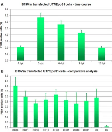
By FISH assay (Fig. 3A), a fraction of positive cells was detected at all time points analyzed, from 1 dpt (2.1%) increasing at 3 dpt (6.7%) and then progressively decreasing (5.6% at 6 dpt, 4.3% at 9 dpt and 2.0% at 12 dpt), indicating that active viral replication could occur in a subset of transfected cells. In these, intensity of staining was maximal at the 6 dpt time point while the distribution of target nucleic acids was mainly nuclear except at later times (Fig. 4A).
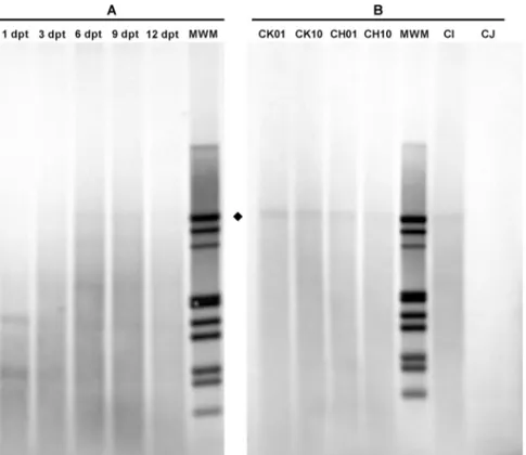
By qPCR assay (Fig. 5A), a high amount of viral DNA was detected in transfected cells, likely due to a high background of input DNA. By Southern Blot analysis (Fig. 6A), a progressive accumulation of DpnI resistant intracellular viral DNA was observed from 6 dpt onwards as a confirmatory indication of active replication of the transfected inserts. A high amount of viral DNA likely due to background was also detected in cell culture supernatants. Benzonase resistant DNA was present in supernatants from 3 dpt, with a significant accumulation occurring from day 6 (+6.9 Log), indicating that a progressively higher fraction of viral DNA had been released from cells encapsidated in mature virions. 3.3.2. Comparative analysis
A series of experiments was carried out to test the functional competence of the genomic inserts in dependence of the different isomer combinations and/or extension of the terminal regions. Inserts CK, CH, CI and CJ were transfected in UT7/EpoS1 cells by the nucleofection technique, then transfected cell cultures were maintained for 6 days post transfection, at what time aliquots of cells and
supernatants were collected and analyzed for viral replication and release of virus from cells by FISH and qPCR assays.
By FISH assay (Fig. 3B), a fraction of positive cells was detected following transfection with all inserts, at different frequencies (CK: 2.3–4.0%; CH:2.3–3.2%, CI: 2.9%; CJ: 0.1%). By qPCR assay (Fig. 5B), as expected, a high and variable background of input DNA was detected. By Southern Blot analysis (Fig. 6B), DpnI resistant molecular forms were observed for all inserts except CJ as an indication of active viral replication. A high and variable amount of viral DNA likely due to background was also present in cell culture supernatants. Benzonase resistant DNA was present in all supernatants with significant differ-ences among the diverse groups of inserts, with highest values for the CK (6.6–6.7 Log geq/10 µL), intermediate for CH and CI (5.8–6.1 Log geq/10 µL), and lowest for CJ (5.0 Log geq/10 µL). Altogether, these results indicate that all the diverse genomic inserts can maintain to some degree functional competence and ability to produce mature virions, and that integrity of the terminal regions has a key role in determining the efficiency of the process.
3.4. Functional competence of B19V synthetic virus
Functional competence of virus released in supernatants of cell cultures, collected in the course of the transfection experiments, was tested by infection of EPCs at day 8 of in vitro differentiation. Following addition of supernatants, cell cultures were maintained for further 6 days post infection. At intervals, 2 hpi, 48 hpi, 6 dpi, aliquots of cells and supernatants were collected and analyzed for viral replication and release of virus from cells by FISH and qPCR assays. By this experimental scheme, both the generation of virus in a time course of transfection, using the insert CK10, as well as the generation of virus from the different genomic inserts, CK, CH, CI and CJ, could be evaluated and compared. Thereafter, to confirm the maintenance of the full competence of virus, a second round of infection of EPCs was carried out by using the different supernatants obtained at 6 dpi in the first round, following the same experimental scheme.
3.4.1. Time course
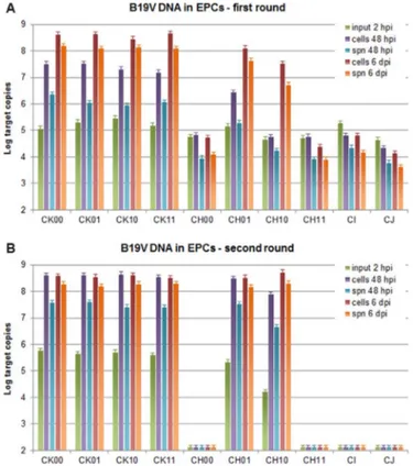
FISH positive cells were detected in EPCs infected with super-natants obtained from the days 6, 9 and 12 post-transfection (Fig. 4B), and the same pattern was maintained in the second round of infection (Fig. 4C). qPCR yielded accurate quantitative information on the extent and dynamics of viral replication within cells and on release of virus in the cell culture supernatant, by comparison of the 48 hpi and 6 dpi time points with respect to input virus at 2 hpi (Fig. 7). In thefirst round of infection (Fig. 7A), no significant increase in the amount of viral DNA was observed for the 1 dpt and 3 dpt samples at any time point, either within cells or in supernatants. Significant increase was observed for the 6–12 dpt samples, at 48 hpi only in cells (in the range +0.5–0.9 Log), at 6 dpi both in cells (in the range +2.6–3.0 Log) and in supernatants (in the range +2.9–3.2 Log). These last values were comparable to what obtained for native virus, used as control at a moi of 104geq/cell (respectively, +2.5, +3.1 Log). Coherently, in the second
round of infection (Fig. 7B), no significant increase in the amount of viral DNA was observed for the 1 dpt and 3 dpt samples at any time point. Significant increase was observed for the 6–12 dpt samples, already at 48 hpi both in cells (in the range +3.4–3.7 Log) and in supernatants (in the range +2.4–2.7 Log), then at 6 dpi not rather in cells (in the range +3.3–3.5 Log) but further in supernatants (in the range +3.9–4.1 Log). These relative increases were significantly higher to what obtained for native virus, used as control at a moi of 104geq/
cell (respectively, +2.1, +2.0, +1.3, +2.3 Log), while total amounts were comparable.
3.4.2. Comparative analysis
FISH positive cells were detected in EPCs infected with super-natants obtained from CK 00/01/10/11 and CH 01/10, but not from
Fig. 3. FISH assay for transfected UT7/EpoS1 cells. A. Fraction (%) of FISH positive UT7/EpoS1 cells following transfection with insert CK10, in a time course experiment (1–12 dpt). B. Fraction (%) of FISH positive UT7/EpoS1 cells following transfection with the indicated inserts, at 6 dpt. Mean % and error bars were determined fromfive replicate samples.
CH 00/11, CI and CJ. The same pattern was maintained in the second round of infection. By qPCR (Fig. 8), in thefirst round of infection (Fig. 8A), significant increases were observed for all CK inserts, already at 48 hpi both in cells (in the range +1.9–2.5 Log) and supernatants (in the range +0.5–1.3 Log), and further at 6 dpi in cells (in the range +3.0–3.6 Log) and supernatants (in the range +2.7–3.2 Log). Comparable increases were observed for CH01/10 at 6 dpi in cells (in the range +2.8–3.0 Log) and supernatants (in the range +2.0–2.5 Log), while no increases were observed for CH 00/11, CI and CJ at any time point either in cells or supernatants. In the second round of infection (Fig. 8B), an increase in the amount of viral DNA was observed for all CK inserts and CH 01/10, evident at 48 hpi as well as 6 dpi, in both cells (in the range +2.8–4.5 Log) and supernatants (in the range +2.5–4.0 Log), but not for CH 00/11, CI and CJ.
Altogether, these results indicate that the functional competence of virus obtained from transfected inserts is critically related to the characteristics of the terminal regions, both in terms of sequence extension and sequence heterogeneity. The potential to reconstitute hairpin structures and provide all cis- elements necessary to replication of the viral genome is different between inserts and leads to different infection outcomes. When inserts extend beyond the axis of dyad symmetry, functional competence is maintained for all the different combinations of terminal isomers. When inserts extend up to the dyad axis functional competence is maintained only if sequence heteroge-neity of terminal isomers is conserved, but is lost if sequence hetero-geneity is not conserved. When inserts extend within the dyad axis, with loss of the potential to form hairpin structure and loss of sequence heterogeneity, and when terminal regions are excluded, functional competence is lost.
4. Discussion
As a result of our work, a new model genetic system for Parvovirus B19 was established, leading from the design of a consensus reference
Fig. 4. FISH assay for transfected UT7/EpoS1 and infected EPCs. Transfected UT7/EpoS1 cells or infected EPCs analyzed by FISH assay for the detection of B19V nucleic acids. A whole length genomic probe for B19V, digoxigenin-labeled, was used and hybrids detected by anti-DIG Fab, FITC conjugated. Positive cells are clearly stained above a low intensity background and show a localized, mainly nuclear, distribution of target. A. UT7/EpoS1 cells transfected with insert CK10, in a time course experiment (1–12 dpt). B. EPCs infected with supernatants from transfected UT7/EpoS1 cell cultures (A). C. EPCs infected with supernatants fromfirst-round infection of EPCs (B).
Fig. 5. B19V DNA from UT7/EpoS1 cells following transfection. B19V DNA amounts in transfected UT7/EpoS1 cells and release in supernatant. A. Cells were transfected with B19V insert CK10 and aliquots of cells and supernatants collected at the indicated days post-transfection (dpt). B. Cells were transfected with the indicated inserts and aliquots of cells and supernatants collected at 6 dpt. The amount of viral DNA was determined by qPCR for the different samples series: cells, amount of intracellular DNA, Log DNA geq/ 104cells; spn, amount of DNA in supernatant, Log DNA geq/10 µL; benz, amount of
genome sequence as a working tool, to molecular cloning of a corresponding synthetic construct in a complete and functional form, to generation of fully competent virus that could be efficiently propagated in erythroid progenitor cells.
In the development of such system, some key achievements might be remarked. A bioinformatic analysis on a set of B19V genomes, genotype I(a), selected to include representative earlier isolates, indicated a low genetic diversity and led to identification of a consensus sequence, possibly representing an ancestral B19V sequence. The choice of a consensus sequence as a working tool, already exploited for the setup of diagnostic assays (Bonvicini et al., 2013), minimizes any stochastic effect related to sequence variability, that may affect biological competence of field isolates or molecular clones obtained from such isolates, and provides a solid benchmark for the study of B19V functional characteristics.
The transition from the in silico designed sequence to DNA was achieved by synthetic techniques. In our strategy, we aimed at the cloning of a complete insert encompassing the whole internal region but only part of the terminal sequences, in order to avoid instability in the vector plasmids. The cloned inserts however extend beyond the sites of dyad symmetry with conservation of the potential to fold back in hairpin structures and preservation offlip/flop sequence heterogeneity, features that were both considered a priori as relevant for the generation of fully competent genomes. Different combinations of terminal isomers and progressively truncated inserts could be excised from vector plasmids and exploited to investigate and compare the respective functional competence and ability to generate infectious virus.
Demonstration of the functional competence of the cloned synthetic genome was obtained by transfection in UT7/EpoS1 cells, as already shown for similar systems in previous works (Filippone et al., 2008; Zhi et al., 2006, 2004). In our system, replication of the viral genome and an enhanced production and release of virus from cells were mainly observed in a prolonged time course and were related to the extension of the terminal regions. Following transfection, the system showed a limited overall efficiency, with low levels of de novo virus production. This effect is possibly linked both to a relatively low efficiency of the transfection technique, and to the semi-permissive characteristics of UT7/EpoS1 cells, relating in particular to restrictive mechanisms affecting the maturation stage of B19V replicative cycle (Wolfisberg et al., 2013).
A clear demonstration of the functional competence of the virus produced in UT7/EpoS1 cells and enhancement of the process efficiency was obtained by amplification in the primary target cells, EPCs (Bua et al., 2016). Two successive rounds of infection, thefirst one with supernatant from transfected cells and the second with supernatant from the first round of infection in EPCs, not only confirmed infectivity and maintenance of infectivity of virus produced, but eventually lead to a viral yield comparable to what could be obtained from native virus in clinical specimens. Within this experi-mental system, functional competence and ability to generate infec-tious virus was observed for the inserts extending beyond the dyad axis of symmetry within terminal regions, irrespective of the terminal isomer combination, and also for inserts extending up to the site of dyad symmetry, in this case provided that the two truncated terminal
Fig. 6. Southern Blot Analysis of B19V DNA from transfected UT7/EpoS1 cells. Molecular forms of B19V DNA in transfected UT7/EpoS1 cells. Southern Blot analysis of B19V DNA obtained from UT7/EpoS1 cells transfected with insert CK10, collected at the indicated days post-transfection (dpt) (A), and from UT7EpoS1 cells transfected with the indicated inserts and collected at 6 dpt (B). Samples were treated by RE DpnI to distinguish de novo synthesized viral DNA (♦) based on different dam methylation pattern and sensitivity. Following cleavage, standard agarose gel electrophoresis and alkaline downward blotting on Nylon membrane, hybridization was carried out by using a full-length digoxigenin-labeled DNA probe, recognized by anti-digoxigenin Fab, alkaline phosphatase conjugated, and NBT/BCIP colorimetric detection (Roche). In the time course, progressive accumulation of DpnI resistant molecular forms was evident from 6 dpt onwards as an indication of loss of the original dam methylation pattern in consequence of active viral replication. In the comparative analysis, such forms were detected for all CK, CH and CI inserts but not for CJ.
regions preserve sequence heterogeneity. Then, both the potential to form hairpin structures and a full representation of cis- elements appear to be critically required for replication of the viral genome, although elucidation of the mechanistic details will require further investigation (Luo and Qiu, 2015).
5. Conclusion
A synthetic genome, whose sequence has been designed to be invariant with respect to the sequence heterogeneity of single isolates, can be used to generate virus with biological properties paralleling those of native virus. A double producer/amplifier cell culture system has been devised for generating competent virus, so that a high relative yield of virus can be obtained in a reasonably simple cell culture system. In fact, although volumes of cell cultures may be kept low to optimize the EPCs system performance, the amount of virus produced per volume unit is of the same order of magnitude of what can be recovered from native isolates, the usual source of virus for in vitro infection experiments.
In perspective, the model genetic system developed offers many advantages. The ease of cloning, possibility of simple manipulation and stability of the cloned genome, make such system ideally amenable to genetic engineering. In turn, the possibility of genetic engineering opens the way for refined sequence/function studies, not only when considering the replicative competence following transfection but also when considering the generation of infectious virus. A virus of defined sequence, and generated in controlled conditions, free from all inter-ference due to contaminating substances in the biological matrices, can constitute a reference tool for investigation of the biological properties
of the virus, and for the research and development of specific antiviral strategies (Bonvicini et al., 2017, 2015; , 2016).
Funding
This research was supported by block grants from the University of Bologna (RFO 2014–2016 to G.G.). This research did not receive any specific grant from funding agencies in the public, commercial, or not-for profit sectors.
Appendix A. Supporting information
Supplementary data associated with this article can be found in the online version atdoi:10.1016/j.virol.2017.05.006.
References
Bonvicini, F., Bua, G., Conti, I., Manaresi, E., Gallinella, G., 2017. Hydroxyurea inhibits parvovirus B19 replication in erythroid progenitor cells. Biochem Pharmacol. Bonvicini, F., Bua, G., Manaresi, E., Gallinella, G., 2015. Antiviral effect of cidofovir on
parvovirus B19 replication. Antivir. Res. 113, 11–18.
Bonvicini, F., Bua, G., Manaresi, E., Gallinella, G., 2016. Enhanced inhibition of parvovirus B19 replication by cidofovir in extendedly exposed erythroid progenitor cells. Virus Res 220, 47–51.
Bonvicini, F., Filippone, C., Delbarba, S., Manaresi, E., Zerbini, M., Musiani, M., Gallinella, G., 2006. Parvovirus B19 genome as a single, two-state replicative and transcriptional unit. Virology 347, 447–454.
Bonvicini, F., Filippone, C., Manaresi, E., Zerbini, M., Musiani, M., Gallinella, G., 2008. Functional analysis and quantitative determination of the expression profile of human parvovirus B19. Virology 381, 168–177.
Bonvicini, F., Manaresi, E., Bua, G., Venturoli, S., Gallinella, G., 2013. Keeping pace with parvovirus B19 genetic variability: a multiplex genotype-specific quantitative PCR assay. J. Clin. Microbiol 51, 3753–3759.
Bua, G., Manaresi, E., Bonvicini, F., Gallinella, G., 2016. Parvovirus B19 Replication and Fig. 7. B19V DNA from EPCs in the time course. B19V replication and release in
supernatant in a time course of infection of EPCs. A. EPCs were infected with supernatants obtained from UT7/EpoS1 cells transfected with insert CK10, at the indicated day post-transfection (dpt). B. EPCs were infected with supernatants obtained at 6 dpi fromfirst round EPCs infection series. Aliquots of cells and supernatants were collected at the indicated time points post-infection (hpi/dpi). The amount of viral DNA was determined by qPCR for the different samples series: cells, amount of intracellular DNA, Log DNA geq/104cells; spn, amount of DNA released in the supernatant, Log DNA
geq/10 µL.
Fig. 8. B19V DNA from EPCs in the comparative analysis. B19V replication and release in supernatant in a time course of infection of EPCs. A. EPCs were infected with supernatants obtained from UT7/EpoS1 cells transfected with the indicated inserts, at 6 dpt. B. EPCs were infected with supernatants obtained at 6 dpi fromfirst round EPCs infection series. Aliquots of cells and supernatants were collected at the indicated time points post-infection (hpi/dpi). The amount of viral DNA was determined by qPCR for the different samples series: cells, amount of intracellular DNA, Log DNA geq/104cells;
Expression in Differentiating Erythroid Progenitor Cells. PLoS One 11, e0148547. Cotmore, S.F., Agbandje-McKenna, M., Chiorini, J.A., Mukha, D.V., Pintel, D.J., Qiu, J.,
Soderlund-Venermo, M., Tattersall, P., Tijssen, P., Gatherer, D., Davison, A.J., 2014. The family parvoviridae. Arch. Virol. 159, 1239–1247.
Filippone, C., Franssila, R., Kumar, A., Saikko, L., Kovanen, P.E., Soderlund-Venermo, M., Hedman, K., 2010. Erythroid progenitor cells expanded from peripheral blood without mobilization or preselection: molecular characteristics and functional competence. PloS One 5, e9496.
Filippone, C., Zhi, N., Wong, S., Lu, J., Kajigaya, S., Gallinella, G., Kakkola, L., Soderlund-Venermo, M., Young, N.S., Brown, K.E., 2008. VP1u phospholipase activity is critical for infectivity of full-length parvovirus B19 genomic clones. Virology 374, 444–452.
Gallinella, G., 2013. Parvovirus B19 achievements and challenges. ISRN Virol..http:// dx.doi.org/10.5402/2013/898730.
Gallinella, G., Manaresi, E., Zuffi, E., Venturoli, S., Bonsi, L., Bagnara, G.P., Musiani, M., Zerbini, M., 2000. Different patterns of restriction to B19 parvovirus replication in human blast cell lines. Virology 278, 361–367.
Guan, W., Wong, S., Zhi, N., Qiu, J., 2009. The genome of human parvovirus B19 can replicate in nonpermissive cells with the help of adenovirus genes and produces infectious virus. J. Virol. 83, 9541–9553.
Hattangadi, S.M., Wong, P., Zhang, L., Flygare, J., Lodish, H.F., 2011. From stem cell to red cell: regulation of erythropoiesis at multiple levels by multiple proteins, RNAs, and chromatin modifications. Blood 118, 6258–6268.
Luo, Y., Qiu, J., 2015. Human parvovirus B19: a mechanistic overview of infection and DNA replication. Future Virol. 10, 155–167.
Manaresi, E., Bua, G., Bonvicini, F., Gallinella, G., 2015. Aflow-FISH assay for the quantitative analysis of parvovirus B19 infected cells. J. Virol. Methods 223, 50–54. Merryweather-Clarke, A.T., Atzberger, A., Soneji, S., Gray, N., Clark, K., Waugh, C.,
McGowan, S.J., Taylor, S., Nandi, A.K., Wood, W.G., Roberts, D.J., Higgs, D.R., Buckle, V.J., Robson, K.J., 2011. Global gene expression analysis of human erythroid progenitors. Blood 117, e96–e108.
Qiu, J., Soderlund-Venermo, M., Young, N.S., 2017. Human parvoviruses. Clin. Microbiol Rev. 30, 43–113.
Tamura, K., Stecher, G., Peterson, D., Filipski, A., Kumar, S., 2013. MEGA6: molecular evolutionary genetics analysis version 6.0. Mol. Biol. Evol. 30, 2725–2729. Wolfisberg, R., Ruprecht, N., Kempf, C., Ros, C., 2013. Impaired genome encapsidation
restricts the in vitro propagation of human parvovirus B19. J. Virol. Methods 193, 215–225.
Wong, S., Brown, K.E., 2006. Development of an improved method of detection of infectious parvovirus B19. J. Clin. Virol.: Off. Publ. Pan Am. Soc. Clin. Virol. 35, 407–413.
Wong, S., Zhi, N., Filippone, C., Keyvanfar, K., Kajigaya, S., Brown, K.E., Young, N.S., 2008. Ex vivo-generated CD36+ erythroid progenitors are highly permissive to human parvovirus B19 replication. J. Virol. 82, 2470–2476.
Zhi, N., Mills, I.P., Lu, J., Wong, S., Filippone, C., Brown, K.E., 2006. Molecular and functional analyses of a human parvovirus B19 infectious clone demonstrates essential roles for NS1, VP1, and the 11-kilodalton protein in virus replication and infectivity. J. Virol. 80, 5941–5950.
Zhi, N., Zadori, Z., Brown, K.E., Tijssen, P., 2004. Construction and sequencing of an infectious clone of the human parvovirus B19. Virology 318, 142–152.