R
EVIEW
T
Insulin Sensitivity in Children
Born Small for Gestational Age (SGA)
Caterina Geremia and Stefano Cianfarani
“Rina Balducci” Center of Pediatric Endocrinology, Department of Public Health and Cell Biology, Tor Vergata University, Via Montpellier 1, 00133 Rome, Italy
Adress correspondence to: Stefano Cianfarani, E-mail: [email protected]
■
Abstract
In the past decade, several epidemiological studies have shown a relationship between intrauterine growth retarda-tion and insulin resistance, type 2 diabetes and cardiovascu-lar disease in adulthood. Although the biological mecha-nisms underlying this association are still largely unknown,
different explanatory hypotheses have been proposed. It seems likely that the various pathways may interact with each other, all contributing at different degrees to the devel-opment of the metabolic disturbances.
Keywords: SGA · gestational age · insulin sensitivity · type 2 diabetes · glucose intolerance · insulin resistance
The child born small for gestational age
(SGA): definition, prevalence, and risks
he term “small for gestational age” (SGA) de- scribes a neonate whose birth weight and/or length is 2 standard deviation (SD) below the mean for the newborn’s gestational age, based on data derived from a reference population [1]. A birth weight or length below the 10th or 5th percentile, though probably less precise, has also been used as the cut-off value to define a SGA neonate. Using the -2 SD cut-off value, it has been calculated that more than 90,000 infants in the United States are born SGA annually [1]. SGA children may have experienced intrauterine growth retardation because of multiple pathophysi-ological mechanisms, such as fetal, maternal, and pla-cental events [2-5]. Fetal factors include chromosome abnormalities and genetic defects. Maternal factors involve age, weight and height, parity, chronic dis-eases, infections, impairment of nutritional status and substance abuse. Placental factors include structural abnormalities and insufficient perfusion [1].Although most children who are born SGA experi-ence catch-up growth and will achieve a height > -2 SD [6-11], intrauterine growth retardation is one of the major causes of short stature. Catch-up growth occurs in early postnatal life and in most children is com-pleted by the age of 2 years [6, 7]. Approximately 10% of SGA children will remain permanently < -2 SD for height [6, 7]. The mechanisms that allow catch-up growth in SGA children or, on the other hand, pre-vent them from achieving a normal height are still unknown [12, 13]. We have suggested that catch-up growth in SGA children might be, at least in part, affected by intrauterine reprogramming of hypotha-lamic-pituitary-adrenal axis, children with increased cortisol secretion being at higher risk of growth failure. During the neonatal period cortisol might act by limit-ing IGFBP-3 proteolysis and, therefore, reduclimit-ing IGF bioavailability [14].
During the last decade, after the first observations of Barker and workers in an adult population co-hort, a number of long-term risks for SGA children were identified, including higher systolic blood
pres-Rev Diabetic Stud (2004) 1:58-65 Copyright © by The SBDR sure [15-17], increased cardiovascular mortality [18],
elevated plasma cortisol [19], glucose intolerance, insu-lin resistance and type 2 diabetes [20-22], premature pubarche and ovarian hyperandrogenism [23, 24].
Underlying mechanisms
Although knowledge of the mechanisms involved in the association between fetal growth impairment and metabolic risks might allow the development of new strategies to lower the prevalence of diabetes and coronary-artery disease, the pathophysiological link is
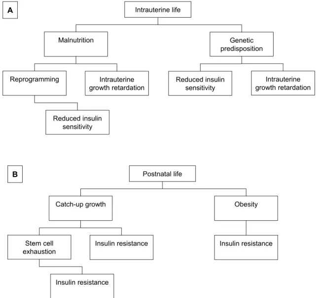
still unclear. Several models have been proposed and it is likely that each hypothesis might represent a differ-ent piece of the same puzzle (Figure 1).
The “thrifty phenotype” hypothesis
The original “thrifty genotype” theory was pro-posed by Neel in 1962 [25] to explain the high preva-lence of type 2 diabetes in Western populations. Neel hypothesized that genes favoring survival during fam-ine would become detrimental when food supply
be-Intrauterine life Malnutrition Reprogramming Intrauterine growth retardation Genetic predisposition Reduced insulin sensitivity Intrauterine growth retardation Reduced insulin sensitivity A Postnatal life Catch-up growth Stem cell
exhaustion Insulin resistance
Obesity
Insulin resistance
Insulin resistance B
Figure 1. A summary of the mechanisms proposed to explain the association between intrauterine malnutrition and
metabolic disturbances. During intrauterine life (A), fetal malnutrition leads to growth retardation and induces a repro-gramming of both the endocrine system and metabolic pathways. When present, genetic predisposition may partly ex-plain both intrauterine growth retardation and reduced insulin sensitivity. In postnatal life (B), rapid catch-up growth in weight and/or length might induce peripheral insulin resistance and lead to an early exhaustion of the tissue stem cell reservoir. Obesity represents the major additional risk factor for the development of insulin resistance.
come abundant, thus explaining the association be-tween malnutrition and later development of metabolic complications.
In 1992 Hales and Barker proposed the “thrifty phenotype” hypothesis [26]. According to the thrifty phenotype model, the growing fetus exposed to nutri-tional deprivation adopts at least two strategies to aid survival [26]. First, it diverts nutrients to the brain to preserve brain growth at the expense of body growth and the development of other organs such as pancreas, liver, and muscle. Second, metabolic reprogramming occurs in a manner that is beneficial to survival under conditions of poor postnatal nutrition. However, if the organism is born into conditions of adequate or over-nutrition, then this may conflict with the earlier repro-gramming and insulin resistance, and, later on, type 2 diabetes may result [26]. The reprogramming process would occur during ‘critical periods’ of embryo-fetal life characterized by high cell proliferation rate in the developing tissues.
The “fetal salvage” hypothesis
This model rises from the observation that SGA children show a far greater insulin response than nor-mal birthweight children. The “fetal salvage” model [27] suggests that the malnourished fetus develops peripheral insulin resistance, in an otherwise normal insulin secretion, which allows a redistribution of nu-trients, such as glucose, in favor of essential organs. This then leads to a permanent reduction in skeletal muscle glucose transporter number or function. This reduced peripheral insulin sensitivity stimulates cells to produce larger amounts of insulin to achieve normal glycaemia and would lead to eventual cell exhaustion. This hypothesis is supported by studies in animal models showing reduced glucose transporter protein concentrations in skeletal muscles of intrauterine growth retarded fetuses and normal concentrations in the brain [28, 29].
The “fetal insulin” hypothesis
Hattersley and Tooke proposed that the association between low birthweight and adult insulin resistance is principally genetically mediated [30]. Genetically de-termined insulin resistance could result in low insulin-mediated fetal growth in utero as well as insulin
resis-of fetal tissues to the effects resis-of insulin [30]. According to this model, polygenic genetic factors that increase insulin resistance, both in utero and in adult life, would produce two phenotypes: a small, thin baby and an adult with insulin resistance and increased risk of car-diovascular disease, particularly in the presence of obesity. Consistent with this hypothesis, Dunger et al. showed an association between common allelic varia-tion (class I or class III) at the variable number of tandem repeat locus in the promoter region of the insulin gene and birth weight [31].
Results from Vassen and co-workers [32] support the hypothesis that genetic variation, by affecting fetal growth, could account for the association between low birth weight and susceptibility to diabetes and cardio-vascular disease in later life. An association between a polymorphism in the promoter region of IGF-I gene and birth weight was found. Individuals who did not have the wild-type allele (192 bp) of the polimorphism had a 215 g lower birth weight than those homozygous for this allele. The same team previously showed that this genetic variation resulted in low circulating IGF-I concentrations, reduced height in adulthood, dimin-ished insulin-secreting capacity, and a high risk of type 2 diabetes and myocardial infarction [33]. Taken to-gether, their findings indicate that association between low birth weight, diabetes, and cardiovascular disease could be a result of genetic variation of the IGF-I gene, affecting both fetal growth and susceptibility to late-onset disease. A role of the polymorphism in the IGF-I gene in the development of fetal growth retar-dation has also been found by Arends and co-workers [34]. Finally, mutations of the IGF-I receptor have recently been described in two children with intrauter-ine growth retardation [35].
The “catch-up growth” hypothesis
SGA children show a rearrangement of the endo-crine system at birth, having low concentrations of insulin, IGF-I, IGFBP-3, and high levels of the growth hormone, IGFBP-1 and IGFBP-2. Normalization of these parameters occurs early during the first trimester of life [12, 13]. On the basis of this rapid adaptation to the extra-uterine environment, we have formulated the ‘catch-up growth’ hypothesis, suggesting that tissues chronically depleted of nutrients and, consequently, insulin and insulin-like growth factors (IGFs) during
Rev Diabetic Stud (2004) 1:58-65 Copyright © by The SBDR tance [36]. According to this model, the first two years
of life, when catch-up growth occurs in about 80% of IUGR children, represent the crucial time for the de-velopment of long-term consequences. Those SGA infants who show early and complete recovery from intrauterine growth retardation would be at higher risk for the occurrence of metabolic disturbances.
Several independent observations strongly support our hypothesis. Data from animals have shown that when fetal growth impairment is followed by catch-up growth postnatally, the lifespan is significantly short-ened [37]. Moreover, the glucose induced insulin re-sponse in infants with catch-up growth is higher than that in children without significant catch-up growth [38]. Furthermore, an association between postnatal catch-up growth in height and/or weight and increased risk of developing type 2 diabetes in later life has been described [39-43]. Recently, Bazaes and co-workers [44] have also found a significant correlation between early postnatal growth rates and insulin sensitivity and secretion in childhood. The same research team in a prospective cohort of SGA and AGA infants, showed that both basal and first-phase insulin release are highly correlated with the extent of centile crossing in weight and length during the first year of life [45]. Finally, we have recently reported that concentrations of adi-ponectin, an adipocytokine with insulin-sensitizing and antiatherogenic properties, are reduced in SGA chil-dren, and are even lower in those with postnatal catch-up growth [46].
The ”stem cell” hypothesis
Another fascinating model stems from the finding that postnatal tissues contain stem cells that, though quiescent, retain their capacity to self-renew and re-generate tissues to fulfill organ demands. We have recently proposed that intrauterine malnutrition re-duces tissue stem cell stores, eventually leading, in adulthood, to an early exhaustion of organ function, especially when demands are increased. According to this model, the ‘critical periods’ would correspond to the time windows of stem cell proliferation, early commitment and migration to the final destination in tissues [47]. Pancreas, endothelium, growth plate and brain might have a reduced stem cell reservoir and, hence, might not be able to face increased biological demands, ultimately exhausting their regeneration and repairing potential [47].
Is glucose metabolism impaired in SGA
chil-dren?
Whilst several epidemiological surveys have con-firmed the association between metabolic disturbances in adulthood and low birth size, few and conflicting data exist for childhood (Table 1). The potential im-pact of the early recognition of altered insulin sensitiv-ity in clinical practice is high, because it might prompt the establishment of appropriate hormone-, diet-, or lifestyle-based strategies to prevent the long-term metabolic consequences of intrauterine growth retar-dation.
Yanjnik and colleagues performed a glucose toler-ance test in 379 4-year-old low birthweight Indian children. 30 minutes after an oral glucose load, subjects with lower birth weights had higher plasma glucose and insulin levels, irrespective of their current size [48].
Law and colleagues carried out an abbreviated oral glucose tolerance test in 7-year-old children, finding that subjects who were thin at birth had higher plasma glucose concentration [49].
Hofman and colleagues [27] investigated insulin sensitivity in short prepubertal SGA children com-pared with short prepubertal children born with a birth weight appropriate for gestational age. They found that the SGA group was less insulin-sensitive than controls, although there were no differences in the plasma glu-cose.
Veening et al. found no significant difference in glucose tolerance and beta-cell function between the SGA and AGA groups. However, the hyperinsu-linemic clamp showed a reduced insulin sensitivity in SGA children, especially in SGA children with catch-up growth and a high BMI [39]. The same research group studied beta-cell capacity and insulin sensitivity in 28 prepubertal SGA children and in 22 prepubertal AGA children. The SGA children showed decreased insulin sensitivity rather than decreased beta-cell capac-ity [50].
Whincup performed a school-based survey of 10- to 11-year-old British children. 590 children were stud-ied in fasting conditions while 547 were investigated 30 minutes after a standard oral glucose load. Neither fasting nor post-load glucose levels showed any rela-tionship with birth weight or ponderal index at birth. After adjustment for childhood height and ponderal index, both fasting and post-load insulin levels de-creased with increasing birth weight [51]. The authors concluded that low birth weight is not related to
glu-cose intolerance at 10-11 years, but may be related to the early development of insulin resistance [51].
After an oral glucose challenge, Potau and colleagues showed the presence of high insulin levels in an early marker of insulin resistance in adulthood [52].
51 children and 49 adolescents born SGA com-pared to normal subjects born AGA and proposed that this elevated insulin concentration might be consid-ered as 1 year of age. SGA subjects were subdivided into children with weight or length catch-up growth.
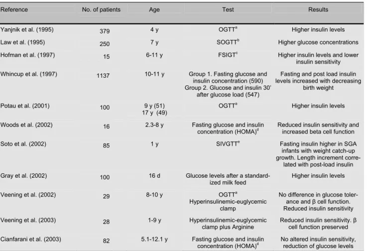
Table 1 Published studies carried out in children
a Oral glucose tolerance test. b Short oral glucose tolerance test. c Frequently sampled IV glucose tolerance test. d Homeostasis model
assessment. e Short IV glucose tolerance test.
Woods et al. demonstrated that short SGA children have reduced insulin sensitivity and increased beta cell function as if higher insulin concentrations were needed to maintain a normal glucidic homeostasis. These two variables were significantly related to over-night GH secretion. An elevated pattern of GH secre-tion, similar to that observed during fasting and malnu-trition, was observed and the authors postulated that resistance to the somatotropic actions of GH and IGF-I in short SGA children may contribute directly
dren that had weight catch-up growth, compared with those who did not catch-up and AGA infants. On the other hand, length increment was the principal deter-minant of post-load insulin secretion. Therefore, fast-ing insulin sensitivity was related to weight catch-up growth and actual BMI, whereas insulin secretion was directly related to length catch-up growth [45].
Gray and colleagues studied 100 premature and/or small for gestational age infants (age range: 1-65 days). Fasting and postprandial glucose and insulin levels
Reference No. of patients Age Test Results
Yanjnik et al. (1995) 379 4 y OGTTa Higher insulin levels
Law et al. (1995) 250 7 y SOGTTb Higher glucose concentrations
Hofman et al. (1997) 15 6-11 y FSIGTc Higher insulin levels and lower
insulin sensitivity Whincup et al. (1997) 1137 10-11 y Group 1. Fasting glucose and
insulin concentration (590) Group 2. Glucose and insulin 30’
after glucose load (547)
Fasting and post load insulin levels increased with decreasing
birth weight
Potau et al. (2001) 100 9 y (51)
17 y (49)
OGTTa
Higher insulin levels Woods et al. (2002) 16 2.3-8 y Fasting glucose and insulin
concentration (HOMA)d Reduced insulin sensitivity and increased beta cell function
Soto et al. (2002) 85 1 y SIVGTTe Fasting insulin higher in SGA
infants with weight catch-up growth. Length increment
corre-lated with post-load insulin Gray et al. (2002) 100 16 d Glucose levels after a
standard-ized milk feed Higher insulin levels
Veening et al. (2002) 29 8-10 y OGTTa
Hyperinsulinemic-euglycemic clamp
No difference in glucose toler-ance and β cell function. Reduced insulin sensitivity
Veening et al. (2003) 28 1-9 y Hyperinsulinemic-euglycemic
clamp plus Arginine
Reduced insulin sensitivity. β cell function preserved Cianfarani et al. (2003) 82 5.1-12.1 y Fasting glucose and insulin
Rev Diabetic Stud (2004) 1:58-65 Copyright © by The SBDR despite similar glucose levels. Postnatal growth velocity
correlated with birth weight and insulin resistance independently of each other. The authors suggested that glucose tolerance of the neonate is determined by weight attained at birth irrespective of gestational age and that maternal blood pressure may influence insulin sensitivity of the newborn. Furthermore, their findings indicate that catch-up growth in neonates is deter-mined by birth weight and insulin sensitivity [54].
Although several reports suggest reduced insulin sensitivity in SGA children, to date very few strict case-control studies have been carried out. We have recently investigated the endocrine status in SGA chil-dren, compared with children born appropriate for gestational age (AGA), strictly matched for age, sex, pubertal status, body mass index (BMI), and height [55]. SGA subjects did not have differences in the indices of insulin sensitivity, but showed significantly lower baseline glucose levels. This finding is consistent with recent studies in animal models using a low-protein diet during pregnancy to produce growth re-striction of the offspring [56]. In young adult life, low-protein offspring have an improved glucose tolerance, compared with controls [57, 58]. This is associated with increased muscle and adipocytes insulin receptors and augmented insulin-stimulated glucose uptake into skeletal muscle [59] and adipocytes [60]. Later on, however, offspring undergo an age-dependent loss of glucose tolerance, such that by 15 months of age, low-protein offspring have a significantly worse glucose tolerance, compared with controls [58]. We speculated that, in human, an early phase of increased insulin sensitivity during childhood might precede the onset of insulin resistance in young adult SGA subjects.
Conclusions
There is increasing epidemiological evidence sug-gesting that certain metabolic disorders in adults such
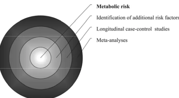
as insulin resistance and type 2 diabetes might originate from in utero malnutrition. Although many hypotheses have been proposed, the molecular mechanisms under-lying this epidemiological association are still un-known. Much less clear is the impact of low birth weight on glucose homeostasis in childhood. Most published reports indicate only subtle abnormalities of insulin secretion and/or insulin sensitivity with no alterations of glucose metabolism in SGA children. A meta-analysis might be appropriate to integrate the results of several independent studies and critically review all the available data for childhood. At the same time, longitudinal case-control studies on large cohorts of children from birth to adolescence are needed to quantify the metabolic risk, identify the critical time windows, and determine the effects of other risk fac-tors, such as obesity or genetic predisposition (Figure 2).
Figure 2. Strategies to ascertain and quantify the metabolic risk in SGA children.
Acknowledgments: The work was supported in part by MIUR, grant COFIN 40%-2003064547.
■
References1. Lee AP, Chernausek SD, Hokken-Koelega ACS,
Czer-nichow P. International small for gestational age advisory
board consensus development conference statement: man-agement of short children born small for gestational age, April 24-October 1 2001. Pediatrics 2003. 111:1253-1261.
2. Markestad T, Vik T, Ahlsten G, Gebre-Medhin M,
Skjaer-ven R, Jacobsen G, Hoffman HJ, Bakketeig LS.
Small-for-gestational-age (SGA) infants born at term: growth and devel-opment during the first year of life. Acta Obstet Gynecol Scand Suppl 1997.165:93-101.
3. Bernstein PS, Divon MY. Etiologies of fetal growth restric-tion. Clin Obstet Gynecol 1997. 40:723-729.
4. Pollack RN, Divon MY. Intrauterine growth retardation: definition, classification and etiology. Clin Obstet Gynecol 1992. 35:99-107.
5. Wollmann HA. Intrauterine growth restriction: definition and etiology. Horm Res 1998. 49(Suppl 2):1-6.
6. Fitzhardinge PM, Stevan EM. The small for date infant later growth patterns. Pediatrics 1972. 49:671-681.
7. Karlberg J, Albertsson-Wickland K. Growth in full-term small-for-gestational-age infants: from birth to final height.
Pe-diatr Res 1995. 38:733-739.
8. Ong KI, Ahmed MI, Emmet PM, Preece MA. For the Avon Longitudinal Study of Pregnancy and Childhood Study Team. Association between postnatal catch-up growth and obesity in childhood: prospective cohort study. BMJ 2000.
320:967-971.
9. Albertsson-Wickland K, Karlberg JPE. Postnatal growth of children born small for gestational age. Acta Ped Suppl 1997. 423:193-195.
10. Karlberg JPE, Albertsson-Wickland K, Kwan EYW, Lam
BCC. The timing of early postnatal catch-up growth in normal,
full term infants born small for gestational age. Horm Res 1997. 48(Suppl 1):17-24.
11. Leger J, Levy-Marchal C, Bloch J. Reduced final height and indication for insulin resistance in 20-year-olds born small for gestational age: regional cohort study. BMJ 1997. 315: 341-347. 12. Leger J, Noel M, Limal KM, Czernichow P. Growth fac-tors and intrauterine growth retardation. Serum growth hor-mone, insulin-like growth factor I, and IGF-binding protein-3, levels in children with intrauterine growth retardation com-pared with normal control subjects: prospective study from birth to two years of age. Pediatr Res 1996. 40:101-107.
13. Cianfarani S, Germani D, Rossi P, Rossi L, Germani A,
Ossicini C, Zuppa A, Argirò G, Holly JMP, Branca F.
In-trauterine growth retardation: evidence for the activation of the insulin-like growth factor (IGF)-related growth-promoting ma-chinery and the presence of a cation-independent IGF binding protein-3 proteolytic activity by two months of life. Pediatr Res 1998. 44:374-380.
14. Cianfarani S, Geremia C, Scott CD, Germani D. Growth, IGF system, and cortisol in children with intrauterine growth retardation: is catch-up growth affected by reprogramming of the hypothalamic-pituitary-adrenal axis? Pediatr Res 2002. 51:94-99.
15. Barker DJ, Osmond C, Golding J, Kuh D, Wadsworth
ME. Growth in utero, blood pressure in childhood and adult
life, and mortality from cardiovascular disease. BMJ 1989. 298(6673):564-567.
16. Barker DJ. The fetal origins of adult hypertension. J
Hyperten-sion 1992. 10:S39-S44.
17. Barker DJ. Fetal origins of coronary disease. BMJ 1995. 311:171-174.
18. Barker DJ, Winter PD, Osmond C, Margetts B,
Sim-monds SJ. Weight in infancy and death from ischaemic heart
disease. Lancet 1989. 2:577-580.
19. Phillips DI, Barker DJ, Fall CH, Seckl JR, Whorwood
CB, Wood PJ, Walker BR. Elevated plasma cortisol
concen-trations: a link between low birth weight and the insulin resis-tance syndrome? J Clin Endocrinol Metab 1998. 83:757-760. 20. Hales CN, Barker DJ, Clark PM, Cox LJ, Fall C, Osmond
C, Winter PD.Fetal and infant growth and impaired glucose tolerance at age 64. BMJ 1991. 303:1019-1022.
21. Valdez R, Athens MA, Thompson GH, Bradshaw BS,
Stern MP. Birth-weight and adult health outcomes in a
bieth-nic population in USA. Diabetologia 1994. 37:624-631.
22. Barker DJ, Hales CN, Fall CH, Osmond C, Phipps K,
Clark PM. Type 2 (non-insulin-dependent) diabetes mellitus,
hypertension and hyperlipidaemia (syndrome X): relation to reduced fetal growth. Diabetologia 1993. 36(1):62-67.
23. Ibanez L, Potau N, Francois I, de Zegher F. Precocious pubarche, hyperinsulinism, and ovarian hyperandrogenism in girls: relation to reduced fetal growth. J Clin Endocrinol Metab 1998. 83:3558-3562.
26. Hales CN, Barker DJ. Type 2 (non-insulin-dependent) diabe-tes mellitus: the thrifty phenotype hypothesis. Diabetologia 1992. 35:595-601.
27. Hofman PL, Cutfield WS, Robinson EM, Bergman RN,
Menon RK, Sperling MA, Gluckman PD. Insulin resistance
in short children with intrauterine growth retardation. J Clin
Endocrinol Metab 1997. 82:402-406.
28. Simmons RA, Gounis AS, Bangalore SA, Ogata ES. In-trauterine growth retardation: fetal glucose transport is dimin-ished in lung but spared in brain. Pediatr Res 1985. 31:59-63. 29. Simmons RA, Flozak AS, Ogata ES. The effect of insulin,
and insulin-like growth factor I on glucose transport in normal and small for gestational age fetal rats. Endocrinology 1993. 133:1361-1368.
30. Hattersley AT, Tooke JE. The fetal insulin hypothesis: an alternative explanation of the association of low birthweight with diabetes and vascular disease. Lancet 1999. 353:1789-1792. 31. Dunger DB, Ong KK, Huxtable SJ, Sherriff A, Woods KA,
Ahmed ML, Golding J, Pembrey ME, Ring S, Bennett ST, et al. Association of the INS VNTR with size at birth.
AL-SPAC study team. Avon longitudinal study of pregnancy and childhood. Nat Genet 1998.19:98–100.
32. Vaessen N, Janssen JA, Heutink P, Hofman A, Lamberts
SWJ, Oostra BA, Pols HA, van Duijn CM. Association
be-tween genetic variation in the gene for insulin-like growth fac-tor-I and low birthweight. Lancet 2002. 23;359(9311):1036-1037.
33. Vaessen N, Heutink P, Janssen JA, Witteman JCM,
Tets-res L, Hofman A, Lamberts SWJ, Oostra BA, Pols HAP, van Duijn CM. A polymorphism in the gene for IGF-I.
Func-tional properties and risk for type 2 diabetes and myocardial in-farction. Diabetes 2001. 50:637-642.
34. Arends N, Johnston L, Hokken-Kolega A, Van Duijn C,
De Ridder M, Savage M, Clark A. Polymorphism in the
IGF-I gene: clinical relevance for short children born small for gestational age (SGA). J Clin Endocrinol Metab 2003. 87:2720-2724.
35. Abuzzahab MJ, Schneider A, Goddard A, Grigorescu F,
Lautier C, Keller E, Kiess W, Klammt J, Kratzsch J, Os-good D, et al. IGF-I receptor mutations resulting in
in-traueterine and postnatal growth retardation. N Engl J Med 2003. 349:2211-2222.
36. Cianfarani S, Germani D, Branca F. Low birth weight and adult insulin resistance: “The catch-up growth” hypothesis.
Arch Dis Child 1999. 81:F71-F73.
37. Hales CN, Desai M, Ozanne SE, Crowther NJ. Fishing in the stream of diabetes: from measuring insulin to the control of fetal organogenesis. Biochem Soc Trans 1996. 24:341-350. 38. Colle E, Schiff D, Andrew G, Bauer CB, Fitzhardinge P.
Insulin responses during catch-up growth of infants who were small for gestational age. Pediatrics 1976. 57:363–371.
39. Veening MA, Van Weissenbruch MM, Delemarre-Van De
Waal HA. Glucose tolerance, insulin sensitivity, and insulin
secretion in children born small for gestational age. J Clin
Endo-crinol Metab 2002. 87:4657–4661.
40. Eriksson JG, Forsen T, Tuomilehto J, Winter PD,
Os-mond C, Barker DJ. Catch-up growth in childhood and death
Rev Diabetic Stud (2004) 1:58-65 Copyright © by The SBDR
42. Ong KKL, Ahmed ML, Emmett PM, Preece MA, Dunger
DB. Association between postnatal catch-up growth and
obe-sity in childhood: prospective cohort study. BMJ 2000. 320:967–971.
43. Singhai A, Fewtrell M, Cole TJ, Lucas A. Low nutrient
intake and early growth for later insulin resistance in adoles-cents born preterm. Lancet 2003. 361:1089–1097.
44. Bazaes RA, Alegria AL, Pittaluga E, Avila A, Iniguez G,
Mericq V. Determinants of insulin sensitivity and secretion in
very-low-birth-weight children. J Clin Endocrinol Metab 2004. 89:1267-1272.
45. Soto N, Bazaes RA, Pena W, Salazar T, Avila A, Iniguez
G, Ong KK, Dunger DB, Mericq V. Insulin sensitivity and
secretion are related to catch-up growth in small-for-gestational-age infants at age one year: results from a prospec-tive cohort. J Clin Endocrinol Metab 2003. 88:3645–3650. 46. Cianfarani S, Martinez C, Maiorana A, Spadoni GL, Scirè
G, Boemi G. Adiponectin levels are reduced in children born
small for gestational age (SGA) and are inversely related to postnatal catch-up growth. J Clin Endocrinol Metab 2004. 89:1346-1351.
47. Cianfarani S. Fetal origins of adult diseases: just a matter of stem cell number? Med Hypoth 2003. 61:401-404.
48. Yajnik CS, Fall CH, Vaidya U, Pandit AN, Bavdekar A,
Bhat DS, Osmond C, Hales CN, Barker DJ. Fetal growth
and glucose and insulin metabolism in four-year-old Indian children. Diabet Med 1995.12(4):330-336.
49. Law CM, Gordon GS, Shiell AW, Barker DJ, Hales CN. Thinness at birth and glucose tolerance in seven-year-old chil-dren. Diabet Med 1995.12(1):24-29.
50. Veening MA, van Weissenbruch MM, Heine RJ,
Dele-marre-van de Waal HA. Beta-cell capacity and insulin
sensi-tivity in prepubertal children born small for gestational age: in-fluence of body size during childhood. Diabetes 2003. 52(7):1756-1760.
51. Whincup PH, Cook DG, Adshead F, Taylor SJ, Walker M,
Papacosta O, Alberti KG. Childhood size is more strongly
re-lated than size at birth to glucose and insulin levels in 10-11-year-old children. Diabetologia 1997. 40(3):319-326.
52. Potau N, Gussinye M, Sanchez Ufarte C, Rique S,
Vicens-Calvet E, Carrascosa A. Hyperinsulinemia in pre- and
post-pubertal children born small for gestational age. Horm Res 2001. 56(5-6):146-150.
53. Woods KA, Van Helvoirt M, Ong KKL, Mohon A, Levy J,
De Zegher F, Dunger DB. The somatotropic axis in short
children born small for gestational age: relation to insulin resis-tance. Pediatr Res 2002. 51:76-80.
54. Gray IP, Cooper PA, Cory BJ, Toman M, Crowther NJ. The intrauterine environment is a strong determinant of glu-cose tolerance during the neonatal period, even in prematurity.
J Clin Endocrinol Metab 2002. 87(9):4252-4256.
55. Cianfarani S, Maiorana A, Geremia C, Scirè G, Spadoni
GL, Germani D. Blood glucose concentrations are reduced in
children born small for gestational age (SGA), and thyroid– stimulating hormone levels are increased in SGA with blunted postnatal catch-up growth. J Clin Endocrinol Metab 2003. 88:2699-2705.
56. Desai M, Crowther NJ, Lucas A, Hales CN. Organ-selective growth in the offspring of protein-restricted mothers.
Br J Nutr 1996. 76(4):591-603.
57. Langley SC, Browne RF, Jiacson AA. Altered glucose toler-ance in rats exposed to maternal low protein diets in utero.
Comp Biochem Phisiol 1994. 109:223-229.
58. Hales CN, Desai M, Ozanne SE, Crowther NJ. Fishing in the stream of diabetes: from measuring insulin to the control of fetal organogenesis. Biochem Soc Trans 1996. 24(2):341-350. 59. Ozanne SE, Wang CL, Coleman N, Smith GD. Altered
muscle insulin sensitivity in the male offspring of protein-malnourished rats. Am J Physiol 1996. 271(6 Pt 1):E1128-1134. 60. Ozanne SE, Nave BT, Wang CL, Shepherd PR, Prins J,
Smith GD. Poor fetal nutrition causes long-term changes in
expression of insulin signaling components in adipocytes. Am J