UNIVERSITÀ DEGLI STUDI DI SALERNO
Dipartimento di Farmacia
PhD Program
in Drug Discovery and Development
XXXI Cycle — Academic Year 2018/2019PhD Thesis in
Target-based study to identify new
bioactive diterpenes
Candidate Supervisors
Lorenzo Fiengo Prof. Nunziatina De Tommasi Prof.Fabrizio Dal Piaz
“Della vita non bisogna temere nulla. Bisogna solo capire"
(Marie Curie)
Index
Abstract... I
- Chapter 1 - ... 1
Introduction ... 3
1.1 Plant small molecules in drug discovery and development ... 3
1.2 Terpenes ... 7
1.3 Diterpenes in cancer ... 9
1.4 Drug discovery today: Chemical Genetics ... 12
1.5 Aim of the thesis ... 14
1.6 Cell-free and cell-based assays ... 14
1.7 Outline of the thesis ... 23
- Chapter 2 - ... 25
Introduction ... 27
2.1 Nucleolin: structure and localizations ... 27
2.1.1 Roles of Nucleolin in physiological and pathological pathways ... 34
2.1.2 Roles of Nucleolin in ribosome biogenesis ... 35
2.1.3 Nucleolin as shuttle between nucleus and cytoplasm ... 38
2.1.4 Nucleolin as receptor on the cell surface ... 39
2.1.5 Nucleolin as tumor marker: therapeutic strategies ... 39
2.1.6 Nucleolin Inhibitors ... 43
2.1.7 Aim of the project ... 46
Results and discussion ... 47
2.2 Screening of diterpenes by Cellular Thermal Shift Assay (CETSA) ... 47
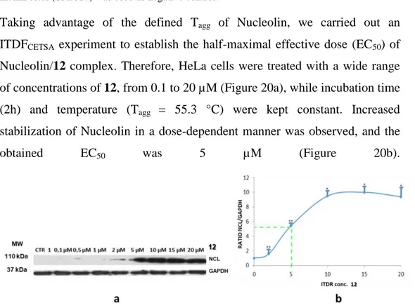
2.2.1 Biological activity of 6,19-dihydroxy-ent-trachiloban-17-oic acid (12) in Jurkat and HeLa cells ... 52
2.2.3 Study of Nucleolin/12 interaction by Drug Affinity Responsive Target
Stability (DARTS) in HeLa cells ... 57
2.2.4 MS-based identification of further targets of 6,19-dihydroxy-ent-trachiloban-17-oic acid ... 60
2.2.5 Investigation of 6,19-dihydroxy-ent-trachiloban-17-oic acid activity in the cell migration: Wound Healing Assay ... 61
2.2.6 Biological effect of 6,19-dihydroxy-ent-trachiloban-17-oic acid on Nucleolin into the cell ... 62
2.2.7 6,19-dihydroxy-ent-trachiloban-17-oic acid activity in subcellular compartments ... 63
2.2.8 RTq-PCR and Western Blot analyses ... 65
2.2.9 Inhibition of Protein Synthesis ... 66
2.2.10 Nucleolin (RBD1-2) expression and purification ... 67
2.2.11 Screening of diterpenes against Nucleolin (RBD 1-2) by Saturation Transfer Difference NMR (STD-NMR) ... 70
2.2.12 Screening of diterpenes against Nucleolin (RBD 1-2) by WaterLOGSY 74 2.2.13 Diterpenes/Nucleolin (RBD 1-2) interaction by SPR ... 77
2.2.14 Conclusions ... 78
Materials and methods ... 79
2.3 Determination of the apparent melting curve (Tm) of Nucleolin in HeLa cells by Cellular Thermal Shift Assay (CETSA). ... 79
2.3.1 Determination of the EC50 of the complex (ITDRFCETSA) ... 80
2.3.2 Drug Affinity Responsive Target Stability (DARTS) experiment ... 80
2.3.3 Wound Healing assay ... 82
2.3.4 RNA isolation and quantitative Real-Time-PCR (qRT-PCR) ... 82
2.3.5 Protein Synthesis assay ... 83
2.3.6 Cell viability and cell cycle analysis... 83
2.3.8 Expression and purification of recombinant NCL (RBD1-2) ... 84
2.3.9 Ligand-binding NMR experiments ... 85
2.3.10 Saturation Transfer Difference NMR (STD-NMR) analysis ... 85
2.3.11 WaterLOGSY experiment ... 86
2.3.12 Surface Plasmon Resonance (SPR) ... 87
2.3.13 Western Blot analysis ... 87
2.3.14 Reagents and Antibodies ... 88
2.3.15 Cytosol and membrane extracts ... 88
2.3.16 Nuclear extracts ... 89
2.3.17 Statistical analysis ... 89
- Chapter 3 - ... 91
Introduction ... 93
3.1 Heat Shock Proteins (HSPs) family ... 93
3.1.1 Heat Shock Protein 70 (Hsp70): structure, localizations and roles ... 94
3.1.2 Hsp70 inhibitors ... 103
3.1.4 Aim of the project ... 104
Results and discussion ... 106
3.2 Identification of Hsp70 ligands by Surface Plasmon Resonance (SPR) ... 106
3.2.1 Epoxysiderol ... 111
3.2.2 MS-based studies on the epoxysiderol-Hsp70 interaction ... 112
3.2.3 Cytotoxic activity of epoxysiderol ... 114
3.2.4 Validation study of epoxysiderol/Hsp70 interaction by DARTS ... 114
3.2.5 Western Blot of Hsp70, Hsp70 co-chaperones and client proteins ... 117
3.2.6 Evaluation of epoxysiderol biological activity in HeLa cells by flow cytometry and WB ... 119
3.2.7 Epoxysiderol effect on Hsp70 levels in subcellular compartments ... 122
3.2.8 Inhibition of Hsp70 ATPase activity mediated by epoxysiderol ... 123
3.2.10 Conclusions ... 126
Materials and Methods... 128
3.3 Surface Plasmon Resonance (SPR) ... 128
3.3.1 Drug Affinity Responsive Target Stability on HeLa cells ... 128
3.3.2 Monodimensional electrophoresis SDS Page ... 129
3.3.3 LC/MS/MS Analysis ... 130
3.3.4 HSP70 peptide mapping ... 130
3.3.5 Cell culture and treatment ... 131
3.3.6 Cell viability and cell cycle analyses ... 131
3.3.7 Determination of apoptosis ... 132
3.3.8 Western Blot analysis ... 132
3.3.9 Cytosol and membrane extracts ... 133
3.3.10 Nuclear extracts ... 133
3.3.11 ATPase activity Assay ... 134
3.3.12 Reagents and Antibodies ... 134
3.3.13 Molecular Docking Studies ... 135
3.3.14 Statistical analysis ... 136
- Chapter 4 - ... 137
Introduction ... 139
4.1 Heat Shock Protein 90: isoforms and localizations ... 139
4.1.1 Hsp90 structure ... 140
4.1.2 Hsp90 ATPase activity and ATP binding Site... 142
4.1.3 Hsp90 client proteins ... 146
4.1.4 Functions of Hsp90 in cancer ... 147
4.1.6 Hsp90 inhibitors: therapeutic targeting of Hsp90 ATPase activity ... 148
Results and discussion ... 150
4.2.1 Antiproliferative activity of fusicoccanes and abietanes tested by MTT
on Jurkat, HeLa and MCF7 cell lines ... 152
4.2.2 Evaluation of the biological activity of the selected diterpenes by flow cytometry and WB ... 154
4.2.3 Effect of 6 and 20 on Hsp90α ATPase activity ... 157
4.2.4 Study of the interaction Hsp90α /6 by molecular docking ... 158
4.2.5 Conclusions ... 159
Materials and methods ... 161
4.3 Reagents and Antibodies ... 161
4.3.1 Surface Plasmon Resonance Analyses ... 161
4.3.2 ATP hydrolysis inhibition ... 162
4.3.3 Cell Culture and Treatment ... 163
4.3.5 Western Blot Analyses ... 164
4.3.6 Statistical Analysis ... 164
4.3.7 Molecular Docking Studies ... 164
4.3.8 Induced Fit Docking ... 165
Bibliography ... 166
Publications ... 187
Abstract
Target identification and mechanism of action studies of plant-derived compounds play a critical role in drug discovery. The knowledge of the bioactivity of natural compounds can lead to a number of advantages: first of all, it is possible to understand their full therapeutic potential; in addition, it can allow the further identification of their side-effects, their toxicity and also structure-activity relationships studies.
This research project is focused on a Reverse Chemical Genetics approach, which relies on the screening of libraries of plant small molecules (provided by the Department of Pharmacy (DIFARMA)-Bioactive Natural Products, University of Salerno (UNISA), Fisciano, Italy) able to bind specific target proteins and on the validation of the ligand/protein interaction. In my PhD project I focused on three protein targets, over-expressed in cancer and identified as potential markers in several tumor cell lines: Nucleolin, Heat Shock Protein 70 (Hsp70) and Heat Shock Protein 90 (Hsp90).
Nucleolin (NCL) is a multifunctional protein involved in many process such as DNA transcription, ribosome biogenesis and regulation of mRNAs of anti-apoptotic and antiproliferative proteins such as AKT1, Bcl2, p53. Firstly, a screening of ent-kaurane and ent-trachilobane library by Cellular Thermal Shift Assay (CETSA) on Jurkat (leukemia T cells) and HeLa (cervical carcinoma) was performed, obtaining as main ligand of Nucleolin the 6,19-dihydroxy-ent-trachiloban-17-oic acid (12) from Psiadia punctulata ((Vatke) Asteraceae). Full length Nucleolin/12 interaction was validated in HeLa (cervical carcinoma) cells by CETSA and Drug Affinity Responsive Target Stability (DARTS). Nucleolin RNA Binding Domains 1-2/12 interaction was investigated by Saturation Transfer Difference NMR (STD-NMR), WaterLOGSY and Surface Plasmon Resonance (SPR): no interaction was
observed with these two domains of the protein. The mechanism of action of the selected diterpene was studied by Flow Cytometry (sub G0/G1 cell cycle arrest), WB analysis (reduction of intracellular AKT1 and Bcl2 levels and pNCL levels on the cell membrane), RTq-PCR (reduction of AKT1 and Bcl2 mRNAs), MTT (IC50: 20 ± 1 µM), Protein Synthesis and Wound Healing assays in HeLa cells (reduction of 20% of migration). Therefore, the 6,19-dihydroxy-ent-trachiloban-17-oic acid (12) may be considered as a new promising modulator of Nucleolin.
The second target protein was the molecular chaperon Heat Shock Protein 70 (Hsp70). A diterpene library was screened by SPR assay, in order to select putative Hsp70 ligands. SPR results showed that the ent-7β-acetoxy,18-hydroxy-15α,16α-epoxikaurane (epoxysiderol or compound 27) from Sideritis spp (Lamiaceae) interacts with Hsp70 (KD: 54 ± 1.2 nM). Epoxysiderol ability to modulate Hsp70 activity was assessed through MS (no covalent binding), DARTS and WB experiments. Moreover, epoxysiderol was tested on HeLa cells by MTT (IC50: 20 ± 0.9 µM), Flow Cytometry (G2/M and subG0/G1 cell cycle arrest), WB for its effect on the intracellular levels of Hsp70, Hsp90, and Hsp70 client proteins (reduction of pAKT1, p38 and p-JNK1) in HeLa cells, and by WB also for Hsp70 cytosolic and cell membrane levels (reduction of Hsp70 levels). Finally, ATPase assay (50% of reduction in dose-dependent manner) and molecular docking studies (interaction with the Hsp70 Nucleolide Binding Domain) were carried out. Therefore, in this study epoxysiderol was identified as a new Hsp70 inhibitor through cell-free and cell-based assays.
Another target object of study in this PhD project was the Heat Shock Protein 90 (Hsp90). Fusicoccane diterpenes from Hypoestes forsskaolii ((Vahl) Acanthaceae), abietane diterpenes form Zhumeria majdae ((Rech.f. & Wendelbo) Lamiaceae) and from different Salvia spp (Lamiaceae) were
screened against Hsp90 by SPR and by MTT in HeLa, Jurkat and MCF7 cells, selecting the 18-hydroxyhypoestenone (6) and lanugon Q (20) as Hsp90 ligands. Subsequently, MTT assay was performed to investigate their cytotoxic and anti-proliferative activity: 18-hydroxyhypoestenone was the most cytotoxic in HeLa cells (IC50: 18 ±1 µM), whereas lanugon Q showed higher activity towards MCF7 (IC50: 20 ± 2 µM). In addition, Flow Cytometry and WB analyses were carried out: G2/M cell cycle arrest and reduction of p-Cdc2, pAKT1 and pERK1 levels were observed in HeLa cells after treatment with 6 (10 µM and 20 µM for 48h); Decrease of pERK, pAKT, cyclin A was observed in MCF7 after 48h of treatment with 20 (18 µM). Selected diterpenes were also tested against Hsp90 by ATPase activity assay: dose-dependent reduction (40%) of hydrolysis was observed with compound 6 (1,5, 10 µM), while no inhibition was induced by 20. Furthermore, molecular docking studies were implemented with compound 6, and the computational analysis of the Hsp90/6 interaction suggested a C-terminal domain. In conclusion, in this study 18-hydroxyhypoestenone and lanugon Q were identified as new Hsp90 interactors, able to modulate its activity and its client proteins levels.
1
3
Introduction
1.1 Plant small molecules in drug discovery and development
Natural compounds play a crucial role in drug discovery and drug development. This is particularly evident in the areas of cancer and infection diseases, whereas over 60% and 75% of therapeutic agents have to be considered of natural origin. This contribution seems impressive, but it can be easily explained in the light of the chemical and evolutionistic proprieties shown by natural products. First of all, they exhibit a wide range of pharmacophores and a high degree of stereochemistry. Therefore, they have been - and they are still - an invaluable source of inspiration for organic chemist to synthesize novel drug candidates,1 since they provide a new starting point for new synthetic compounds with diverse structures and often with multiple stereocenters that can be challenging synthetically. Indeed, many structural features common to natural products (e.g., chiral center, aromatic rings, complex ring system, degree of molecule saturation and number and ratio of heteroatoms) are expected to contribute to the ability of natural products to provide hits even against the more difficult screening targets, such as protein-protein interactions.2 Moreover, they may have the additional advantage of being natural molecules: compounds that are efficient as drugs have been suggested to have the propriety of ‗metabolite-likness‘.3
Therefore, natural compounds are a good starting point for the setting up of libraries to test for drug discovery, not only for their complex and diversified chemical space but also for their ability to interact with biomolecules.
Although natural products have not been developed to bind to human proteins, they can do it. There are two main theories to explain this phenomenon: the first, widely accepted, is based on their ability to bind human proteins as a
4
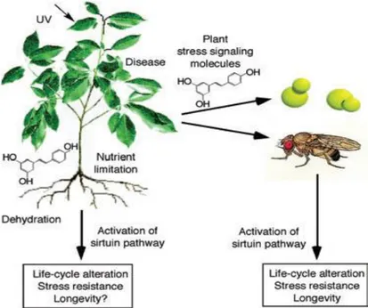
result of a long-term co-evolution within biological communities; interacting organisms, that evolved in close proximity to one another, developed compounds that could influence the biological process of neighboring species.4 The second theory, advanced by Howitz and Sinclairand called xenohormesis, (Fig. 1) is based on the hypothesis that there have been common ancestors of plants and animals able to synthesize a large number of stress- induced secondary metabolites; animals and fungi, that feed on plants, gradually lost the capacity to synthesize these low-weight molecular compounds while plants retained this ability. Animals and fungi only retained the ability to sense these chemical cues in plants, possibly in order to detect when plants were stressed and gain an early warning of changing environmental condition. 5
Figure 1. Xenohormesis hypothesis. Sirtuin enzymes evolved early in life‘s history to increase
somatic maintenance and survival during times of adversity. The xenohormesis hypothesis of Howitz and Sinclair proposes that primordial species synthesized polyphenolic molecules to stimulate sirtuins during times of stress. Only plants have retained this ability. Survival pathways in fungi and animals, instead, have retained the ability to respond to plant stress signalling molecules because they provide useful prediction about the state of the environment and/or food supply. This ability would allow organisms to prepare for and survive adversity when they might otherwise perish. Adopted from Lamming DW et al. 2004; Molecular
5
In fact, natural products are evolutionary preselected, owning structural requirements to be bound to proteins. Their structural scaffolds represent the biologically relevant fractions of chemical space explored by nature so far.6 Moreover, recently Stuart Schreiber, Paul Clemons and their colleagues at the Broad Institute in Boston, performed a bioinformatic analysis of natural product targets, thus demonstrating that natural molecules statically tend to target proteins with a high number of protein–protein interactions that are particularly essential for an organism.7 This observation is consistent with the common role played by natural products as chemical weapons against predators or competitors.
The long-lasting experience of traditional folk medicines may facilitate the identification of novel agents. Although in industrialized countries medicinal herbs gradually lost importance, replaced by the chemical progress during the 20th century, bioactive plant compounds are recently experiencing an impressing revival.
The plant world possesses an uncountable number of specialized metabolites, characterized by different chemical scaffolds and a wide range of biological properties such as protective agents against herbivores and various pathogens or growth regulators. The rationale in exploring plant-derived products relies on the assumption that their chemical scaffolds have been under a continuous co-evolutionary selection process to achieve interactions with biological macromolecules. Therefore, it is reasonable to assume that they might have more suitable structures to interact with cellular targets compared with chemicals.8 The use of plant small molecules to fight human diseases is a centuries old practice that has led to the discovery of more than half of the all ―modern‖ pharmaceuticals, becoming an essential source of leads for drug discovery.9
6
Nowadays, researchers still focus on the identification and structural characterization of new plant compounds and on the discovery of their detailed mechanism of action. To have an idea of the significance of the ethnobotanic use of plant drugs, it is sufficient just to consider the wide consumption of medicinal plants in the traditional Chinese medicine to cure and prevent diseases.10
Despite the success of plant molecules as drugs and the large number of benefits they show, they are losing favor among drug developers. One reason is the perceived ―dirtiness‖ of plant molecules: a molecule is considered ―dirty‖ if it interacts with numerous endogenous proteins. Such compounds presumably are more likely to have negative ―off target‖ effects than a molecule that specifically targets a single protein. Indeed, there are examples of plant molecules that, despite interacting with multiple human enzymes and receptors, are surprisingly safe:11 for example, salicylic acid, capsaicin or curcumin are surprisingly powerful and nontoxic, although they are multi-target molecules. In fact, having multi-multi-target molecules could be a winning strategy to face different diseases. It has been increasingly recognized that, in several pathologies, there is a large number of mutated genes and/or modified proteins that disrupt multiple pathways, which normally exhibit extensive biological cross-talk and redundancy. Therefore, the development of drugs that bind selectively to single protein targets also appears less clinically useful, since, interfering with a single target and/or pathway may not abrogate the disease. Moreover, a promising strategy for mitigating an acquired drug resistance or suppress disease, is to simultaneously inhibit multiple molecular pathways, either by using several agents in combination or by using a single agent that concurrently blocks multiple targets or pathways.
Even if natural products enjoy a privileged position in drug discovery, they can exhibit multiple behaviors that could interfere in assay redouts, such as
7
metal chelation, redox cycling and protein reactivity. For these reasons several natural molecules were termed Pan Assay INterference compoundS (PAINS). 12
These compounds possess reactive moieties, such as:
- catechols (e.g. apomorphine, droxidopa): cathecols have a high propensity to be redox active and can also variously chelate metals as well as being reactive in the oxidized form to nucleophiles present in the side chain of proteins such as cysteine and lysine.
- quinones (e.g. geldanamycin), because of the redox-active and reactive quinone moiety.
- phenolic Mannich bases and hydroxyphenilhydrazones (e.g. topotecan, rifampicin).
1.2 Terpenes
Among the plant specialized metabolites, terpenes are an important class which constitute a vast family of natural substances structurally different from each other, whose starting elements for the biosynthesis are the isoprene units.
They are produced by diverse organisms to perform an assortment of biological functions in varying ecological contexts and they are derived biosynthetically from units of isoprene (2-methyl-1,3-butadiene). Isoprene itself does not undergo the building process, whereas the activated forms, isopentenyl pyrophosphate (IPP or also isopentenyl diphosphate) and dimethylallyl pyrophosphate (DMAPP or also dimethylallyl diphosphate), are the actual components in the biosynthetic pathway (Fig. 2). There are two metabolic pathways that create terpenes:mevalonic acid pathway (MVA) and the 2-C-methyl-D-erythritol 4-phosphate/1-deoxy-D-xylulose 5-phosphate pathway (MEP/DOXP pathway), also known as non-mevalonate pathway. In both MVA and MEP pathways, IPP is isomerized to DMAPP by the enzyme isopentenyl pyrophosphate isomerase. IPP and DMAPP condense to give
8
geranyl pyrophosphate, the precursor to monoterpenes and monoterpenoids. Geranyl pyrophosphate is also converted to farnesyl pyrophosphate and geranylgeranyl pyrophosphate, respectively C15 and C20 precursors to sesquiterpenes and diterpenes (as well as sesequiterpenoids and diterpenoids).13 Their biosynthesis is mediated by the terpene synthase.14
Figure 2. Isopentenyl pyrophosphate (IPP) and dimethylallyl pyrophosphate (DMAPP)
condense to produce geranyl pyrophosphate, precursor to all terpenes and terpenoids.
Although all terpenes are synthesized from two five-carbon building blocks, the structures and functions vary widely. Many terpenes have shown several biological activities. Isoprenoids and derivatives play a critical role in all living systems: the cell structure, systems of electron transport in cell-cell signals (steroids, abscisic acid, gibberellic acid, phytol ecc.), in the structure of organisms and interactions between them. Some isoprenoids play a role in plant defense systems against attack by micro-organisms and insects, act as allelopathic compounds in plant-insect and plant-environment.15 They are also used for the treatment of human diseases. In fact, there is a wide spectrum of bioactivities for these types of substances such as antibacterial, antifungal, antiparasitic, anti-inflammatory, cytotoxic and antitumor.16 Although they show all of these activities, approximately 35,000 terpenes have been
9
identified and the majority of the possible functions or biological activity of these molecules are unknown. Then, it would be interesting to study this class of natural molecules to find out its potential biological effects and uses in therapy.
1.3 Diterpenes in cancer
Among plant terpenes, diterpenes form a vast class of more than 10000 structures (Fig.3). A number of potential anticancer diterpenes and their derivatives have been introduced already, such as taxanes (e.g. taxol and paclitaxel),17,18 kauranes (e.g. oridonin),19 andrographolide,20 diterpenes from coffee (e.g. cafestol, cafeic acid and kahweol) 21 and so on.
10
11
As we all know, cancers are considered one of the most lethal causes of death throughout the world.22 The use of medicinal preparations with plant-derived products is antique and, more recently, they have continued to enter in clinical trials or to provide leads for compounds in clinical trials, particularly as anticancer agents.23
In Figure 4 most of the known diterpenes involved in anti-cancer pathways such as oxidative stress, ER stress, proliferation/differentiation, autophagy, cell cycle arrest and apoptosis, are reported.
Figure 4. Anticancer pathways of diterpenes.
Several diterpenes are reported to act in a number of cancer cells such as T leukemia (Jurkat), cervical carcinoma (HeLa), breast cancer (MCF7), human embryonic kidney (HEK293) cell lines, while either alone or combined to other diterpenes or anticancer drug, have been found to work synergistically.
12
Altogheter, we can conclude that diterpenes may be one of the leading therapeutic molecules in cancer.24
1.4 Drug discovery today: Chemical Genetics
Chemical genetics is an emerging field that can be used to study the interactions of chemical compounds, including natural products, with proteins. Chemical genetics uses chemical compounds, including plant molecules, that may modulate the activity of target proteins. 24, 25 This field comes from the classical genetics, widely used to study biology by manipulating the biological system at the level of the gene.
Chemical Genetics offers several advantages over its classical counterpart and allows the study of unexplored biological space. For example, genes essential for survival or development cannot be studied using classical genetics; this can only be done using chemical genetics. Thus, the instantaneous effects of small molecules can be characterized using chemical genetics. It also makes it possible to study mammals whereas classical genetic techniques are more complicated to apply due to their diploid genome, physical size and slow reproduction rate.26 Other benefits of using chemical compounds are the temporal control and reversibility of the inhibition of protein function.27 Classical genetics is divided into forward genetics (involving random mutations followed by phenotypic screening and gene identification) and reverse genetics (involving mutation of a specific gene and phenotype characterization). So, genetics in the ‗forward‘ direction is from phenotype to gene; in the ‗reverse‘ direction it is from gene to phenotype. As classical genetics, two approaches can be taken also to chemical genetics (Fig. 5): Forward Chemical Genetics (FCG) and Reverse Chemical Genetics (RCG).27
13
Figure 5. Chemical Genetics approaches: a) Forward chemical genetics;
b) Reverse Chemical Genetics
1) In FCG, typical random mutagenesis is replaced by a screening of a library of not targeted small molecules against multiple potential targets simultaneously. Compounds that induce a phenotype of interest can be selected and then the target protein of this compound is identified.26 Forward chemical genetics requires three components: a) a collection or library of small molecules; b) a biological assay with a quantifiable phenotypic output, usually performed using living cells or complex cellular extracts and c) a strategy to join an active compound to its biological target. Therefore, the last goal of FCG is the target identification.
2) In RCG, a known target protein is screened using small molecule library to identify functional ligands that either stimulate or inhibit the target protein. Once a specific ligand that produces a change in the protein function is identified, it is introduced into a cell or organism and the resulting changes in
14
the phenotype are studied.27 Also RCG requires three components: a) specific protein target; b) a library of small molecules; c) cell-free or cell-based assays to validate the interaction and to understand the mechanism of action of the target bound to the ligand. Therefore, the goal of RCG is the validation of the interaction and of the protein target functions. 1.5 Aim of the thesis
This PhD project was mainly focused on a RCG approach,starting from the screening of a library/collection of diterpenes able to bind a specific protein target. The molecules emerging from the screening were then studied to validate the screening results by cell-free and cell-based assays. In my PhD project were adopted:
Cell-based tecniques, such as: Cellular Thermal Shift Assay (CETSA), Drug Affinity Responsive Target Stability (DARTS), Western Blotting (WB), Flow Cytometry, RTq-PCR, Wound Healing, Apoptosis and Protein Synthesis assays.
Cell-free tecniques, such as: Mass Spectrometry (MS), Surface Plasmon Resonance (SPR), Saturation transfer difference NMR (STD-NMR), WaterLOGSY and ATPase activity assays.
1.6 Cell-free and cell-based assays
a) Cellular Thermal Shift Assay (CETSA)
CETSA allows to carry out qualitative and quantitative analyses of the direct interaction of a drug candidate to a target protein in the cells 28. In particular, this approach offers the opportunity to firmly link the observed phenotypic
15
response to a compound with a particular target engagement. CETSA, set up by Molina and coworkers in 2013, is based on the principle of thermodynamic stabilization inferred to the protein as a result of the ligand binding, which can be used for the estimation of binding free energies as well other thermodynamic properties for isolated systems at equilibrium. The shift in thermal stability is estimated by measuring the amount of remaining soluble target protein at different temperatures for treated and control samples.
CESTA protocol starts with the treatment of cells with either molecule of interest and vehicle as control, followed by cell heating (to denaturate and precipitate the protein of interest), cell lysis, removal of cell debris and aggregates through centrifugation and, finally, detection of the protein by WB studies (Fig. 6).
Figure 6. Schematic illustration of CETSA approach.
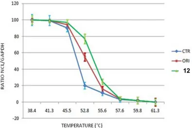
The apparent aggregation temperature (Tagg or Tm) observed in the presence or in the absence of the drug can be compared; the occurrence of significant shifts in these parameters following cells treatment with the drug, demonstrate the actual stabilization of the target protein by the molecule. Therefore, a typical output form CESTA experiment is a comparison between apparent melting curves (or, more accurately, temperature-induced aggregation curves)
16
measured in treated and control cells; these curves report the amount of residual target protein detected at different temperatures and a potential thermal stabilization can be assessed and identified as a shift of the melting curve on the right side of the graphic (Fig. 7, in purple).
Alternatively, an isothermal dose-response fingerprints CETSA (ITDRFCETSA) could be generated, in which the stabilization of the protein can be followed as a function of increasing ligand concentration. This latter experiment requires knowledge of the temperature at which the protein denatures and precipitates (Tagg or Tm).
Figure 7. Principle of Cellular Thermal Shift Assay (CETSA)
b) Drug Affinity Responsive Target Stability (DARTS)
DARTS is a chemical proteomic techology performed to identify and study protein-ligand interactions.29 It is based on the idea that small molecule binding its target protein may induce a protein stabilization, resulting in the resistance of a proteolytic enzyme (Fig. 8). This enhanced stability is postulated to result into a shift of the thermodynamic landscape of the protein
17
to favor the ligand-bound state, which prevents much of the protein‘s innate flexibility and movement from being realized. Therefore, DARTS can be applied to the initial identification of the target proteins of small molecules (Forward Chemical Genetics), but can also be used to validate potential protein-ligand interactions predicted or identified by other means and to estimate the affinity of interactions. This approach can be performed using intact cells, crude cell lysates and other complex protein mixtures (without requiring purified proteins), and in addition, it uses native and unmodified small molecules.
Figure 8. Principle of Drug Affinity Responsive Target Stability (DARTS).
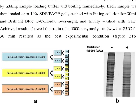
As shown in Figure 8, DARTS protocol starts from the incubation of cell lysates or of intact cells (which will be lysated after the incubation) with only vehicle (DMSO) and with the compound of interest. Subsequently, the samples undergo limited enzimatic digestion using a non specific protease (such as subtilisin), followed by electrophoresis 1D SDS-PAGE. Digested proteins will be then investigated by WB and/or MS. Finally, if the drug interacts with the protein of interest, the final output will be higher levels of the protein in the treated sample than the control. There are different proteases that can be used for DARTS (e.g subtilisin, thermolysin and so on); in our
18
experiments subtilisin was selected, since we needed a rapid kinetic of hydrolysis and a protease with a broad specificity; thermolysin, another enzyme widely used for this technique, is not very stable and there is a significant proportion of the proteome that is not highly resistant to its proteolytic activity.29
c) Surface Plasmon Resonance (SPR)
SPR is a non-invasive optical technique, based on the evanescent wave phenomenon, used to study the interactions of proteins with small molecules, protein conjugates, nucleic acids, lipid micelles and even larger particles such as viruses and whole cells.30 The binding between a compound in solution and its ligand immobilized on the surface of a sensor chip results in a change of the refractive index, that could be monitored in real time allowing the measurement of association and dissociation rates. Different concentrations of the compound in solution are used against the putative protein target, singularly immobilized on sensor chips .31
A glass prism is coated with a thin layer of a noble metal (usually gold) to create a biosensor surface (Fig. 9A). The biomolecule (named ―ligand‖) is immobilized on the sensor surface. The metal layer possesses surface mobile electrons. At certain incidence angle the incoming beam of light disturbs these electrons causing changes in surface plasmon waves. Electrons ―resonate‖ giving a name to SPR phenomena, this particular incidence angle is called SPR angle. At the SPR angle, reflectivity drops to a minimum. However, the binding of the protein to a free compound in solution onto sensor‘s surface affects SPR conditions: refractive index increases and a shift of SPR angle occurs (Fig. 9B). The process is shown in real time by sensorgrams, where association (kon) and dissociation (koff) phases can be observed (Fig. 9C). The responsive units (RU) can be used to measure the binding affinity (KD).
19
Figure 9. Surface Plasmon Resonance (SPR)-based instrument. A) SPR- biosensor surface;
B) shift of SPR angle; C) SPR Sensorgram.
d) Saturation Transfer Difference NMR (STD-NMR)
The STD NMR experiment, at first described by Mayer & Meyer in 1999, is a spectroscopic technique used to study interactions, in solution, between a large molecule (protein) and a medium-small sized molecule (ligand).32 It is based on the Nuclear Overhauser Effect (NOE) and on the observation and analysis of the resonances of the ligand protons.
The experiment is carried out by first registering a reference spectrum under conditions of thermal equilibrium with the irradiation frequency set at a value that is far from any ligand or protein signal (e.g. 40 ppm), i.e, the so-called
20
off-resonance spectrum (Fig. 10, top), which is used as reference with signal intensities I0.
Figure 10. Scheme of the STD-NMR experiment showing the protein in surface
representation and the non-exchangeable protons of the ligand as spheres. (Top) A 1D standard NMR experiments show only equilibrium intensities of the ligand in the free state (I0). (Middle) Upon selective saturation of some receptor signals, this is efficiently spread throughout the protein (yellow surface) by spin diffusion (intra-molecular NOEs). The fast exchange (transient binding) between the free and bound ligand states allows the transfer of magnetization (inter-molecular NOEs) from the receptor to the ligand protons in contact with the protein surface (salmon spheres, on-resonance spectrum). (Bottom) The difference spectrum (I0-Isat) only contains the ligand signals perturbed upon binding, whose intensities reflect the proximity of each proton to the protein surface. Adopted from Doctoral thesis of Garcia JCM, Alvarez JA, Nieto Mesa PM, insights on the structure and dynamics of glycosaminoglycans and their interactions with langerin: NMR and computational studies. 2013.
A second experiment is then recorded, in which the protein is selectively saturated (on-resonance spectrum; Fig. 10, middle), giving rise to ligand signals with Isatintensities. In general, the selective irradiation consists of a cascade of Gaussian or Adiabatic-shaped pulses (low power) that saturate only a region of the spectrum that contains a few protein resonances (but not ligand signals), e.g., the aliphatic (from 0 to -1 ppm) or aromatic region (around 7
21
ppm), for a specific period of time (saturation time; typically from 0.5 to 5-6 seconds). The selective saturation is transferred to the whole protein via spin diffusion through the vast network of intra-molecular 1H-1H cross-relaxation pathways (intra-molecular NOE; see Fig. 10, middle), being a quite efficient processes due to the typical large molecular weight of the receptor. Also, saturation is transferred from the protein to the bound ligand via spin diffusion through inter-molecular NOEs. The dissociation of the ligand will then transfer this saturation into the bulk solution where it accumulates during the saturation time of the experiment, as a result of the much slower relaxation in the unbound that the bound state. In particular, as in fast-exchanging protein-ligand systems the enthalpic relaxation (R1) of fast-tumbling molecules (small) in the free state is much slower than the kinetic off-rate constant of binding (koff>> R1), the accumulation of ligands molecules containing some of their resonances perturbed (NOE of large molecule) results in the macroscopic detection of transferred saturation on the ligand signals in the saturated STD NMR spectrum (Isat; Fig.10, middle). Furthermore, for those hydrogen atoms of the ligand establishing close contacts to the protein surface (4-5 Å) these Isat values will be lower than the the I0 intensities, negative inter-molecular NOE, due to the transfer of the relaxation properties of the macromolecule to the small ligand in the bound state (Fig. 10, middle). By subtracting the off-resonance from the on-off-resonance spectrum (I0-Isat) the difference or STD spectrum is obtained, which will just contain the proton signals of the ligand in close contact to the protein surface (Fig. 10, bottom), and where any signal coming from non-binding compounds is cancelled out. So, if a non-binder is present in solution, its resonances will not appear in the STD spectrum. Therefore, the difference spectrum, or the STD spectrum (ISTD = I0 - ISAT) yields only those resonances that experienced saturation in the on-resonance experiment, the receptor resonances and the ones from the binding ligands.32
22
e) WaterLOGSY
WaterLOGSY is a widely applied 1D ligand-observation technique for the detection of protein–ligand interactions. As the STD-NMR approach, WaterLOGSY is based on the NOESY experiment, and implies transfer of magnetization via a intermolecular NOE and spin diffusion. The originality of WaterLOGSY comes from the intervention of water molecules in the transfer pathway. 33 (Fig. 11a)
Figure 11. a) WaterLOGSY principle; b) WaterLOGSY spectrum in comparison with Normal
1D 1H-NMR spectrum. Adopted from FragmentTech.univ-lyon1.fr
The bulk water magnetization is excited and transferred during the NOESY mixing time to the bound ligand via different mechanisms. The WaterLOGSY spectrum, which is recorded for the free ligand, contains the bound-state perturbed magnetization as long as the relaxation time T1 of the ligand is
23
greater than the dissociation rate constant koff. The inverted water magnetization can be transferred via different pathways to the bound ligand: a) direct transfer from water molecules immobilized in the protein binding site (water residence times greater than nanoseconds);
b) chemical exchange between excited water and protein labile protons (amide, hydroxyl, amino, etc.) and propagation of the inverted magnetization to the ligand by intermolecular dipole–dipole crossrelaxation as well as spin diffusion via the protein– ligand complex;
c) transfer from the water molecules found in the protein surface via the protein–ligand complex;
In all the pathways, the ligands interact with water via water–ligand–protein or protein–ligand complexes, whose rotational correlation times yield negative cross-relaxation rates and exhibit a negative NOE with water (Fig. 11b waterlogsy spectrum). By contrast, small molecules that only interact with bulk water (non-binders) will experience much faster tumbling, which translates into a positive NOE (Fig. 11b Normal 1D spectrum). Therefore, opposite signs for signals from free versus protein-bound ligands are observed in a WaterLOGSY spectrum, which enables one to easily discriminate binders and non-binders.33
1.7 Outline of the thesis
The PhD thesis is organized in three chapters:
- in Chapter 2, a screening by CETSA of a collection of ent-kaurane and ent-trachilobane diterpenes against Nucleolin is described. Once identified a diterpene ligand of Nucleolin, cell-free and cell-based assays were implemented to validate the interaction and to study the diterpene biological activity in cancer cells;
- in Chapter 3, ent-kaurane, ent-trachilobane, fusicoccane, labdane and clerodane diterpenes were screened by SPR against Heat Shock Protein
24
70 (Hsp70). Aiming to expand and validate our knowledge about the selected compound, multidisciplinary approaches were adopted, either to validate Hsp70/diterpene binding, carried out by MS, DARTS and molecular docking or to identify its biological activity, proved by WB, Flow Cytometry and ATPase assay;
- in Chapter 4, the target protein is the Heat Shock Protein 90 (Hsp90). A collection of fusicoccane and abietane diterpenes was double screened by SPR against Hsp90 and by MTT. The compounds emerging from the screening were investigated for their biological activity by Flow Citometry, WB, ATPase activity assays and molecular docking experiments.
25
27
Introduction
2.1 Nucleolin: structure and localizations
Nucleolin (Fig. 1) is a protein encoded in humans by the NCL gene. The human NCL gene is located on chromosome 2 and consists of 14 exons, 13 introns and is long approximately 11kb. The intron 11 of the NCL gene encodes a small nucleolar RNA (U20).34
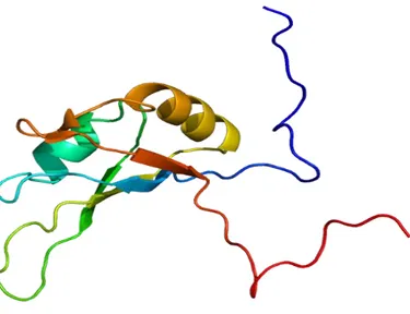
Figure 1. Structure of RNA Binding Domain 1 (RBD1) of Nucleolin. Adopted from Allain
FH et al. 2000; J Mol Biol. 303(2):227-41.
Firstly, Nucleolin was described by Orrick et al. (1973) and called C23 because of its mobility on a two dimensional gel.35 The name Nucleolin is now widely used for this protein, which can represent as much as 10% of total nucleolar proteins.36 This protein is also often described as a 100-110 kDa protein; however, cloning of the cDNA of hamster Nucleolin revealed that it contained 713 amino acids, giving rise to a predicted molecular mass of 77 kDa. This difference was attributed to the amino acid composition of the N-terminal domain of Nucleolin.37
28
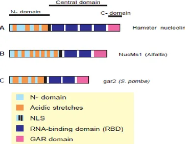
Proteins homologous to human NCL were identified in rat, mouse, chicken and Xenopus laevis.38 Nucleolin is highly phosphorylated, methylated, and can also be ADP-ribosylated.39,40 Analysis of its amino acid sequence revealed the presence of three different structural domains (Fig. 2):
1) The N-terminal domain, showing highly acidic regions interspaced with basic sequences and containing multiple phosphorylation sites.
2) The central domain, called RNA Binding Domain (RBD) or RNA Recognition Motif (RRM), where four RNA binding domains are localized. 3) The C-terminal domain, called GAR or RGG domain, rich in glycine, arginine and phenylalanine residues. This domain contains high levels of NG,NG-dimethylarginines.41
Several nucleolar proteins of different eukaryotic species exhibit a similar tripartite structural organization (Table 1).
Figure 2. Schematic representation of the organization of three Nucleolin and ‗Nucleolin-like
proteins‘. RBDs are defined from the 1 and 4 strand. (A) Organization of hamster Nucleolin: mouse, rat chicken, human and Xenopus laevis Nucleolin have the same organization. (B) Alfalfa and pea ‗Nucleolin-like protein‘ have the same organization. (C) Organization of gar2. Nsr1p possesses only 3 long acidic stretches. N-, N-terminal; , C-terminal.
Adopted from Ginisty H, Sicard H, Roger B, Bouvet P, structure and functions of Nucleolin. 1999; Journal of Cell Science 112: 761-772.
29
1) The N-terminal domain
The length of this domain is highly variable among the different Nucleolin-like proteins (Fig. 2). Highly acidic regions separated from each other by basic sequences represent a tipical feature of this domain. Acidic domains have been proposed to bind histone H1, and could be responsible for a displacement of H1 from its interaction with linker DNA, thus inducing chromatin decondensation. Indeed, the presence of the basic and repeated octapeptide motifs (XTPXKKXX, X being a non-polar residue) bears strong similarity to an analogous sequence of histone H1 and could be responsible for the ability of Nucleolin to modulate DNA condensation in chromatin.42 Besides, the N-terminal domain of Nucleolin has been involved in many other protein-protein interactions, such as those with some ribosomal proteins.43
The N-terminal domain of Nucleolin is highly phosphorylated,38 being this protein a substrate for several kinases such as casein kinase II (CK2), 44 p34cdc2 45
and protein kinase C-δ.46 CK2 catalizes in vitro and in vivo phosphorylation of some Nucleolin serine residues in the acidic regions,44 whereas p34cdc2 phosphorylation occurs on threonine residues within the basic TPXKK repeat. Phosphorylation of Nucleolin by CK2 and p34cdc2 is highly regulated during the cell cycle. Extensive phosphorylation by CK2 occurs in interphase and by p34cdc2 in mitosis. This regulated phosphorylation of Nucleolin probably regulates its function during the cell cycle.45
2) The RNA-binding domains
Nucleolins from hamster, mouse, rat, human, chicken and Xenopus laevis possess four RNA-binding domains RBDs, also known as RRMs (Fig. 2 and Table 1). These domains, found in a large number of proteins implicated in various functions, are known to confer an RNA-binding specificity to the
30
protein they belong to.47 According to their RBDs sequence identity, Nucleolins and Nucleolin-like proteins can be divided in three groups. The first group includes Nucleolin from hamster, mouse, rat, human, chicken, Xenopus laevis and fish; in the second group there are yeast proteins such as gar2 and Nsr1p, and in the third group plant proteins such as NucMs1 and Nucleolin from pea.48 RNAs associated with Nucleolin possess a small stem-loop structure composed of a short stem (5 base pairs) and a 7-10 nt stem-loop containing the motif U/G CCCGA, obtaining a minimal RNA-binding site named Nucleolin Recognition Element (NRE).49 The factors responsible for the binding affinity difference are not identified. Taken separately, none of the four individual RBDs interact significantly with RNA targets, but a peptide that contains the first two RBDs (RBD 1-2) is sufficient to account for Nucleolin/RNA binding specificity and affinity.50 Both domains participate in a joint interaction with the NRE using a different surface to contact the RNA. The determination of the structure of this RNA/protein complex has been investigated by NMR and X-ray high-resolution crystallography. The presence of several RBDs in Nucleolin structure suggests that Nucleolin could interact with multiple RNA targets.
Table 1. Percentage of sequence identity of the RBD domain of Nucleolin from different
species
Hamster Mouse Human Chicken Xenopus
laevis Carp RBD 1 100 96 85 62 53 ND RBD 2 100 99 83 64 53 51 RBD 3 100 93 96 70 67 64 RBD 4 100 100 99 89 85 85
31
3) The C-terminal GAR/RGG domain
The C-terminal domain is defined as Arg-Gly-Gly (RGG) repeats interspaced with other - often aromatic - amino acids; the motif RGG is particularly frequent in nucleolar proteins. The length of the GAR/RGG domain is variable among Nucleolins, with its sequence and arrangement of the repeats not well conserved. For example, plant Nucleolin-like proteins have a longer GAR domain than mammalian Nucleolins.51 The presence of this domain in a protein is associated with the presence of an RNA-binding domain (RBD or others). The presence of this GAR domain does not influence the binding affinity and specificity for the NRE sequence, but it could be involved in the interaction of Nucleolin RBD domains with targets located within large and complex RNA, such as rRNA.49, 50 The GAR domain protein-protein interactions are not well defined. It is not known how the GAR domain mediates protein-protein interactions, but since the GAR domain of Nucleolin interacts with only a subset of ribosomal proteins, these interactions seem to be specific. 43 After its discovery, it was found that Nucleolin contained high levels of NG,NG-dimethylarginines; this post-translational modification is found on arginines located in the GAR domain.37,41 This modification could be an important signal for the regulation of GAR/RNA interactions, the stability of the protein, or its localization. It is not known if this post-translational modification is reversible. 43,50
The intracellular localization of Nucleolin has been extensively studied by electron microscopy analysis and/or immunofluorescence ultrastructural localization in vertebrates,41 plants,52 and yeast cells.53 It is localized in the nucleolus, in the cytoplasm 54 and on the cell membrane 55(Fig. 3). This different localizations are linked to changes in Nucleolin isoelectric point and/or post-translational modifications, such as glycosylation, ADP-ribosylation, acetylation and most importantly phosphorylation.39, 40 In fact,
32
Nucleolin translocation into the nucleus improves when it is dephosphorylated whereas pophorilated nucleolin is mainly moved into the cytoplasm.
Biochemical fractionation of the nucleoli has shown that the nucleolar fraction of Nucleolin represents more than 90% of the Nucleolin cellular pool, while the nucleoplasmic compartment does not represent more than 5% of the protein. 56 The cytoplasmic fraction of Nucleolin is difficult to estimate as it depends on the quality of the fractionation, or sensibility of the detection techniques that are used; however, it probably does not represent more than a few % of the total protein. In specific cytoplasmic structures, such as the centrosomes, centrosomal Nucleolin represents less than 0.1% of the total protein. 57 S. Nisole et al. determined that the cell surface Nucleolin is less than 20% of the nuclear-free cytoplasmic fraction.58 In the different cell compartments, Nucleolin has different molecular targets that confer distinct functions (Fig. 3).
Figure 3: localizations of Nucleolin (in red).
Adopted from Kotb Abdelmohsen & Myriam Gorospe RNA-binding protein nucleolin in disease. 2012; RNA Biology. 9:799-808.
The predominant localization in the nucleolus represent an important aspect of Nucleolin profiling of its functions.59, 60 Nucleolin is mainly observed in the Dense Fibrillar Compartment (DFC), whreas it is less abundant in the
33
Granular Compartment (GC), and almost absent in the Fibrillar Center (FC).61 How Nucleolin accumulates in the nucleolus remains an important question: several studies were performed to identify the Nucleolin domains responsible for the nucleolar localization62 but, the obtained results showed that Nucleolin does not contain a single, linear nucleolar targeting signal. The well-defined
bipartite nuclear localization signal (NLS) of Nucleolin
(KRKKEMANKSAPEAKKKK) is used to enter in the nucleus and then the protein probably accumulates within the nucleolus as a consequence of its binding to other nucleolar components (probably rRNA) via the RNA binding domains and the C-terminal RGG domain. The RNA-binding domains of nucleolin are crucial for its nucleolar localization, but are unable by themselves to target hybrid proteins to the nucleolus. The C-terminal GAR domain allows total nucleolar accumulation when it is associated with at least one RBD.63
Nucleolin has also been observed in the plasma membrane of several cell lines, using immunofluorescence and electron microscopy. This localization is surprising, because Nucleolin neither possesses a transmembrane hydrophobic domain nor a plasma membrane targeting sequence.64 Ding et at. demonstrated that Nucleolin traslocation to the cell membrane is mediated by Heat Shock Protein 70 (Hsp70). This chaperon interacts with Nucleolin in endothelial cells via its peptide-binding domain and the RNA-binding domain 3 and 4 of Nucleolin. Phosphorylation of Nucleolin by either protein kinase C-ξ or casein kinase 2 mediates Nucleolin interaction with Hsp70 and the surface expression. Moreover, Hsp70 regulates Nucleolin translocation via stabilizing it and enhancing its interaction with nonmuscle myosin heavy chain 9. On the plasma membrane, Nucleolin interacts with several proteins involved in cell proliferation, apoptosis and angiogenesis.65
34
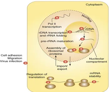
2.1.1 Roles of Nucleolin in physiological and pathological pathways
The multiple sub-cellular localizations of Nucleolin are directly connected to its physiological and pathological functions (Fig. 4):
- in the nucleolus, it is directly involved in ribosome biogenesis,66 chromatin remodeling,67 transcriptional regulation 68 and telomerase activity;69 - in the nucleoplasm, Nucleolin interacts with several RNAs and proteins (such as trasctription factors) and it is involved in regulation of the cellular response to stress;70
- in the cytoplasm; Nucleolin constantly shuttles between nucleus and cytoplasm where it is involved in many non nucleolar functions, e.g. import/export of ribosomal proteins, centrosome duplication 59 as well as post-transcriptional and translational regulation of various mRNAs 71 including p53 72, Bcl2 73 and AKT;74
- on the cell surface Nucleolin behaves as receptor, binding to several proteins, thus inducing tumorigenesis (cell migration, adesion, angiogenesis), and mediating inflammation and viral infections; 75
35
Figure 4. The cellular compartment locations of Nucleolin (red dots). (a) In the nucleolus,
association of Nucleolin with nucleolar chromatin (rDNA) could be involved in the regulation of Pol I transcription. (b) Nucleolin has been seen on nascent pre-rRNA transcripts and is believed to participate in co-transcriptional pre-rRNA folding, (c) maturation at the first processing site and (d) assembly of pre-rRNA with ribosomal proteins. (e) The shuttling of Nucleolin between the nucleus and the cytoplasm might participate in the import and/or export of several nucleolar components or proteins, such as ribosomal proteins. (f) In the nucleoplasm, Nucleolin has been also found associated with several genes transcribed by Pol II and with mRNAs, with functions from (g) the regulation of translation to (h) mRNA stability. (i) A large number of reports have identified nucleolin on the cell membrane, with potential roles in cell migration and adhesion and virus infection.
Adopted from Fabien Mongelard and Philippe Bouvet.Nucleolin: a multiFACeTed protein.
TRENDS in Cell Biology. 2007; 17: 80-86.
2.1.2 Roles of Nucleolin in ribosome biogenesis
In the nucleolus, Nucleolin plays many functions, specially in ribosome biogenesis (Fig. 4):
- it is a chromatin co-remodeler, as it facilitates the interaction between nucleosomes and the remodeling complex of chromatin; 67
- it is a histone chaperone, with functional similarity to the ―facilitates chromatin transcription‖ complex (FACT); 67
- it is involved in the regulation of RNA polymerase I trascription and RNA polymerase II activity; 76
36
- it is invoved in the proper folding and maturation of the preribosomal RNA (pre-rRNA);
- it participates in the ribosome assembly; 77
According to its histone chaperone activity, Nucleolin promotes RNA transcription through nucleosomes. Histone chaperones are key factors for the dynamic organization of chromatin template. They mediate:
- histone storage;
- translocation of histones to the nucleus;
- exchange and deposition of histones onto the DNA during replication-dependent chromatin assembly;
- chromatin reorganization not only during elongation, but also during initiation of RNA transcription;
Chromatin assembly is a two-step process: deposition of a H3–H4 tetramer on DNA followed by the deposition of two H2A–H2B dimers. Various histone chaperones and ATPdependent chromatin remodeling factors facilitate the organization of nucleosomes into a regularly spaced array. Nucleolin directly binds to H2A–H2B dimers 76 and facilitates the assembly of nucleosomes on naked DNA, presumably by incorporating H2A–H2B dimers into auto-assembled tetrasomes ((H3–H4)2 tetramer acceptor). This effect of Nucleolin on the dynamics of H2A–H2B dimers facilitates the passage of the RNA polymerase II through nucleosomes. The ability of Nucleolin in increasing H2A–H2B turnover is a key factor to understand its co-remodeling activity. The spontaneous or Nucleolin-induced disassembly of one H2A–H2B unit would be expected to lead to a destabilization of the histone octamer particles. Nucleolin also shows FACT-like activities (Fig. 5).
37
Figure 5. The histone chaperone and FACT-like activities of Nucleolin. In vitro, Nucleolin
can, similar to FACT, destabilize nucleosomes to promote the formation of hexasomes (loss of one H2A–H2B dimer). This helps the passage of polymerase II through chromatin templates. The histone chaperone activity of Nucleolin helps the reformation of nucleosomal structures after the passage of the polymerase.
Adopted from Fabien Mongelard and Philippe Bouvet.Nucleolin: a multiFACeTed protein.
TRENDS in Cell Biology. 2007; 17: 80-86.
The ―facilitates chromatin transcription‖ (FACT) complex is required to overcome the nucleosomal barrier and to promote a modest but significant level of transcription elongation. Therefore, FACT is also itself a histone chaperone and facilitates the loss of an H2A–H2B dimer upon polymerase II transcription, enabling the passage of the polymerase through the nucleosomal particle.78
Nucleolin is also involved in the regulation of rRNA transcription. Indeed, following inhibition of rRNA transcription, a rapid release of Nucleolin from the dense fibrillar zone of the nucleolus is observed,79 demonstrating that its presence in this subnucleolar compartment is dependant upon rRNA transcription. A model related to Nucleolin ability to interact with rRNA has been proposed by Bouche et al., where Nucleolin is involved in the regulation of rRNA transcription elongation through the binding of its RNA-binding domains with the nascent rRNA transcript. The N-terminal domain, instead, interacts with the polymerase I machinery, which would block transcription elongation. In this model, the phosphorylation of Nucleolin by CK2 during interphase would be required to proteolyse Nucleolin (between the N- and
38
central domains) and to release the transcription complex, while the central and C-terminal domains of Nucleolin could remain bound to the rRNA and participate in the pre-ribosome assembly.80
The ability of Nucleolin in vitro to promote the formation of secondary structures in complex RNAs, 81 and its association with preribosomal particles in vivo,82 suggest that Nucleolin could be required for the correct assembly of these particles. Therefore, Nucleolin can play a role as an assembly factor, bringing together a correctly folded rRNA and the other components necessary for rRNA maturation and/or assembly of the ribosome. The GAR domain of Nucleolin is implicated in some of these interactions. 43 The interaction of Nucleolin with these ribosomal proteins further supports its role at an early step of ribosome assembly.
2.1.3 Nucleolin as shuttle between nucleus and cytoplasm
Despite Nucleolin is found almost exclusively into the nucleolus, assays based on interspecies heterokaryons have shown that Nucleolin shuttles between nucleus and cytoplasm. However, a very small amount of Nucleolin is able to migrate to the cytoplasm. In the heterokaryon system, the N-terminal domain of Nucleolin is required for efficient shuttling, but it is unable to increase the nuclear export of an heterologous protein, indicating that Nucleolin does not contain a positively acting export signal.83 In addition, its cytoplasmic localization is related to its phosphorylation by p34cdc2, whereas the nuclear translocation is correlated with a dephosphorylation of Nucleolin. These data show that phosphorylation of its N-terminal domain can regulate the function of Nucleolin NLS.63 Regarding to the C-terminal domain, it was shown that the GAR domain of Nucleolin reduced slightly the export process.83 The discovery that this protein is able to shuttle, even under specific conditions, has raised the possibility that it can be involved in the nuclear import of
39
ribosomal components (like ribosomal proteins), or in the nuclear export of the ribosomal particles.
2.1.4 Nucleolin as receptor on the cell surface
Nucleolin has been observed at the plasma membrane of several cell lines using immunofluorescence and electron microscopy.84 The expression of Nucleolin at the plasma membrane is increased in several tumor cells 85 and in endothelial cells during angiogenesis.86 There are several evidences indicating that Nucleolin located in plasma membrane can facilitate the binding of HIV on host cell surface, and its entrance. Moreover, Nucleolin has been shown to promote inflammation, enhancing the internalization of lipopolysaccharides from the cell membrane to the cytosol.75 Therefore, Nucleolin is believed to be a useful signature for cancer, inflammation and viral infection diagnostics and can be an interesting target for the development of new drugs.
2.1.5 Nucleolin as tumor marker: therapeutic strategies
An altered expression and function of Nucleolin has been observed in several cancers. For instance:
- an increase of Nucleolin expression is observed in tumor tissue from colorectal cancer human patients;87
- it is also over-expressed in tumor tissues from gastric cancer, and a high cytoplasmic amount of Nucleolin is associated with worse prognosis for the patients;88
- in human breast cancer tumors, Nucleolin is over-expressed (from 3 to 6 fold increase in human breast cancer cell lines compared to normal breast cells);89
- higher Nucleolin mRNA or protein levels are also observed in leukemia cells as studied in different patients cohort;90
40
- Nucleolin expression is also increased in lung cancer tissues from human patients and associated with largest tumors;91
- in glioblastoma cells, the presence of glycosylated Nucleolin at the cell surface increases with the malignancy of the human tumor;85
- in human melanoma cells, a specific glycosylated form of Nucleolin is observed and the inhibition of cell surface Nucleolin in a mouse model, delays the emerging of melanoma and significantly prevents subsequent visceral metastasis;92
- in cervical cancer, Nucleolin expression is directly linked to human papillomavirus type 18 induced carcinogenesis by favoring the expression of viral oncoproteins;93
A major challenge is to understand the contribution of Nucleolin to the development of these cancers. In some cases, Nucleolin deregulation may just be the consequences of the pathological state, whereas in other situations the deregulation of Nucleolin may contribute to the initiation or the progression of the disease.
As shown in figure 6, Nucleolin can play several functions in cancer progression:
- in the nucleoplasm, Nucleolin binds promoters of genes that are over-expressed in cancer (such as c-myc) and promotes their transcription;
- in the cytoplasm, Nucleolin affects mRNA turnover by interacting with the 3′-untranslated region of several target mRNAs of proteins important for cancer development (p53, Bcl2 or AKT1 mRNAs). Particularly, binding to Bcl2 mRNA,73 Nucleolin prevents its proteolysis inferred by exosoms, increasing Bcl2 levels and improving anti-apoptosis. In addition, interacting with AKT1 mRNA,74 Nucleolin enhances its translation increasing cell proliferation. Nucleolin also interacts with miRNAs to affect the expression of oncogenes and tumor suppressor genes;
41
- at the plasma membrane, Nucleolin interacts with several proteins involved in cell proliferation, apoptosis and angiogenesis;
- Nucleolin interacts with Fas, member of the tumor necrosis factor superfamily of apoptosis receptors, inhibiting apoptosis;94
- Nucleolin interaction with ErbB195 and Ras96 at the plasma membrane favors cell proliferation;
- it can bind to the growth factors Pleiotrophin (PTN) 97 (inducing cell migration) and midkine 98 (promoting cell survival);
- numerous other key proteins involved in tumorigenesis and angiogenesis have been shown to interact with Nucleolin and act through cell surface Nucleolin as for instance Hepatocyte Growth Factor (HGF),99 Vascular Endothelial Growth Factor (VEGF)100 and tumor necrosis factor alpha inducing protein (Tipα) ;101