Universit`a Politecnica delle Marche
Scuola di Dottorato di Ricerca in Scienze dell’Ingegneria Facolt`a di Ingegneria
Per quelli che han vissuto con la coscienza pura, il cuore tenero e gli occhi contenti, alla mia meravigliosa e libera Zia, al mio caro e forte Massimo al mio habibi e dolce Khaled
Abstract
The endothelium is the largest organ in the human body and covers all the vessels in the cardiovascular system (heart, arteries, veins, capillaries and lym-phatic system). It plays a role of primary importance in the modulation of vessel tone and blood flow, as well as other numerous functions such as the regulation of inflammatory and immune processes and vascular permeability. The alteration of this organ leads to and endothelial dysfunction is known to be implicated in the pathogenesis and clinical course of all known cardiovascular diseases (CVDs). CVDs refer to several disorders of the heart and blood ves-sels and include coronary, cerebrovascular, rheumatic-heart diseases and other conditions that could lead to heart attacks and strokes, with consequent prema-ture death. A possible strategy to prevent premaprema-ture deaths is identifying the individuals at highest risk of Cardiovascular Diseases (preventive healthcare) to ensure that they receive appropriate treatment. The aim of this thesis it is develop a new medical screening device to detect the endothelial dysfunction. The main contributions of this Thesis can be summarised as follows:
• Design and development of a new medical device for ED screening; • Design and development of data-driven approaches for the analysis of
signals acquired with the new device;
• Design and development of a new Clinical Trial Study and Protocol for the ED device in the oncology context (IOT project).
• The new Clinical Decision Support Systems for ED clinical evaluation The device design and development has been a complex process rife with regulations, specifications, application requirements, and end user needs and all of which are balanced and adhered to for a successful product.
Results prove the correctness of the design intuition trough the real device implementation, the effectiveness of the biomedical data processing technique and of the Clinical Decision Support System applied to a real dataset and real patients, using the proposed device. The application to Oncology Telecare is suitable and the use of the overall approach on real clinical trials will apply the proposed device and methodology to the oncological care follow-up.
Part of the research presented in this thesis was carried out at the Depart-ment of Biomedical Engineering of the University of Lund. The scientific con-tributions of this work have been presented at an international conference and one is being reviewed in an international journal. Other results are not pre-sented because has been protected the intellectual property of this project, indeed the project is patent pending. For this reason, the non-disclosure of this work was requested for a period of 18 months. All the work described in this thesis was supervised by Strumedical s.r.l. who co-founded of this research project.
Sommario
L’endotelio `e il pi`u grande organo del corpo umano e ricopre tutti i vasi del sistema cardiovascolare (cuore, arterie, vene, capillari e sistema linfatico). Con-siderato in passato come semplice tessuto, svolge invece un ruolo di primaria importanza nella modulazione del tono vasale e del flusso ematico, oltre ad altre numerose funzioni come la regolazione dei processi infiammatori ed immunitari e la permeabilit`a vascolare. Un’alterazione dell’endotelio comporta una condi-zione patologica caratterizzata da una ridotta vasodilatacondi-zione, contribuendo a diverse alterazioni cardiovascolari come l’aterosclerosi, l’ipertensione e la trom-bosi. Tale condizione, prende il nome di disfunzione endoteliale ed `e e alla base di numerosi fattori di rischio cardiovascolare come il fumo, la dislipidemia, il diabete, l’obesit`a, il sedentarismo, oltre ad essere presente in alcune patologie cardiovascolari come la cardiopatia ischemica e lo scompenso cardiaco. La dia-gnosi precoce della disfunzione endoteliale, pu`o giocare un ruolo fondamentale nella prevenzione di eventi cardiovascolari. Ad oggi la metodica gold standard, oltre ad essere costosa ed altamente operatore dipendente, richiede l’impie-go di personale specializzato, limiti che la rendono molto lontana dall’essere considerata una metodica di screening.
Partendo da questa premessa, questa tesi mira a sviluppare un nuovo dispo-sitivo di screening per valutare la funzione endoteliale. L’ambizione finale `e quella di rivolgere l’utilizzo del dispositivo a tutta la popolazione, cos`ı da age-volare le azioni di prevenzione nel ridurre gli eventi cardiovascolari, rendendo quindi l’esame pi`u accessibile alla pratica clinica.
L’attenzione `e stata rivolta allo sviluppo del dispositivo con tecnologia af-fidabile e riproducibile e allo sviluppo di algoritmi sofisticati per valutare la funzione endoteliale. Questo lavoro intende fornire uno strumento medicale da poter testare e validare in un ambiente ospedaliero, anche in campi ad oggi sconosciuti. Proprio rispetto a quest’ultimo punto, un’applicazione altamente innovativa `e l’utilizzo del nuovo dispositivo sui pazienti oncologici per moni-torare l’effetto dei farmaci chemioterapici. Questo aspetto `e stato affrontato nel contesto di un progetto regionale chiamato Intelligent Oncology Telecare (IOT), dove il dispositivo in questione svolge un ruolo centrale del sistema di monitoraggio del paziente oncologico.
Sommario
Per la progettazione del dispositivo medicale sono stati eseguiti i seguenti passaggi: (a) studio delle tecnologie normalmente adoperate per lo studio del-la disfunzione endoteliale, (b) scelta deldel-la tecnologia, (c) definizione di tutte le specifiche a livello hardware, software e di design, (d) sviluppo di un siste-ma di supporto alle decisioni con applicazione basate su tecniche di ”Machine Learning” (f) sviluppo degli algoritmi per rilevare dal segnale pletismografico biomedico la disfunzione endoteliale.
Contestualmente, `e stata seguita tutta l’attivit`a di ricerca relativa al dispo-sitivo nel contesto del progetto IOT, che ha permesso lo sviluppo del primo prototipo che verr`a testato e validato presso l’ospedale di Fabriano. Parte del-l’attivit`a di ricerca presentata in questa tesi `e stata svolta presso il Dipartimen-to di Ingegneria Biomedica dell’Universit`a di Lund. I due contributi scientifici prodotti da questo lavoro sono stati presentati uno a conferenza internazionale ed uno `e in revisione in una rivista internazionale. Altri risultati saranno pre-sentati una volta tutelata la propriet`a intellettuale di questo progetto, a questo scopo il progetto `e patent pending. Per questo motivo `e stato richiesto, per un periodo di 18 mesi, la non divulgazione di questo lavoro. Tutto il lavoro descritto in questa tesi `e stato supervisionato dalla Strumedical s.r.l. che ha cofinanziato questo progetto di ricerca.
Indice
Sommario xi Abbreviations xxi 1 Introduction 1 1.1 Objective . . . 1 1.2 Thesis structure . . . 3 1.3 Thesis contribution . . . 4 2 Background of Endothelium 5 2.1 The Vascular Endothelium . . . 62.1.1 Endothelial function . . . 8
2.1.2 Endothelial dysfunction . . . 10
2.2 Method of evaluating the Endothelial Function . . . 11
2.2.1 Invasive technique . . . 11
2.2.2 Non Invasive technique . . . 11
2.2.3 Limitation of the assessment of endothelial function . . 14
2.3 A new approach for the assessment of the Endothelial Dysfunction 16 2.3.1 Photoplethysmography . . . 16
2.3.2 PPG and ED . . . 18
3 ED in the oncology context 19 3.1 Introduction and scopes of the IOT project . . . 19
3.2 IOT Design Activities . . . 25
3.3 Impact of the IOT project and future prospective . . . 27
4 Conceptual Design, Hardware e Software 29 4.1 Conceptual Design . . . 29
4.1.1 Hardware and Software design: materials and methods . 31 4.2 Hardware Design . . . 40
4.2.1 Introduction . . . 40
4.2.2 Material and methods . . . 41
4.3 Software Design . . . 43
4.3.1 Introduction . . . 43
4.3.2 Material and Methods . . . 43 xiii
Indice
4.3.3 IOT cloud-based healthcare architecture . . . 48
4.4 Results . . . 49
5 Development of a novel algorithm for detect endothelial dysfunction 57 5.1 Introduction . . . 57
5.2 Material and methods . . . 59
5.2.1 Data collection . . . 59
5.3 PPG analysis . . . 65
5.3.1 Preprocessing . . . 65
5.3.2 Algorithm 1: New method for assessment of ED based on PPG . . . 68
5.3.3 Algorithm 2: Semi-Automatic identification of fiducial points . . . 72
5.4 Results . . . 81
6 CDSS and Endothelial Dysfunction 87 6.1 Introduction . . . 87
6.2 Methods . . . 89
6.2.1 Endothelial-Dysfunction screening methodology . . . 89
6.2.2 Experimental protocol . . . 92
6.3 Results . . . 93
7 Discussion and conclusions 97 Appendices 109 A.1 Clinical Trial Protocol procedure . . . 109
A.2 Function Analysis System Technique . . . 110
A.3 3D printed chassis . . . 115
Elenco delle figure
1.1 Graphical map of Ph.D. thesis, the background of endothelium in the Chapter 2 (red),the IOT project and the phases of the design in the Chapter 3 (red). The Chapter 4 (purple) describe the development of the hardware and software about the devi-ce. The Chapter 5 (green) explain the algorithm to detect the endothelial dysfunction, while the classification of the dataset by Machine Learning approach will be describe in the Chap-ter 6 (blue). Moreover, with the green arrows are indicated the scientific contributions and in blue the technical contributions. Finally are reported the logos about the co-financiers, Strumedi-cal s.r.l. (company that it is the owner of the new mediStrumedi-cal device and co-financier of the this doctorate), IOT that is the project co-funded by the Marche region program POR-FESR 2014-2020 explain in the Chapter 3. . . 3 2.1 Cardio Vascular System . . . 6 2.2 Blood vessel structure, where are displayed the tunica intima,
media and adventitia for arteries, veins and capillaries (image from [1]). . . 7 2.3 Flow Mediated Dilatation procedure . . . 12 2.4 EndoPATTM
, Itamar Medical, Israel . . . 14 2.5 PPG sensors placed on finger . . . 17 2.6 Pulse oximetry waveform illustrating pulsatile and non-pulsatile
absorption components (image from [2]) . . . 17 2.7 Percentage changes in the brachial artery diameter with the PPG
signal (dotted) and the FMD (solid line) in the healthy patient during the hyperaemia phase (images from[3]). . . 18 3.1 IOT project schema . . . 24 4.1 Basic steps to obtain the House of Quality . . . 30 xv
Elenco delle figure
4.2 House of Quality of the new medical device. The colours are based on the weights given for each characteristic, green (weights ¿ 3), yellow (weights ¿1) and red (weights=1). The blue colour indicates the most important information extracted from the
House of Quality. . . 35
4.3 Abstraction process . . . 37
4.4 Abstraction base level . . . 38
4.5 Abstraction process first level . . . 39
4.6 Hardware device units . . . 41
4.7 Activity diagram with a series of actions and the flow control of the device. . . 44
4.8 Use case diagram where are describe a set of actions (use cases) for the different users: Admin, Doctor and Operator and their collaborations. . . 47
4.9 The cloud-based healthcare infrastructure . . . 48
4.10 The main computer board . . . 51
4.11 The interfaces . . . 51
4.12 Measure Unit . . . 52
4.13 Power Management Unit . . . 52
4.14 Some screenshot about the first version of the software in the new medical device to detect the ED . . . 54
4.15 Design Concept . . . 55
4.16 Design of the new medical device . . . 56
5.1 Experimental setup . . . 60
5.2 PPG-signal acquisition setup: (a) VenoScreen (medis) withR two photoplethysmography sensors and (b) sphygmomanometer with cuff. Acquired signals were processed by the VenoScreen (medis)R software. . . 63
5.3 Example of PPG signal recorded for a subject. Follow the proce-dure three phases are highlighted: pre-occlusion (normal blood flow), occlusion (flow is occluded), post-occlusion (restore of the flow). . . 63
5.4 The block diagram about the procedure to develop the new algorithm . . . 65
5.5 Example of raw signal for a patient (top), the transmittance of the sensor that change in the time (middle) and the signal considering the transmittance . . . 66 xvi
Elenco delle figure 5.6 The block diagram shows the process of the preprocessing of the
PPG signal. At the first stage we remove the powerline interface, then the Butterworth filter is applied to remove the motion ar-tefact, and after that the Moving Average Algorithm is applied for baseline drift removal, at the end the specific algorithm is performed to remove the outliers. . . 67 5.7 Noise filtering. The original PPG signal (top), the output signal
after the pre-processing steps (bottom). . . 68 5.8 Envelope of the waveform during the pre-occlusion (top) and
envelope of the waveform during the post-occlusion (bottom). . 69 5.9 PPG signal in the hyperaemic stress (top) and in the arm
un-blocked (bottom). . . 70 5.10 Parameters extracted from the morphological analysis of PPG. 70 5.11 Example of PPG: a pulse wave from a PPG signal, where the
Systolic Peak, the Diastolic Peak and the Dicrotic Notch can be easily identified. The signal includes two different cardiac cycle phases, the systolic and diastolic phases. . . 72 5.12 Scheme of the proposed fiducial points detection methodology . 73 5.13 PPG waveform and derivatives for a patient. (a) PPG original
waveform, (b) first derivative wave, (c) second derivative wave. From the first derivative it is possible to identify automatical-ly the following fiducial points: A,B,C,D,E. Where A it is the Systolic Peak, C is the Dicrotic Notch and E it is the Diastolic Peak. . . 74 5.14 Waveform of the PPG of the patient and its characteristics
fidu-cial points. . . 75 5.15 Example of two different patients, on the left the younger patient
where it is possible to define the dicrotic notch and on the right the older patient where the dicrotic notch is impossible to define only by means of morphological characteristics . . . 76 5.16 Waveform of the PPG of the patient and its characteristics
fidu-cial points. . . 76 5.17 Example of the fiducial points identified in eighteen beats for a
patient, in this case, we chose randomly ten beats after excluded the number 16 and 17 where the morphology of the wave or some artefact lead to an error identifications.The points are: sistolic (red), dicrotic notch (green), diastolic (purple) . . . 78 5.18 Parameters extracted from the morphological analysis of PPG. 79 5.19 Linear regression evaluation of the relation between IRH and
FMD of the brachial artery in all fifteen patients. In red and in blue the ED and healthy patients respectively (r=0,73, P<0.001) 81
Elenco delle figure
5.20 Bland Altman plot of the difference between the method used to calculate the new index IRH and the gold standard otherwise
the FMD. In red the patients with the ED, in blue the healthy
patients . . . 82
5.21 PPG signal in healthy patient . . . 83
5.22 PPG signal in patient with ED . . . 83
6.1 Workflow of the proposed learning-based approach to endothe-lial dysfunction (ED) screening from photoplethysmographic and anthropometric data. . . 89
6.2 (a) The three phases of the photoplethysmography (PPG) si-gnal acquisition: pre-occlusion (normal blood flow), occlusion (occluded flow), post-occlusion (restored flow). Dotted lines hi-ghlights a zoomed signal portion. (b) From the zoomed signal portion, the parameters useful for computing PPG features are highlighted. . . 90
6.3 Confusion matrices (CMs) obtained when classifying photople-thysmography features (H1) with (a) K-Nearest Neighbor (KNN), (b) Random Forest (RF) and (c) Support Vector Machine (SVM) classifier. (d) CM for SVM obtained when testing H2 (both photoplethysmography and anthropometric features). . . 94
1 Clinical Study Protocol procedure, for this part had collabora-ted the Vivisol, Meccano, Strumedical companies and the local health authority ”Azienda Sanitaria Unica Regionale Marche” 109 2 Function Analysis System Technique and Abstraction in Con-ceptual Design Process . . . 111
3 Abstraction process second level . . . 112
4 Abstraction process third level . . . 113
5 Abstraction process fourth level . . . 114
6 3D printed chassis, view from above (a) and below (b) . . . 116
Elenco delle tabelle
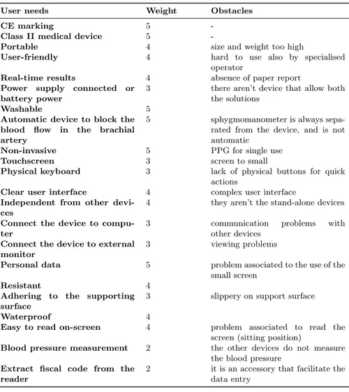
2.1 Advantages and Disadvantages of the most commonly used tech-niques to evaluate the Endothelial Function.* EndoPAT device 15 4.1 User needs, weight from 1 to 5 for each need (defined by the
users) and the obstacles recognised during the analysis of the context of use of the product . . . 31 4.2 The list of requirements divided in basic,technical and attractive 32 4.3 List of tasks that the user can perform thanks to the help of the
device, that covering the implicit and explicit needs identified 33 4.4 List of the technical requirements and relative MoSCow analysis 46 5.1 EDD description . . . 61 5.2 Dataset with fifteen patients and relative FMD. ED indicate
patients with Endothelial Dysfunction, NED the healthy patients. 64 5.3 Investigated parameters and their values . . . 84 5.4 The results based on the patient’s age. The first row the patients
have calculated the parameters, the second-row patients without the dicrotic notch identification and the last one indicate the patient excluded for some morphology anomalies. . . 85 5.5 The mean and the standard deviation for each parameters are
reported in this table, with the minimum and maximum value. 85 6.1 PPG feature mean (± Standard Deviation (SD)) of the PPG
endothelial dysfunction dataset (ppgEDD). . . 92 6.2 Anthropometric feature mean (± Standard Deviation (SD)) of
the PPG endothelial dysfunction dataset (ppgEDD) . . . 92 6.3 Investigation of H1: Classification performance obtained when
classifying PPG features with K-Nearest Neighbor (KNN), Ran-dom Forest (RF) and Support Vector Machine (SVM) classifiers. Classification accuracy (Acc), recall (Rec) and precision (P rec) are reported. . . 95
Elenco delle tabelle
6.4 Investigation of H2: Classification performance obtained when classifying photoplethysmography and anthropometric features with KNN, Random Forest (RF) and SVM classifiers. Classi-fication accuracy (Acc), recall (Rec) and precision (P rec) are reported. . . 95
Abbreviations
The following abbreviations are used in this manuscript:
BMI Body Mass Index
CDSS Clinical Decision Support Systems CMs Confusion Matrices
CV Cross Validation CVDs Cardiovascular Diseases DT Decision Tree
ED Endothelial Dysfunction
EDD Endothelial Dysfunction Dataset EHRs Electronic Health Records
FAST Function Analysis System Technique FMD Flow Mediated Dilatation
IRH Hyperaemic Reactive Index
IOT Intelligent Oncology Telecare IPA Pulse Area
KNN K-Nearest Neighbor LLO Leave-One-Out ML Machine Learning
NED Not Endothelial Dysfunction NO Nitric Oxide
PI Pulse interval PP Pulse Pressure
PPG Photoplethysmography
QFD Quality Function Deployment (QFD) RF Random Forest
SA Systolic Amplitude
SI Large Artery Stiffness Index P rec Precision
Rec Recall
RT Recovery Time
SVMs Support vector machines US Ultrasound
Capitolo 1
Introduction
1.1 Objective
In 2017, the European Heart Network (EHN) affirmed that Cardiovascular Di-seases (CVDs) cause 3.9 million deaths in Europe and over 1.8 million deaths in the European Union (EU) and it costs the EU economy 210 billion Euro a year [4]. CVDs refer to several disorders of the heart and blood vessels and in-clude coronary, cerebrovascular, rheumatic-heart diseases and other conditions that could lead to heart attacks and strokes, with consequent premature death. A possible strategy to prevent premature deaths is identifying the individuals at highest risk of CVDs (preventive healthcare) to ensure that they receive appropriate treatment.
In this context, Endothelial Dysfunction (ED) is achieving increasing im-portance, because it may improve the identification of individuals at risk: in Widlansky et al. [5], the endothelial function is defined a “barometer” for cardiovascular health. ED is observed in the early stage of most CVDs, athero-sclerosis or other disorders, and for this reason an effective screening and earlier diagnosis of ED is extremely important.
There are several (both invasive and non-invasive) techniques for endothe-lial assessment (Sec. 2.2), with limitations such as high incisiveness and/or costs, not being suitable for screening purposes. For example, the Eco-Doppler method, which is considered the gold standard among the non-invasive techni-ques, is operator dependent, expensive and requires a skilled medical specialist. The EndoPATTM
, (Itamar Medical, Israel) is an innovative device that tries to overcome some of these issues [6], [7], but still presents several limitations: the probes are disposables, which implicates high costs, and it is neither automated or standalone.
To date, a device that satisfy all the requirements of an ideal screening device to detect the ED does not exist and the state of the art does not reports fully automated data processing and Clinical Decision Support Systems the ED diagnosis.
Capitolo 1 Introduction
The purpose of this Ph.D. work is to develop and test a new screening device to detect the ED, this idea was inspired by Doctor Mario Rabuini (Cardiologist, Italy) and Eng. Massimo Pergolesi (Strumedical s.r.l.)1
.
Hence, this work is based on the development of an ideal screening device which is able to fulfil all the following requirements:
- Non-invasive ; - Cheap ; - Safe ; - Automated ; - Repeatable ; - Operator-independent ; - Portable ; - Stand-alone ;
- Printer-integrated to release immediately the report ; - Usable in home care ;
- Usable by any operator (medical doctors, nurses, pharmacists), not only by cardiologists.
The device may contribute to the early detection of ED, preventing cardiova-scular complications and lowering the number of premature deaths, using also a novel data processing method inside a Clinical Decision Support System for assisted diagnosis.
The device may contribute to the early detection of ED, preventing cardio-vascular complications and lowering the number of premature deaths, using also a novel data processing method inside a Clinical Decision Support Sy-stem for assisted diagnosis. Although the device is designed to be used for this application, the project has also been extended to an oncological context. The innovative idea consists in the use of the new device on cancer patients to monitor the action of chemotherapeutic drugs. In this way, it is possible to follow the cancer treatments also from the cardiology perspective. Indeed the toxic effects caused by cancer treatments may lead to endothelium alteration which may result in cardiovascular complications. For this reason, the design of the device, object of this thesis, has been included in the regional project Intelligent Oncology Telecare (IOT).
This Ph.D. work is included in the Italian Intelligent Oncology Telecare (IOT) project (Chapter 3). IOT is co-funded by the Marche Region program POR-FESR 2014-2020, with ten cooperatating companies, two departments of research of the “Universit`a Politecnica Delle Marche”, one department of the “Universit`a Degli Studi di Camerino” and the local health authority “Azienda Sanitaria Unica Regionale Marche”.
1
Strumedical s.r.l. is a company specialized in selling, assistance, importing and marketing electromedical equipment for diagnosis and monitoring. It is situated in Via Tambroni, 28, 62010 Montecassiano (MC), Italy
1.2 Thesis structure
1.2 Thesis structure
Figura 1.1: Graphical map of Ph.D. thesis, the background of endothelium in the Chapter 2 (red),the IOT project and the phases of the design in the Chap-ter 3 (red). The ChapChap-ter 4 (purple) describe the development of the hard-ware and softhard-ware about the device. The Chapter 5 (green) explain the algorithm to detect the endothelial dysfunction, while the classification of the dataset by Machine Learning approach will be describe in the Chap-ter 6 (blue). Moreover, with the green arrows are indicated the scientific contributions and in blue the technical contributions. Finally are repor-ted the logos about the co-financiers, Strumedical s.r.l. (company that it is the owner of the new medical device and co-financier of the this doc-torate), IOT that is the project co-funded by the Marche region program POR-FESR 2014-2020 explain in the Chapter 3.
Figure 1.1 shows the Ph.D. thesis structure and emphasises the scientific and technical contributions of this thesis.
The thesis is organised in seven chapters, which describe the design and de-velopment of the new medical device to detect ED. Chapter 2 explains the endothelium and reviews the state-of-the-art techniques for assessing ED and their limitations. Chapter 3 briefly describes the ED in the oncology context and the IOT project Chapter 4 describe the phases to develop the device (conceptual design, analysis and design of software and hardware, including the investigation of the ergonomic design of the device and the assembly of 3
Capitolo 1 Introduction
all components). Chapter 5 describes the development of the new index to discriminate the presence of the ED and the new algorithm to define the fi-ducial points useful for the parameters calculation from the PPG. Chapter 6 explains the machine learning approaches to analyse and improve the diagnosis of ED. Finally, conclusions are reported in Chapter 7 , as well as some future research directions.
1.3 Thesis contribution
The main contributions of this Thesis can be summarised as follows: • Design and development of a new medical device for ED screening; • Design and development of data-driven approaches for the analysis of
signals acquired with the new device;
• Design and development of a new Clinical Trial Study and Protocol for the ED device in the oncology context (IOT project).
• The new Clinical Decision Support Systems for ED clinical evaluation The device design and development has been a complex process rife with regulations, specifications, application requirements, and end user needs and all of which are balanced and adhered to for a successful product.
Results prove the correctness of the design intuition trough the real device implementation, the effectiveness of the biomedical data processing technique and of the Clinical Decision Support System applied to a real dataset and real patients, using the proposed device. The application to Oncology Telecare is suitable and the use of the overall approach on real clinical trials will apply the proposed device and methodology to the oncological care follow-up.
Capitolo 2
Background of Endothelium
This chapter is mainly focused on the endothelium functions and anatomy to better understand the overall project focus and the specific bio-medical me-chanism that are the base of the project design and data driven methodologies that are the main results of this thesis.
In the last decades, the endothelium was considered a simple barrier between the circulating blood and the vascular wall. It is now considered, instead, a predominant player in the control of blood fluidity, platelet aggregation and vascular tone, and it is recognised as the most important autocrine organ for the regulation of pathogenesis of thrombosis and atherosclerosis[8],[9].
The endothelium is a mono-layer of endothelial cells covering the lumen of blood vessels (arteries, veins and capillaries) and the lymphatic system, and therefore is in direct contact with the blood or lymph and the circulating cells[10]. In 1865 [11], the Swiss anatomist, Wilhelm His, introduced the term endothelium to define the barrier between the blood and the other tissues . At the end of the 70’s, thanks to the advent of new techniques how electron microscopy and the culture of tissues, Florey and collaborators give the new foundations to study the morphology and the function of endothelial cells [12]. Since then, several discoveries have occurred, in particular the endothelium vasculature modulating function and its specific agent, nitric oxide. In 1998 Drs. Furchgott, Ignarro, and Murad received the Nobel Prize for their discovery of the role of nitric oxide in cardiovascular regulation[13][14][15]. To understand the importance of endothelium and its functions, it is necessary to start with anatomy and physiology of it.
Capitolo 2 Background of Endothelium
2.1 The Vascular Endothelium
The vascular endothelium is an extensive network of cells covering the entire cardiovascular system. The essential components of the human cardiovascular system are the heart, blood and blood vessels. The hearth is the system’s pump and the blood is carried through the body via blood vessels (Figure 2.1). There are three major types of blood vessels:
- the arteries, which carries blood away from the hearth where it branches into smaller vessels
- the capillaries where nutrients, wastes and gases are exchanged between the blood and the tissues
- the veins which carry blood from capillaries to the heart.
Figura 2.1: Cardio Vascular System
The arteries and veins share the same general features, have three distinct tissue layers from the most interior layer called tunics, the most interior is the tunica intima, then there is the tunica media and the outer is the tunica adventitia (Figure 2.2). Each tunica has different characteristics, as follows:
- Tunica adventitia: it is mainly composed of collagen and surrounded by an external elastic lamina to anchor vessels with surrounding tissues. 6
2.1 The Vascular Endothelium - Tunica media: it is made up of smooth muscle cells, elastic tissue and
collagen.
- Tunica intima: it is the thinnest layer it is made up of one continuous layer of endothelial cells that are in direct contact with the blood flow. This layer is supported by a subendothelial connective tissue and then is surrounded by a thin membrane comprised of elastic fibres running parallel to the vessel.
Figura 2.2: Blood vessel structure, where are displayed the tunica intima, media and adventitia for arteries, veins and capillaries (image from [1]).
The capillaries walls consist of a single layer of endothelial cells and the smallest have a single endothelial cell wrapped around to join with itself.
Therefore lining the tunica intima is the squamous endothelial cells (called the endothelium), which is continuous throughout the entire vascular system, including the lining of the chambers of the endocardium (the innermost layer of the heart) and the lymphatic vessels (that carry lymph).
The endothelial cells are not all alike, in fact, the shape of the cells varies across the vascular tree, normally they are thin and slightly elongated, 50 -7
Capitolo 2 Background of Endothelium
70 µm long, 10 - 30 µm wide and 0.1 - 10 µm thick. In the blood vessel wall, endothelial cells are orientated along the axis of the vessel, minimizing the shear stress forces exerted by the flowing blood. In the adult human, the endothelium is composed of approximately 10 to 60 x 1012
endothelial cells that occupy a surface (blood/endothelium interface) measuring approximately 300 to 1000 m2
. For this reason the endothelium it is considered the largest organ in the human body [10].
2.1.1 Endothelial function
Endothelial cells act to maintain vascular homeostasis through multiple com-plex interactions with cells in the vessels wall and lumen including [10]:
• Barrier function - The endothelium forms a semi-selective barrier bet-ween the vessel lumen and surrounding tissue, controlling the passage of materials.
• Leukocyte trafficking - The interaction between leukocytes and the vascular endothelium is a physiological and pathophysiological process. The endothelium regulates the passage of white blood cells into and out of the bloodstream. Excessive or prolonged increases in permeability of the endothelial mono-layer, as in cases of chronic inflammation, may lead to tissue edema/swelling.
• Hemostasis - The hemostasis processes do not involve only the forma-tion of platelets, but also the fibrin formaforma-tion or blood coagulaforma-tion, even if both processes strongly interact. When the integrity of the vascular endothelium is interrupted, the subendothelial matrix and the collagen fibres are exposed, consequently, the circulating platelets adhere to these structures and start the hemostatic process.
• Balancing blood fluidity (thrombosis and fibrinolysis) - The endothe-lium prevent adhesion, aggregation and activation of platelets and promo-te plapromo-telet de-aggregation. Instance normally the endothelium provides a non-thrombogenic surface because it contains, for example, heparan sul-fate which acts as a cofactor for activating antithrombin, a protease that inactivates several factors in the coagulation cascade.
• Metabolism and catabolism - The endothelium can metabolize or con-versely activate many circulating factors and have essential roles during vessel formations, such as glycolysis, fatty acid oxidation, polypeptide hormones, amines, nucleotides, lipoproteins, metabolites of arachidonic acid and reactive oxygen species.
2.1 The Vascular Endothelium • Regulate smooth muscle cell growth - The endothelium it is able to stimulate or inhibit the proliferation of the underlying smooth muscle cells.
• Regulate the immune response - The endothelium play a key role in the regulation of immune response.
• Control of the vascular inflammatory process - the endothelium has the capacity to produce cytokines and adhesion molecules that regulate and direct the inflammatory process.
• Control of blood pressure - The endothelium plays an important role in the control of blood pressure through the regulation of vascular tone. • Regulation of Vascular Tone - The endothelium regulates vascular
tone by balancing the production of vasodilators including nitric oxi-de (NO), prostaciclina (PGI2) e l’endothelium oxi-derived hyperpolarizing factor, and vasoconstrictors.
As this PhD thesis mainly deals with the assessment of endothelial function based on the capacity of the blood vessels to self-regulate vascular tone, the regulation of it will be discussed more in detail hereafter.
The role of Nitric Oxide in the regulation of vascular tone
In the 1980 Furchgott and Zawadski, discovered that the endothelium is able to produce the vasodilatation substance that called the endothelium-derived-relaxing-factor (EDRF) [13]. Later researches identify this vasodilator with the Nitric Oxide (NO), that play a crucial role in the regulation of vascular tone. NO is a soluble gas continuously released by the endothelium with a half-life of 630 s, synthesized from the amino acid L-arginine in endothelial cells by the constitutive calcium-calmodulin- dependent enzyme nitric oxide synthesis (NOS)[16][17][18][19][20].
The production of this enzyme may be induced by many stimuli that opera-te on specific receptor situaopera-ted on the surface of endothelial cells, and consist of different endogenous substance such as acetylcholine, bradykinin, Substan-ce P, Histamine and the serotonin. However, the principal stimulus for the NOS and release from the endothelium is a physical stimulus, in particular the shear stress of blood flowing over the surface of the vessel by a non-receptor-dependent mechanics [21][22][23][24].
Regardless of the stimulus, when the Nitric Oxide is released, spreading through the membrane of endothelial cells and inside in the smooth muscle cells where the NO stimulus soluble guanylyl cyclase, producing an increased concentration of cyclic guanosine monophosphate (GMP) that regulate many 9
Capitolo 2 Background of Endothelium
functions, such as the relaxation of the vascular smooth muscle and the platelet function[25][8].
To summarize, the main roles of NO are vasomotor action, inflammation pro-cess, antioxidant activity, and has multiple beneficial effects including, maintain vascular homeostasis, regulation of local cell growth, inhibition of leukocyte ad-hesion, and protection of the vessel from platelets whole and cells circulating in the blood [10][26]. A limited production of the NO and a reduced bioa-vailability of NO, it is the early anatomical alteration of endothelium due to the atherosclerotic process and consequently may induce other cardiovascular diseases.
2.1.2 Endothelial dysfunction
Under normal conditions, the endothelium preserves normal vascular tone and blood fluidity and all the endothelial functions are balanced. When cardio-vascular risk factors appear, a chronic inflammatory process starts. Disease conditions could worsen and provoke a loss of vasodilator and anti-thrombotic factors and an increase in vasoconstrictor and pro-thrombotic products. This situation implies an unbalanced condition of the normal endothelial functions and, in the first stages, the principal endothelial alteration is solely at the anatomical level [5][27]. In this conditions, the defence mechanism of vascu-lar endothelium is compromised, and the endothelium is liable to Endothelial Dysfunction (ED).
ED leads to atherosclerotic lesion formation, inflammation, hypertension, heart failure and is strongly correlated with classical and novel risk factors for CVDs [28]. Commonly-recognized risk factors are ageing, smoking, dyslipi-demia, diabetes, postmenopause state, hypertension, to which novel risk fac-tors are added, e.g. obesity, homocysteine, infection or inflammation, physical inactivity, post-prandial state [5][29].
An endothelium characterized by a dysfunction (maybe due to the presence of risk factors) is marked by a decrease of the NO bioavailability, and other protective substances. In addition, the endothelium may transform into a harmful organ because it is induced to synthesize mainly vasoconstricting, pro-aggregating, pro-inflammatory substances. The first consequence is the beginning of atherosclerotic or thrombosis processes. Furthermore, patients with diabetes, obesity, dyslipidemia and hypertension are characterised by ED. There are many interventations to limit these complications, some of these may contribute to the reverse of endothelial dysfunction. Thus for instance antioxi-dant therapy (typically vitamin C, vitamin E), dietary modifications, lifestyle modification, reduction of cholesterol, quitting smoking and the assumption of some substances to improve the NO synthase.
2.2 Method of evaluating the Endothelial Function
2.2 Method of evaluating the Endothelial Function
During the last years, several techniques have been developed to evaluate the endothelial function, both invasive (that are considered the gold standard for early detection of endothelial dysfunction) and non-invasive (that are giving promising results and show good reproducibility)[30][31][32].2.2.1 Invasive technique
• Intracoronary Infusions:
This method consists of the intracoronary infusion of some vasodilatory substances (such as acetylcholine) to measure the changes in vessel dia-meter using quantitative coronary angiography. The aim is checking how the coronary arteries dilate after the infusion, to quantify the endothelial function. In particular, in presence of endothelial dysfunction, the acetyl-choline may lead to a decreased vasodilatory response, on the other hand, the arteries dilate in a dose-dependent way. This technique is invasive, more expensive. Moreover there are some risks correlated with the ca-theterisation and for these limitations it is not be considered a screening procedure.
• Intrabrachial Infusions:
In this technique, the infusion it is more accessible than the coronary because it is applied in the brachial artery. This procedure like the pre-vious method gives a direct quantification of endothelial function (at the level of brachial artery ) and it consists in the evaluation of dose-response relations of endothelial agonists and antagonists. This method is higher reproducible and the catheterisation may be less complicated but may be dangerous for the median nerve. Nevertheless, it is invasive and expensive and can causes infections.
2.2.2 Non Invasive technique
Flow Mediated Dilatation
In 1992, Celermajer et al. developed a new non-invasive method to evalua-te the endothelial function [33], called Flow Mediaevalua-ted Dilatation (FMD) that describes the increase of the arterial diameter, as a consequence of the reac-tive hyperemia, and is comparing it with the baseline diameter and expressed simply as a percentage of this baseline diameter (% FMD). The exam is con-ducted with the ultrasound system by a highly specialised doctor, usually a cardiologist. The procedure in detail is reported in the ”Guidelines for the 11
2.2 Method of evaluating the Endothelial Function 2. Compressive phase: This phase lasts 5 minutes and consists in the inflation of the sleeve with a pressure of at least 50 mmHg above the systolic blood pressure, in any case, not exceeding 200 mmHg.
3. Post-compressive or hyperaemic phase: This phase lasts 5 minutes and starts immediately after the quick deflation of the sleeve. After the first minute, the video recording begins for 1 minute with the reference ECG and with the positioning of the sample volume of the pulsed Doppler in the centre of the brachial artery (always on the longitudinal plane with correction of the angle lower than 60 degree ) to highlight the hyper flow blood. The measurements of the arterial diameter in 5 samples, in tele-diastole, coinciding with the R wave of the ECG. The average of these 5 measurements, will be considered as the Post Hyperaemia Diameter. FMD is calculated as a percentage increase in the diameter of the artery during the reactive hyperemia phase compared to baseline values, how the ratio:
F M D = P ostHyperemiaDiameter − BaselineDiameter
BaselineDiameter ∗ 100 (2.1)
The patient with normal endothelial function has an FMD greater than 7% while the patient with endothelial dysfunction less than that value.
Pulse Wave Velocity
Aortic Pulse Wave Velocity (PWV) is considered an index of aortic stiffness, and is defined as the velocity of pressure pulse wave, generated by the systolic contraction of the heart, propagate along the arterial tree. PWV is normally measured between the carotid and femoral artery calculating the distance from the arrival of the pressure pulse wave in these two places. The pulse waves travel through the arteries and its velocity depends on the vessel. Indeed, all the factors including the endothelial dysfunction may lead to the arterial stiffness and therefore the lower vessel dilatation and compliance [37][38][39].Typically, the pulse wave is detected by pressure transducers or arterial tonometry. Peripheral Arterial Tone
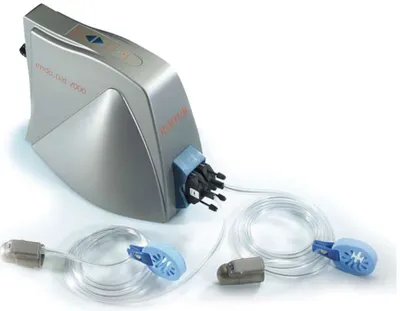
The Peripheral Arterial Tone (PAT) signal is a proprietary technology mea-sured by the EndoPATTM
Israel device (Itamar Medical) and represents the arterial tone changed in peripheral arterial vessels. In detail, EndoPATTM
is a non-invasive device for endothelial function assessment, is approved by the Federal Drug Administration and it was developed and distributed by Itamar Medical, Israel. The device (Figure 2.4) records the PAT by unique photoelec-tric fingertips, different from the normal photoplethysmography sensors because 13
Capitolo 2 Background of Endothelium
they are provided by an external casing that containing inflatable chambers. This solution is thought to apply uniform pressure to the surface of the distal finger, to excluded the venous flow. The exam follows the same procedure of the FMD method. In detail, the exam consists in the application of two finger-tips one for the hand that will be subjected to the transient ischemia (created by a normal pressure cuff situated on the upper arm) and one in the other hand for control. To assess endothelial function, the blood flow after occlusion (hyperemic condition) is compared to the baseline flow and the ratio is com-puted by the EndoPATTM
software that gives the EndoPAT index, called also RHI-PAT [6],[40][41][42] [43][44].
Figura 2.4: EndoPATTM
, Itamar Medical, Israel
2.2.3 Limitation of the assessment of endothelial function
Table 2.1 summarises the advantages and disadvantages of both the invasive and non-invasive procedures”.
It is worth noticing that these techniques are time-expensive, require both manual intervention (from specialized heathcare operators) and expensive de-vices, that are not standalone. All these disadvantages make it difficult to consider these devices suitable for the screening and prevention, and indeed they are not easily included in the routine examination.
Starting from such a premise, this thesis addresses the topic of developing a new medical device to assessment the endothelial function, to prevent the 14
2.2 Method of evaluating the Endothelial Function cardiovascular complications. Following are reported some requirements: non-invasive, cheap, safe, automated, repeatable, operator-independent, portable, stand-alone, printer integrated to release immediately the report, usable in home-care, usable from any operator (medical doctors, nurses, pharmacists), not only cardiologists.
Techniques Advantages Disadvantages
Intracoronary infusion
- direct quantification of endothelial function in the coronary arteries
- direct connection with the infusion of nitric oxide
- invasive - expensive
- risk associated with the catheterisation infection, vascular injury,stroke
- not screening test
Intrabrachial infusion
- brachial artery is easily accessible then the coronary arteries
- cannulation has less complications
- invasive - expensive
- risk associated with the catheterisation (infection, vascular injury)
- risk of median nerve - not screening test
FMD
- non invasive - safer
- faster then the other techniques
- highly operator-dependent - expensive
- variability in measurements - the arteries smaller than 2,5 mm in diameter are difficult to measure - requires a skilled operator - size of the equipment
Pulse Wave Velocity
- non invasive - safer
-requiring less training - operator independent
- it is not stand alone - variability in measurements PAT* - non invasive - safer - new technology - operator-independent - expensive
- probes can be used only one time - it is not standalone
- not automated - size of the equipment
Tabella 2.1: Advantages and Disadvantages of the most commonly used techniques to evaluate the Endothelial Function.* EndoPAT device
Capitolo 2 Background of Endothelium
2.3 A new approach for the assessment of the
Endothelial Dysfunction
This paragraph describe the technique used to develop a new medical device. Although each of the many functions performed by the endothelium can be examined to detect the endothelial dysfunction, the one that is simpler to evaluate, and which appear more standardized, is the regulation of vascular tone (the same issue evaluated by the FMD and EndoPAT)[34][45]. When endothelial damage occurs the vascular tone is the first function that can be compromised because may depend by the anatomical alteration of endothelial cells, and therefore useful to detect previously. For this reason, the study of volume-blood changes was identified as a possible method to detect the endothelial dysfunction.
Among the different methodologies to assesment the endothelial function, the attention was focused on the photoplethysmography. There are several studies that were conducted to investigate the utility and efficacy of the photoplethy-smographic signals in the assessing of endothelial function. In particular, some of these were conducted to investigate if there is any comparison with the gold standard (Doppler ultrasound via flow-mediated dilatation)[3],[46],[47],[48],[49]. Based on these results, that show the high correlation between the exams of ED by PPG and the gold standard and considering the limitations of the normal techniques that have already been explained, to satisfy all the requirements of the new medical device the PPG method was chosen.
2.3.1 Photoplethysmography
Photoplethysmography (PPG) is a noninvasive, optical technique that can be used to determine the changes in blood volume as a function of time, in the microvascular bed of tissue. To obtain a PPG signal, a light emitting diode (LED) illuminates tissue with two different wavelengths (red and infrared) and a photodiode on the other side of the tissues measures the intensity of the non-absorbed light at each wavelength, Figure 2.5. This technique was developed by Takuo Aoyagi and is an indispensable clinical tool for non-invasive monitoring of blood oxygen levels. The technology it is based on two principles:
• Light absorbance is different for oxygenated and non-oxygenated haemo-globin at the two wavelengths used
• Based on the wavelengths there are two components (Figure 2.6) – The absorbance has a pulsatile (AC) component which reflects the
pulsations from the cardiac cycle, indeed represent the changes in the blood volume that occurs between the systolic and diastolic phases. 16
Capitolo 2 Background of Endothelium
2.3.2 PPG and ED
As mentioned in the paragraph 2.3, in literature different studies demonstrated that there is a correlation between the PPG and the Flow Mediated Dilata-tion (gold standard) to assessment the ED [3],[46],[47],[48],[49]. In particular, Zahedi et al. [3] [50] demonstrated a significant association between PPG amplitude and the FMD to evaluate the endothelial function, they applied the same procedure of the Flow Mediated Dilatation with the acquisition of PPG. In detail, for measuring the PPG signal, they recorded three minutes in the baseline condition, four minutes during the occlusion of the brachial artery (by the cuff placed above the elbow), five minutes during the hyperaemia. The fi-gure 2.7 takes from the paper Zahedi et al,[3] represents the PPG wave respect the FMD wave measured with Eco-Doppler of a healthy patient.
Figura 2.7: Percentage changes in the brachial artery diameter with the PPG si-gnal (dotted) and the FMD (solid line) in the healthy patient during the hyperaemia phase (images from[3]).
It was thus decided to develop a new medical device to detect the endothe-lial dysfunction based on photoplethysmography technology, for the following advantages:
- There is the correlation between the PPG and FMD (gold standard) - Less expensive respect the other methods
- Possibility to extract some morphological features - Non-Invasive
Capitolo 3
ED in the oncology context
In recent years, thanks to advances in early diagnosis and above all substantial improvements in therapy, there has been a noticeable prolongation of patients survival affected by neoplasms. However, cancer patients showed some car-diovascular complications which represent an increasing problem in modern cardiology and oncology clinic practice. In particular, ventricular dysfunction and heart failure are the adverse effects common to many categories of cancer drugs. For this reason, a careful assessment of the patient’s risk profile throu-gh close cooperation between doctors and patient monitoring is necessary for successful treatments, in a way to prevent and control complications. In this context, the idea is follow cancer patients during chemotherapeutic treatments, with the new medical device, objective of this thesis. The introduction of this device allow to identify previously the ED which, has some studies have shown, may be caused by the toxic effects that comes from the assumption of cancer drugs. At this purpose a project called IOT has been developed with the aim to constantly monitor patients following chemotherapy treatments with the ED detection tool.
3.1 Introduction and scopes of the IOT project
The project Intelligent Oncology Telecare (IOT) integrated the research activities of this thesis. IOT is a project co-funded by indirect Europoean Commission funding managed by the Marche Region under the program POR-FESR 2014-2020. In the project are involved ten companies, three university departments and the local public health authority.The IOT project targets the development of a new management model for chronically illness oncology, through the development and testing of advan-ced technological systems that allow home management of the sick under the supervision of qualified medical team that works remotely through a control centre. Today the improvement of efficiency and optimization of the health system requires of the decision-makers that they respond to a growing demand for services while maintaining the same level of service. In Italy the number 19
Capitolo 3 ED in the oncology context
of cancer patients are increasing (in 2014 there were 365,000 malignant tumor diagnoses, in the latest ISTAT data (2012) there were 1322 cases in the Mar-che Region) and at the same time the mortality rate of this disease is actually decreasing, so that today the term ”chronic” allows for living with the disease for a longer and longer period, engaging more therapeutic resources, through both hospital and domestic care since several studies conducted show that the treatment administered to chronic patients is far more effective if administered at the patient home. As a matter of fact, experimental studies only 4% under-going domiciliary palliative care resort to hospital admissions. The innovative organisational and management model of home care for the cancer sick will be through the development of new telecare devices and technological communi-cation infrastructure for the continuous monitoring of the patient, controlled drug intake, psychological and clinical care as well as the creation of a data sharing environment related to the particular regime of treatment. From the point of view of hardware new SMART devices will be developed that will cau-se a paradigm shift in the quality of life of a terminal cancer patient enabling treatment at home with all the required medical and technological support. The system will avail of devices for the detection of important physiological parameters to determine the patient’s health that can be easily deployed in the home environment. In addition, the cancer patient would have to undergo diagnostic tests to check for side effects of a therapy, which leaves scope in this project for the design and development of such innovative devices (System for evaluation of endothelial dysfunction and electrocardiograph) that will allow examinations at home with analysis and reporting conducted through autho-rized doctors remotely. In patient management oncology at home the project will also produce a device for the administration of drugs according to appro-priate protocols through a smart dispenser that is an innovation compared to the current scientific and technical state of the art. The release of the drug will be processed automatically by generating alarms at two levels: the first is effected through a simple acoustic reminder mechanism and / or voice. Failing this warning the device is able to alert a caregiver that the medicine has not been taken through the delivery of a push message. The use of this device safeguards the patient against a loss of the correct prescribed care, without in-terruption of family life at a difficult time. It also simultaneously facilitates the optimization of cost and management of delivered health care. The monitoring and diagnostic devices integrated constantly send all the medical information in encrypted form to a Cloud facility that will allow it to be available in real time. Furthermore, this same data and can be integrated with the regional health records, a facility not available today. In this way it will effectively per-mit the creation of a real home control centre of the physiological parameters required to assess the state of health of the patient by carrying out ”remote
3.1 Introduction and scopes of the IOT project visits” to the clinician who will be able to evaluate the effects of the drug use via the dispenser, thus providing a totally innovative, immediately accessible mode of administration. The scientific information produced by research com-bined with those produced in real time and transmitted from the devices on the network from the dispenser, generate large amounts of data, the complexity of the phenomenon, requires the development of a inferential semantic system for oncology. The system is designed as a powerful and effective innovative tool to be used by the clinician or physician to acquire, integrate, develop, make available and enhance an impressive wealth of knowledge, based on the integra-tion and correlaintegra-tion with multiple levels of both clinical and scientific grounds, both real and experimental. The architectural solution will be service oriented (REST), in order to offer to the user community, in a short time, an innovative and advanced service, which can be continually improved on the basis of its own results.
IOT partnership
The project leader is the VIVISOL Srl, one of the leading European groups ope-rating in home healthcare, that through the means of this project intends to invest in research and development of new models of specialized care for cancer patients, and thus enter into a new market. The rest of the partnership consists of a pool of highly competent knowledge based companies working intensively for years in the field of medical and electromedical devices. Aditech, Systemic and Strumendical are involved in the project for the construction of new mo-nitoring devices of physiological parameters, home diagnostic devices and the new dispenser thereby taking the opportunity to introduce new products in the healthcare market. Ataena, manufacturer of machinery for the packaging of the drug, recognizes the potential of the dispenser by intervening in the de-velopment of a new packaging format for drugs in order to reduce waste. Again with reference to the dispenser design, the intervention of AV Consulting will provide the project with valuable experience in the area of biomedical design. The software for the device will instead be drawn up by companies Win Italia and On Demand. Charry Lab will provide for communication between the dif-ferent devices in order to enhance the character of interoperability allowing the sharing of data and warnings that are managed by a web platform developed by Bookerang that has already participated in other projects in the health sector and intends to go to market with a developed product product. In the part-nership, taking the role of consulting will be three public research companies (Unicam, UNIVPM and further to these in the same role one public-private company (Meccano). The main competitive advantage attained by companies is expected be the definition of a model of care of cancer patients standardized and normalised so that it can be replicated and exported on a national and 21
Capitolo 3 ED in the oncology context
international scale. This represents a real opportunity for companies to enter their systems and devices within standardized protocols that can be sold as a single system or package. Further, entire territories might adopt on a trial basis such systems to provide certain types of users (from the chronically ill) who may come from abroad to choose a type certified nursing which enables high standards of quality and comfort. In addition, companies will be able to look out at a potential market which is offered through the involvement of private investigators. The companies will commit to producing products in 18 months, with the exception of the endothelial device that requires more time for the certification of the biomedical device and this is expected to release at month 24. In any case, the duration will be from 18 to 12 months. The ex-perimental phase of the project will be conducted on ASUR AREA VASTA 2, the territorial zone of Fabriano and Senigallia (Italy), will validate the clinical aspect making it possible to identify a business model and measurement of the ROI of such technologies within the public and private health systems. This is thanks to the involvement of VIVISOL, which will handle the technological validation.
IOT Clinical Trial
The trial will be carried out thanks to the voluntary cooperation of 100 cancer patients treated at the Medical Oncology Unit of Fabriano and Senigallia. The population will be composed of individuals who show a higher cure prospective greater than six months, sub categorized through a sufficiently representative sample by age, tumour type and treatment. The purpose is to validate a social welfare model of cancer patients through a trial that compares two groups of patients. In fact the first half of the sample (approximately 50 subjects) will be treated through an evolved care model with all the instrumentation developed within the project, the other half will only be treated with a traditional home care system with the aim of putting comparing the results obtained in terms of cost benefit. This assessment, which will be realized through statistical studies, will be based on symptom control and the quality of life of patients, the use of hospital resources in the active treatment phase and the end of life, the acceptability of the technologies by patients and their family members.
3.1 Introduction and scopes of the IOT project IOT goals
The benefits that this new model would bring the needs of the community are manyfold and all aimed at improving home care resulting in more efficient pu-blic spending: i) greater comfort for care received in the home environment will lead to a reduction in hospital admissions and emergency calls; ii) through the platform and the sensors it will be possible to provide new daily data availa-ble to the general practitioner and clinical staff allowing them to evaluate and to appropriate care prescribed based on the extensive monitoring by reducing hospital admissions and facilitating optimisation of drug consumption.
The project started in 2017 and will last three years. The idea is to propose a new and efficient home-based management system for the oncologist to im-prove the quality of care through an innovative tele-care devices and the use of advanced data methods.
The main project goals related to this thesis are (figure 3.1):
- overseeing the cancer patients every hour at home with a new, low cost and easy to use screening device (object of this thesis) and collect a list of other basic health parameters and data (electrocardiogram, blood pressure, etc) to a cloud infrastructure.
- design a data driven decision support system (DSS) on a cloud computing architecture concerning oncologist medicine, to facilitate the sharing of health data and coordinate their streams.
- give the possibility to the clinical team, to administrate the drugs, check the action of the chemotherapeutic treatment identify previously the toxic effects and eventually change the therapies, based on the DSS.
In this context, the new screening device to detect endothelial dysfunction plays a key role: it should follow the patients during the cancer treatment, mo-nitoring the action of chemotherapeutic agents. In this way, it is possible to fol-low the cancer treatment also from the cardiology perspective. Previous studies linked the toxic effects caused by the radiotherapy (RT) and/or anthracycline-containing chemotherapy and the endothelium alteration. This may result in an increased risk of cardiovascular complications, as coronary heart disease, valvular heart disease, and heart failure (HF) in survivors of tumours [51] [52] [53][54][55][56].
The following paragraphs describe all the activities of the IOT project cor-related with the development of the new medical device. Whereas, all the following phases, are coordinated and supervised from the undersigned that has covered the role of project manager. All these activities are coordinated by the collaboration of Strumedical s.r.l. 1
(the owner of the device) and the
1
Strumedical s.r.l. Via Tambroni, 28, 62010 - Montecassiano (MC).
Capitolo 3 ED in the oncology context
Department of Information Engineering , Universit`a Politecnica Delle Marche
2
.
Figura 3.1: IOT project schema
2
Department of Information Engineering, DII, Universit`a Politecnica delle Marche, Ancona (AN)
3.2 IOT Design Activities
3.2 IOT Design Activities
The IOT project is divided into two main activities: The development of the prototypes, and the clinical trial for the bio-medical technique validation. The following list reports all the project activities with the specific partners involved:
• Design and development of the device prototype, in cooperation with: – Department of Industrial Engineering and Mathematical Sciences,
Universit`a Politecnica Delle Marche3
for the conceptual design (de-scribed in the paragraph 4.1).
– KUNST Engineering s.r.l. 4
company to design and produce the hardware and firmware (described in the paragraph 4.2 ).
– Department of Informatics of the ”Universit`a di Camerino” (UNI-CAM)5
and the Department of Information Engineering , Universit`a Politecnica Delle Marche (UNIVPM)6
to develop the software and the Electronic Health Record (described in the paragraph 4.3). – Eng. Raul Frolla 7
and the TECNE 90 s.p.a.8
company for the in-dustrial design and the 3D printing models respectively (described in the paragraph 4.4).
• Clinical Trial: The aim of this step is to compare the results from the device to the gold standard, i.e. the Flow-Mediated-Dilatation by Eco-Color-Doppler exam. For this part, it became necessary to contribute to defining the statistical analysis and the correct way to compare the two exams. Moreover, it was necessary describing in detail the new medi-cal device, as the exam procedure, the algorithm implemented and other features to define the clinical investigation plan, used by the Ethics Com-mittee and Ministry of Health. This phase is obligatory to validate the algorithm and the device, and fundamental eventually for the next step, otherwise the EC certificate according to the Directive 92/42 EEC. Sche-matically the clinical trial is reported in the Appendix (Figure 1). In this task are involved the following structures:
3
Department of Industrial Engineering and Mathematical Sciences, Universit`a Politecnica Delle Marche Via Brecce Bianche, polo Monte Dago, 60121 - Ancona (AN)
4
KUNST Engineering s.r.l. Via Remo Stortoni, 20, 62019 - Recanati (MC)
5
Department of Mathematics and Computer Science, University of Camerino - Camerino (MC)
6
Department of Information Engineering, DII, Universit`a Politecnica delle Marche, Ancona (AN)
7
Eng. Raul Frolla, Via Vittorio Veneto, 9 , 63839 - Servigliano (FM)
8
TECNE 90 s.p.a., Via Firmino Giulietti, 3, 62010 - Montelupone (MC)
Capitolo 3 ED in the oncology context – Hospital of Fabriano9
, where the clinical trial will start in January and after six month will be concluded, the study will be supervi-sed by local health authority ”Azienda Sanitaria Unica Regionale Marche”.
– VIVISOL s.p.a. 10
(a SOL group company), operates in the fra-mework of home care in the health care setting by managing medical and curative therapies at home. They will be follow part of the Cli-nical trial in the Hospital and then they will replicate the procedure in the home care environment.
– MECCANO s.p.a. 11
expert in the sector of the device certification process, they supported all the activities necessary to obtain the CE Marking.
Furthermore, has been necessary for this project add some technical requi-rements respect the first version of the device because were necessary some integrations, how the communication with health platform (provide clinical health care from a distance) and the interconnection with other devices as the blood pressure monitor. For this part, it is involved the Department of Informa-tion Engineering (UNIVPM), and the Department of Informatics (UNICAM). After this phase, when the measure will be validated, begins the second phase the monitoring of cancer patients at home with the device to detect the ED. For this phase will be necessary to continuously monitor the patients involved by providing them with a basic technological infrastructure for the detection of physiological parameters. For this purpose, the work that Vivisol (a SOL group company, operates in the framework of home care in the health care setting by managing medical and curative therapies at home) makes at the pa-tient’s home it is crucial, thanks to the constant nursing medical support, were collected all the measurements of physiological parameters. At the same time, the ASUR Marche with the oncology associations follows the home care service to the cancer patient’s home. Finally, the monitoring and diagnostic devices that are used at patient’s home, constantly send all the medical information in encrypted form to a Cloud, to be available in real time the patient’s data to the clinician.
9
”E. Profili” Hospital, Intensive Care Unit, 60044 - Fabriano (AN).
10
VIVISOL, Via Agostino Novello 1, 60035 - Jesi (AN)
11
Meccano s.p.a., Via G. Ceresani, 1, 60044 - Fabriano (AN)

![Figura 2.2: Blood vessel structure, where are displayed the tunica intima, media and adventitia for arteries, veins and capillaries (image from [1]).](https://thumb-eu.123doks.com/thumbv2/123dokorg/2967809.27051/29.680.131.567.274.668/figura-blood-vessel-structure-displayed-adventitia-arteries-capillaries.webp)