La presente tesi è stata prodotta nell’ambito della Scuola di Dottorato in Scienze
Biomediche dell’Università degli Studi di Sassari, a.a. 2011/2012 – XXVII ciclo,
con il supporto di una borsa di studio finanziata con le risorse del P.O.R.
SARDEGNA F.S.E. 2007-2013 - Obiettivo competitività regionale e occupazione,
Asse IV Capitale umano, Linea di Attività l.3.1.
U
NIVERSITÀ DEGLIS
TUDI DIS
ASSARISCUOLA DI DOTTORATO DI RICERCA IN SCIENZE BIOMEDICHE
Direttore della Scuola: Prof. Andrea Fausto Piana
INDIRIZZO IN ODONTOSTOMATOLOGIA PREVENTIVA
XXVII CICLO
Clinical evaluation of a self-adhering material
as desensitizing agent
in xerostomic patients for head and neck cancer.
Direttore:
Prof. Andrea Fausto Piana
Tutor:
Prof.ssa Egle Milia
Tesi di dottorato di:
Dott. Roberto Pinna
Roberto Pinna,
Clinical evaluation of a self-adhering material as desensitizing agent in xerostomic patients for head and neck cancer.,
Tesi di Dottorato in Odontostomatologia preventiva, tudi di Sassari.
2
Abstract
Xerostomia is a common clinical symptom that may suffer patients with Head and Neck
Cancers during and after radiotherapy. The aim of the present thesis were therefore: 1)
to review the current state of knowledge of pathology, clinical complications and
radiotherapeutic patient management, 2) to evaluate the aetiology of dentine
hypersensitivity in conditions of reduced salivary flow resulting in the radiation
exposure, 3) to evaluate the effectiveness of the materials commonly used in the
treatment of hypersensitivity, when they work in conditions of hyposalivation.
Paper I is systematic review of actual management strategies for radiation-induced
hypofunction and xerostomia in head and neck cancer patients. Paper II and III are
based on the data of two split-mouth randomized clinical trial, where the efficacy of 4
different kinds of desensitizers has been assessed in the short and long term on patients
with normal salivary flow. Paper IV is a long term evaluation based on the same
experimental protocol applied on xerostomic patients.
The results showed that Dentine hypersensitivity is one among the multiple
complications in the oral cavity that is possible diagnose in patients affected by
xerostomia post radiotherapy. It may occur as a result of the combination between the
typical etiologic factors and the reduction in salivary flow, that have a essential
protective role for teeth and oral mucosa health. Dentine hypersensitivity arise from the
tubular dentine exposure as a result of enamel loss and/or gingival root surface exposure
and their occlusion is the first choice for the treatment. Unfortunately, there is still no
gold standard for therapy of DH available today. In addiction, there is in the literature a
lack of information about DH in xerostomic patients after radiotherapy.
The main conclusions from this thesis are that: 1) the radiation-induced xerostomia
could be considered a multifactorial disease. It could depend on the type of cancer
treatment and the cumulative radiation dose to the gland tissue. A preventive approach
and the correct treatment of the particular radiotherapeutic patient can help to improve
the condition of xerostomia. 2)The experimental data obtained from patients with
normal salivary flow show that all the materials tested produced a reduction of dentine
permeability. However, after 12-week controls, there was no significant statistical
difference in the efficacy. 3) In xerostomic condition all the materials tested produced a
significant reduction in the dentine sensibility. In light of the observed data, after
12-week controls there is no statistically significant difference between the desensitizers
and they show a less stable behaviour compared to the normal salivation condition.
Key words: Xerostomia, head and neck cancers, radiotherapy, dentine hypersensibility,
desensitising agents.
Roberto Pinna,
Clinical evaluation of a self-adhering material as desensitizing agent in xerostomic patients for head and neck cancer.,
Tesi di Dottorato in Odontostomatologia preventiva, tudi di Sassari.
3
Contents
Original Papers ……… 4
Introduction ………. 5
Rational of the study ………... 13
Aims ……….. 14
Material and Methods ………. 15
Results ……….. 25
Discussion ………. 32
Conclusions ……….. 38
References ……… 39
Roberto Pinna,
Clinical evaluation of a self-adhering material as desensitizing agent in xerostomic patients for head and neck cancer.,
Tesi di Dottorato in Odontostomatologia preventiva, tudi di Sassari.
4
Original Papers
This thesis is based on the following four papers, which will be referred to in the text by
their Roman numerals:
I.
Xerostomia induced by radiotherapy: an overview of the physio-pathology,
clinical evidence and management of the oral damage.
Pinna R, Campus G, Cumbo E, Mura I, Milia E.
Ther Clin Risk Manag. 2015;11.
II.
Short-term response of three resin-based materials as desensitizing agents
under oral environmental exposure.
Milia E, Castelli G, Bortone A, Sotgiu G, Manunta A, Pinna R, Gallina G.
Acta Odontol Scand 2013;71:599-609.
III.
Clinical evaluation of the efficacy of one self-adhesive composite in dental
hypersensitivity.
Pinna R, Bortone A, Sotgiu G, Dore S, Usai P, Milia E.
Clin Oral Investig. 2014; In press.
IV.
Clinical evaluation of a self-adhering material as desensitizing agent in
xerostomic patients for head and neck cancer.
Pinna R, Dore S, Sotgiu G, Milia E.
Ready for press.
Roberto Pinna,
Clinical evaluation of a self-adhering material as desensitizing agent in xerostomic patients for head and neck cancer.,
Tesi di Dottorato in Odontostomatologia preventiva, tudi di Sassari.
5
Introduction
Xerostomia induced by radiotherapy in head and neck cancers.
Xerostomia is a term used to describe the subjective symptoms of a dry mouth often
deriving from a lack of saliva. A large variety of causes can lead to xerostomia e.g.
radiotherapy and chemotherapy(1-4), the chronic use of drugs (5-7), rheumatic and
dysmetabolic diseases (8,9). Major salivary glands contribute to most of the secretion
volume and electrolyte content of saliva (the parotid, submandibular, and sublingual
glands, which account for 90% of saliva production), whereas minor salivary glands
contribute little secretion volume and most of the blood-group substance
(10).
Most patients diagnosed with head and neck cancer (HNC) receive radiotherapy as part
of their cancer treatment. Head and neck cancer (HNC) actually includes many different
malignancies. The most common type of cancer in the head and neck is squamous cell
carcinoma, which originates in the cells that line the inside of the paranasal sinuses,
nasal cavity, salivary glands, oral cavity, esophagus, pharynx and larynx (11).
Worldwide, lip and oral cavity cancer along with thyroid cancer have the highest
incidence; esophagus cancer is the most aggressive presenting a 4.9% mortality rate
(Table 1). Similar findings regarding the incidence, mortality and prevalence of cancer
in the European Union have been reported. The highest mortality rate belongs again to
esophagus cancer with a predominance of 2.3% (Table 2).
HNC patients receive radiotherapy before, during, or after surgery as part of their
cancer treatment. Routinely, HNC patients receive a dose between 50 and 70 Gy once a
day for five days a week, (2 Gy per fraction) (12); on the other hand, if the radiotherapy
protocol is just pre-operative, the total amount of radiation is usually lower. Conformal
radiotherapy (CRT) is the most common type of radiotherapy used for the treatment of
HNC; a special attachment to the radiotherapy machine carefully arranges the radiation
beams to match the shape of the cancer, reducing the radiation to the surrounding
healthy cells. Another similar type of radiotherapy used against HNC, known as
intensity-modulated radiotherapy (IMRT), allows a more accurate delivery of specific
radiation to be distributed to the tumor mass according to its location and severity,
sparing the tissue and organs at risk, e.g., salivary glands (10). This radiation dose
normally is used to destroy malignant cells and very often leads to the onset of salivary
gland hypofunction and chronic xerostomia (13), that are the most common
complications and occur to some degree in up to 100% of patients, severely impairing
their quality of life (14).
The main problem, which correlates the xerostomia to radiotherapy is the anatomical
location of the salivary glands. In fact, the salivary glands are superficially located
compared to most head and neck tumors, and thus, the ionizing radiation has to pass
through the salivary glands to effectively treat the tumor (15). There are differences
among the various type of salivary glands; in fact, the submandibular gland is less radio
Roberto Pinna,
Clinical evaluation of a self-adhering material as desensitizing agent in xerostomic patients for head and neck cancer.,
Tesi di Dottorato in Odontostomatologia preventiva, tudi di Sassari.
6
Ta b . 1 – Wo rl d In ci de nc e, M or ta li ty a nd 5 -ye ar pr eva le nc e of H ea d and N ec k C anc er . Ca nc er In ci de nc e Mo rt al it y 5-ye ar pr eva le nc e Nu mb er (% ) AS R (W ) Nu mb er (% ) AS R (W ) Nu mb er (% ) Pr op Li p, o ral cav it y 300373 2. 1 4. 0 145328 1. 8 1. 9 702149 2. 2 13. 5 Na so ph ar yn x 86691 0. 6 1. 2 50828 0. 6 0. 7 228698 0. 7 4. 4 Ot he r pha rynx 142387 1. 0 1. 9 96090 1. 2 1. 3 309991 1. 0 6. 0 Oe so ph ag us 455784 3. 2 5. 9 400156 4. 9 5. 0 464063 1. 4 8. 9 La ry nx 156877 1. 1 2. 1 83376 1. 0 1. 1 441675 1. 4 8. 5 Th yr oi d 298102 2. 1 4. 0 39769 0. 5 0. 5 1206075 3. 7 23. 2 (% ) = R is k of g et ti ng o r dy in g fr om th e di se as e be fo re a ge 7 5 (% ) AS R (W ) = Ag e-st an da rd is ed ra te (W ) Ta b . 2 – Eu ro pe an U ni on I nc id en ce , M or ta li ty a nd 5 -ye ar pr eva le nc e of H ea d and N ec k C anc er . Ca nc er In ci de nc e Mo rt al it y 5-ye ar pr eva le nc e Nu mb er (% ) AS R (W ) Nu mb er (% ) AS R (W ) Nu mb er (% ) Pr op. Li p, o ra l cav it y 43847 1. 6 4. 9 14467 1. 1 1. 5 121633 1. 7 28. 4 Na so ph ar yn x 3267 0. 1 0. 4 1494 0. 1 0. 2 9283 0. 1 2. 2 Ot he r ph ar ynx 26585 1. 0 3. 2 12583 1. 0 1. 4 67590 0. 9 15. 8 Oe so ph ag us 34777 1. 3 3. 4 29845 2. 3 2. 8 38086 0. 5 8. 9 La ry nx 28336 1. 1 3. 1 12248 1. 0 1. 2 94193 1. 3 22. 0 Th yr oi d 37 440 1. 4 5. 4 3637 0. 3 0. 3 149044 2. 1 34. 8 (% ) = R is k of g et ti ng o r dy in g fr om th e di se as e be fo re a ge 7 5 (% ) AS R (W ) = Ag e-st an da rd is ed ra te (W )Roberto Pinna,
Clinical evaluation of a self-adhering material as desensitizing agent in xerostomic patients for head and neck cancer.,
Tesi di Dottorato in Odontostomatologia preventiva, tudi di Sassari.
7
sensitive than the parotid gland
(16). From this point of view, the most severe and
irreversible forms of salivary gland hypofunction result from the damage/loss of
salivary acinar cells, giving rise to rapid and predictable compositional changes,
reduction in saliva production and in the quality of the flow.
Radiotherapy can cause some temporary side effects. Although these may be worse if
the treatment is combined with chemotherapy, they gradually disappear after the
treatment has finished. Most radiotherapy side effects occur towards the middle and end
of the course of treatment and continue during the first couple of weeks after the
treatment. The effects can be mild or more troublesome, depending on the dose of
radiotherapy and the length of treatment. Thus, the quantitative and qualitative salivary
changes predispose the irradiated patient to a variety of problems.
The final degree of damage to gland tissue depends on individual patient characteristics,
such as pre-treatment already done, age, and sex.
Xerostomia may affect the 80% of the patients who need radiotherapy as a primary
treatment, as an adjunct to surgery, in combination with chemotherapy, or as palliation
(17-19). Hyposalivation represents the biggest acute side effect in HNC radiotherapy.
The reduced secretion rates and the alteration in the quality of saliva in irradiated
patients are due to irreversible fibrosis and atrophy of the gland parenchyma (20), as
well as damage to the extra glandular blood vessels or nerve structures (21). The major
reduction of salivation after radiotherapy is observed in the period from the onset of
radiotherapy to three months after completion. During radiotherapy, the first ten days
are the worst ones as a massive decrease in saliva production occurs; especially in the
first week, it could reduce by 50% to 60%
(Fig. 1) (22). After this period the flow rate is
reduced by less than 10% of the initial conditions (Fig. 2) (23). The salivary
composition may change and it becomes more viscose than usual, so its colour may turn
yellow, brown or even white (Fig. 3).
As a consequence of a reduction in the rate of saliva flow, which is correlated to the
amount of radiation given to the patient, oral complications occur (16). The buccal
mucosa has a dry and sticky appearance (Fig. 2). The normally moist, glistening
appearance of the oral cavity is often replaced with a thin, pale, cracked appearance that
is more susceptible to gingivitis and bleeding. Another frequent acute side effect is oral
mucositis, which can be experienced by more than 50% of patients receiving HNC
radiotherapy (Fig. 1). Some typical side effects are onset of erythema, edema and pain
in the oral mucosa (24). Furthermore, the lack of saliva may lead to angular cheilitis,
cracked lips (Fig. 4), periodontal disease, aching of the mouth and halitosis.
When part or all of the mouth is treated, the sense of taste may change quickly during
the radiotherapy and some patients may even either lose their sense of taste completely
or find that everything tastes the same (usually rather metallic or salty). Changes in taste
are correlated to the direct irradiation of the taste buds, and also to the reduction in
salivary flow rate that alters the ionic composition of saliva that is related to the
sensation of taste (25).
Roberto Pinna,
Clinical evaluation of a self-adhering material as desensitizing agent in xerostomic patients for head and neck cancer.,
Tesi di Dottorato in Odontostomatologia preventiva, tudi di Sassari.
8
Fi g. 1 Co nd it io n of th e or al c av it y du ri ng th e fi rs t w ee k of r ad ia ti on th er ap y. Fi g. 2 Co nd it io n of th e or al c av it y af te r th re e m on th s of r ad io th er ap y.Roberto Pinna,
Clinical evaluation of a self-adhering material as desensitizing agent in xerostomic patients for head and neck cancer.,
Tesi di Dottorato in Odontostomatologia preventiva, tudi di Sassari.
9
Fi g. 3 Ap pe ar an ce o f sa li va a ft er r ad io th er ap y. Fi g. 4 Or al a nd d en ta l l es io ns a ft er r ad io th er ap y.Roberto Pinna,
Clinical evaluation of a self-adhering material as desensitizing agent in xerostomic patients for head and neck cancer.,
Tesi di Dottorato in Odontostomatologia preventiva, tudi di Sassari.
10
Moreover, the loss of saliva compromises mastication and nutrition. Some patients lose
their appetite as a general effect of radiotherapy. Dryness of the mouth and lips can
cause discomfort, ranging from a mild irritation to a severe burning sensation with
difficulties in normal eating habits, particularly eating spicy or acidic food.
A sore, dry mouth can also make eating and swallowing difficult, because moistening of
food is insufficient and oral mucosa surfaces are not wet and not lubricated enough (26).
Furthermore an insufficient lubrication, due to a diminished salivary output, causes
intolerance to prosthetic appliances, so more friction is present between the mucosa and
the resin that can injure the delicate irradiated epithelial layer. In addition, the
inadequate presence of saliva weakens the stability of prostheses in the mouth.
Ulceration is more likely because the dry mucosa is more vulnerable to trauma.
A further complication that tends to occur later in irradiated patients is the increased
risk of developing dental caries and oral infections, due to the alterations in the saliva
flow and consequently in oral microflora (27). The decay is most often recurrent or
primary and located at sites generally not usually susceptible to caries such as the
cervical margins, incisal margins or the tips of teeth (Fig. 4).
Dentine Hypersensitivity in xerostomic patients after radiotherapy.
Saliva plays an essential role for the health condition of the oral cavity (28). Saliva
components interact in related functions in the following general areas:
1) bicarbonates, phosphates, and urea act to modulate pH and the buffering capacity of
saliva;
2) macromolecule proteins and mucins serve to cleanse, aggregate, and/or attach oral
microorganisms and contribute to the dental plaque metabolism;
3) calcium, phosphates, and proteins work together as an antisolubility factor and
modulate demineralization and remineralization of tooth surfaces;
4) immunoglobulins, proteins, and enzymes provide antibacterial action.
Thanks to the properties to humidify and lubricate the soft and hard tissue, saliva plays
protective effects of the tissues, among which the preventing of mechanical damage. As
regard to the tooth structural integrity, the buffering effect of the saliva is very
important in the control of demineralization/remineralisation process (28-30).
Physiologically saliva is supersaturated with respect to the tooth mineral content.
Among the inorganic components, bicarbonate is related to saliva buffering capacity,
while calcium, fluoride and phosphate are necessary for remineralisation allowing for
the maintenance of tooth mineral integrity (31).
As a consequence of a reduction in the rate of saliva flow, which is correlated to the
amount of radiation given to the patients, oral complications will occur (32). An
increase of Dentine Hypersensitivity (DH) may represent one of the most common
manifestations that affects patients after radiotherapy (33-36).
Roberto Pinna,
Clinical evaluation of a self-adhering material as desensitizing agent in xerostomic patients for head and neck cancer.,
Tesi di Dottorato in Odontostomatologia preventiva, tudi di Sassari.
11
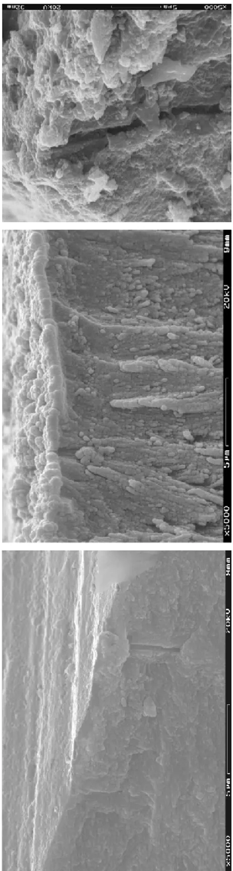
DH is characterized by a short and sharp sensation of pain arising from the tubular
dentine exposure as a result of enamel loss and/or gingival root surface exposure due to
attrition, abrasion, erosion, abfraction or gingival recession (37) (Fig 2). Any thermal,
osmotic and mechanical stimuli induced by the application of tooth brushing, sweet and
acid foods, hot or cold drinks may provoke pain referred to fluid shifts in the exposed
a ubu w h ac a o of h pu p , acco o “B ä öm’
hy o y am c h o y” (37-40) (Fig. 5).
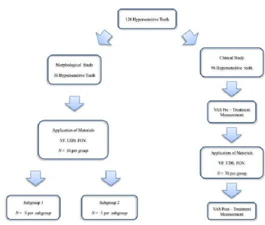
Therefore, the occlusion of the tubules by different materials may reduce the fluid
movement inside the dentinal tubules and the clinical symptoms of DH (39). When
reducing fluid movement by fully or partially occluding open dentine tubules,
hypersensitivity could be diminished (41). Consequently, most desensitizing agents
have been designed to cover the dentine surface with occlusion of the exposed tubules
or penetration in the tubules, coating and sealing them (39,26-30) (Fig 6).
However, the efficacy of desensitizing agents is quite variable in long term, as reported
in our previous studies and other clinical outcomes conditions (42 – 46). Clinical data
show that the desensitizing capacity has been correlated to the ability of the material to
resist in front of the interactions of saliva and other oral ambient interferences (46).
Moreover, differences in the efficacy were attributed to the different chemistries of the
materials and application modalities required by the desensitizer itself (47-50)..
Several different formulations of resin-based materials have being used in DH
treatment. Four different kind can be summarized: 1) varnishes, usually with fluoride,
creating a coat of calcium fluoride precipitates on the exposed surface and dentinal
tubules (51,,52-54); 2) adhesive monomeric systems, with or without the etching phase,
able to seal the exposed surface by a layer of interdiffusion in dentine and tubular resin
plugs (47-49, 55); 3) resin sealants and 4) flowable resin composites able to form
covers on the dentine surface (56) which sealing capacity in the time is influenced by
the resin composition and the coupling between filler and matrix (57).
Roberto Pinna,
Clinical evaluation of a self-adhering material as desensitizing agent in xerostomic patients for head and neck cancer.,
Tesi di Dottorato in Odontostomatologia preventiva, tudi di Sassari.
12
Fi g. 5 SE M m ic ro gr ap h of d en ti na l t ub ul es . Fi g. 6 – SE M m ic ro gr ap h of o cc lu si on o f th e ex po se d tu bu le s or p en et ra ti on in th e tu bu le s by d if fe re nt d en ta l m at er ia lsRoberto Pinna,
Clinical evaluation of a self-adhering material as desensitizing agent in xerostomic patients for head and neck cancer.,
Tesi di Dottorato in Odontostomatologia preventiva, tudi di Sassari.
13
Rational of the study.
Twenty-four patients about to start radiotherapy for HNC were subjected to dental
check up at the Dental Clinic of the University of Sassari, during and after the treatment
in 2013, alongside with an on-going evaluation study of DH patients with normal
salivary flow. Few months after the end of radio-exposition, 8 patients began to
complain DH. Our research team started to study if a correlation between their health
status and the clinical symptoms has been already described in the literature. With a
systematic approach, a literature search for articles related to the Radiotherapy
Xerostomia and DH, published between 01/01/1990 and 31/06/2013, was conducted in
the databases MEDLINE/PubMed, Scopus and The Cochrane Library, using
combinations of the MeSH terms: [Head and Neck Cancer] OR [Salivary
Hyposalivation] OR [Xerostomia] OR [Radiotherapy] AND [Dentin Hypersensitivity].
Th c o c a ch ’ fy a y u a h a o h ap u c
xerostomia and dentin hypersensitivity.
In the light of the results obtained, the research team decided to change the experimental
protocol of the on-going study, adapting it to the clinical condition of patients
undergoing radiation therapy.
The aim of the new clinical evaluation was to evaluate the 3-month efficacy of 4 kinds
of dental materials used as desensitizing agents, especially focusing on the differences
in DH reduction between the tested materials after the observation period. In addition,
the difference of the desensitizing agent efficacy among xerostomic patients and
patients with normal salivary flow was evaluated.
Roberto Pinna,
Clinical evaluation of a self-adhering material as desensitizing agent in xerostomic patients for head and neck cancer.,
Tesi di Dottorato in Odontostomatologia preventiva, tudi di Sassari.
14
Aims
The owerall aim of this thesis was to collect knowledge about radiation-induced
hypofunction, xerostomia and dentine hypersensitivity in head and neck cancer patients.
In more detail, the aims of this thesis were:
To review the current state of knowledge of pathology, clinical complications
and radiotherapeutic patient management
To evaluate the aetiology of dentine hypersensitivity in conditions of reduced
salivary flow resulting in the radiation exposure
To evaluate the effectiveness of the materials commonly used in the treatment of
hypersensitivity, when they work in conditions of hyposalivation
Roberto Pinna,
Clinical evaluation of a self-adhering material as desensitizing agent in xerostomic patients for head and neck cancer.,
Tesi di Dottorato in Odontostomatologia preventiva, tudi di Sassari.
15
Material and Methods
Paper I
Systematic Review methodology
Search strategy
A first systematic literature search for articles published between 01/01/1970 and
30/06/2013 was conducted in the databases MEDLINE/PubMed and The Cochrane
Library, using combinations of the MeSH terms: [Saliva] OR [Salivary Glands] OR
[Saliva Flow] OR [Salivation] OR [Salivary Gland Diseases] OR [Xerostomia] OR
[Saliva in Xerostomia] OR [Dry Mouth] OR [Oral Dryness] OR [Composition Saliva
Xerostomia] AND [Head and Neck Cancer] OR [Radiotherapy] OR [Radiation-induced
Xerostomia] OR [Parotid-Sparing Intensity-Modulated Radiotherapy] AND [Quality of
Life Analysis-Xerostomia] OR [Management Strategies Salivary Gland Hypofunction]
OR [Prevention Xerostomia] OR [Treatment Xerostomia]. The search results were
imported into a computerized database Review Manager 5.2. The search results from
each of the electronic databases of MEDLINE/PubMed and The Cochrane Library were
combined, and duplicated publications were eliminated. Subsequently, an update to
include studies published up to 30/06/2013 was performed.
Criteria for selecting studies
After completing the search, articles for review were selected based on:
•
English language
•
Original data of cancer therapies protocols
•
Oral complications associated with cancer therapies
•
Human
Exclusion criteria
The reasons for exclusion were defined as follows:
•
Studies without original and/or actual data
•
Studies with data from previous publications
•
Opinion papers
•
Editorials
In this way, a preliminary set of potentially relevant publications, removing irrelevant
citations according to the criteria was created. Two reviewers (RP and GC)
independently screened the registered title and abstracts, author and references in two
separate files (one for included abstracts and one for excluded abstracts) using a
screening guide based on eligibility criteria. Studies rejected at this or subsequent stages
were reported in the table of excluded studies. The full text of all potentially eligible
Roberto Pinna,
Clinical evaluation of a self-adhering material as desensitizing agent in xerostomic patients for head and neck cancer.,
Tesi di Dottorato in Odontostomatologia preventiva, tudi di Sassari.
16
studies in at least one screening was retrieved. Reviewers then evaluated the full text for
inclusion using a screening guide and a second reviewer (RP) screened all the findings.
When disagreement occurred, a third reviewer (IM) was consulted. For each review, the
following information was recorded: Year, Authors, Journal, Aim and Number of
Papers Reviewed; and for Clinical Trial Papers included: Year, Authors, Journal, Aim,
Number of Patients and Results. All studies meeting the inclusion criteria then
underwent validity assessment. Two examiners (RP and GC) read the papers
independently. The qualities and relevance of each study were graded as follows: high
(+++), medium (++) or low (+) using a study-quality checklist. External validity,
internal validity and study precision were analysed to obtain an overall assessment of
quality. The assessment was used as a basis for the discussion between the two
examiners to grade the studies. In the case of disagreement, all authors discussed the
paper until a consensus was reached.
Paper II
Elemental analysis
The elemental composition of Vertise Flow
TM, Universal Dentine Sealant and
Flor-Opal® Varnish was investigated using an X-ray energy dispersive spectrometer (EDX)
(INCA-X-acta, Oxford Instruments, Tubney Woods Abingdon, Oxfordshire, UK) in
conjunction with an environmental scanning electron microscope (ESEM) (EVO
®LS
25, Zeiss, Oberkochen, Germany). EDX was carried out using an accelerating voltage
of 20 kV and ESEM was used for imaging of each sample at standardized magnification
(200X, 1000X).
For the semi-quantitative X-ray analysis VF, UDS and FOV (0.5 mL) were weighed,
placed in a thin layer over Perspex
®slabs mounted on aluminum stubs (Agar Scientific,
Stansted, UK). Three stubs were made for each tested material and the analysis was
performed twice for each sample. The elemental analysis (weight % and atomic %) was
performed in low-vacuum conditions (20 Pa). Atomic number, absorption, and
fluorescence corrections were applied during the analysis with the ZAF correction
method.
Experimental design
Subjects who had hypersensitive teeth were selected from an ongoing program of
evaluating desensitizing agents at the Dental Clinic of the University of Sassari. Two
clinicians selected patients complaining about hypersensitivity and who had reported
this to the Department of Periodontology at the Dental Clinic. The protocol and
informed consent forms were approved by the ethics committee at the University of
Sassari (n° 1000/CE). The medical and dental history of the patients was collected, and
Roberto Pinna,
Clinical evaluation of a self-adhering material as desensitizing agent in xerostomic patients for head and neck cancer.,
Tesi di Dottorato in Odontostomatologia preventiva, tudi di Sassari.
17
sensitive teeth were differentiated from other clinical conditions which frequently
f w h DH. A h ubj c w ho ou h y fo m abou h u y’ pu po ,
risks, and benefits. A total of 86 patients with hypersensitive teeth were collected after
an intake period of 8 months. The study inclusion/exclusion criteria were the following:
1) patients were considered suitable for the study if they had sensitive teeth showing
abrasion, erosion or recession with the exposure of the cervical dentine; 2) teeth with
subjective or objective evidence of carious lesions, pulpitis, restorations, premature
contact, cracked enamel, active periapical infection, or which had received periodontal
surgery or root-planning up to 6 months prior to the investigation were excluded from
the study. Other exclusion criteria were professional desensitizing therapy during the
previous 3 months, or use of desensitizing toothpaste in the last 6 weeks. Patients were
also excluded if they were under significant medication that could have interfered with
pain perception (e.g., antidepressants, anti-inflammatory drugs, sedatives, and muscle
relaxants). As a consequence, the total study population included in the program was of
74 subjects, 43 female and 31 male, aged 27- 75 years (mean age ± standard deviation:
53 ± 7 years) with a total of 286 hypersensitive teeth (mean teeth for patient 2 ± 1). The
level of sensitivity experienced by the patient was considered as independent of the
position of the hypersensitive tooth in the oral cavity.
Morphological study
VF, D a FOV’ , ability to occlude dentine tubules and their morphology on
dentinal surfaces were evaluated in 30 selected patients, 18 female and 12 male, part of
the total sample of 74 subjects with hypersensitive teeth. Patients had 30
hypersensitivity teeth (11 premolars, 13 incisors, 6 cuspids), whose Grade III mobility
and significantly reduced response to periodontal treatment suggested the need for
extraction.
A full medical and dental history was taken and all the teeth were carefully examined to
confirm the diagnosis of DH. The nature and scope of the study was explained, and
informed consent was obtained.
A week before treatment, patients received oral prophylaxis and were randomly
assigned to three experimental groups (N=10 per group). The treatments were carried
out at random by one of the clinicians while the other assisted. The teeth were isolated
with cotton rolls and the treatment with VF, UDS and FOV was performed as
summarized in Table 1. As recommended, a halogen curing light (Optilux 501, Kerr
Corporation, USA; 11mm exit window) under the standard curing mode (output
wavelength range: 400–505 nm; output irradiance: 580–700mW/cm2) was used to
allow light curing of VF. After the treatment, teeth were immediately extracted (N=5
per subgroup), subgroup 1, and after 7 days post-treatment (N=5 per subgroup),
subgroup 2.
After extraction, samples were rinsed with
distilled water at 37°C and fixed in a solution
of 2.5% glutaraldehyde in 0.1 M PBS buffer (pH 7.2) for 72 h. In each sample, the
Roberto Pinna,
Clinical evaluation of a self-adhering material as desensitizing agent in xerostomic patients for head and neck cancer.,
Tesi di Dottorato in Odontostomatologia preventiva, tudi di Sassari.
18
treated cervical dentine was sectioned from the remaining crown and roots of the tooth
with a water-cooled saw (Isomet low-speed saw; Buehler, Lake Bluff, IL, USA) and
then fractured into two halves in order to analyze the buccal surface and the longitudinal
surface of the material-treated dentine surfaces. Samples were post-fixed in 1% osmium
tetroxide, dehydrated in increasing concentrations of acetone (25% 100%), dried by
critical point drying, and metal-coated. Specimens were then observed using a scanning
electron microscope (SEM) (Zeiss, DSM 962, Oberkochen, Germany). Observations
were recorded at standardized magnifications (1000×, 3000×, 5000X).
Clinical study
The study population consisted of another 36 patients, 19 females and 17 males who
were randomly selected from the total population of 74 subjects who had hypersensitive
teeth. A total of 90 teeth (30 premolars, 44 incisors and 16 cuspids constituted the
group of hypersensitive teeth for the clinical effectiveness of VF, UDS and FOV.
A week before the experiment, patients received oral prophylaxis. Non-fluoride
toothpaste, soft toothbrush and oral hygiene instructions were also provided in order to
have standardized habits during the period of the study.
Teeth were randomly assigned to three groups (N=30 per group) for the treatment with
the three desensitizing agents (Table 1). At the baseline visit, they were reassessed for
dentine hypersensitivity using the Visual Analogue Scores (VAS) of pain. Treatment
was performed by one examiner, while the pain stimulus was given by the other
examiner with the same equipment yielding similar air pressure each time.
The VAS scale consisted of a horizontal line that was 100 mm long, on which "no pain"
was marked on the right-hand extremity and "unbearable pain" on the other. The
patients expressed the intensity of the pain experienced by placing a mark at any point
along the continuum. The distance, expressed in millimeters, from the right edge of "no
pain" was used as the VAS score. Each patient was asked to rate the perception of
discomfort after the application of air via a dental syringe at 45 to 60 psi, 1cm at the
cervical third of the tooth after removing supragingival plaque with a low-speed
handpiece with pumice powder and without fluoride. The adjacent teeth were covered
by cotton rolls. The stimulus was delivered until reaction or up to a maximum duration
of 10 seconds by the same examiner with the same equipment yielding similar air
pressure each time. The subject's response was considered as the baseline measurement
(PRE-1) -mean±standard deviation VAS score: 5.3±2.1. Before the application of the
material (PRE-1), immediately after (POST-1), and after 7 days of oral environment
(POST- 2), the same clinician carried out the sensitivity test.
To compare the efficacy of the treatments, teeth were evaluated as a statistical unit
rather than a subject. Data were elaborated using parametric tests (ANOVA for more
ha wo amp a ju acco o ak’ mu p ) w h a 5% f ca c
level.
Roberto Pinna,
Clinical evaluation of a self-adhering material as desensitizing agent in xerostomic patients for head and neck cancer.,
Tesi di Dottorato in Odontostomatologia preventiva, tudi di Sassari.
19
Fig. 7 - Summary of the experimental design to collect hypersensitivity teeth to test the efficiency of
desensitising materials during the clinical study.
Paper III
Partecipants
The study was designed as a split-mouth randomized clinical trial. The protocol and
informed consent forms were approved by the ethics committee at the University of
Sassari (n° 1000/CE). Subjects who had hypersensitive teeth were selected from an
on-going program of evaluating desensitizing agents at the Dental Clinic of the University
of Sassari, Italy.
Two examiners selected patients complaining about hypersensitivity and who had
reported this to the Department of Periodontology at the Dental Clinic. The medical and
dental history of the patients was collected, and sensitive teeth were differentiated from
other clinical conditions that frequently interfere with DH. To participate in the study,
the subjects had to have two or three teeth that were hypersensitive to the stimulation
with a blast of air.
All the subjects were thorough y fo m abou h u y’ pu po , k , a
benefits. A total of 86 patients with hypersensitive teeth were collected. The study
Roberto Pinna,
Clinical evaluation of a self-adhering material as desensitizing agent in xerostomic patients for head and neck cancer.,
Tesi di Dottorato in Odontostomatologia preventiva, tudi di Sassari.
20
inclusion/exclusion criteria were the following: 1) patients were considered suitable for
the study if they had sensitive teeth showing abrasion, erosion or recession with the
exposure of the cervical dentine; 2) teeth with subjective or objective evidence of
carious lesions, pulpitis, restorations, premature contact, cracked enamel, active
periapical infection, or which had received periodontal surgery or root-planning up to 6
months prior to the investigation were excluded from the study. Other exclusion criteria
were professional desensitizing therapy during the previous 3 months, or use of
desensitizing toothpaste in the last 6 weeks. Patients were also excluded if they were
under significant medication that could have interfered with pain perception (e.g.,
antidepressants, anti-inflammatory drugs, sedatives, and muscle relaxants). As a
consequence, the total study population included in the program consisted of 46
patients, 27 females and 19 males who were randomly selected from the total
population of 74 subjects who had hypersensitive teeth. A total of 116 teeth (52
incisors, 38 premolars, and 26 cuspidates) were included in the study.
Clinical Procedure
VF self adhering composite was compared to: Universal Dentin Sealant (UDS)
(Ultradent Products Inc., South Jordan, UT, USA), a biocompatible, non-polymerizable,
high molecular weight resin sealant in alcohol solvent, Clearfil Protect Bond (CPB),
(Kuraray Noritake Dental, Osaka, Japan) a methacrilate-based resin, self-etching
adhesive system, and Flor-Opal® Varnish (FOV), (Ultradent Products Inc., South
Jordan, UT, USA), a fluoride-based varnish.
A week before the experiment, patients received oral prophylaxis. Non-fluoride
toothpaste (Biorepair, Coswell), soft toothbrush (Oral-B Sensitive Advantage, Procter &
Gamble) and oral hygiene instructions were also provided in order to have standardized
habits during the period of the study.
In view of the treatment with the desensitizing agents, teeth were randomly assigned in
to four groups (N=29 per group) (Fig. 8). The level of sensitivity experienced by each
patient was considered as independent of the position of the hypersensitive tooth in the
oral cavity 11]. The pain experience was assessed using a Visual Analogue Scores
(VAS) graded from 1 to 10, according to the same procedure of a previous study (56).
The pain stimulus was given by one examiner with the same equipment yielding similar
air pressure each time, while the other one performed the treatments. The subject's
response was considered before the application of the material (PRE-1), immediately
after (POST-1), after 1 week (POST- 2), 4 weeks (POST- 3) and 12 weeks (POST-4) of
oral environment, the same operator carried out the sensitivity test. None of the
participants failed to complete the study, and none of them reported any adverse
reactions.
Roberto Pinna,
Clinical evaluation of a self-adhering material as desensitizing agent in xerostomic patients for head and neck cancer.,
Tesi di Dottorato in Odontostomatologia preventiva, tudi di Sassari.
21
Statistical Analysis
Shapiro-Wilk normality test was used to assess the normality distribution of the
collected variables. Median and inter-quartile ranges were used as measures of central
tendency and variability to describe quantitative variables. Statistical differences in the
Visual Analogue Scale (VAS) values of VF, UDS, CPB and FOV were performed using
the Kruskall-Wallis analysis at the different time-points, adjusting statistical
significance for the multiple comparisons (Bonferroni correction). Statistical differences
at the baseline VAS value and the other time-points were calculated performing the
Mann–Whitney U test. Statistical analysis was carried out using IBM® SPSS®
Statistics, Version 21.0 (IBM Corporation ©, Armonk, NY, USA) and STATA®13
(StataCorp, College Station, TX, USA).
Fig. 8 - Summary of the experimental design to collect hypersensitivity teeth to test the efficiency of
Roberto Pinna,
Clinical evaluation of a self-adhering material as desensitizing agent in xerostomic patients for head and neck cancer.,
Tesi di Dottorato in Odontostomatologia preventiva, tudi di Sassari.
22
Paper IV
Partecipants
The study was designed as a split-mouth randomized clinical trial. The protocol and
informed consent forms were approved by the ethics committee at the University of
Sassari (n° 1000/CE). Radio-therapeutic patients who had hypersensitive teeth were
selected from an on-going program of evaluating desensitizing agents at the Dental
Clinic of the University of Sassari, Italy.
During 2013, a total of 48 patients were visited at the Department of Radiology. 24
patients, which needed radiotherapy for HNC were collected. These groups of patients
were subjected to a dental check-up with eventual teeth treatments, during and after the
radiotherapy. Few months later the end of the radio-exposition, 8 patients began to
complain HD.
To pa c pa h u y a h ubj c w ho ou h y fo m abou h u y’
purpose, risks, and benefits.
The study inclusion criteria were the following:
A relative good general health status;
A clinical reduction of salivary flow;
Two or three teeth that were hypersensitive to the stimulation with a blast of air.
In addition, patients were considered suitable for the study if they had sensitive teeth
showing abrasion, erosion or recession with the exposure of the cervical dentine.
The study exclusion criteria were:
teeth with subjective or objective evidence of carious lesions, pulpitis,
restorations, premature contact, cracked enamel, active periapical infection;
received periodontal surgery or root-planning up to 6 months prior to the
investigation;
professional desensitizing therapy during the previous 3 months
use of desensitizing toothpaste in the last 6 weeks.
Patients were also excluded if they were under significant medication that could have
interfered with pain perception (e.g., antidepressants, anti-inflammatory drugs,
sedatives, and muscle relaxants).
Clinical Procedure
Saliva collection
All salivary assessments were performed in the absence of acute sialadenitis. The flow
rate was determined in every person according to the method described by Sreebny
(58). Saliva was collected in a standardised manner. Patients were instructed not to eat,
drink, or smoke for 90 minutes before the sialometric assessment. All assessments were
performed at a fixed time of the day, between 10 am and 1 pm, in order to minimise
fluctuations related to a circadian rhythm of salivary secretion and composition. All
Roberto Pinna,
Clinical evaluation of a self-adhering material as desensitizing agent in xerostomic patients for head and neck cancer.,
Tesi di Dottorato in Odontostomatologia preventiva, tudi di Sassari.
23
assessments were performed by the same observer. Whole saliva was collected in
pre-weighed plastic tubes using an electronic scale.
Unstimulated salivary secretions were collected for 5 min with the patient seated in an
upright position and with the tilted head. When possible the tongue, cheeks and lips
movements were limited during the procedure. At the end of the collection period, the
patient had to expectorate saliva into the test-tube.Stimulated whole saliva was collected
asking to patients to chew a small block of paraffin wax or chewing gum. All the saliva
secreted for 5 min was then collected in the test-tube.Measuring vessels were weighed
after each collection using an electronic scale, and salivary flow rate was expressed in
ml/min, which is nearly equivalent to g/min (59). A secretion rate < 0.1-0.2 ml/min for
unstimulated flow and < 0.5-0.7 ml/min for stimulated flow was considered as an
objective sign of hyposalivation.
Assessment of hypersensitivity and desensitizing agents application.
A week before the experiment, patients received oral prophylaxis. Non-fluoride
toothpaste (Biorepair, Coswell), soft toothbrush (Oral-B Sensitive Advantage, Procter &
Gamble) and oral hygiene instructions were also provided in order to have standardized
habits during the period of the study. The level of sensitivity experienced by each
patient was considered as independent of the position of the hypersensitive tooth in the
oral cavity (30). The pain experience was assessed using a Visual Analogue Scores
(VAS) according the methodology described in the previous studies.
The following dental materials were used following manufacture instructions: Vertise
Flow
TM(VF) (Kerr Corporation, Orange, CA, USA), a self-adhering composite;
Universal Dentin Sealant (UDS) (Ultradent Products Inc., South Jordan, UT, USA), a
biocompatible, non-polymerizable, high molecular weight resin sealant in alcohol
solvent; Clearfil Protect Bond (CPB), (Kuraray Noritake Dental, Osaka, Japan) a
methacrilate-based resin, self-etching adhesive system, and Flor-Opal® Varnish (FOV),
(Ultradent Products Inc., South Jordan, UT, USA), a fluoride-based varnish.
All 8 patients were considered eligible and agreed to take part in the study. In view of
the treatment with the desensitizing agents, teeth were randomly assigned into four
groups (N= per group) (Fig. 9). None of the participants failed to complete the study
neither reported any adverse reactions.
Statistical Analysis
Shapiro-Wilk normality test was used to assess the normality distribution of the
collected variables. Median and inter-quartile range were used as measures of central
tendency and variability to describe quantitative variables. Statistical differences
between Visual Analogue Scale (VAS) values of Vertise FlowTM, Universal Dentine
Sealant, Clearfil Protect Bond and Flor-Opal® Varnish were evaluated performing
Kruskall-Wallis analysis at different time points, adjusting statistical significance for the
multiple comparisons (Bonferroni correction). Statistical differences between baseline
Roberto Pinna,
Clinical evaluation of a self-adhering material as desensitizing agent in xerostomic patients for head and neck cancer.,
Tesi di Dottorato in Odontostomatologia preventiva, tudi di Sassari.
24
VAS values and those obtained at other time-points were calculated performing the
Mann–Whitney U test. Statistical analysis was carried out using IBM® SPSS®
Statistics, Version 21.0 (IBM Corporation ©, Armonk, NY, USA) and STATA®13
(StataCorp, College Station, TX, USA).
Statistical differences between VAS values of xerostomic group and normo-salivation
group were calculated performing the Mann–Whitney U test. Statistical analysis was
carried out using STATA®13 (StataCorp, College Station, TX, USA).
Fig. 9 - Summary of the experimental design to collect hypersensitivity teeth to test the efficiency of
Roberto Pinna,
Clinical evaluation of a self-adhering material as desensitizing agent in xerostomic patients for head and neck cancer.,
Tesi di Dottorato in Odontostomatologia preventiva, tudi di Sassari.
25
Results
Review research result
(Paper I)
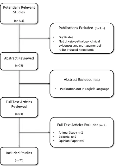
The electronic searches identified about a thousand titles and abstracts, and after
reviewing the titles 411 studies were evaluated. Subsequently, during the review of the
abstract, 336 studies were excluded. The final analysis included 70 articles that
conformed to the criteria for the present review (Fig. 10). Although animal studies have
been excluded, important information regarding the experimental results on two of the
papers was considered useful and therefore they were discussed.
Roberto Pinna,
Clinical evaluation of a self-adhering material as desensitizing agent in xerostomic patients for head and neck cancer.,
Tesi di Dottorato in Odontostomatologia preventiva, tudi di Sassari.
26
Laboratory and Clinical Analysis of short-term evaluation of DH treatment
(Paper II)
Elemental analysis
VF treatment left a layer of highly visible randomly distributed 5 to 40 µm particles.
Spectra of silicion (Si), ytterbium (Yb) alumina (Al) were highest in the layer in which
also phosphorus (P), calcium (Ca), barium (Ba) and fluoride (F) were found.
UDS treatment left fine, dispersed particles of about 0.5 µm in a thin and smooth layer.
Spots on these particles showed very high pecks of Ca and chlorine (Cl). The
semi-quantitative analysis obtained by scanning different areas of the matrix highlighted Ca
and Cl associated with Si and other oxides of Al, iron (Fe), chrome (Cr), potassium (K),
sulphur (S), magnesium (Mg), titanium (Ti) and zinc (Zn). FOV treated samples
showed a layer of particles embedded in a smooth matrix rich in sodium (Na) and F
peaks and with traces of Si and P.
Morphological study
On the surface of the exposed dentine (ED) to the oral fluids, VF formed a thick,
irregular coat that completely masked the underlying tubular dentine. Cracks were also
noted in ED. Longitudinal sections showed a coating about 3 µm thick composed of a
matrix with crystal-like particles of different sizes. Tubule orifices were tightly blocked
by the material and plugs of resin-like material were found inside the tubules. After 7
days of exposure to the oral environment (subgroup 2), tubular orifices were still not
visible on ED treated dentin surface which showed cracks and gap formations.
Crystal-like precipitates were dissolving, but the tubular apertures remained occluded.
UDS formed a smooth amorphous layer that contained particles about 0.5 µm in
diameter, over dentine. Particles had a tendency to form clusters and adhered to the
underlying dentine completely occluding the tubular orifices. Longitudinal sections
showed the dentine surface covered by a coating of UDS that was about 0.4 µm thick,
and plug-like structures in the tubules. After exposure to oral environment for 7 days
(subgroup 2), the dentine surface treated with UDS showed a residual coating of dentine
with different representations of crystal-like particles. Longitudinal sections showed a
thick granular surface and peritubular dentine masking the intratubular space.
Occasionally, small areas of separation between the surface coating and the dentine
subsurface demonstrated the presence of a barrier-like structure with tag-like structures
reproducing the tubular dentine.
FOV treated dentine surface exhibited an amorphous layer with dispersed particles
leaving most of the tubules partially occluded. Transverse sections of exposed dentine
revealed a thick coating of varnish almost blocking the tubular apertures. After 7 days
of exposure to the oral environment (subgroup 2), ED showed areas of solubilization of
a surface coating with disclosure of the underling smear layer. The solubilization
Roberto Pinna,
Clinical evaluation of a self-adhering material as desensitizing agent in xerostomic patients for head and neck cancer.,
Tesi di Dottorato in Odontostomatologia preventiva, tudi di Sassari.
27
process involved the tubular blocks of varnish on ED simultaneously showing
crystal-like precipitates with reduction of the tubular diameter.
Clinical Study
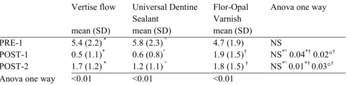
The mean VAS scores are shown in Table 3. There was no difference among baseline
VAS scores of all groups (P > 0.05). After treatment, all teeth exhibited statistically
significant reductions in VAS in Post-1. Teeth treated with VF had lower VAS scores
immediately after Post-1 control (VF vs. FOV: P =0.034). After 7 days of exposure to
oral fluids (POST-2) there was no significant difference among tested materials,
acco o ak’ mu p a ju m . How , wh compa w h
baseline data, all the VAS scores at post-treatment evaluation points were significantly
decreased (P < 0.05).
Tab. 3 – Visual Analogue Scale (VAS) values measured in 30 patients baseline and post-treatment.
Vertise flow mean (SD) Universal Dentine Sealant mean (SD) Flor-Opal Varnish mean (SD)
Anova one way
PRE-1 5.4 (2.2) * 5.8 (2.3) ° 4.7 (1.9) NS
POST-1 0.5 (1.1)* 0.6 (0.8)° 1.9 (1.5)† NS*° 0.04*† 0.02°† POST-2 1.7 (1.2) * 1.2 (1.1) ° 1.8 (1.5) † NS*° 0.01*† 0.03°†
Anova one way <0.01 <0.01 <0.01
Values expressed as means and standard deviation.
Clinical Analysis of long-term evaluation of DH treatment
(Paper III)
The sample size based on the initial assumptions showed a statistical power higher than
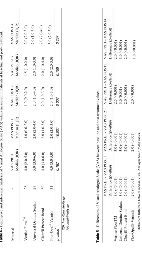
80%. Table 4 shows the median VAS scores at the different time-points.
At baseline (VAS score), no significant statistical differences were observed (p-value
>0.05) among the groups. After the applications of the materials, statistical significant
decrease of the VAS values was observed proceeding from Post-1 to Post-4 control.
Teeth treated with VF showed lower VAS scores at Post-1 control when compared to
UDS (p-value >0.001), CPB (p-value =0.001), and FOV (p-value >0.001), while at
Post-2, a significant statistical reduction of the value was demonstrated in VF in
comparison to UDS (p-value =0.001) and FOV (p-value =0.001). As far as the Post-3
and Post-4 controls, no significant differences were detected in VF efficiency in respect
to any other materials. Also, post-treatment values showed a significant decrease in the
VAS score in all of the groups in comparison to the baseline values (Tab. 5).
Roberto Pinna,
Clinical evaluation of a self-adhering material as desensitizing agent in xerostomic patients for head and neck cancer.,
Tesi di Dottorato in Odontostomatologia preventiva, tudi di Sassari.
28
Ta b le 4 De sc ri pt iv e an d in fe re nt ia l a na ly si s of Vi su al An al og ue S ca le ( VAS ) va lu es m ea su re d in p at ie nt s at b as el in e an d po st -tr ea tm en t. Ma te ri al n VAS P R E 1 Me di an ( IQ R ) VAS P OS T 1 Me di an ( IQ R ) VAS P OS T 2 Me di an ( IQ R ) VAS P OS T 3 Me di an ( IQ R ) VAS P OS T 4 Me di an ( IQ R ) Ve rt is e F lo w TM 28 4. 0 (2. 0-5. 0) 1. 0 (0. 0-2. 0) 1. 0 (0. 0-2. 0) 1. 5 (1. 0-2. 0) 2. 0 (2. 0-3. 0) Un iv er sa l De nt in e S ea la nt 27 5. 0 (3. 0-6. 0) 2. 0 (2. 0-4. 0) 2. 0 (1. 0-4. 0) 2. 0 (1. 0-3. 0) 2. 0 (1. 0-3. 0) Cl ea rf il P ro te ct Bo nd 30 4. 0 (3. 0-6. 0) 2. 0 (1. 0-3. 0) 2. 0 (1. 0-3. 0) 2. 0 (1. 0-4. 0) 2. 0 (2. 0-4. 0) Fl or -Op al ® Va rn is h 31 4. 0 (3. 0-5. 0) 2. 0 (2. 0-3. 0) 2. 0 (1. 0-3. 0) 2. 0 (2. 0-3. 0) 3. 0 (2. 0-3. 0) p-va lu e * 0. 187 <0 .0 01 0. 002 0. 156 0. 297 IQ R : In te r-Qu ar ti le R an ge *Kr us ka ll -Wa ll is te st Ta b le 5 Di ff er en ce s of Vi su al An al og ue S ca le ( VAS ) be twe en b as el in e an d po st -tr ea tm en t v alu es . VAS P R E 1 - VAS P OS T 1 Di ff er en ce ( p-va lu e) VAS P R E 1 - VAS P OS T 2 Di ff er en ce ( p-va lu e) VAS P R E 1 - VAS P OS T 3 Di ff er en ce ( p-va lu e) VAS P R E 1 - VAS P OS T 4 Di ff er en ce ( p-va lu e) Ve rt is e F lo wT M 3. 0 (< 0. 001) 3. 0 (< 0. 001) 2. 5 (< 0. 001) 2. 0 (< 0. 001) Un iv er sa l De nt in e S ea la nt 3. 0 (< 0. 001) 3. 0 (< 0. 001) 3. 0 (0. 001) 3. 0 (< 0. 001) Cl ea rf il P ro te ct Bo nd 2. 0 (< 0. 001) 2. 0 (< 0. 001) 2. 0 (< 0. 001) 2. 0 (< 0. 001) Fl or -Op al ® Va rn is h 2. 0 (< 0. 001) 2. 0 (< 0. 001) 2. 0 (< 0. 001) 1. 0 (< 0. 001) Di ff er en ce : Di ff er en ce b et we en me di an Vi su al An al og ue S ca le ( VAS ) va lu es .Roberto Pinna,
Clinical evaluation of a self-adhering material as desensitizing agent in xerostomic patients for head and neck cancer.,
Tesi di Dottorato in Odontostomatologia preventiva, tudi di Sassari.
29
Clinical Analysis of long-term evaluation of DH treatment in radio-induced
xerostomia
(Paper IV)
The mean basal salivary flow rate was 0.24 ml/min (minimum 0.06 – maximum 0.42)
while the stimulated rate was of 0.54 ml/min (minimum 0.29 – maximum 0.86).
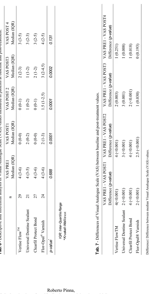
The median VAS scores at different time-points is shown in Table 6. No statistically
significant differences between the baseline VAS scores were observed (p-value >0.05).
Following the exposure to the materials, a statistically significant VAS decreases was
observed from Post-1 to Post-3; no statistical differences were detected in the final
point. Teeth treated with Vertise FlowTM and Universal Dentine Sealant showed lower
VAS scores at Post-1 in in comparison to those treated with Clearfil Protect Bond
(p-value <0.0001), and Flor-Opal® Varnish (p-(p-value <0.0001). On the other hand,
statistically significant lower VAS values were showed for Vertise FlowTM and
Universal Dentine Sealant in Post-2 when compared to Flor-Opal® Varnish (p-value
=0.0002 and p-value<0.0001, respectively). Significantly higher VAS values were
reported in regard to Flor-Opal® Varnish, Universal Dentine Sealant (p-value =0.0003)
and Clearfil Protect Bond (p-value =0.0002). Conversely, no significant differences
were detected at Post-4. In the case of Universal Dentine Sealant and Clearfil Protect
Bond, the baseline, Pre-1, and the post-treatment values Post-4 showed significant VAS
co c a , Th wa ’ h ca of V F owTM a F o -Opal® Varnish
(Tab. 7).
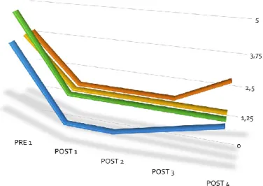
Moreover, no statistically significant differences (p-value >0.05) were detected at the
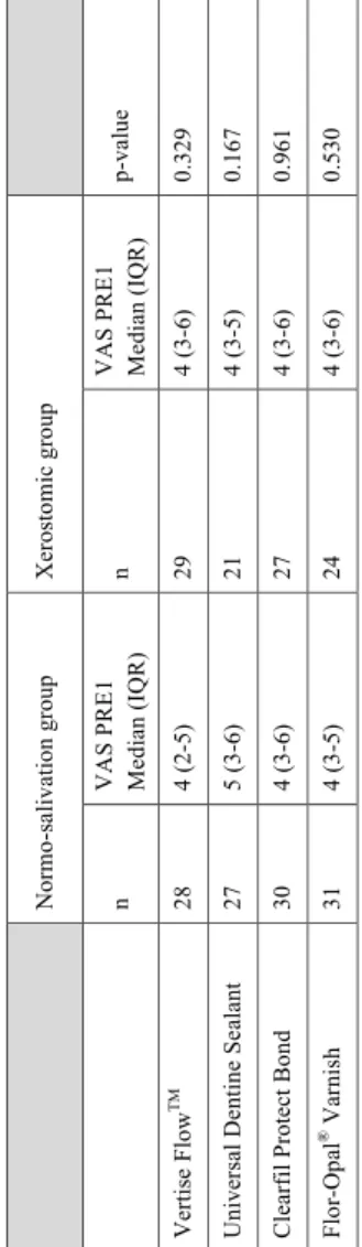
baseline VAS when the xerostomic group was compared to the healthy (Table 8).
Statistically lower VAS values were showed in the normo-salivation group treated with
Vertise FlowTM , Clearfil Protect Bond and Flor-Opal® Varnish at Post-4 (p-value
<0.05) (Table 9).
Roberto Pinna,
Clinical evaluation of a self-adhering material as desensitizing agent in xerostomic patients for head and neck cancer.,
Tesi di Dottorato in Odontostomatologia preventiva, tudi di Sassari.
30
Ta b . 6 De sc ri pt iv e an d in fe re nt ia l a na ly si s of Vi su al An al og ue S ca le ( VAS ) va lu es m ea su re d in p at ie nt s at b as el in e an d po st -tr ea tm en t.IQ R : In te r-Qu ar ti le R an ge *Kr us ka ll -Wa ll is te st Ta b . 7 Di ff er en ce s of Vi su al An al og ue S ca le ( VAS ) be twe en b as el in e an d po st -tr ea tm en t v alu es . Di ff er en ce : Di ff er en ce b et we en me di an Vi su al An al og ue S ca le ( VAS ) va lu es . n VAS P R E 1 Me di an ( IQ R ) VAS P OS T 1 Me di an ( IQ R ) VAS P OS T 2 Me di an ( IQ R ) VAS P OS T 3 Me di an ( IQ R ) VAS P OS T 4 Me di an (IQ R ) Ve rt is e F lo w TM 29 4 (3 -6) 0 (0 -0) 0 (0 -1) 2 (2 -3) 3 (3 -5) Un iv er sa l De nt in e S ea la nt 21 4 (3 -5) 2 (0 -2) 1 (0 -2) 1 (1 -2) 3 (2 -3) Cl ea rf il P ro te ct Bo nd 27 4 (3 -6) 0 (0 -0) 0 (0 -1) 2 (1 -2) 3 (2 -5) Fl or -Op al ® Va rn is h 24 4 (3 -6) 2 (1 -3) 1. 5 (1 -2. 5) 3 (2 -4. 5) 4 (2 -5) p-va lu e * 0. 688 0. 0001 0. 0001 0. 0002 0. 131 VAS P R E 1 - VAS P OS T 1 Di ff er en ce ( p-va lu e) VAS P R E 1 - VAS P OS T 2 Di ff er en ce ( p-va lu e) VAS P R E 1 - VAS P OS T 3 Di ff er en ce ( p-va lu e) VAS P R E 1 - VAS P OS T 4 Di ff er en ce ( p-va lu e) Ve rt is e F lo wT M 4( < 0. 001) 4 (< 0. 001) 2 (0. 003) 1 (0. 251) Un iv er sa l De nt in e S ea la nt 2 (< 0. 001) 3 (< 0. 00 1) 3 (0. 001) 1 (0. 008) Cl ea rf il P ro te ct Bo nd 4 (< 0. 001) 4 (< 0. 001) 2 (< 0. 001) 1 (0. 018) Fl or -Op al ® Va rn is h 2 (< 0. 001) 2. 5 (< 0. 001) 1 (0. 030) 0 (0. 193)
Roberto Pinna,
Clinical evaluation of a self-adhering material as desensitizing agent in xerostomic patients for head and neck cancer.,
Tesi di Dottorato in Odontostomatologia preventiva, tudi di Sassari.