• Non ci sono risultati.

Recurrent glioma and crossed cerebellar diaschisis in a patient examined with 18F-DOPA and 18F-FDG PET/CT

N/A
N/A
Protected

Academic year: 2021

Condividi "Recurrent glioma and crossed cerebellar diaschisis in a patient examined with 18F-DOPA and 18F-FDG PET/CT"

Copied!
2
0
0

Testo completo

(1)

Recurrent Glioma and Crossed Cerebellar Diaschisis in a Patient

Examined With

18

F-DOPA and

18

F-FDG PET/CT

Ferdinando Calabria, MD,* and Orazio Schillaci, MD*Þ

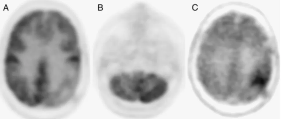
Abstract: Two years after resection of a left parietal glioma, a 46-year-old woman underwent18F-FDG and18F-DOPA brain PET/CT. FDG showed left

parietal hypometabolism with crossed cerebellar diaschisis. No abnormally increased FDG activity was seen.18F-DOPA PET/CT scan demonstrated focal

left parietal uptake. Recurrent glioma was confirmed by surgical biopsy.

18

F-DOPA may demonstrate abnormal amino acid metabolism in evaluation of recurrence of glioma.

Key Words:18F-DOPA,18F-FDG, PET/CT, brain tumor recurrence, crossed

cerebellar diaschisis

(Clin Nucl Med 2012;37: 878Y879)

REFERENCES

1. Tripathi M, Sharma R, Varshney R, et al. Comparison of F-18 FDG and C-11 methionine for the evaluation of recurrent primary brain tumors. Clin Nucl Med. 2012;37:158Y163.

2. Kanhaiya LA, Bhagwant RM, Anish B, et al. Crossed cerebellar diaschisis on F-18 FDG PET/CT. Indian J Nucl Med. 2011;26:102Y103.

3. Shih WJ, Huang WS, Milan PP. F-18 FDG PET demonstrates crossed cerebel-lar diaschisis 20 years after stroke. Clin Nucl Med. 2006;31:259Y261. 4. Gonzalez-Forero M, Prieto E, Domingues I, et al. Dual time point18F-DOPA

PET as a tool for characterizing brain tumors. Rev Esp Med Nucl. 2011; 30:88Y93.

5. Fueger BJ, Czernin J, Cloughesy T, et al. Correlation of 6-18F-fluoro-L-DOPA

PET uptake with proliferation and tumor grade in newly diagnosed and re-current gliomas. Clin Nucl Med. 2011;36:650Y655.

6. Tripathi M, Sharma R, D’Souza M, et al. Comparative evaluation of F-18 FDOPA, F-18 FDG and F-18 FLT-PET/CT for metabolic imaging of low grade gliomas. Clin Nucl Med. 2009;34:878Y883.

7. Santra A, Kumar R, Sharma P, et al. Detection of recurrence in glioma: a comparative study between Tc-99m GHA SPECT and F-18 FDG PET/CT. Clin Nucl Med. 2011;36:650Y655.

8. Chen W, Silverman DH, Delaloye S, et al.18F-FDOPA PET imaging of brain

tumors: comparison study with18F-FDG PET and evaluation of diagnostic

accuracy. J Nucl Med. 2006;47:904Y911.

9. Ledezma CJ, Chen W, Sai V, et al.18F-DOPA PET/MRI fusion in patients with

primary/recurrent gliomas: initial experience. Eur J Radiol. 2009;71:242Y248. 10. Calabria F, Chiaravalloti A, Di Pietro B, et al. Molecular imaging of brain tumors with 18F-DOPA PET and PET/CT. Nucl Med Commun. 2012; 33:563Y570.

I

NTERESTING

I

MAGE

878

www.nuclearmed.com

Clinical Nuclear Medicine

&

Volume 37, Number 9, September 2012

Received for publication February 16, 2012; revision accepted May 30, 2012. From the *Department of Nuclear Medicine and Molecular Imaging, IRCCS

Neuromed, Pozzilli (IS); and †Department of Biopathology and Diagnostic Imaging, University ‘‘Tor Vergata,’’ Rome, Italy.

Conflicts of interest and sources of funding: none declared.

Reprints: Ferdinando Calabria, MD, Department of Nuclear Medicine and Molecular Imaging, IRCCS Neuromed, Via Atinense 18, 86077 Pozzilli (IS), Italy. E-mail: ferdinandocalabria@hotmail.it.

Copyright* 2012 by Lippincott Williams & Wilkins ISSN: 0363-9762/12/3709-0878

(2)

FIGURE 1. In a patient submitted 2 years before to surgical intervention in the left posterior parietal lobe and subsequent

radiotherapy for a malignant glioma, axial

18

F-FDG PET view shows area of hypometabolism in the surgical site (A). No foci of

increased uptake of the tracer were detected in the rest of the brain, whereas corresponding CT images, obtained for attenuation

correction,

1

did not show significant morphological abnormalities, with the exception of diffuse hypodensity in the same area,

considered as a postsurgical lacuna. A further area of hypometabolism was detected in the right cerebellar hemisphere as shown in

axial

18

F-FDG PET view (B). Considering that the limitation of the excitatory output from an injured brain region could reduce

the physiological activity in connecting the cerebellar region contralateral to the lesion site,

2,3

we interpreted this findings as

indicative of crossed cerebellar diaschisis.

18

F-DOPA PET/CT of the brain, subsequently performed, exhibited in the left posterior

parietal lobe an area of focal increased uptake, with SUVmax = 5,

4

suggestive for recurrence of glioma,

5

as evident in the axial detail

(C). No other abnormalities were detected with this radiopharmaceutical.Histological examination confirmed our suspicion and

diagnosed tumor recurrence.

18

F-FDG PET/CT was not able to identify the tumor recurrence. In fact, some previous articles

on this topic well described the limits of this tracer in the evaluation of tumor recurrence, compared with the feasibility of

18

F-DOPA

and

18

F-FLT,

6

or the effectiveness of SPECT with

99m

Tc-glucoheptonate.

7

Therefore,

18

F-FDG may play a limited role in this field

for the intrinsic property of metabolic characterization, allowing the detection of functional disorders such as crossed cerebellar

diaschisis.On the other hand, this report confirms the feasibility of

18

F-DOPA acting as a tracer of amino acid metabolism in the

evaluation of recurrence of brain tumors after surgery and/or radiotherapy, as well known in various articles, because of the

possibility to distinguish recurrent neoplasm from radiation necrosis, especially in comparison with

18

F-FDG PET,

8

and by virtue of

the low physiological uptake in normal white and gray matter, considering the exception of the basal ganglia.

9,10

Clinical Nuclear Medicine

&

Volume 37, Number 9, September 2012

18

F-DOPA and

18

F-FDG PET/CT for Recurrent Glioma

* 2012 Lippincott Williams & Wilkins

www.nuclearmed.com

879

Figura

FIGURE 1. In a patient submitted 2 years before to surgical intervention in the left posterior parietal lobe and subsequent radiotherapy for a malignant glioma, axial 18 F-FDG PET view shows area of hypometabolism in the surgical site (A)

Riferimenti

Documenti correlati

Non è questa la sede per ripercorrere le voci critiche, di contestazione e di protesta, che si sono levate, in tutto il mondo, e che hanno indotto governi, istituzioni

Chiaramente, è difficile tenere in considerazione per ogni allievo tutti gli aspetti che possono determinare un mancato coinvolgimento nella pratica sportiva, ma è dovere del

Il che sollecita un'ulteriore domanda: al di là del settore di diritto privato, cosa può accadere per tutti quegli altri settori (e ci sono, a quanto pare) nei quali risulta che

Metodologia di ottimizzazione aerodinamica per l’estrattore di una vettura di alte prestazioni, Tesi di laurea Specialistica, Dipartimento di Ingegneria Aerospaziale, Università

Finally, the idea was to work on the high power EOM (Chapter 4 ) and the FI (Chapter 5 ) used in the injection part of Advanced Virgo, and especially, using those fibers, to design

Quando un campione di suolo è inserito in una soluzione più densa dell’acqua (possono essere usate soluzioni di ac- qua-benzene, acqua-etere, acqua-solfato di magnesio,

The total systematic uncertainty is dominated by the uncertain- ties in the modeling of the t¯t þ ≥ 1b background, the second-largest source being the limited number of events in

Di medioevo Zieger parla anche in altre opere di sintesi di minore spes- sore quali Il contrasto fra il principato vescovile di Trento e i conti del Tirolo (1957) e Storia