Electrophysiological investigations on the role of
retinal HCN channels
A Thesis submitted for the degree of Philosophiæ Doctor by:
Luca Della Santina
([email protected])Under the supervision of:
Prof. Luigi Cervetto
University of Pisa Dept. of Scienze Fisiologiche
Had I the heavens embroidered cloths, Enwrought with golden and silver light, The blue and the dim and the dark cloths Of night and light and the half-light,
I would spread the cloths under your feet: But I, being poor, have only my dreams; I have spread my dreams under your feet; Tread softly because you tread on my dreams.
He Wishes For the Cloths of Heaven William Butler Yeats (1865 ∼ 1939)
Declaration
The work described in this thesis is included in the following papers:
Luca Della Santina, Muriel Bouly, Antonella Asta, Gian Carlo Demontis, Luigi Cervetto and Claudia Gargini
Effect of HCN Channel Inhibition on Retinal Morphology and Function in Normal and Dystrophic Rodents.
Submitted to Invest. Ophtalmol. Vis. Sci., manuscript ID: IOVS-09-3680
Lorenzo Cangiano, Claudia Gargini, Luca Della Santina, Gian Carlo Demontis, Luigi Cervetto
High-Pass Filtering of Input Signals by the Ih Current in a Non-Spiking
Neuron, the Retinal Rod Bipolar Cell. PLoS ONE, 2(12):e1327, 2007
Abstract
Hyperpolarization-activated cyclic nucleotide-modulated (HCN) channels are a particular class of voltage-gated ionic channels, mediating the If current in the
cardiac tissue and Ih in the central nervous system (CNS). They were initially
described in cardiac pacemaker cells, where they play a key role in the gen-eration and regulation of cardiac rhythmicity. In the CNS they participate in processes like dendritic integration, network synchronization, long-term poten-tiation and motor learning. Because of their role in the cardiac tissue, HCN channels reprepresent a target in clinical pharmacology, but a percentage of pa-tients treated with HCN inhibitors report visual side effects. This is possibly due to interaction with HCN channels expressed in the retinal tissue, where all the four HCN isoforms are present. In this thesis the role of HCN channels in visual processing was investigated.
HCN distribution was evaluated in the murine retina using immunohisto-chemical techniques. The functional role of these channels in retinal signalling was investigated in details, by means of perforated patch clamp recordings from rod bipolar cells (RBCs) in retinal sections, and by non-invasive recording of the electroretinogram (ERG) in rats, wild-type mice and in mice in which the hcn1 gene was deleted (hcn1-ko mice).
RBCs exhibit the HCN-driven Ih current in response to hyperpolarizing
voltage steps. When this current is active, the cellular impedance profile has band-pass properties. Application of the selective HCN blocker ZD7288 inhibits Ih, changing the impedance profile to low-pass mode. These effects are predicted
by a mathematical model of the RBC where it is assumed that Ih is the only
active conductance. The model also predicts that Ihquickens the light response
in RBC.
The in-vivo inhibition of HCN channels by the organic blocker Ivabradine induces a slight prolongation of the ERG response to weak flashes. At the same time, it triggers a consistent change of the frequency-response relationship from band-pass to low-pass mode, following to both acute or prolonged drug administration; these effects are reversible upon interruption of treatment.
In the hcn1-ko mice line, ERG responses to flashes are prolonged with respect to control mice. In response to sinusoidal stimulation, hcn1-ko mice exhibit a partial attenuation of the band-pass behavior, but not a complete switch to low-pass mode like that induced by the organic channel blocker.
Taken together these data point out the importance of HCN channels in the early steps of rod retinal signalling. This helps in building a framework that may soon allow to explain the role of Ihin visual side effects experienced during
the clinical use of HCN inhibitors. In addition, experiments on hcn1-ko mice deliver the first clue that preserving a partial Ihfunctionality in the retina, may
be an effective way to mitigate the side effects these drugs induce on the visual system.
Sommario
I canali attivati dalla iperpolarizzazione e modulati da nucleotidi ciclici (HCN) sono una particolare classe di canali ionici voltaggio-dipendenti, che mediano la corrente If nel tessuto cardiaco e la Ih nel sistema nervoso centrale
(SNC). Sono stati descritti per la prima volta nelle cellule pacemaker cardia-che, dove rivestono un ruolo chiave sia nella generazione che nella regolazione del ritmo cardiaco. Nel SNC essi partecipano in processi quali l’integrazione dendritica, la sincronizzazione delle reti neurali, il potenziamento a lungo ter-mine e la memorizzazione di compiti motori. A causa del loro ruolo cardiaco, i canali HCN rappresentano un target per la farmacologia clinica, ma purtroppo una percentuale dei pazienti trattati con inibitori degli HCN riportano effetti collaterali a livello visivo. Essi sono probabilmente dovuti all’interazione coi canali HCN presenti nella retina, dove sono espresse tutte e quattro le isoforme. Questa tesi indaga il ruolo dei canali HCN nella funzione visiva.
La distribuzione dei canali HCN `e stata valutata nella retina murina me-diante tecniche immunoistochimiche. Il ruolo funzionale degli stessi nella tra-smissione del segnale visivo `e stato indagato in dettaglio mediante patch-clamp perforato su cellule bipolari dei bastoncelli (RBC) in sezioni retiniche, e tramite la tecnica non invasiva dell’elettroretinogramma (ERG) in ratti, topi wild type e topi knockout per il gene hcn1 (hcn1-ko).
Le RBC esibiscono la Ihin risposta a step iperpolarizzanti. Laddove questa
corrente `e attiva, il profilo d’impedenza di queste cellule assume caratteristiche passa-banda. L’applizazione del bloccante HCN selettivo ZD7288 inibisce la Ih, trasformando il profilo d’impedenza in passa-basso. Questi comportamenti
vengono predetti da un modello matematico della RBC dove si assume che Ih
sia l’unica conduttanza di tipo attivo. Il modello predice anche che l’Ih`e capace
di accelerare la risposta alla luce nelle RBC.
L’inibizione dei canali HCN in-vivo mediante somministrazione del bloccan-te Ivabradine induce un leggero prolungamento della risposta a flash di bassa intensit`a luminosa. Allo stesso tempo provoca una consistente modifica della re-lazione frequenza - ristposta da un profilo passa-banda ad uno passa-basso, sia a seguito di somministrazione acuta che prolungata; questi effetti sono reversibili mediante interruzione del trattamento.
Nei topi hcn1-ko le risposte ERG da flash si presentano prolungate rispetto agli animali di controllo. In risposta a stimoli sinusoidali la delezione di hcn1 pro-voca una parziale attenuazione del comportamento passa banda, senza arrivare per`o ad un comportamento passa-basso come osservato dopo somministrazione del bloccante organico.
Considerati nel loro insieme, questi dati mettono in luce l’importanza dei canali HCN nei primi stadi della trasmissione del segnale visivo dei bastoncelli. Fornendo cos`ı un bagaglio di conoscenze che presto potrebbe spiegare il ruolo della Ih negli effetti collaterali riportati durante l’uso clinico degli inibitori dei
Contents
Table of contents ii
List of figures iii
1 Introduction 1
1.1 Retina . . . 1
1.1.1 Morphological structure of the retina . . . 1
1.1.2 Features of the visual systems of mouse and rat . . . 3
1.2 HCN channels . . . 7
1.2.1 Molecular properties of HCN channels . . . 7
1.2.2 Ih current properties . . . 13
1.2.3 Physiological expression and role of HCN channels . . . . 14
1.2.4 HCN disruption insight on isoform relevance . . . 16
1.2.5 Ih current in the retinal tissue . . . 18
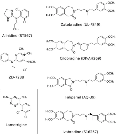
1.2.6 HCN as therapeutic target for novel drugs . . . 20
2 Materials and Methods 22 2.1 Animals . . . 22
2.2 Patch Clamp . . . 22
2.2.1 Slice preparation . . . 22
2.2.2 Pipettes . . . 23
2.2.3 Cell recording and identification . . . 23
2.2.4 Light stimulation . . . 23
2.2.5 Ih characteristics evaluation . . . 24
2.2.6 Input impedance measurement . . . 24
2.2.7 Input impedance modeling . . . 25
2.3 Immunohistochemistry . . . 25 2.3.1 Slice preparation . . . 25 2.3.2 Immunoreaction . . . 25 2.4 Electroretinogram . . . 26 2.4.1 Animal preparation . . . 26 2.4.2 Drug delivery . . . 26 2.4.3 Light stimulation . . . 27 3 Results 29 3.1 In vitro studies . . . 29 3.1.1 Murine RBCs exhibit Ih . . . 29
3.1.3 Influence of Ih on light responses . . . 34
3.1.4 HCN distribution in the murine retina . . . 36
3.2 In vivo studies . . . 39
3.2.1 Acute Ih inhibition in rats . . . 39
3.2.2 Prolonged Ih inhibition in rats . . . 42
3.2.3 hcn1 knockout mice . . . 44
4 Discussion 47 4.1 Properties of RBCs in physiological conditions . . . 47
4.2 Frequency tuning . . . 48
4.3 HCN subtype expression and relative role . . . 49
4.4 Future directions . . . 50
Nomenclature 53
List of Figures
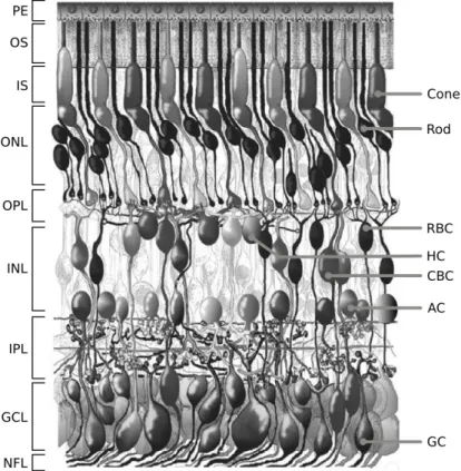
1.1 Stratification and principal classes of retinal neurons . . . 2
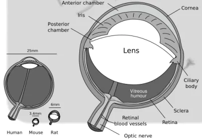
1.2 Schematic representation of murine eye . . . 3
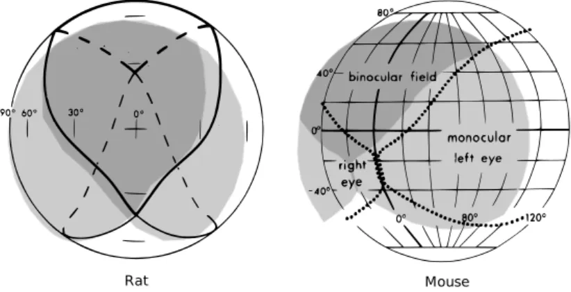
1.3 Visual field in mice and rats . . . 4
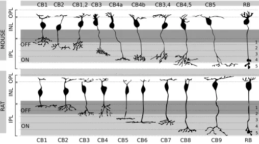
1.4 Type of bipolar cells . . . 5
1.5 Cellular density in the murine retina . . . 5
1.6 General structure of voltage-gated potassium channels . . . 7
1.7 Structure of HCN1 channel . . . 8
1.8 Conserved regions in murine HCN channel isoforms . . . 12
1.9 Hypothesis of HCN4 reserve-deposit in heart rate modulation . . 16
1.10 Retinal distribution of HCN . . . 18
1.11 Chemical structure of HCN drugs . . . 21
3.1 Functional and morphological properties useful to identify RBCs 29 3.2 Voltage clamp hyperpolarizing/depolarizing protocols . . . 30
3.3 Ihidentification with ZD7288 . . . 31
3.4 Sinusoidally modulated stimulation in current-clamp mode . . . . 32
3.5 Input impedance properties and voltage dependence . . . 33
3.6 Impedance profile switches to low-pass with ZD7288 . . . 35
3.7 Influence of Ih on light responses . . . 35
3.8 HCN distribution in the wild-type murine retina . . . 37
3.9 HCN2 distribe on RBCs’ dendrites, aligned to rod synapses . . . 38
3.10 Effect of acute HCN blockade on Flash ERG’s b-wave . . . 39
3.11 Example of flash ERG response during acute HCN blockade . . . 40
3.12 Frequency-response profile changes after HCN blockade . . . 41
3.13 Ratio R3Hz/R0.3Hz and heart rate for acute HCN blockade . . . 41
3.14 Flash ERG responses after chronic treatment with HCN blocker . 42 3.15 FRC and heart rate following prolonged Ihinhibition . . . 43
3.16 Ihis lacking in hcn1-ko rods . . . 44
3.17 Flash ERG response in wild type and hcn1-ko mice . . . 45
3.18 Frequency-response profiles for wild type and hcn1-ko mice . . . 46
3.19 Time-course of Ih inhibition in mice . . . 46
4.1 Effect of finite seal resistance on membrane potential and Ibp . . 48
Chapter 1
Introduction
Any road followed precisely to its end leads precisely nowhere. Climb the mountain just a little bit to test that it’s a mountain. From the top of the mountain, you cannot see the mountain.
Family Commentaries by the Princess Irulan
1.1
Retina
1.1.1
Morphological structure of the retina
Among the five senses, vision is especially important in human beings since most of the perceptual experience starts by looking to the external world. The eye is dedicated to capture light reflected by the scene surrounding the subject, to encode it into electrical signals and efficiently transfer them to the brain. At first approximation the eye takes the shape of a sphere, in which the neural portion, the retina, is placed in the backmost hemisphere.
Like many other structures in the central nervous system, a vertical section of the retina reveals an organization structured into morphological and functional layers. From the outmost to the innermost with respect to the ocular bulb’s surface they are:
• Pigmented epithelium (PE): Placed at the very periphery of the retina, these highly pigmented cells prevents residual light from scattering back into the photosensitive retina and are involved in key processes like phago-cytosis of aged photoreceptor’s outer segments and in regeneration of the light-sensitive pigment.
• Photoreceptors’ outer segments (OS): The most distal part of photore-ceptors where the multi-stage process known as phototransduction begins with the light-induced isomerization of a pigment tightly packed into vesi-cles (cones) or membrane invaginations (rods). The whole phototransduc-tive machinery is contained in here.
• Outer nuclear layer (ONL): Contain all the photoreceptor nuclei, as well as the terminal processes of M¨uller glia.
• Outer plexiform layer (OPL): Zone of synaptical contact between photore-ceptors and second-order neurons
• Inner nuclear layer (INL): In this layer resides the cellular bodies of all the second-order neurons: rod- and cone-bipolar cells, amacrine cells, hor-izontal cells; and also the non-neural M¨uller glia.
• Inner plexiform layer (IPL): Zone of synaptical contact between second-order neurons and ganglion cells.
• Ganglion cell layer (GCL): Ganglion cell and displaced amacrine cellular bodies reside in this layer. The former represents the third-order neurons, the only neurons generating spike potentials.
• Optic nerve fiber layer (NFL): Here axons of ganglion cells, packed to-gether, make their way out of the retina toward the brain. Together they form the optic nerve, which at the level of optic disc assumes the usual myelination.
Figure 1.1: Morphological representation of the retina in a vertical section of the adult human eye. Retinal layers on the left. Principal neuronal classes on the right. RBC: rod bipolar cell, HC: horizontal cell, CBC: cone bipolar cell, AC: amacrine cell, GC: ganglion cell
1.1.2
Features of the visual systems of mouse and rat
Mice (Mus musculus) and rats (Rattus norvegicus) are currently among the most widely used animal species in vision research, especially since the availability of knockout mice lines reproducing phenotypical and/or genotypical features of human pathologies. Despite the great usefulness of these models, some major differences between the visual system of rodents and that of humans must be taken into account during design and interpretation of the experimental work.6mm 3.4mm
Human Mouse Rat
25mm Vitreous humour Iris Anterior chamber Cornea Posterior chamber Lens Sclera Retina Ciliary body Retinal blood vessels Optic nerve
Figure 1.2: Schematic representation of murine eye. Inset: Relative proportion of the eye of human, rat and mouse (image adapted from wikimedia, original author Rhcastillos)
With respect to human eye, the one of rats and mice is quite smaller, re-spectively 1/4 and 1/7 in diameter. The general structure remains quite similar but the relative proportion of components varies among species. The cornea and lens of mice and rats takes about 60% of the axial eye length [88]. Both mice and rats are nocturnal rodents with laterally-positioned eyes, leading to a small amount of intersection in their monocular visual fields compared to di-urnal species with frontally-positioned eyes, yielding a binocular field of about 30-40◦ (fig. 1.3). Accordingly, the relative proportion of ipsilaterally projecting retinal ganglion cells is small: 2-3% in mice and 3% in rats, compared to 25-30% of cats or 40% of macaques [11].
The retina of these two rodents is strongly rod-dominated (∼97% of pho-toreceptors are rods in mice, ∼99% in rats), tuning vision for a mostly nocturnal activity. The small population of cones contain two kind of visual pigment, one maximally sensitive to ultraviolet (UV) light (peak at 360 nm) while the other one maximally sensitive to medium(M)-wavelength light (peak at 508 nm) [75]. The dorsal retina exhibits the highest density of M-cones while in the ventral
Mouse Rat
Figure 1.3: Visual field in mice and rats, assuming the animal is sitting in the center of the sphere facing the zero intersection of horizontal and vertical meridians. Schematics for rat and mouse respectively from [45] and [23].
In mice and rats, the neural network carrying the visual signal from photore-ceptors to ganglion cells resembles the one of other mammals. For rods, three possible pathways have been described, likely to operate under different light conditions:
• Signal from rods is passed to the unique class of rod bipolar cell (see fig. 1.4). RBCs make synapses with A2 amacrine cells which plug the signal into the evolutionary preexisting cone pathway, by making sign-inverting synapses with OFF cone bipolars and sign-preserving synapes with ON cone bipolars. Cone bipolars carry the signal directly to ganglion cells.
• Rod signal can be passed via gap junctions to cones and thus use the preexisting cone-processing network.
• Rods can make direct synaptic contact with one type of OFF bipolar cells, described as a pathway for driving scotopic signals to OFF ganglion cells by [102].
Cones make synapses with cone bipolar cells (CBCs), which directly contact ganglion cells. Nine subtypes of CBCs are known at the present time (see fig. 1.4) each one characterized by a peculiar morphology and a precise axonal stratification into the IPL. Thus generating an OFF and ON pathway, optimized to process decrements or increments of light. CBCs with axons stratifying within the first two fifth of the IPL belong to the OFF pathway and they functionally present light-evoked hyperpolarizing responses; while those one stratifying in the remaining three fifth belong to the ON pathway and depolarize in response to light stimulation, just like rod bipolar cells.
The relative density of neurones in the murine retina decreases from the most central portion of the retina to the periphery. Rods account for the majority of cells in the murine retina (near 6.4 millions, 97.2% of photoreceptors), while cones are near 180.000 (see fig.1.5). Inside the INL, the relative proportion is quite conserved among species: in mice 41% of cell bodies are bipolar cells, 39% are amacrine, 16% M¨uller cells and only 3% are horizontal cells. Inside
1 2 3 4 5 ON OFF IP L IN L O P L CB1 CB2 CB1,2 CB3 CB4a CB4b CB3,4 CB4,5 CB5 RB 1 2 3 4 5 ON OFF IP L IN L O P L CB1 CB2 CB3 CB4 CB5 CB6 CB7 CB8 CB9 RB M O U S E R A T
Figure 1.4: Type of bipolar cells in the murine (upper panel) and rat’s (lower panel) retina. CB: Cone Bipolar, RB: Rod Bipolar. In dark gray the outmost 2/5 of IPL also known as sublamina a. Below, in light gray the sublamina b. Image adapted from [84] (mouse) and [37] (rat).
the ganglion cell layer, the majority of cells are displaced amacrine cells (59%) while the remaining 41% are proper ganglion cells [52].
Rods AC BC Cones GC HC 103 104 105 106 Cells / mm2
Figure 1.5: Cellular density in the murine retina: for each cell class values are expressed as range spanning from the less populated portion (around the retinal perimeter) to the most populated (in the neighborhood of the optic disc). BC: Bipolar Cells, AC: Amacrine Cells, GC: Ganglion Cells HC: Horizontal Cells. Population data from [52].
Retinal ganglion cells (RGCs) in mice have a very heterogeneous morphology, at present time up to 14 different classes of RGC were identified in the murine retina basing on cellular dimension, morphology and dendritic stratification in the IPL [97], [14]. RGCs receive visual input from cone bipolar cells, in majority as input from both UV- and M-cones, 18% only from UV-cones and a 3% only from M-cones. Around 2% of RGC receive input from UV- and M- cones whose pathway has opposite polarity (ON and OFF) [25], thus representing a possible way to carry chromatic information in these animals. Some ON and ON-OFF ganglion cells exhibit a preferential response to the movement’s direction a visual
both mice and rats, but also identified in cats and humans [42] [16] [92]. This class of ganglion cells is intrinsically sensitive to light since these neurons contain an opsin-like pigment, named melanopsin. These RGCs project their axons to the suprachiasmatic nucleus, and are involved in the regulation of circadian rhythms as well as pupillary reflexes. The number of melanopsin RGCs is about 700 cells in the mouse retina and 2300-2600 in the rat’s (corresponding to 2.3% of the total number of RGCs).
1.2
HCN channels
1.2.1
Molecular properties of HCN channels
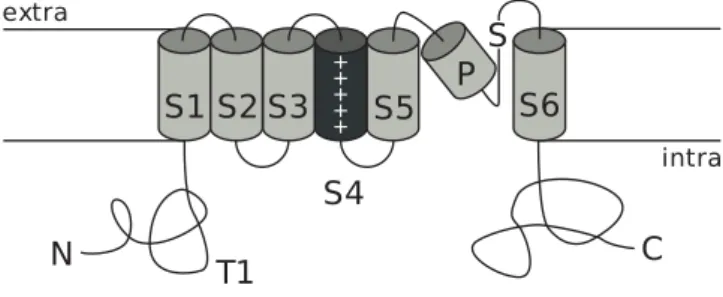
Hyperpolarization-activated, cyclic Nucleotide-modulated (HCN) channels be-long to the family of voltage-gated potassium channels (for a review see [72] [2] [112]). In this respect they share a common general membrane topology com-prising six transmembrane segments (S1-S6), a signature sequence placed in the loop between S4 and S5 is shared with minor variations among all the chan-nels belonging to this family. In the H5 loop resides the pore-forming region. The S4 segment presents every three or four hydrophobic aminoacids a lysine or arginine; which are positively charged at physiological pH and act as voltage sensor. N- and C-terminals reside in the intracellular membrane side; while in proximity to the first transmembrane segment, close to the intracellular side, a T1 region acts as anchor point for multimeric assembly of the complete channel.
C
S
S1 S2 S3
S4
S5
S6
N
extra + + + + +T1
P
intraFigure 1.6: General structure of voltage-gated potassium channels. S1-S6: transmembrane segments, P: pore-forming region, S: signature sequence, T1: region involved in subunit assembly. Figure adapted from [72].
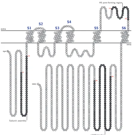
HCN channels have all these features common to their superfamily, plus a cytoplasmic region placed between S6 and the C-terminal, which acts as cyclic nucleotide binding domain (CNBD) (fig. 1.7), capable of binding cAMP and cGMP (the latter with less affinity), that act as modulators on channel proper-ties. The CNBD is connected to the S6 segment by an α-helical linker region. The carboxyl terminal portion of the protein, downstream to the CNBD, is also involved in trafficking toward the cellular membrane; HCN channels can distribute in a non-uniform manner along the cellular membrane by interacting via PDZ-binding domain with scaffold proteins like Tamalin, S-SCAM, Mint2 and Filamin A [55] [38]. The complete HCN channel is formed by homo- or hetero-tetramerization of four subunits, which also contribute together in defin-ing the pore region. At present time, four genes are known to code for HCN1-4 channel subunit isoforms in mammals [62] [63]. These four subunits present high level of similarity in the portion enclosed between the beginning of S1 and the end of the CNBD, with the majority of differences localized in correspondence of N- and C-terminals. This evidence suggests that HCN subtypes arose from early duplication of a single ancestral gene [51] and subsequently differentiated during evolution.
formation) are the most preserved regions. Also their CNBDs present an high percentage of homology, even if some isoform-specific residues are expressed in the median and terminal portion, possibly being the molecular origin for the different sensibility to cyclic nucleotide modulation. A 52 aminoacid portion present at the N-terminal is implied in subunit tetramerization, since its lacking results in a non-functional protein which remains localized in the perinuclear re-gion, instead of being correctly trafficked to membrane [101]. The most variable portions are the N- and C-termini also in mice.
extra intra H2N–
•
◦
M•
◦
E•
◦
G•
◦
G•
◦
G•
◦
K•
◦
P•
◦
N•
◦
S•
◦
A•
◦
S•
◦
N•
◦
S•
◦
R•
◦
D•
◦
D•
◦
G•
◦
N•
◦
S•
◦
V•
◦
F•
◦
P•
◦
S•
◦
K•
◦
A•
◦
P•
◦
A•
◦
T•
◦
G•
◦
P•
◦
V•
◦
A•
◦
A•
◦
D•
◦
K•
◦
R•
◦
L•
◦
G•
◦
T•
◦
P•
◦
P•
◦
G•
◦
G•
◦
G•
◦
A•
◦
A•
◦
G•
◦
K•
◦
E•
◦
H•
◦
G•
◦
N•
◦
S•
◦
V•
◦
C•
◦
F•
◦
K•
◦
V•
◦
D•
◦
G•
◦
G•
◦
G•
◦
G•
◦
•
◦
E E•
◦
P•
◦
A•
◦
G•
◦
S•
◦
F•
◦
E•
◦
D•
◦
A•
◦
E•
◦
G•
◦
P•
◦
R•
◦
Q•
◦
Y•
◦
G•
◦
F•
◦
M•
◦
Q•
◦
R•
◦
Q•
◦
F•
◦
T•
◦
S•
◦
M•
◦
L•
◦
Q•
◦
P•
◦
G•
◦
V•
◦
N•
◦
K•
◦
F•
◦
S•
◦
L•
◦
R•
◦
M•
◦
G•
◦
S•
◦
Q•
◦
K•
◦
A•
◦
V•
◦
E•
◦
K•
◦
E•
◦
Q•
◦
E•
◦
R•
◦
V•
◦
K•
◦
T•
◦
A•
◦
G•
◦
F•
◦
W•
◦
I•
◦
I•
◦
H•
◦
P•
◦
Y•
◦
S•
◦
F•
◦
R•
◦
F•
◦
Y•
◦
W•
◦
•
◦
D L•
◦
I•
◦
M•
◦
L•
◦
I•
◦
M•
◦
M•
◦
V•
◦
V•
◦
•
◦
G•
◦
N•
◦
L I•
◦
I•
◦
P•
◦
V•
◦
G•
◦
I•
◦
T•
◦
F•
◦
F•
◦
T•
◦
E•
◦
Q•
◦
T•
◦
T•
◦
T•
◦
P•
◦
W•
◦
I•
◦
I•
◦
F•
◦
N•
◦
V•
◦
A•
◦
S•
◦
D•
◦
T•
◦
V•
◦
F•
◦
L•
◦
L•
◦
D•
◦
L•
◦
I•
◦
M•
◦
N•
◦
F•
◦
R•
◦
T•
◦
G•
◦
T•
◦
V•
◦
N•
◦
E•
◦
D•
◦
S•
◦
•
◦
S E•
◦
I•
◦
I•
◦
L•
◦
D•
◦
P•
◦
K•
◦
V•
◦
I•
◦
K•
◦
M•
◦
N•
◦
Y•
◦
L•
◦
•
◦
K S•
◦
W•
◦
F•
◦
V•
◦
V•
◦
D•
◦
F•
◦
•
◦
IP•
◦
•
◦
S•
◦
S•
◦
I V•
◦
D•
◦
Y•
◦
I•
◦
F•
◦
L•
◦
I•
◦
V•
◦
•
◦
E K•
◦
G•
◦
M•
◦
D•
◦
S•
◦
E•
◦
V•
◦
Y•
◦
K•
◦
T•
◦
A•
◦
R•
◦
A•
◦
L•
◦
R•
◦
I•
◦
V•
◦
R•
◦
F•
◦
T•
◦
K•
◦
I•
◦
L•
◦
S•
◦
L•
◦
L•
◦
R•
◦
L•
◦
L•
◦
R•
◦
L•
◦
S•
◦
R•
◦
L•
◦
I•
◦
R•
◦
Y•
◦
I•
◦
H•
◦
Q•
◦
W•
◦
E•
◦
E•
◦
I•
◦
F•
◦
H•
◦
M•
◦
T•
◦
Y•
◦
D•
◦
L•
◦
A•
◦
S•
◦
A•
◦
V•
◦
V•
◦
R•
◦
I•
◦
F•
◦
•
◦
N L•
◦
I•
◦
G•
◦
M•
◦
M•
◦
L•
◦
L•
◦
L•
◦
D•
◦
C•
◦
•
◦
H•
◦
W G•
◦
C•
◦
L•
◦
Q•
◦
F•
◦
L•
◦
V•
◦
P•
◦
•
◦
L L•
◦
Q•
◦
D•
◦
F•
◦
P•
◦
P•
◦
D•
◦
C•
◦
W•
◦
•
◦
V S•
◦
L•
◦
N•
◦
E•
◦
M•
◦
V•
◦
N•
◦
D•
◦
S•
◦
W•
◦
•
◦
G K•
◦
Q•
◦
S•
◦
Y•
◦
A•
◦
L•
◦
F•
◦
K•
◦
A•
◦
M•
◦
S•
◦
M•
◦
•
◦
L C•
◦
I•
◦
G•
◦
Y•
◦
G•
◦
A•
◦
Q•
◦
A•
◦
V•
◦
S•
◦
M•
◦
S•
◦
D•
◦
L•
◦
W•
◦
I•
◦
T•
◦
M•
◦
L•
◦
S•
◦
M•
◦
I•
◦
V•
◦
G•
◦
A•
◦
T•
◦
C•
◦
Y•
◦
A•
◦
M•
◦
F•
◦
V•
◦
G•
◦
H•
◦
A•
◦
T•
◦
A•
◦
L•
◦
I•
◦
Q•
◦
S•
◦
L•
◦
D•
◦
S•
◦
S•
◦
R•
◦
R•
◦
Q•
◦
Y•
◦
Q•
◦
E•
◦
K•
◦
Y•
◦
K•
◦
Q•
◦
V•
◦
E•
◦
Q•
◦
Y•
◦
M•
◦
S•
◦
F•
◦
H•
◦
K•
◦
L•
◦
P•
◦
A•
◦
D•
◦
M•
◦
R•
◦
Q•
◦
K•
◦
I•
◦
H•
◦
D•
◦
Y•
◦
Y•
◦
E•
◦
H•
◦
R•
◦
Y•
◦
Q•
◦
G•
◦
K•
◦
I•
◦
F•
◦
D•
◦
E•
◦
E•
◦
N•
◦
I•
◦
L•
◦
S•
◦
E•
◦
L•
◦
N•
◦
D•
◦
P•
◦
L•
◦
R•
◦
E•
◦
E•
◦
I•
◦
V•
◦
N•
◦
F•
◦
N•
◦
C•
◦
R•
◦
K•
◦
L•
◦
V•
◦
A•
◦
T•
◦
M•
◦
P•
◦
F•
◦
A•
◦
N•
◦
A•
◦
D•
◦
P•
◦
N•
◦
F•
◦
V•
◦
T•
◦
A•
◦
M•
◦
L•
◦
S•
◦
K•
◦
L•
◦
R•
◦
F•
◦
E•
◦
V•
◦
F•
◦
Q•
◦
P•
◦
G•
◦
D•
◦
Y•
◦
I•
◦
I•
◦
R•
◦
E•
◦
G•
◦
A•
◦
V•
◦
G•
◦
K•
◦
K•
◦
M•
◦
Y•
◦
F•
◦
I•
◦
Q•
◦
H•
◦
G•
◦
V•
◦
A•
◦
G•
◦
V•
◦
I•
◦
T•
◦
K•
◦
S•
◦
S•
◦
K•
◦
E•
◦
M•
◦
K•
◦
L•
◦
D•
◦
G•
◦
S•
◦
Y•
◦
F•
◦
G•
◦
E•
◦
I•
◦
C•
◦
L•
◦
L•
◦
T•
◦
K•
◦
G•
◦
R•
◦
R•
◦
T•
◦
A•
◦
S•
◦
V•
◦
R•
◦
A•
◦
D•
◦
T•
◦
Y•
◦
C•
◦
R•
◦
L•
◦
Y•
◦
S•
◦
L•
◦
S•
◦
V•
◦
D•
◦
N•
◦
F•
◦
N•
◦
E•
◦
V•
◦
L•
◦
E•
◦
E•
◦
Y•
◦
P•
◦
M•
◦
M•
◦
R•
◦
R•
◦
A•
◦
F•
◦
E•
◦
T•
◦
V•
◦
A•
◦
I•
◦
D•
◦
R•
◦
L•
◦
R•
◦
I•
◦
G•
◦
K•
◦
K•
◦
N•
◦
S•
◦
I•
◦
L•
◦
L•
◦
Q•
◦
K•
◦
F•
◦
Q•
◦
K•
◦
D•
◦
L•
◦
N•
◦
T•
◦
G•
◦
V•
◦
F•
◦
N•
◦
N•
◦
Q•
◦
E•
◦
N•
◦
E•
◦
I•
◦
L•
◦
K•
◦
Q•
◦
I•
◦
V•
◦
K•
◦
H•
◦
D•
◦
R•
◦
E•
◦
M•
◦
V•
◦
Q•
◦
A•
◦
I•
◦
P•
◦
P•
◦
I•
◦
N•
◦
Y•
◦
P•
◦
Q•
◦
M•
◦
T•
◦
A•
◦
L•
◦
N•
◦
C•
◦
T•
◦
S•
◦
S•
◦
T•
◦
T•
◦
T•
◦
P•
◦
T•
◦
S•
◦
R•
◦
M•
◦
R•
◦
T•
◦
Q•
◦
S•
◦
P•
◦
P•
◦
V•
◦
Y•
◦
T•
◦
A•
◦
T•
◦
S•
◦
L•
◦
S•
◦
H•
◦
S•
◦
N•
◦
L•
◦
H•
◦
S•
◦
P•
◦
S•
◦
P•
◦
S•
◦
T•
◦
Q•
◦
T•
◦
P•
◦
Q•
◦
P•
◦
S•
◦
A•
◦
I•
◦
L•
◦
S•
◦
P•
◦
C•
◦
S•
◦
Y•
◦
T•
◦
T•
◦
A•
◦
V•
◦
C•
◦
S•
◦
P•
◦
P•
◦
I•
◦
Q•
◦
S•
◦
P•
◦
L•
◦
A•
◦
T•
◦
R•
◦
T•
◦
F•
◦
H•
◦
Y•
◦
A•
◦
S•
◦
P•
◦
T•
◦
A•
◦
S•
◦
Q•
◦
L•
◦
S•
◦
L•
◦
M•
◦
Q•
◦
Q•
◦
P•
◦
Q•
◦
Q•
◦
Q•
◦
L•
◦
P•
◦
Q•
◦
S•
◦
Q•
◦
V•
◦
Q•
◦
Q•
◦
T•
◦
Q•
◦
T•
◦
Q•
◦
T•
◦
Q•
◦
Q•
◦
Q•
◦
Q•
◦
Q•
◦
Q•
◦
Q•
◦
Q•
◦
Q•
◦
Q•
◦
Q•
◦
Q•
◦
Q•
◦
Q•
◦
Q•
◦
Q•
◦
Q•
◦
Q•
◦
Q•
◦
Q•
◦
Q•
◦
Q•
◦
Q•
◦
Q•
◦
Q•
◦
Q•
◦
Q•
◦
Q•
◦
Q•
◦
Q•
◦
Q•
◦
Q•
◦
Q•
◦
Q•
◦
Q•
◦
Q•
◦
Q•
◦
P•
◦
Q•
◦
T•
◦
P•
◦
G•
◦
S•
◦
S•
◦
T•
◦
P•
◦
K•
◦
N•
◦
E•
◦
V•
◦
H•
◦
K•
◦
S•
◦
T•
◦
Q•
◦
A•
◦
L•
◦
H•
◦
N•
◦
T•
◦
N•
◦
L•
◦
T•
◦
K•
◦
E•
◦
V•
◦
R•
◦
P•
◦
L•
◦
S•
◦
A•
◦
S•
◦
Q•
◦
P•
◦
S•
◦
L•
◦
P•
◦
H•
◦
E•
◦
V•
◦
S•
◦
T•
◦
L•
◦
I•
◦
S•
◦
R•
◦
P•
◦
H•
◦
P•
◦
T•
◦
V•
◦
G•
◦
E•
◦
S•
◦
L•
◦
A•
◦
S•
◦
I•
◦
P•
◦
Q•
◦
P•
◦
V•
◦
A•
◦
A•
◦
V•
◦
H•
◦
S•
◦
T•
◦
G•
◦
L•
◦
Q•
◦
A•
◦
G•
◦
S•
◦
R•
◦
S•
◦
T•
◦
V•
◦
P•
◦
Q•
◦
R•
◦
V•
◦
T•
◦
L•
◦
F•
◦
R•
◦
Q•
◦
M•
◦
S•
◦
S•
◦
G•
◦
A•
◦
I•
◦
P•
◦
P•
◦
N•
◦
R•
◦
G•
◦
V•
◦
P•
◦
P•
◦
A•
◦
P•
◦
P•
◦
P•
◦
P•
◦
A•
◦
A•
◦
V•
◦
Q•
◦
R•
◦
E•
◦
S•
◦
P•
◦
S•
◦
V•
◦
L•
◦
N•
◦
T•
◦
D•
◦
P•
◦
D•
◦
A•
◦
E•
◦
K•
◦
P•
◦
R•
◦
F•
◦
A•
◦
S•
◦
N•
◦
L HOOC– 78•
◦
R Subunit assembly•
◦
F 129•
◦
D 334•
◦
Y H5 pore-forming region•
◦
H 355•
◦
P 464•
◦
L cAMP binding•
◦
T 581•
◦
D S1 S2 S3 S4 S5 S6Figure 1.7: Structure of murine HCN1 channel (910 AA). Protein data from UniProtKB/Swiss-Prot, sequence id: O88704. Membrane topology rendered with TEXtopo [6]
HCN1 ...MEGGGKPNSAS 11
HCN2 ...MDARGGGGRPGDSP 14
HCN3 ... 0
HCN4 MDKLPPSMRKRLYSLPQQVGAKAWIMDEEEDGEEEGAGGRQDPSR 45
HCN1 NSRDDGNSVFPSKAPATG... 29
HCN2 GTTPAPGPPPPPPPPAPPQPQPPPAPPPNPTTPSHPESADEPGP. 58
HCN3 ... 0
HCN4 RSIRLRPLPSPSPSVAAGCSESRGAALGATESEGPGRSAGKSSTN 90
HCN1 ...PVAADKRLGTPPG 42
HCN2 ...RARLCSRDSACTPGAAKGGANGECGRGEPQCSPEGP...ARG 97
HCN3 ...MEEEARPAAGA 11
HCN4 GDCRRFRGSLASLGSRGGGSGGAGGGSSLGHLHDSAEERRLIAAE 135
HCN1 GGAAGKEHGNSVCFKVDGGGGEEPAGSFEDAEGPR... 77
HCN2 PKVSFSCRGAASGPSAAEEAGSEEAGPAGEPRGSQ... 132
HCN3 GEAATPARETPP.AAPAQARAASGGVPESAPEPKR... 45 HCN4 GDASPGEDRTPPGLATEPERPATAAQPAASPPPQQPPQPASASCE 180 HCN1 ...RQYGFMQRQFTSMLQPG 94 HCN2 ...ASFLQRQFGALLQPG 147 HCN3 ...RQLGTLLQPT 55 HCN4 QPSADTAIKVEGGAAAIDHILPEAEVRLGQSGFMQRQFGAMLQPG 225 | {z } subunit assembly
HCN1 VNKFSLRMFGSQKAVEKEQERVKTAGFWIIHPYSDFRFYWDLIML 139
HCN2 VNKFSLRMFGSQKAVEREQERVKSAGAWIIHPYSDFRFYWDFTML 192
HCN3 VNKFSLRVFGSHKAVEIEQERVKSAGAWIIHPYSDFRFYWDLIML 100
HCN4 VNKFSLRMFGSQKAVEREQERVKSAGFWIIHPYSDFRFYWDLTML 270
| {z } | {z }
subunit assembly S1
HCN1 IMMVGNLVIIPVGITFFTEQTTTPWIIFNVASDTVFLLDLIMNFR 184
HCN2 LFMVGNLIIIPVGITFFKDETTAPWIVFNVVSDTFFLMDLVLNFR 237
HCN3 LLMVGNLIVLPVGITFFKEENSPPWIVFNVLSDTFFLLDLVLNFR 145
HCN4 LLMVGNLIIIPVGITFFKDENTTPWIVFNVVSDTFFLIDLVLNFR 315
| {z } | {z }
S1 S2
HCN1 TGTVNEDSSEIILDPKVIKMNYLKSWFVVDFISSIPVDYIFLIVE 229
HCN2 TGIVIEDNTEIILDPEKIKKKYLRTWFVVDFVSSIPVDYIFLIVE 282
HCN3 TGIVVEEGAEILLAPRAIRTRYLRTWFLVDLISSIPVDYIFLVVE 190
HCN4 TGIVVEDNTEIILDPQRIKMKYLKSWFVVDFISSIPVEYIFLIVE 360
| {z }
HCN1 KG..MDSEVYKTARALRIVRFTKILSLLRLLRLSRLIRYIHQWEE 272 HCN2 KG..IDSEVYKTARALRIVRFTKILSLLRLLRLSRLIRYIHQWEE 325 HCN3 LEPRLDAEVYKTARALRIVRFTKILSLLRLLRLSRLIRYIHQWEE 235 HCN4 TR..IDSEVYKTARAVRIVRFTKILSLLRLLRLSRLIRYIHQWEE 403 |{z} | {z } S3 S4 HCN1 IFHMTYDLASAVVRIFNLIGMMLLLCHWDGCLQFLVPLLQDFPPD 317 HCN2 IFHMTYDLASAVMRICNLISMMLLLCHWDGCLQFLVPMLQDFPSD 370 HCN3 IFHMTYDLASAVVRIFNLIGMMLLLCHWDGCLQFLVPMLQDFPSD 280 HCN4 IFHMTYDLASAVVRIVNLIGMMLLLCHWDGCLQFLVPMLQDFPHD 448 | {z } S5 Signature z }| { HCN1 CWVSLNEMVNDSWGKQYSYALFKAMSHMLCIGYGAQAPVSMSDLW 362
HCN2 CWVSINNMVNHSWSELYSFALFKAMSHMLCIGYGRQAPESMTDIW 415
HCN3 CWVSMNRMVNHSWGRQYSHALFKAMSHMLCIGYGQQAPVGMPDVW 325
HCN4 CWVSINGMVNNSWGKQYSYALFKAMSHMLCIGYGRQAPVGMSDVW 493 | {z } H5 pore-forming HCN1 ITMLSMIVGATCYAMFVGHATALIQSLDSSRRQYQEKYKQVEQYM 407 HCN2 LTMLSMIVGATCYAMFIGHATALIQSLDSSRRQYQEKYKQVEQYM 460 HCN3 LTMLSMIVGATCYAMFIGHATALIQSLDSSRRQYQEKYKQVEQYM 370 HCN4 LTMLSMIVGATCYAMFIGHATALIQSLDSSRRQYQEKYKQVEQYM 538 | {z } S6
HCN1 SFHKLPADMRQKIHDYYEHRYQGKIFDEENILSELNDPLREEIVN 452
HCN2 SFHKLPADFRQKIHDYYEHRYQGKMFDEDSILGELNGPLREEIVN 505
HCN3 SFHKLPADTRQRIHEYYEHRYQGKMFDEESILGELSEPLREEIIN 415
HCN4 SFHKLPPDTRQRIHDYYEHRYQGKMFDEESILGELSEPLREEIIN 583
HCN1 FNCRKLVATMPLFANADPNFVTAMLSKLRFEVFQPGDYIIREGAV 497
HCN2 FNCRKLVASMPLFANADPNFVTAMLTKLKFEVFQPGDYIIREGTI 550
HCN3 FTCRGLVAHMPLFAHADPSFVTAVLTKLRFEVFQPGDLVVREGSV 460
HCN4 FNCRKLVASMPLFANADPNFVTSMLTKLRFEVFQPGDYIIREGTI 628
| {z }
cAMP binding
HCN1 GKKMYFIQHGVAGVITKSSKEMKLTDGSYFGEICLLTKGRRTASV 542
HCN2 GKKMYFIQHGVVSVLTKGNKEMKLSDGSYFGEICLLTRGRRTASV 595
HCN3 GRKMYFIQHGLLSVLARGARDTRLTDGSYFGEICLLTRGRRTASV 505
HCN4 GKKMYFIQHGVVSVLTKGNKETRLADGSYFGEICLLTRGRRTASV 673
| {z }
HCN1 RADTYCRLYSLSVDNFNEVLEEYPMMRRAFETVAIDRLDRIGKKN 587
HCN2 RADTYCRLYSLSVDNFNEVLEEYPMMRRAFETVAIDRLDRIGKKN 640
HCN3 RADTYCRLYSLSVDHFNAVLEEFPMMRRAFETVAMDRLRRIGKKN 550
HCN4 RADTYCRLYSLSVDNFNEVLEEYPMMRKKNSILLHKVQHDLN..S 716
| {z }
cAMP binding
HCN1 SILLQKFQKDLNTGVFNNQENEILKQIVKHDREMVQAIPPINYPQ 632
HCN2 SILLHKVQHDLSSGVFNNQENAIIQEIVKYDREMVQQAE.LGQRV 684
HCN3 SILQRKRSEPSPG....SSGGVMEQHLVQHDRDMARGVRGLAPGT 591
HCN4 GVFNYQENE...IIQQIVRHDREMAHCAHRVQAAA 748
HCN1 MTALNCTSSTTTPTSRMRTQSPPVYTATSLSHSNLHS... 669
HCN2 GLFPPPPPPQVTSAIATLQQAVAMSFCPQVARPLVG... 720
HCN3 GARLSGKPVLWEPLVHAPLQAAAVTSNVAIALTHQR... 627
HCN4 SATPTPTPVIWTPLIQAPLQAAAATTSVAIALTHHPRLPAAIFRP 793
HCN1 ... 669
HCN2 ... 720
HCN3 ... 627
HCN4 PPGPGLGNLGAGQTPRHPRRLQSLIPSALGSASPASSPSQVDTPS 838
HCN1 ...PSPSTQTPQPSAILSPCSYTTAVCSPPI 697
HCN2 ...PLALGS... 726 HCN3 ...GPLPLSPDS...PATL 640 HCN4 SSSFHIQQLAGFSAPPGLSPLLPSSSSSPPPGACGSPPAPTPSTS 883 HCN1 QSPLATRT...FHYASPTA 713 HCN2 .PRLVRRA...PP 735 HCN3 LARSARRS...AGSPA 653 HCN4 TAAAASTTGFGHFHKALGGSLSSSDSPLLTPLQPGARSPQAAQPP 928 HCN1 SQLSLMQQPQQQLPQSQVQQTQTQTQQQQQQQQQQQQ... 750 HCN2 GPLPPAASP...GPPAASPPAAPSS... 757 HCN3 SPLVPVRAG...PLLARGPWASTSRLPAPP... 680 HCN4 PPLPGARGGLGLLEHFLPPPPSSRSPSSSPGQLGQPPGELSLGLA 973 HCN1 ...QQQQ 754 HCN2 ... 757 HCN3 ... 680 HCN4 AGPSSTPETPPRPERPSFMAGASGGASPVAFTPRGGLSPPGHSPG 1018 HCN1 QQQQQQQQQQQQQQQQQQQQQPQTPGSSTPKNEVHKSTQALHNTN 799
HCN2 .PRAPRTSPYGVPGSPATRVGP..ALPARRLSR..ASRP... 791
HCN3 .ARTLHASLSRTGRSQVSLLGPPPGGGARRLGP..RGRP... 716
HCN1 LTKEVRPLSASQPSLPHEVSTLISRPHPTVGESLASIPQPVAAVH 844
HCN2 ...LSASQPSLPHG...VPAPSP 808
HCN3 ...LSASQPSLPQR...ATG 730
HCN4 LTQDLKLISASQPALPQDGAQTLRRASPHSSGESVAAFSLYPRAG 1108
HCN1 STGLQAGS...RSTVPQRVTLFRQMSSGAIPPNRGVPPAP 881 HCN2 AAS...ARPASSSTPRLGPAPTART 830 HCN3 DGS...PRRKGSGSERLPPSGLLAK 752 HCN4 GGSGSSGGLGPPGRPYGAIPGQHVTLPRKTSSGSLPPPLSLFGAR 1153 HCN1 PPPAAVQRESPSVLNTDP....DAEKPRFASNL 910 HCN2 AAPSPDRRDSASPGAASGLDPLDSARSRLSSNL 863 HCN3 PPGTVQPPRSSVP...EP...VTPRGPQISANM 779
HCN4 AASSGGPPLTTAAPQREPGARSEPVRSKLPSNL 1186
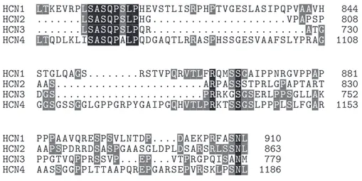
Figure 1.8: Conserved regions in murine HCN channel isoforms. Protein data from UniProtKB/Swiss-Prot, sequence id: O88704 (HCN1), O88703 (HCN2), O88705 (HCN3), O70507 (HCN4). Residue alignment performed with ClustalW 2.0.10. Similarity shading rendered with TEXshade[5], gray shading for con-served residues, black shading for completely matching regions.
1.2.2
I
hcurrent properties
HCN channels have the peculiar feature to be opened in response to an hyperpo-larizing stimulus. When this event happens a slowly activating, non-inactivating ionic current flows; this current takes the name of If in the cardiac district or
Ih in the central nervous system (CNS). Ih exhibits both time- and
voltage-dependency and drives a cationic flow across the membrane, mainly sodium and potassium flow with a relative permeability of 1:3 [108], causing an influx of positive charges into the cell, which leads to a depolarization. In case Ih is
active at resting potential, this negative feedback mechanism is effective also with depolarizing stimuli, since the induced depolarization of the cellular mem-brane closes HCN channels, resulting in a hyperpolarization that counteracts the disturbing stimulus.
A major component of this stabilizing effect is due to Ih’s voltage-dependant
gating: opposite to voltage-gated calcium or sodium channels which are maxi-mally activated at their reversal potential, Ih activation curve falls in its basal
values close to the reversal potential, endowing this current with intrinsically negative feedback properties. Activation kinetics depend upon the isoform com-bination in the tetramer, for homomeric channels kinetics slow down according to: HCN 1 > HCN 2 > HCN 3 > HCN 4 from a few millisecond for the faster isoform to hundreds of milliseconds and even seconds for the slowest one (at voltages more negative than -100 mV) [32].
HCN channels may be modulated by cyclic nucleotides; in the presence of cAMP and cGMP (the latter with lesser affinity), activation kinetics speed up and the activation curve of these channels is shifted toward more positive po-tentials. The same shift occurs in the absence of CNBD, suggesting that the CNBD acts by limiting channel kinetics, and this constrain can be removed by cyclic nucleotide binding. Different channel isoforms exhibit different sensitiv-ity to cAMP modulation, in particular: HCN 2, HCN 4 > HCN 1 > HCN 3 [1]. Modulation of Ih properties occurs also in dependance of the intracellular
lev-els of phosphoinositides like phosphatidylinositol-4,5-biphosphate (P IP2). This
class of molecules is able to shift the activation curve of Ih toward ∼ 20 mV more positive potentials. This regulatory mechanism operates in a manner in-dependent from cyclic nucleotides and is equally effective on HCN 1,2,4 isoforms [115]. Regulation by phosphorilation is also reported for HCN channels, since in some preparations the cAMP modulation on Ih is completely abolished by
applying protein kinase inhibitors. In a similar way, Ser/Thr inhibitors shift the activation curve toward more positive potentials, in particular PKA-mediated phosphorilation seems to preferentially happen [32].
The first HCN channel blocker known is the inorganic ion cesium, which is non-selective With respect to channel isoform and also acts as blocker for some potassium channels. More specific organic inhibitors are now available, developed mainly for their potential use as heart-rate reducing agents (see 1.2.4 for a detailed discussion). Among organic drugs, only the novel antiepilectic Lamotrigine shifts the activation curve of Ih in pyramidal neurons toward more
1.2.3
Physiological expression and role of HCN channels
HCN channels play a major role in the cardiac tissue, where they control rhyth-micity in sinus-atrial node (SAN) pacemaker cells. In the SAN Ih is activatedby membrane hyperpolarization leading to a progressive depolarization that, along with the contribution other calcium and sustained inward currents (ICaT,
ICaL, Ist), takes the name of diastolic depolarization. In the cardiac tissue,
channel isoforms mainly expressed are HCN4 and HCN2 (with the respective proportion of 80% to 20%). The other major function of Ih in cardiac tissue is
the regulation of heart rate, operated by autonomous nervous system. Sympa-thetic stimulation of β-adrenergic receptors leads to an activation of Ihfollowing
to an increment in cAMP production. By contrast, Parasympathetic stimula-tion of muscarinic receptors induces a decrement of intracellular cAMP which negatively modulates Ih.
HCN channels are also expressed in dorsal root ganglion (DRG) neurons. In these cells HCN1 is the most abundant isoform, expressed by almost all the large- and medium-size neurons (A-type) at the level of their somatic mem-brane. HCN2 expression is lower, almost a half compared to HCN1 in every DRG neuron. HCN2 is mainly located on the membrane of large-body cells while intracellularly in small- and medium-body neurons, in the latter case it usually co-localizes with CGRP, the marker of nociceptive neurons [53]. HCN3 and HCN4 expression is much lower. Involvement of Ih in chronic neuropathic
pain, a disease characterized by spontaneous pain, hyperalgesia and allodynia, is correlable to multiple aspects.
Ectopic potentials caused by some experimental models (sciatic nerve ligation, chronic constriction injury, DRG chronic compression) are reported to happen along with an up-regulation of Ih in these neurons, while application of the
HCN channel blocker ZD-7288 significantly prevents ectopic discharges from in-jured neurones [12]. When discharges from inin-jured neurons happen, adjacent nerves experience hyperexcitability, which results in an exaggerate perception of noxious stimuli as well as an increment in spontaneous activity. Few studies showed possible involvement of HCN channels as pro-nociceptive agents, since intraplantar injection of ZD-7288 alleviated thermal hyperalgesia in rats [17]. Ih involvement in regulating the axonal conduction and synaptic transmission
in spinal dorsal horns is somewhat controversial and requires more clues (see [53]).
Within the CNS, HCN channels are widely expressed. HCN1 isoform is present in hippocampus, neocortex, cerebellar cortex and brainstem. HCN2 is expressed ubiquitously, but with particular abundance in brainstem nuclei and thalamus. HCN3 is present in retinal cones, only inside the olfactory bulb and in some hypothalamic nuclei its presence is medium to high. HCN4 is particu-larly expressed in thalamic nuclei and in the olfactory bulb, at level of mitral cell layer [73] [90] [79]. Near the resting potential HCN channels are usually open in these neurons and the resulting Ihcontributes in setting and stabilizing
the resting potential. Stabilization by constitutively open HCN channels acts in two distinct ways: by lowering the membrane resistance, therefore every input current induces a smaller change in membrane potential; and by actively coun-teracting to depolarizing or hyperpolarizing stimuli, acting as a slow “voltage clamp” mechanism able to suppress low-frequency fluctuations of the membrane potential. In dendrites of CA1 hippocampal neurons Ih influences the
ampli-tude and kinetic properties of excitatory post-synaptic potentials (EPSPs) [69] [70]. In the prefrontal cortex neurons it was shown that HCN co-localize with α2a receptors for noradrenaline on dendritic spines, this made the authors
sug-gest that decrements of cAMP produced by α2a activation can modulate and
reduce the open probability of HCN channels, augmenting the cellular suscep-tibility to synaptic inputs [104]. In cerebellar Purkinje cells Ih counteracts
hyperpolarizing stimuli arriving to dendrites, stabilizing the membrane below the threshold for spontaneous spiking, this helps these neurons in maintaining an input-output relationship independent from previous activity’s history. This, along with evidence that hcn1 knockout mice present deficits in learning motor tasks like rotarod or swimming, highlights an implication of HCN channels in motor learning [78].
Another major role for Ih in brain is to enable resonance in single neurons or
modulate resonant behavior in neural networks. Resonance in a neuron is the property of responding preferentially to inputs of a defined frequency; to gener-ate resonance a neuron needs to have properties of both low-pass and high-pass filter. The membrane properties account in every neuron for the low-pass filter. A current that opposes to changes in membrane potential it’s needed to endow high-pass filter properties; and it’s activation kinetic must be slower than the membrane time constant [49]. In this respect, Ihexhibits this feature and thus
is a good candidate to generate high-pass filter properties. In fact several studies report Ih contribution in generating resonance in CNS neurons [103] [105] [47].
Inside known neural networks, Ih contribution is implied in generating theta
oscillations [77] and also is an important player in generating γ-oscillations in hippocampus [30] [15], synchronized oscillations in the inferior olive [3] and subthreshold oscillations in enthorinal cortex [22] [39].
Peripheral expression of HCN was detected in the enteric nervous system, where all but HCN3 isoforms were detected [111]. An increment in Ih
suscep-tibility to cesium blockade by AH neurons in distant colon was also observed in inflammed tissue [59]. HCN expression in non-neuronal cells is reported for pancreatic β-cells [26] and α-cell lines [113], in the latter case application of the HCN blocker ZD7288 enhanced glucagon secretion independently from glucose concentration.
1.2.4
HCN disruption insight on isoform relevance
Important insights on Ih-related disfunctions and pathologies comes from
exper-iments of genetic deletion of hcn genes. HCN4 knockout mice die prematurely in-utero during day 9.5-11.5 of embryonic development, due to a deficiency in SAN [95]. Recording before the deadly period showed a cardiac If reduction by
85%, and an heart rate reduction by 40%. Most importantly these hearts are not sensitive to cAMP stimulation; this highlighted HCN4 as crucial in cyclic nucleotide modulation even in conditions where this isoform is not essential for heart beat generation. By creating Tamoxifen-inducible knockout mice for hcn4 it was possible to investigate the relative importante of HCN4 in adult cardiac tissue [44]. In these animals most of the sinoatrial If was eliminated,
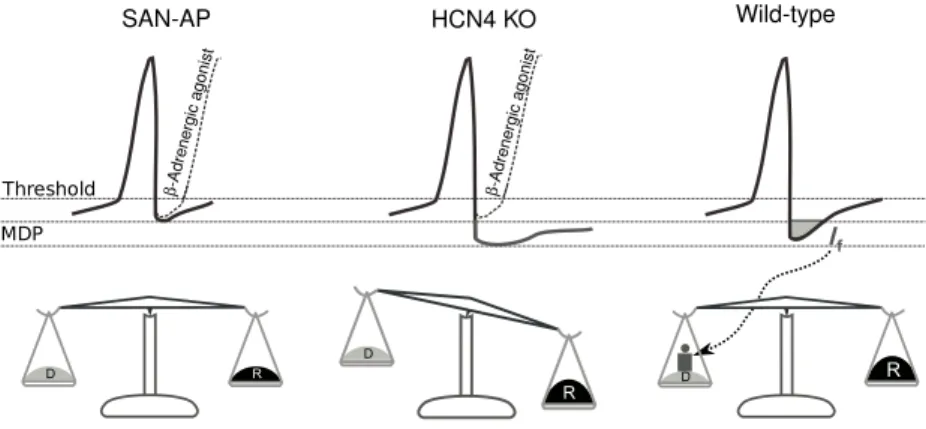
resulting in recurrent sinus pauses; on the other hand, heart rate is surpris-ingly still sensitive to sympathetic stimulation. Authors suggest that HCN4 may not be involved in physiological heart rate modulatory mechanisms, but could be instead a necessary component for maintaining a stable rhythmicity and to counteract parasympathetic heart rate decrements or transitions from stimulated to basal cardiac states (see fig. 1.9) [43].
SAN-AP HCN4 KO Wild-type
β-Adrene rgic agonist β-Adrene rgic agonist Threshold MDP
Figure 1.9: Hypothesis of HCN4 reserve-deposit in heart rate modulation. SAN-AP is the regular SAN action potential in wild type mice, whose length is short-ened by noradrenergic stimulation. In HCN4 knockout noradrenergic stimu-lation still behaves as in wild type but following an increase in repolarizing currents (symbolized by R in the balance) If is required to join depolarizing
currents (symbolized with D) in order to recover the balance. Figure adapted from [43]
Mice knockout for hcn2 are characterized by an hypoactive behavior and ataxic gait [61]. Electroencephalographic recordings and video analysis show occurrence of spike waves in correspondence to behavioral arrest, demonstrat-ing that these mice suffer from absence epilepsy. Generation of spikes is proba-bly related to increased excitability of thalamocortical neurones, since injection of positive currents into these neurons does not elicit single spike but results instead in burst firing. Calcium imaging in thalamic slices revealed oscilla-tory activity. These events are probably related to recovery of T-type calcium channels, normally inactivated at resting potential, by the induced hyperpolar-ization caused by lack of HCN2. At cardiac level these mice presents normal heart rate, with also a normal regulation of heart rate in response to motor
ex-ercise or administration of β-blockers. The only cardiac phenotype is a varying RR interval, resulting in sinus arrhythmia. Recordings from SAN cells showed a 25% reduction of If compared to wild type, resulting in a more
hyperpolar-ized maximum diastolic potential. The mechanism hypotheshyperpolar-ized to cause sinus arrhythmia identifies HCN2 as useful in setting the diastolic potential toward more depolarized levels, a contribution that may become crucial in response to excessive hyperpolarization.
Deletion of hcn1 gene results in a viable phenotype, which presents deficit in learning motor tasks [78], especially the motor learning of fast movements (evaluated by rotarod and swimming tests). In these mutants spontaneous ac-tivity of Purkinje cells is not altered, demonstrating that HCN1 is not crucial for pacemaking in this neurones. However, in response to injection of hyperpolariz-ing currents, hcn1 knockout Purkinje cells display a profound hyperpolarization (normally counteracted by Ih) that strongly reduces firing. In addition, the
Purkinje cell’s firing independence on previous state is lost, and in knockout mice firing is much reduced if these cells experienced a previous silent state. The other major difference found in CNS resides at the level of hippocampal CA1 neurones, where deletion of hcn1 results in an enhancement of spatial learn-ing and memory. This improvement affects both short- and long-term memory, and has an electrophysiological correlate in the enhancement of long-term po-tentiation (LTP) at the direct perforant path input to the more distal dendrite of CA1 neurones. No cardiac-specific phenotypes are reported for this mice. Creation of hcn3 knockout mice has been made, but no clues on either cardiac nor brain phenotype are reported so far [68].
1.2.5
I
hcurrent in the retinal tissue
The presence of a hyperpolarization-activated current was described in many of the retinal cells: photoreceptors, bipolar cells, amacrine and ganglion cells.
Ihis present in both rods and cones of amphibians, fish, molluscs and
mam-mals; since the resting potential of these neurons is depolarized in dark condi-tions, Ih activates and plays a major role in shaping light response especially
following a bright light. In rods, immunolabelling shows that the HCN1 isoform is particularly expressed in these neurons at the level of inner segment; while in the outer segment HCN are absent and thus no Ih may be detected recording
from isolated outer segments [20]. This conductance in rods has the functional role to quicken the recovery of dark potential after a light response, providing a way to overcome the inherently slow speed of the rod phototransduction. In addition Ihfilters the signal from noise before it reaches the synapse to bipolar
cells [19] [18]. In cones the presence of HCN3 channels is reported at the level of terminal pedicles, while staining for HCN1 is present at the level of inner segment and somata [74].
HCN1,2,3,4 HCN1,2 HCN1,2 HCN1,3 HCN1 Rod Cone RBC AC CBC HCN1,2,4 GC HC
Figure 1.10: HCN distribution in retinal neurons. Rods [20], Cones [74], Cone Bipolar [50], Amacrine [57], Rod Bipolar [9] [56], Ganglion [74][57]
Retinal bipolar cells do also express HCN channels, in the mouse HCN1,2,4 isoforms were found to be expressed by cone bipolar cells [74][50][34]. In
par-ticular HCN4 are expressed by type3 bipolars, while type5 express both HCN1 and HCN4. Rod bipolar cells are reported to exhibit Ih in response to
hy-perpolarizing steps and this has been correlated with the expression of HCN2 channels in their axonic terminals [74]. Even if widely distributed among this neuronal class, some bipolar cells are also reported in literature to not exhibit Ih, in these neurons a time-independent potassium inward rectifier current is
visible upon hyperpolarization [66]. Other papers report the presence of an hyperpolarization-activated current in bipolar cells also in lower-vertebrates [54] [13].
Amacrine cells of the mouse show Ihalong with other potassium inward
rec-tifiers in their dendritic compartment, as documented by recordings on horizon-tally sectioned retinae by Koizumi et al.[57]. Single-cell RT-PCR revealed that HCN1 and HCN2 are the only isoforms expressed by these cells. Ih stabilizes
the membrane potential and has been proposed that the dendritic localization of HCN isolates functionally dendritic voltage changes evoked by synaptic inputs. This hypothesis, already formulated for hippocampal and cortical pyramidal neurons, has been validated even for smaller neurons like retinal amacrine cells by computational modelling of the cellular conductances, in starburst amacrine cells [57] [110].
Ganglion cells are also reported to exhibit Ih in both rat’s [58] and
gold-fish’s [100] preparation of isolated RGC. In this neuron Ih is suggested by the
authors to play a role in both sustained and transient responses. Ih enables
the RGC’s membrane potential to sag in response to sustained light-induced hyperpolarization, and to recover in an accelerated manner at the termination of hyperpolarizing synaptic inputs. Additionally, since Ih reversal potential is
more positive than the threshold of regenerative N a+ and LVA Ca++ currents, it’s activation may also increase the cell’s excitability. Additional clues of HCN presence in this neuronal class come both from immunoistochemical studies, which showed HCN1 labelling among the ganglion cell layer[74], and from sin-gle cell RT-PCR where all of the four HCN isoform transcripts are found in the analyzed RGC[57].
![Figure 1.10: HCN distribution in retinal neurons. Rods [20], Cones [74], Cone Bipolar [50], Amacrine [57], Rod Bipolar [9] [56], Ganglion [74][57]](https://thumb-eu.123doks.com/thumbv2/123dokorg/7343655.92263/26.892.300.594.506.961/figure-distribution-retinal-neurons-bipolar-amacrine-bipolar-ganglion.webp)