Development/Plasticity/Repair
Nuclear Factor
B-Dependent Neurite Remodeling Is
Mediated by Notch Pathway
Sara Anna Bonini,
1Giulia Ferrari-Toninelli,
1Daniela Uberti,
1Mery Montinaro,
1Laura Buizza,
1Cristina Lanni,
2Mariagrazia Grilli,
3,4and Maurizio Memo
11Department of Biomedical Sciences and Biotechnologies, University of Brescia, 25123 Brescia, Italy,2Department of Drug Sciences, Centre of Excellence in Applied Biology, University of Pavia, 27100 Pavia, Italy, and3Laboratory of Neuroplasticity and Pain, Department of Chemical, Food, Pharmaceutical, and Pharmacological Sciences, University of Piemonte Orientale “A. Avogadro,” and4Drug and Food Biotechnology Center, 28100 Novara, Italy
In this study, we evaluated whether a cross talk between nuclear factor
B (NF-B) and Notch may take place and contribute to regulate
cell morphology and/or neuronal network in primary cortical neurons. We found that lack of p50, either induced acutely by inhibiting p50
nuclear translocation or genetically in p50
⫺/⫺mice, results in cortical neurons characterized by reduced neurite branching, loss of
varicosities, and Notch1 signaling hyperactivation. The neuronal morphological effects found in p50
⫺/⫺cortical cells were reversed after
treatment with the
␥-secretase inhibitor DAPT (N-[N-(3,5-difluorophenacetyl)-1-alanyl 1]-S-phenylglycine t-butyl ester) or Notch RNA
interference. Together, these data suggested that morphological abnormalities in p50
⫺/⫺cortical neurons were dependent on Notch
pathway hyperactivation, with Notch ligand Jagged1 being a major player in mediating such effect. In this line, we demonstrated that the
p50 subunit acts as transcriptional repressor of Jagged1. We also found altered distribution of Notch1 and Jagged1 immunoreactivity in
the cortex of p50
⫺/⫺mice compared with wild-type littermates at postnatal day 1. These data suggest the relevance of future studies on
the role of Notch/NF-
B cross talk in regulating cortex structural plasticity in physiological and pathological conditions.
Introduction
Functional and structural plasticity, often referred to as
“neuro-plasticity,” is a fundamental brain property involving chemical,
electrical, molecular, and cellular responses and leading to
connec-tion reorganizaconnec-tion within and/or between brain regions.
Poten-tially, a better understanding of the molecular events participating in
neuroplasticity may provide relevant information for innovative
therapeutic approaches in a variety of neurological disorders,
in-cluding Alzheimer’s disease, depression, autism, and schizophrenia,
which have been associated with deregulated neuroplasticity (van
Spronsen and Hoogenraad, 2010).
Among others, two important players involved in neuroplasticity
regulation are nuclear factor
B (NF-B) and Notch signaling
path-ways. NF-B proteins are ubiquitously expressed transcription
fac-tors that play different roles depending on the cellular context in
which they act and the dimer subunit composition. Their
functional roles include regulation of nerve cell survival and
plasticity, immune and inflammatory responses, proliferation,
neurogenesis, apoptosis, angiogenesis, and oncogenesis (Grilli
and Memo, 1999; Mattson and Meffert, 2006; Kaltschmidt and
Kaltschmidt, 2009). NF-
B-dependent transcriptional activity
becomes relevant in neurons when axons and dendrites grow and
synapses are formed, and remains high under basal conditions in
most regions of the adult brain (Mattson, 2005). NF-B signaling
is also known to promote neurite outgrowth and to enhance the
size and complexity of neuronal processes in the developing
ner-vous system and in cultured neurons (Gutierrez et al., 2005;
Ga-valda` et al., 2009; Gutierrez and Davies, 2011). As for many other
complex biological processes, the NF-B signaling pathway is
likely to work in concert with others to regulate nerve cell survival
and plasticity.
Notch is recognized as a key player in neurodevelopment
(Artavanis-Tsakonas et al.,1990; Williams et al., 1995; de la
Pompa et al., 1997), whereas in adulthood it appears to be
in-volved in synaptic plasticity regulation and in “morphological
maturation” of terminally differentiated neurons (Berezovska et
al., 1999; Redmond et al., 2000; Wang et al., 2004). It has been
previously shown that Notch activation induces neurite
remod-eling in different experimental models of neuronal cells by acting
on cytoskeletal structures and modulates the expression of genes
whose products are responsible for contact-dependent inhibition
of dendrite outgrowth (Sˇestan et al., 1999; Ferrari-Toninelli et al.,
2008, 2009).
Together, both Notch and NF-B signalings may potentially
contribute by a reciprocal cross talk interaction to regulate
neu-ronal functional and structural plasticity in physiological and
pathological conditions. Numerous cellular contexts have been
described wherein interaction of Notch and NF-
B signaling
pathways takes place (Bash et al., 1999; Wang et al., 2001;
Nick-oloff et al., 2002; Espinosa et al., 2003; Shin et al., 2006; Osipo et
Received March 3, 2011; revised May 30, 2011; accepted June 1, 2011.
Author contributions: M. Memo designed research; S.A.B., G.F.-T., D.U., M. Montinaro, L.B., and C.L. performed research; S.A.B. and M.G. analyzed data; S.A.B. wrote the paper.
This work was supported by grants from the Italian Ministry of Education, University and Research (PRIN 2007) and Regione Lombardia (Network-Enabled Drug Design). We thank Dr. Giovanna Cenini for critical discussions and comments on this manuscript, and Dr. Annamaria Lanzillotta and Dr. Valeria Bortolotto for technical support.
Correspondence should be addressed to Prof. Maurizio Memo, Department of Biomedical Sciences and Biotech-nologies, University of Brescia, Viale Europa 11, 25123 Brescia, Italy. E-mail: [email protected].
DOI:10.1523/JNEUROSCI.1113-11.2011
al., 2008; Cao et al., 2010), but clear indications of their functional
cross talk in cortical neurons and its potential relevance in CNS
disorders remain to be demonstrated (Ang and Tergaonkar,
2007).
In this study, we evaluated whether a cross talk between
NF-
B and Notch may take place and contribute to regulate cell
morphology and/or neuronal network in primary cortical
neu-rons. The results obtained from in vitro and in vivo experiments
support the idea that modulation of Notch/NF-B cross talk may
represent a potential target for pharmacological treatment of
de-regulated neuroplasticity.
Materials and Methods
Primary cortical neurons cultures. NF-B p50⫺/⫺(B6;129P2-Nfkb 1tm 1
Bal/J; The Jackson Laboratory) and wild-type (wt) mice (B6;129PF2; The Jackson Laboratory) were maintained in high-efficiency particulate air-filtered THOREN units (THOREN Caging Systems) at the University of Piemonte Orientale animal facility, were kept three to four per cage, and had ad libitum access to food and water (Denis-Donini et al., 2008). Animal treatments were performed in accordance with the National Institutes of Health guidelines and approved by the local institutional animal care and use committee. To obtain primary cortical neurons, embryonic day 15 (E15) cortices were isolated from p50⫺/⫺and wt mice, pooled, mechanically dissociated into a single-cell suspension in Neuro-basal Medium (Invitrogen) containing 2% B27 supplement (N-B27 me-dium; Invitrogen), 500 Mglutamine, 100 U/ml penicillin, and 100 g/ml streptomycin (Sigma-Aldrich), and centrifuged for 5 min at 200 ⫻
g. Cells were plated onto poly-D-lysine (Sigma-Aldrich)-coated glass
cov-erslips or dishes (5⫻ 104cells/cm2) and cultured in N-B27 medium for
7 d in vitro (DIV). We assessed cell viability using the trypan blue exclu-sion assay, which provides a measure of cell membrane integrity. At 7 DIV, the medium was removed from the dishes and the attached cells were washed three times with PBS (Sigma-Aldrich). Cells were harvested with trypsin–EDTA (Invitrogen) and resuspended in medium. The cells were mixed 1:1 with 20% trypan blue and counted using a hemocytom-eter. The number of blue (dead) cells was calculated and subtracted to the total cell number. Four separate experiments were performed from four different cell preparations.
Immunofluorescence and confocal analysis. Cells were plated at a density of 5⫻ 104cells/cm2in a 24-well plate, grown on glass coverslip (coated
with poly-D-lysine; Sigma-Aldrich), and then fixed. Cells were incubated in PBS (Sigma-Aldrich) containing 1% bovine serum albumin (BSA) (Sigma-Aldrich) and 0.2% Triton X-100 overnight at 4°C with the ap-propriate antibody. After rinses, cells were incubated with the secondary antibody in PBS for 1 h at room temperature. Slice were mounted using the Dako Fluorescent Mounting Medium and examined by Zeiss LSM 510 META confocal laser-scanning microscope (Carl Zeiss). Three-dimensional images were performed with the LSM Image Browser soft-ware on z-stack scansion images (1m interval sections) taken from fixed cells. For in vivo analysis, postnatal day 1 (P1) mice of either sex were killed, and brains were fixed by immersion in 4% buffered formalin. After overnight postfixation, brains were cryopreserved by dehydration in 30% sucrose solution, mounted in embedding medium, and cut in 15-m-thick serial coronal brain sections using the cryostat. Brain sec-tions were then incubated with primary antibody in PBS solution con-taining 3% BSA and 0.3% Triton X-100 at 4°C overnight. After rinses, brain sections were incubated with Alexa Fluor-conjugated (Invitrogen) secondary antibody in PBS/BSA (1%) for 1 h at room temperature. Slice were mounted and examined by confocal microscopy.
Branching study.III-Tubulin-labeled neurons were visualized and
digitally acquired using a Zeiss LSM 510 META confocal laser-scanning microscope (Carl Zeiss). To quantify the extent and complexity of neu-ronal processes, the Sholl profile analysis was undertaken (Sholl, 1953). Briefly, a series of concentric rings with regular radial increments (20 m) centered in the neuronal soma were traced, bifurcation (Bi) and terminal (Ti) points of processes were counted in each ring and the number (Xi) of processes intersecting each ring was calculated using the iterative equation Xi⫽ Xi ⫺ 1 ⫹ Bi ⫺ Ti (Gutierrez and Davies, 2007).
Antibodies. The following antibodies were used: monoclonal anti-III-Tubulin (Promega) (working dilution: 1:1000 for immunofluores-cence); monoclonal anti-GAPDH (Millipore) (working dilution: 1:1000 for immunoblotting); monoclonal anti-Notch1 [which recognizes both Notch1 full-length and Notch intracellular domain (NICD)] (Sigma-Aldrich) (working dilution: 1:100 for immunofluorescence and 1:1000 for immunoblotting); polyclonal anti-Jagged1 (Abcam) (working dilu-tion: 1:500 for immunoblotting, 1:100 for immunofluorescence).
Conjugated CY3, CY2 (Jackson ImmunoResearch Laboratories), and FITCH (Sigma-Aldrich) were used as secondary antibodies.
RNA isolation. Total RNA was isolated from 7 DIV wt and p50⫺/⫺
cortical cells (⬃2.5 ⫻ 106cells) using the RNeasy kit (QIAGEN) and
digested with the RNase-free DNase set (QIAGEN), according to the manufacturer’s protocol. Quality of RNA samples was tested by RNA electrophoresis to ensure nucleic acid integrity.
Quantitative real-time PCR. One microgram of total RNA from wt and p50⫺/⫺cortical cells was transcribed into cDNA using murine leukemia virus reverse transcriptase (Promega) and oligo-dT(15–18) as a primer (final volume, 50l). Parallel reactions containing no reverse transcrip-tase were used as negative controls to confirm the removal of all genomic DNA. Murine-specific primers were designed using the Primer3 software (http://frodo.wi.mit.edu) (Rozen and Skaletsky, 2000). The oligonucle-otide sequences of the primers (M-Medical) used are as follows: Notch1, forward primer, 5⬘-TGA GAC TGC CAA AGT GTT GC-3⬘, reverse primer, 5⬘-GTG GGA GAC AGA GTG GGT GT-3⬘; Jagged1, forward primer, 5⬘-CAG TGC CTC TGT GAG ACC AA-3⬘, reverse primer, 5⬘-AGG GGT CAG AGA GAC AAG CA-3⬘; Hes1, forward primer, 5⬘-CCC ACC TCT CTC TTC TGA CG-3⬘, reverse primer, 5⬘-AGG CGC AAT CCA ATA TGA AC-3⬘; GAPDH, forward primer, 5⬘-AAC TTT GGC ATT GTG GAA GG-3⬘, reverse primer, 5⬘-ACA CAT TGG GGG TAG GAA CA-3⬘. Amplification and detection were performed with the iCY-CLER iQ Q-RT-PCR Detection System (Bio-Rad); the fluorescence sig-nal was generated by SYBR Green I. Samples were run in triplicate in a 25 l of reaction mix containing 12.5 l of 2⫻ SYBR Green Master Mix (Bio-Rad), 12.5 pmol of each forward and reverse primer, and 2l of diluted cDNA. Each PCR experiment included serial dilutions of a posi-tive control for construction of the calibration curve, a posiposi-tive and a negative DNA sample, and water blanks. The PCR program was initiated by 10 min at 95°C followed by 40 cycles, each one of 15 s at 95°C and 1 min at 56 – 62°C. A subsequent dissociation curve analysis verified the
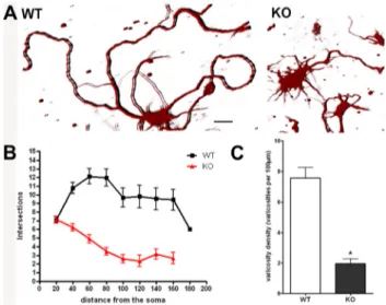
Figure 1. Neurite branching and varicosities are reduced in p50⫺/⫺cortical neurons. Re-duced complexity and varicosity density of neuronal processes in p50⫺/⫺(KO) compared with wild-type (WT) cortical cells. A, Representative 3D images of cortical neurons from WT (left) and KO mice (right) stained with anti-III-Tubulin antibody. Scale bar, 20 m. B, Graphic repre-sentation of branching quantification performed using the Sholl profile analysis. Data are ex-pressed as mean⫾ SEM. C, Quantification of varicosity loss in KO neurons compared with WT. Varicosity density was measured counting the number of varicosities in 100mneuritelength. Data are expressed as mean⫾ SEM. *p ⬍ 0.0001.
products specificity. Gene expression levels are presented as fold change in target gene expression in the test (p50⫺/⫺cells) normalized to the internal control gene (GAPDH) and relative to the calibrator (wt cells). Results were estimated as Ct values; the⌬Ct was calculated as the mean of
the Ct for the target gene minus the mean of the Ct for the internal control gene. The⌬⌬Ct represented the mean difference between the ⌬Ct of the test minus the ⌬Ct of the calibrator. The N-fold differential expression in the target gene of the test compared with calibrator was expressed as 2⫺⌬⌬Ct. Data analysis and graphics were performed using GraphPad Prism 4 software and were the results of a single experiment run in triplicate for each gene.
Western blots. Protein extracts were prepared from 7 DIV cortical cells from wt and p50⫺/⫺embryos. Western blotting was performed using 6 –12% SDS polyacrylamide gels with 20 –50g of protein ex-tract loaded per lane. Nitrocellulose filters were incubated with pri-mary antibodies raised against Notch1 and Jagged1 overnight at 4°C, followed by HRP-conjugated secondary antibodies. Membranes were stripped and reprobed with anti-GAPDH antibody. Densitometric analysis was performed using Quantity One software system (Bio-Rad), and each band was normalized to GAPDH signal in each lane
and expressed as percentage.
Cell treatments. Ten micromolar␥-secretase
inhibitor N-[N-(3,5-difluorophenacetyl)-1-alanyl 1]-S-phenylglycine t-butyl ester (DAPT) (Calbiochem/EMD Biosciences) was added to the culture medium of 4 DIV cortical cells, and cells were analyzed after 72 h. The DAPT treat-ment resulted in 34% reduction in Notch1 pro-tein levels and 48% reduction in NICD propro-tein levels.
siRNA probes targeted to Notch1 receptor were purchased from Dharmacon. The mouse-specific Notch1 interference was performed using an Accell SMARTpool siRNA mixture (DHE-041110-00) containing a mixture of four siRNAs targeting the Notch1 gene (NM_ 008714). A nontargeting Accell siRNA pool (DHD-001910-10) was used as a control in all siRNA transfection experiments. Primary cor-tical neurons were transfected with Accell siRNAs in Neurobasal-B27 medium to preserve cell viability. Primary cortical neurons were treated with Accell siRNA probes after 4 DIV. Cells were cultured for 3 additional days after transfection with 1MsiRNA, according to the manufacturer’s instructions, then treated, lysed, and subsequently analyzed for mRNA and pro-tein contents. siRNA treatment effectiveness and specificity were validated by measuring Notch1 mRNA levels and using a scrambled siRNA (siRNA⫺).ThesiRNAtreatmentresultedin58% reduction in Notch1 mRNA levels, 48% reduc-tion in Notch1 protein levels, and 30% reducreduc-tion in NICD protein, compared with vehicle-treated cells.
NF-B SN-50 cell-permeable inhibitor pep-tide (Calbiochem/EMD Biosciences) was added to the culture medium at the concentration of 10 Mfor 48 h. To prove SN-50 specificity, a mutant SN-50 (SN-50 mut) (Calbiochem/EMD Biosci-ences) was used at the concentration of 10 Mfor 48 h.
TNF-␣ (Invitrogen) was added to the cul-ture medium at the concentration of 10 ng/ml for 24 h.
Chromatin immunoprecipitation analysis. Chromatin immunoprecipitation (ChIP) anal-ysis was performed essentially as described by Lanni et al. (2010). Protein complexes were cross-linked to DNA in living cells by adding formaldehyde directly to the cell culture medium at 1% final concentra-tion. Chromatin extracts containing DNA fragments with an average size of 300 bp were incubated overnight at 4°C with milk shaking using
poly-Figure 2. Notch1 pathway is upregulated in p50⫺/⫺cortical cells. Q-RT-PCR was per-formed using RNA extracted from WT and KO cortical cells to evaluate Notch1, Jagged1, and HES1 expression levels. Data are expressed as fold change of target gene expression in WT and KO cortical cells, normalized to the internal control gene (GAPDH). Data were analyzed accord-ing to the comparative Ct method.
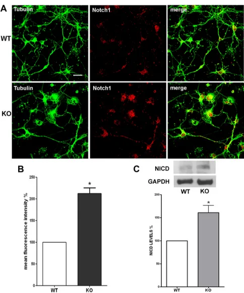
Figure 3. Notch1 protein expression levels are increased in p50⫺/⫺cortical cells. A, Representative confocal images of cortical cells stained with anti-III-Tubulin (green) and anti-Notch1 (red) antibodies. Pictures show an increase of Notch1-positive stain-ing in KO compared with WT cortical cells. Scale bar, 20m. B, Quantification of Notch1 immunostaining fluorescence intensity. Values are expressed as percentage of mean fluorescence intensity (% mean⫾ SEM) and are from at least 40 cellular fields. *p ⬍ 0.001 versus WT. C, Representative immunoblot of WT and KO cortical cell lysates using an anti-Notch1 antibody. Graph: Densito-metric analysis of NICD levels measured by Western blot in WT and KO cortical cells. Data are normalized to the GAPDH signal, expressed as mean⫾ SEM, and are obtained from three experiments run with two different cell preparations. *p ⬍ 0.01.
clonal anti-p50 and anti-p65 antibodies (Santa Cruz). DNA–protein complexes were recovered with protein A/G-agarose (Santa Cruz). Before use, protein A/G was blocked with 1 g/l sheared herring sperm DNA and 1g/l BSA overnight at 4°C, and then incubated with chro-matin and antibodies for 3 h at 4°C. PCR was performed using immunoprecipitated DNA and promoter-specific primers flanking the NF-B site for Jagged1 (M-Medical) and specific primers for 36B4, used as housekeeping gene (kind gift from Dr. Delbarba, University of Milano, Milano, Italy). Primer sequences used were as fol-lows: Jagged1 promoter, forward primer, 5⬘-TTC AGG GGT GAT CAA GGA AG-3⬘, reverse primer, 5⬘-TGG CAT ACT GGG AAT GTC AA-3⬘; 36B4, forward primer, 5⬘-AGG ATA TGG GAT TCG GTC TCT TC-3⬘, reverse primer, 5⬘-TCA TCC TGC TTA AGT GAA CAA ACT-3⬘. Immunoprecipitation with nonspecific Igs (no Ab) was performed as negative controls. PCR products were run on a 2.5% agarose gel and vi-sualized with ethidium bromide staining using UV light. The amount of precipitated chromatin measured in each PCR was normalized with the amount of chromatin present in the input of each immunoprecipitation.
Statistical analysis. A t test analysis or one-way ANOVA followed by Bonferroni’s multiple-comparison test as post hoc analysis was per-formed. Data are expressed as mean⫾ SEM of n⫽ 3–5 experiments. A value of p ⬍ 0.05 was considered to be statistically significant.
Results
Neurite branching and varicosities are
reduced in p50
ⴚ/ⴚcortical neurons
Previous studies demonstrated that NF-B
signaling is involved in regulation of neurite
outgrowth, size, and complexity of cortical
neuron arborizations (Gutierrez et al., 2005,
2008; Gavalda` et al., 2009). To gain insight
into the role of NF-
B in neuronal
differen-tiation, we have taken advantage of the
availability of mice carrying a homozygous
deletion of the NF-B1 gene encoding the
p50 subunit (Sha et al., 1995; Denis-Donini et al., 2008). Brain
cor-tical neuroblasts were isolated from E15 wt and p50
⫺/⫺embryos.
Cells were grown on poly-
D-lysine-coated glass coverslips and
main-tained in culture for 7 d. Cells were then smain-tained with an
anti-III-Tubulin antibody. Immunoreactivity and morphology were
analyzed by confocal microscopy. We observed a remarkable
differ-ence in neurite branching of wt and p50
⫺/⫺cultures. As shown in a
representative image in Figure 1A, cortical cells from wt mice
showed a complex and extended neuritic network, reminiscent of
mature and differentiated cortical neurons. Conversely, the p50
⫺/⫺derived cortical cells displayed short neurites and a poor branching
phenotype (Fig. 1A, right panel). We then performed Sholl analysis,
a valuable and widely used method for quantifying the extent and
complexity of neuronal processes (Sholl, 1953; Gutierrez and Davies,
2007). An overall reduction in the spatial complexity of the neuritic
arborizations was demonstrated in p50
⫺/⫺derived cortical cells
(Fig. 1B), with a significant decrease in neurite length and number of
bifurcations when compared with wt derived cells.
The presence of varicosities is a well recognized
morphologi-cal feature of differentiated cortimorphologi-cal cells. They are regarded as
presynaptic, dynamic structures that are able to remodel their
morphology in response to a variety of stimuli (De Paola et al.,
2003; Nikonenko et al., 2003; Udo et al., 2005; Umeda et al., 2005;
Ferrari-Toninelli et al., 2009). Varicosities appear as membrane
swellings of various size. They can represent more stable
struc-tures, giving origin to secondary branching neurites,
filopodia-like structures, or en passant varicosities, fast-turnover structures
containing secretory granules (De Paola et al., 2003). We
com-pared the presence of varicosities in wt and p50
⫺/⫺cortical cells.
Wt cortical cells in culture appeared to have long, thin, and
var-icose neurites, whereas p50
⫺/⫺cortical cell neurites were short,
thicker, and “smooth,” since they were lacking varicosities. We
quantified these differences by measuring the density of
varicos-ities in 100
m neurite length. Varicosity densities were 7.57 ⫾
0.68 and 1.96
⫾ 0.33 in wt and p50
⫺/⫺derived cortical cells,
respectively (Fig. 1C).
Hyperactivation of Notch1 pathway in absence of p50 subunit
The morphological features observed in cortical neurons from
p50-deficient mice were highly reminiscent of cortical cells in
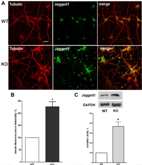
Figure 4. Jagged1 protein expression levels are increased in p50⫺/⫺cortical cells. A, Representative confocal images of cortical cells stained with anti-III-Tubulin (red) and anti-Jagged1 (green) antibodies. Pictures show increased Jagged1 immunostaining in KO com-paredwithWTcorticalcells.Scalebar,20m.B,QuantificationofJagged1immunostainingfluorescenceintensity.Valuesareexpressedas percentage of mean fluorescence intensity (% mean⫾ SEM) and are from at least 40 cellular fields. *p ⬍ 0.001. C, Representative immunoblot of cortical cell lysates obtained from WT and KO mice using an anti-Jagged1 antibody. Graph: Densitometric analysis of Jagged1 levels measured by Western blot in WT and KO cortical cells. Data are normalized to the GAPDH signal, expressed as mean⫾SEM, and are obtained from three experiments run with two different cell preparations. *p⬍0.0001.
cultures after Notch pathway stimulation by a synthetic Jagged1
ligand (Ferrari-Toninelli et al., 2008). We therefore investigated
by quantitative real-time PCR (Q-RT-PCR) the expression levels
of three major participants in the Notch signaling pathway,
namely the Notch1 receptor, its ligand Jagged1, and the
down-stream bHLH family member HES1, a Notch1 target gene
com-monly used to detect Notch1 pathway activation (Iso et al., 2003),
in wt and p50
⫺/⫺cortical cells. As reported in Figure 2, Notch1
and HES1 mRNA levels were increased by 83 and 73% in p50
⫺/⫺compared with wt cultures. Remarkably, Jagged1 mRNA levels
increased by 17-fold in p50
⫺/⫺compared with wt cultures.
Notch1 and Jagged1 protein expression was then evaluated by
immunofluorescence. Cortical cells were double-stained with an
anti-Notch1 antibody, which recognizes both full-length Notch
and its C-terminal intracellular fragment (NICD), and an
anti-III-Tubulin antibody. After immunostaining, we analyzed at
least 40 fields (15–20 cells/field), derived from three different
culture preparations. As shown in Figure 3A, Notch1
immuno-reactivity appeared to be increased in p50
⫺/⫺compared with wt
cortical cells, considering both the number of Notch1-positive
nuclei and the overall fluorescence intensity (Fig. 3A, merge). The
total fluorescence intensity was quantified and expressed as
per-centage of mean fluorescence intensity. As reported in Figure 3B,
fluorescence intensity was increased by twofold in p50
⫺/⫺com-pared with wt cultures. Western blot analysis using a specific
anti-NICD antibody further supported the finding of an
in-creased Notch pathway activation in p50
⫺/⫺cortical cells. As
reported in Figure 3C, NICD protein levels were significantly
increased (approximately
⫹60%) in p50
⫺/⫺compared with wt
derived cortical cells.
Similarly, to evaluate Jagged1 protein levels, wt and p50
⫺/⫺cortical cells were double-stained with an anti-Jagged1 and an
anti-III-Tubulin antibodies (Fig. 4A). Fluorescence intensity of
Jagged1 immunostaining was quantified, and data were reported
in Figure 4 B. Fluorescence intensity was increased by 127% in
p50
⫺/⫺compared with wt cortical cells. Data were confirmed by
Western blot analysis: the results from three different
experi-ments demonstrated a significant increase (
⫹269%) in Jagged1
level in p50
⫺/⫺cultures (Fig. 4C).
Overall, these results demonstrate that Notch1 receptor,
NICD, and Jagged1 are significantly upregulated in the absence of
p50 subunit.
Notch1 pathway inhibition restores neurite branching and
varicosities in p50
ⴚ/ⴚcortical cells
To investigate whether the observed morphological features of
p50
⫺/⫺cortical neurons were correlated to Notch1 pathway
hy-peractivation, the Notch1 pathway was inhibited either
pharma-cologically, using the
␥-secretase inhibitor DAPT (Kanungo et
al., 2008), or genetically, using the Notch1 siRNA approach
(siRNA NOTCH).
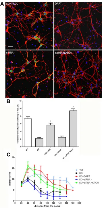
Notch pathway inhibition by DAPT or siRNA NOTCH resulted
in dramatic changes in p50
⫺/⫺cortical neuron morphology. In
par-ticular, as shown by representative pictures in Figure 5A, 3-d-long
treatment with DAPT or siRNA NOTCH (but not vehicle or
scram-bled siRNA) resulted in increased neurite branching and varicosities
in p50
⫺/⫺cells that became indistinguishable from wt cortical
neurons. Varicosity density and branching were quantified in
DAPT/siRNA NOTCH-treated p50
⫺/⫺cells and compared with
those of p50
⫺/⫺vehicle or scrambled siRNA-treated cells.
Quan-titative data confirmed increased varicosity density (Fig. 5B) and
branching (Fig. 5C) in DAPT and siRNA NOTCH-treated
p50
⫺/⫺cortical neurons. Together, these data suggest that
mor-phological abnormalities in p50
⫺/⫺cortical neurons are
depen-dent on Notch pathway hyperactivation.
No morphological changes were observed in wt cortical
neu-rons treated with either DAPT or siRNA Notch (data not shown).
p50 NF-
B subunit is directly involved in Notch pathway
modulation
NF-
B signaling was acutely manipulated using SN-50 peptide
(Lin et al., 1995). Treatment of wt cells with 10
MSN-50 for 48 h
Figure 5. Notch1 blockade restores neurite branching and varicosities in p50⫺/⫺cortical neu-rons. A, Representative confocal images of cortical cells stained with anti-III-Tubulinantibody(red), DAPI (blue), and anti-Notch1 antibody (green). KO cortical neurons were exposed to the␥-secretase inhibitor DAPT, the siRNA NOTCH, or the scrambled siRNA probe (siRNA⫺)for72h.Scalebar,20m. B, Quantification of varicosity density in WT and KO neurons after different treatments, as indicated. Varicosity density was measured counting number of varicosities in 100mneuritelength.Dataare expressed as mean⫾ SEM. *p ⬍0.001 versusKO.C, Graphicrepresentationof branching quantifi-cation performed using the Sholl profile analysis in WT and KO neurons after different treatments, as indicated. Data are expressed as mean⫾SEM.
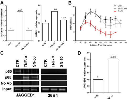
resulted in increased Jagged1 (⫹191%) and
Notch1 (
⫹66%) mRNA levels, compared
with vehicle- or mutated SN-50 (SN-50
mut)-treated cells (Fig. 6A). Branching
studies by Sholl analysis revealed that SN-50
significantly reduced neurite length and
complexity in wild-type neurons (Fig. 6B).
Also, varicosities were reduced after SN-50
treatment: varicosity densities were 7.53
⫾
0.41, 8.24
⫾ 0.42, and 3.00 ⫾ 0.26 in
vehicle-, SN-50 mut-, and SN-50-treated
cortical cells, respectively.
We evaluated the role of p50 in
regulat-ing Jagged1 expression in mouse neuronal
cortical cells using the ChIP approach. In
line with previous data (Johnston et al.,
2009), we found that vehicle-treated cells
yielded a strong signal for p50 and only a
weak p65 signal on the Jagged1 promoter
(Fig. 6C). In sharp contrast to control cells,
this ratio is reversed in TNF-treated cells, in
which we found a weak p50 signal and a
strong p65 signal, correlating with the
higher transcriptional activity of the Jagged1
gene in TNF-treated cells (Fig. 6D). A
simi-lar pattern of results was obtained in cells
treated with SN-50. Together, the TNF-␣
and SN-50 results suggest that, in resting
cells, the NF-B site is likely occupied
mostly by p50 homodimers, whereas in
TNF-␣- or SN-50-treated cells there is a
shift toward p65-containing complexes, and
this event is associated with enhanced
Jag-ged1 transcription. As it is not entirely clear
what the consequences of deletion of p50
throughout embryogenesis may have for
NF-B signaling in cortical neurons, we
evaluated p65 protein levels in p50
⫺/⫺and
wt cortical neurons. We found that p50
⫺/⫺neurons expressed significantly higher protein level (
⫹152%)
com-pared with wt.
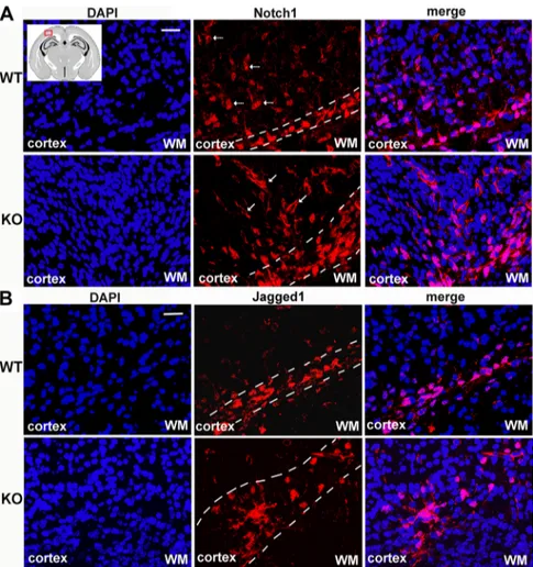
Localization of Notch1- and Jagged1-immunoreactive
neurons in p50
ⴚ/ⴚP1 mice cortex
Finally, we investigated whether Notch1 hyperactivation observed in
p50
⫺/⫺embryonal cortical neurons was also observed in vivo. To
this purpose, brains from P1 wt and p50
⫺/⫺mice were collected,
fixed, cut in 15-
m-thick coronal sections, and immunostained for
Notch1 and Jagged1 proteins. In P1 wt mice, Notch1
immunoreac-tivity was detected in the basal zone of the motor/somatosensory
cortex and gradually decreased in intensity toward the upper cortical
layers, as shown in the representative images in Figure 7A, top panel.
Conversely, in p50
⫺/⫺brain sections, Notch1-immunoreactive cells
were detected throughout all cortical layers (Fig. 7A, bottom panel).
A similar expression pattern was also observed for Jagged1
immu-nofluorescence in p50
⫺/⫺compared with wt brains:
Jagged1-positive cells were mainly localized in the basal cortical layer in P1 wt
brain sections, whereas in p50
⫺/⫺mice, Jagged1-positive cells were
spread throughout the upper layers (Fig. 7B).
Discussion
In the CNS, activation of NF-
B has been reported to be involved
in neuronal cell differentiation and survival (Maggirwar et al.,
1998; Kova´cs et al., 2004), inflammatory response (Li and Verma,
2002), neurogenesis (Denis-Donini et al., 2005, 2008),
prolifera-tion, and apoptosis (Grilli and Memo, 1999; Kucharczak et al.,
2003). More recently, emerging data demonstrated its crucial
involvement in regulating the growth and complexity of
neuro-nal arborizations (Gutierrez et al., 2005; Gavalda` et al., 2009;
Russo et al., 2009; Gutierrez and Davies, 2011) and in synaptic
plasticity and memory in the adult brain (Albensi and Mattson,
2000; Kaltschmidt et al., 2006). Nevertheless, the mechanisms by
which NF-B may exert such a role is largely unknown.
p50
⫺/⫺mice were chosen as an experimental model to
inves-tigate the contribution of NF-
B in regulating cortical cell
mor-phology and to identify the intracellular pathway(s) involved in
such effects. p50
⫺/⫺mice have been extensively used in an
at-tempt to clarify the role of NF-B in developing, adult, and aged
nervous system. Indeed, this mouse model has been shown to
display accelerated aging (Lu et al., 2006) and a dramatic
impair-ment in hippocampal neurogenesis linked to a selective cognitive
deficit in hippocampal-dependent spatial short-term memory
(Denis-Donini et al., 2008). Mice lacking p50 subunit also display
an increased exploratory activity and reduced anxiety behavior
(Kassed and Herkenham, 2004).
In the present study, measuring parameters commonly used
to define the stage of neuron differentiation during brain
devel-Figure 6. Acute NF-B signaling manipulation induces Jagged1 upregulation and morphological changes. A, Q-RT-PCR was performed on cDNA from WT cortical cells after 10MSN-50 or SN-50 mut treatment at 48 h. Data are presented as fold change of target gene expression normalized to the internal control gene (GAPDH). Data were analyzed according to the comparative Ct method. SN-50 treatment resulted in an upregulation of Jagged1 mRNA expression compared with the vehicle-treated cells (CTR). An SN-50 mutant peptide (SN-50 mut) was used to validate SN-50 specificity. B, Graphic representation of branching quantifica-tion performed using the Sholl profile analysis in WT neurons after SN-50 treatment. Data are expressed as mean⫾ SEM. C, ChIP experiments were performed with anti-p50 and anti-p65 antibodies on WT cortical neurons treated with 10 ng/ml TNF-␣for24h or 10MSN-50 for 48 h. PCR analysis was performed on the immunoprecipitated DNA samples using specific primers for the Jagged1 promoter. A sample representing linear amplification of the total input chromatin (Input) was included as control. Additional controls included immunoprecipitation performed with nonspecific Igs (no Ab). Amplification of 36B4 housekeeping gene was used as control of p50 and p65 binding specificity to the Jagged1 promoter. D, Q-RT-PCR was performed on cDNA from WT cortical cells after 24 h of TNF-␣treatment(10ng/ml).Dataarepresentedasfoldchangeoftargetgeneexpressionnormalized to the internal control gene (GAPDH). Data were analyzed according to the comparative Ct method. TNF-␣treatmentresultedinan upregulation of Jagged1 mRNA expression compared with the vehicle-treated cells (CTR).
opment, we unraveled marked morphological and biochemical
differences between wt and p50
⫺/⫺cortical cells in culture. We
demonstrated that neuritic branching and varicosity density were
significantly reduced in p50
⫺/⫺neurons. Our results on the
NF-
B role in neurite growth regulation are in line with data
from Gutierrez et al. (2007, 2008) in nodose and sensory neurons.
These authors reported that different protein subunits contribute
to NF-B-mediated effect on neurites, and, in particular, they
described a remarkable inhibitory effect of SN-50, a peptide that
interferes with p50 nuclear translocation, on neuritic branching.
We now further extend these observations, demonstrating the
role of NF-B on neuronal branching and differentiation also in
cortical neurons.
We were interested in investigating the mechanism by which
NF-
B is able to act on neuritic morphology. Using primary
murine hippocampal cultures, Salama-Cohen et al. (2005)
pro-vided evidence that Notch and NF-
B signaling pathways
con-verge at the level of Hes1/5 to regulate dendritic growth. In
previous studies, we characterized the effects of Notch1 in
re-modeling neuronal processes, analyzing its effect on
cytoskele-ton, varicosities, and neurotransmitter release (Ferrari-Toninelli
et al., 2008, 2009). Surprisingly, the reduced varicosity density
and neurite ramifications observed in
p50
⫺/⫺neurons strongly resembled the
morphology seen in cortical neurons or
differentiated SH-SY5Y cells after Notch1
receptor stimulation by its ligand Jagged1.
Since the relevance of Notch signaling in
development and synaptic transmission,
it appeared reasonable to analyze the
Notch1 pathway in p50
⫺/⫺cortical cells.
Notch1 is well known for its
involve-ment in important cellular processes like
neurogenesis (Artavanis-Tsakonas et al.,
1999), cell fate determination (Louvi and
Artavanis-Tsakonas, 2006), and neural
stem cell maintenance in the adult brain
(Imayoshi et al., 2010). As recently
re-viewed by Ables et al. (2011), several
stud-ies suggested that Notch signaling may
serve important functions in the
regula-tion of neurite outgrowth and plasticity.
Both Sˇestan et al. (1999) and Berezovska
et al. (1999) found that Notch1 activation
inhibits neurite outgrowth or causes their
retraction, whereas Notch1 signaling
in-hibition promotes neurite extension.
Redmond et al. (2000) demonstrated that
NICD translocates to the nucleus during
neuronal differentiation where it
regu-lates the expression of genes whose
prod-ucts influence dendritic morphology.
Here, we collected evidence indicating
that the Notch signaling is hyperactivated
in p50
⫺/⫺cortical neurons. Remarkably,
Jagged1 transcript was increased by
17-fold in p50
⫺/⫺cells compared with wt
cells, suggesting that it might be directly
affected by the lack of p50 subunit. We
manipulated acutely NF-
B signaling in
wt neurons. Specifically, wt cortical
cul-tures were treated with SN-50 and
re-sulted in a significant increase of both
Jagged1 and Notch1 mRNA levels. SN-50-treated cells also
dis-played a significant reduction in neurite branching and varicosity
density. Using the ChIP approach, we found that, in resting cells,
the NF-B site within Jagged1 promoter is occupied mostly by
p50 homodimers, whereas in TNF-
␣ or SN-50 treated cells there
is a shift toward p65-containing complexes, which correlates with
enhanced Jagged1 transcription. This is in line with the data by
Johnston et al. (2009) demonstrating that, in human endothelial
cells, Jagged1 transcription is repressed by p50 homodimers. Our
data suggest that, in wt culture system, p50-containing dimers
may act as repressor of Jagged1 gene expression. A vast array of
information is available in the literature (Zhong et al., 2002;
Driessler et al., 2004; Grundstro¨m et al., 2004) describing the
specific role of p50 homodimers as transcriptional repressors
within the family.
We propose Jagged1 as one of the major players in Notch
pathway hyperactivation. Its interaction with the receptor may
indeed result in massive translocation of NICD to the nucleus
and thereby in increased transcription of the Notch target gene
HES1. HES1 is a Notch1-responsive bHLH family member
im-plicated in controlling the maintenance of undifferentiated cells
and timing of cell differentiation (Akimoto et al., 2010). Since
Figure 7. In vivo increase of Notch1- and Jagged1-positive neurons in p50⫺/⫺mouse cortex. Representative confocal images of cerebral cortex from P1 WT and KO mouse brain sections stained with anti-Notch1 (red) or anti-Jagged1 (red) antibody and DAPI (blue). Inset, Scheme of a P1 mouse brain section, with the indication (red mark) of the region of interest. A, Top panel, Represen-tative images of WT cortex with Notch1-positive neurons localized mainly in the basal layer and few Notch1-stained cells in the upper cortical layers (arrows). Bottom panel, Representative images of KO mouse cortex with Notch1-expressing neurons both in the basal cortical layer and in the whole cortical stratifications (arrows). B, Top panel, Representative images of WT cortex with positive neurons localized in the basal layer. Bottom panel, Representative images of KO mouse cortex with Jagged1-expressing neurons that exceed the basal layer and spread through other cortical layers. WM, White matter. Scale bar, 20m.
different Notch receptors and ligands have been described, we
cannot exclude the possible involvement of other members of the
Notch pathway in this effect. However, in our in vitro model the
increased NICD nuclear levels and HES1 expression
unequivo-cally testify for a Notch pathway hyperactivity.
We were then interested in evaluating the possible functional
link between morphological features and Notch pathway activity.
To test this hypothesis, the Notch pathway was inhibited and
morphology of cortical neurons was studied. Two different
ap-proaches were undertaken: a pharmacological treatment with the
␥-secretase inhibitor DAPT, and Notch1 gene silencing. DAPT is
a well known Notch1 pharmacological inhibitor (Sastre et al.,
2001; Geling et al., 2002; Crawford and Roelink, 2007),
prevent-ing Notch1 intracellular cleavage, which is required for NICD
nuclear translocation and signaling activation. DAPT exerts a
strong inhibitory effect on Notch1 activation, but it has poor
selectivity, because of the high number of
␥-secretase substrates
(Lleo´, 2008). siRNA NOTCH is a Notch1 gene interference tool
that specifically downregulates Notch1 transcription. Both
ex-perimental treatments resulted effective in inhibiting the Notch1
signaling. Interestingly, treatment of p50
⫺/⫺cortical cells with
either DAPT or siRNA NOTCH was able to recover varicosities
loss and increase neurite branching. These findings indicated that
the morphological alterations present in cortical neurons lacking
the NF-B p50 subunit are dynamic and reversible. More
impor-tantly, these results also suggested that Notch hyperactivation
mediated the morphological features observed in the absence of
the NF-
B p50 subunit.
Notch effects on neurite plasticity might be relevant to the
generation of highly refined neuronal circuits (Luo and O’Leary,
2005). Axonal and dendrite architecture is indeed responsible for
polarization and migration of neuronal cells and crucial for
cor-rect lamination during cortex development. The analysis of
p50
⫺/⫺and wt mouse brains, at postnatal day 1, revealed an
altered distribution of Notch1- and Jagged1-positive cells in the
cortex. In control brains, Notch1- and Jagged1-positive cells were
uniformly localized in the basal layer of the cortex, with few cells
in the upper layers. Conversely, p50
⫺/⫺mice showed an
in-creased number of both Notch1- and Jagged1-expressing cells,
with a distribution not only in basal but also in upper layers. In
this regard, it should be noted that the Notch pathway has been
found to be implicated in cortical neuron migration and in
cor-tical layering, mainly through the cross talk with the Reelin
sig-naling pathway (Gaiano, 2008; Hashimoto-Torii et al., 2008).
Although young and adult p50
⫺/⫺mouse brains do not show
overt structural alterations, our data suggest that future studies
will be needed to address the possibility that in absence of p50
very subtle defects may occur in neuronal networks and in
par-ticular in cortex and that they may potentially contribute to
be-havioral alterations.
In summary, we found that lack of p50, either induced acutely
by inhibiting p50 nuclear translocation in wt cortical cells or
genetically in p50
⫺/⫺mice, results in cortical neurons
character-ized by reduced neurite branching, loss of varicosities, and
Notch1 signaling hyperactivation. The neuronal morphological
effects caused by lack of p50 were reverted by inhibition of Notch
signaling.
A direct link between Notch and NF-
B pathways is also
sup-ported by ChIP data showing that activation of Jagged1 gene
tran-scription is associated with removal of p50 homodimers bound to
the NF-B-responsive element in the Jagged1 promoter region.
These data demonstrate that a cross talk between NF-
B and
Notch1 signaling in primary neuronal cortical cells does exist.
Our data also support a role for this interaction in the regulation
of cortical neuron remodeling.
References
Ables JL, Breunig JJ, Eisch AJ, Rakic P (2011) Not(ch) just development: Notch signalling in the adult brain. Nat Rev Neurosci 12:269 –283. Akimoto M, Kameda Y, Arai Y, Miura M, Nishimaki T, Takeda A, Uchinuma
E (2010) HES1 is required for the development of craniofacial structures derived from ectomesenchymal neural crest cells. J Craniofac Surg 21:1443–1449.
Albensi BC, Mattson MP (2000) Evidence for the involvement of TNF and NF-B in hippocampal synaptic plasticity. Synapse 35:151–159. Ang HL, Tergaonkar V (2007) Notch and NF-B signaling pathways: do
they collaborate in normal vertebrate brain development and function? Bioessays 29:1039 –1047.
Artavanis-Tsakonas S, Delidakis C, Fehon R, Hartley D, Herndon D, Johan-sen K, Markopoulou K, Preiss A, Rebay I, Scottgale N (1990) Notch and the molecular genetics of neuroblast segregation in Drosophila. Mol Re-prod Rev 27:23–27.
Artavanis-Tsakonas S, Rand MD, Lake RJ (1999) Notch signaling: cell fate control and signal integration in development. Science 284:770 –776. Bash J, Zong WX, Banga S, Rivera A, Ballard DW, Ron Y, Ge´linas C (1999)
Rel/NF-B can trigger the Notch signaling pathway by inducing the ex-pression of Jagged1, a ligand for Notch receptors. EMBO J 18:2803–2811. Berezovska O, McLean P, Knowles R, Frosh M, Lu FM, Lux SE, Hyman BT (1999) Notch1 inhibits neurite outgrowth in postmitotic primary neu-rons. Neuroscience 93:433– 439.
Cao Q, Li P, Lu J, Dheen ST, Kaur C, Ling EA (2010) Nuclear factor-B/p65 responds to changes in the Notch signaling pathway in murine BV-2 cells and in amoeboid microglia in postnatal rats treated with the␥-secretase complex blocker DAPT. J Neurosci Res 88:2701–2714.
Crawford TQ, Roelink H (2007) The Notch response inhibitor DAPT en-hances neuronal differentiation in embryonic stem cell-derived embryoid bodies independently of sonic hedgehog signaling. Dev Dyn 236: 886 – 892.
de la Pompa JL, Wakeham A, Correia KM, Samper E, Brown S, Aguilera RJ, Nakano T, Honjo T, Mak TW, Rossant J, Conlon RA (1997) Conserva-tion of the Notch signaling pathway in mammalian neurogenesis. Devel-opment 124:1139 –1148.
Denis-Donini S, Caprini A, Frassoni C, Grilli M (2005) Members of the NF-B family expressed in zones of active neurogenesis in the post-natal and adult mouse brain. Brain Res Dev Brain Res 154:81– 89.
Denis-Donini S, Dellarole A, Crociara P, Francese MT, Bortolotto V, Qua-drato G, Canonico PL, Orsetti M, Ghi P, Memo M, Bonini SA, Ferrari-Toninelli G, Grilli M (2008) Impaired adult neurogenesis associated with short-term memory defects in NF-B p50-deficient mice. J Neurosci 28:3911–3919.
De Paola V, Arber S, Caroni P (2003) AMPA receptors regulate dynamic equilibrium of presynaptic terminals in mature hippocampal networks. Nat Neurosci 6:491–500.
Driessler F, Venstrom K, Sabat R, Asadullah K, Schottelius AJ (2004) Mo-lecular mechanisms of interleukin-10-mediated inhibition of NF-B ac-tivity: a role for p50. Clin Exp Immunol 135:64 –73.
Espinosa L, Ingle´s-Esteve J, Robert-Moreno A, Bigas A (2003) IB␣ and p65 regulate the cytoplasmic shuttling of nuclear corepressors: cross-talk be-tween Notch and NFB pathways. Mol Biol Cell 14:491–502.
Ferrari-Toninelli G, Bonini SA, Bettinsoli P, Uberti D, Memo M (2008) Mi-crotubule stabilizing effect on Notch activation in primary cortical neu-rons. Neuroscience 154:946 –952.
Ferrari-Toninelli G, Bonini SA, Uberti D, Napolitano F, Stante M, Santoro F, Minopoli G, Zambrano N, Russo T, Memo M (2009) Notch activation induces neurite remodeling and functional modifications in SH-SY5Y neuronal cells. Dev Neurobiol 69:378 –391.
Gaiano N (2008) Strange bedfellows: Reelin and Notch signaling interact to regulate cell migration in the developing neocortex. Neuron 60:189 –191. Gavalda` N, Gutierrez H, Davies AM (2009) Developmental switch in NF-B
signaling required for neurite growth. Development 136:3405–3412. Geling A, Steiner H, Willem M, Bally-Cuif L, Haass C (2002) A
gamma-secretase inhibitor blocks Notch signaling in vivo and causes a severe neurogenic phenotype in zebrafish. EMBO Rep 3:688 – 694.
Grilli M, Memo M (1999) Nuclear factor-B/Rel proteins. Biochem Phar-macol 57:1–7.
Grundstro¨m S, Anderson P, Scheipers P, Sundstedt A (2004) Bcl-3 and NFB p50–p50 homodimers act as transcriptional repressors in tolerant CD4⫹ T cells. J Biol Chem 279:8460–8468.
Gutierrez H, Davies AM (2007) A fast and accurate procedure for deriving the Sholl profile in a quantitative studies of neuronal morphology. J Neu-rosci Methods 163:24 –30.
Gutierrez H, Davies AM (2011) Regulation of neural process growth, elab-oration and structural plasticity by NF-B. Trends Neurosci 34:316–325. Gutierrez H, Hale VA, Dolcet X, Davies A (2005) NF-B signaling regulates the growth of neural processes in the developing PNS and CNS. Develop-ment 132:1713–1726.
Gutierrez H, O’Keeffe GW, Gavalda` N, Gallagher D, Davies AM (2008) Nuclear factor B signaling either stimulates or inhibits neurite growth depending on the phosphorylation status of p65/RelA. J Neu-rosci 28:8246 – 8256.
Hashimoto-Torii K, Torii M, Sarkisian MR, Bartley CM, Shen J, Radtke F, Gridley T, Sˇestan N, Rakic P (2008) Interaction between Reelin and Notch signaling regulates neuronal migration in the cerebral cortex. Neu-ron 60:273–284.
Imayoshi I, Sakamoto M, Yamaguchi M, Mori K, Kageyama R (2010) Es-sential roles of Notch signaling in maintenance of neural stem cells in developing and adult brains. J Neurosci 30:3489 –3498.
Iso T, Kedes L, Hamamori Y (2003) HES and HERP families: multiple ef-fectors of the Notch signaling pathway. J Cell Physiol 194:237–255. Johnston DA, Dong B, Hughes CC (2009) TNF induction of jagged-1 in
endothelial cells is NF-B-dependent. Gene 435:36–44.
Kaltschmidt B, Kaltschmidt C (2009) NF-B in the nervous system. Cold Spring Harb Perspect Biol 1:a001271.
Kaltschmidt B, Ndiaye D, Korte M, Pothion S, Arbibe L, Pru¨llage M, Pfeiffer J, Lindecke A, Staiger V, Israe¨l A, Kaltschmidt C, Me´met S (2006) NF-B regulates spatial memory formation and synaptic plasticity through pro-tein kinase A/CREB signaling. Mol Cell Biol 26:2936 –2946.
Kanungo J, Zheng YL, Amin ND, Pant HC (2008) The Notch signaling inhibitor DAPT down-regulates cdk5 activity and modulates the distri-bution of neuronal cytoskeletal proteins. J Neurochem 106:2236 –2248. Kassed CA, Herkenham M (2004) NF-B p50-deficient mice show reduced
anxiety-like behaviours in test of exploratory drive and anxiety. Behav Brain Res 154:577–584.
Kova´cs AD, Chakraborty-Sett S, Ramirez SH, Sniderhan LF, Williamson AL, Maggirwar SB (2004) Mechanism of NF-B inactivation induced by survival signal withdrawal in cerebellar granule neurons. Eur J Neurosci 20:345–352.
Kucharczak J, Simmons MJ, Fan Y, Ge´linas C (2003) To be, or not to be: NF-B is the answer. Role of Rel/NF-B in the regulation of apoptosis. Oncogene 22:8961– 8982.
Lanni C, Nardinocchi L, Puca R, Stanga S, Uberti D, Memo M, Govoni S, D’Orazi G, Racchi M (2010) Homeodomain interacting protein kinase 2: a target for Alzheimer’s beta amyloid leading to misfolded p53 and inappropriate cell survival. PLoS One 5:e10171.
Li Q, Verma IM (2002) NF-B regulation in the immune system. Nat Rev Immunol 2:725–734.
Lin YZ, Yao SY, Veach RA, Torgerson TR, Hawiger J (1995) Inhibition of nuclear translocation of transcription factor NF-B by a synthetic peptide containing a cell membrane-permeable motif and nuclear localization sequence. J Biol Chem 270:14255–14258.
Lleo´ A (2008) Activity of gamma-secretase on substrates other than APP. Curr Top Med Chem 8:9 –16.
Louvi A, Artavanis-Tsakonas S (2006) Notch signaling in vertebrate neural development. Nat Rev Neurosci 7:93–102.
Lu ZY, Yu SP, Wei JF, Wei L (2006) Age-related neural degeneration in nuclear-factorB p50 knockout mice. Neuroscience 139:965–978. Luo L, O’Leary DD (2005) Axon retraction and degeneration in
develop-ment and disease. Annu Rev Neurosci 28:127–156.
Maggirwar SB, Sarmiere PD, Dewhurst S, Freeman RS (1998) Nerve growth
factor-dependent activation of NF-B contributes to survival of sympa-thetic neurons. J Neurosci 18:10356 –10365.
Mattson MP (2005) NF-B in the survival and plasticity of neurons. Neu-rochem Res 30:883– 893.
Mattson MP, Meffert MK (2006) Roles for NF-B in nerve cell survival, plasticity, and disease. Cell Death Differ 13:852– 860.
Nickoloff BJ, Qin JZ, Chaturvedi V, Denning MF, Bonish B, Miele L (2002) Jagged-1 mediated activation of notch signaling induces complete matu-ration of human keraticocytes through NF-B and PPAR␥. Cell Death Differ 9:842– 855.
Nikonenko I, Jourdain P, Muller D (2003) Presynaptic remodeling contrib-utes to activity-dependent synaptogenesis. J Neurosci 23:8498 – 8505. Osipo C, Golde TE, Osborne BA, Miele LA (2008) Off the beaten pathway:
the complex cross talk between Notch and NF-B. Lab Invest 88:11–17. Redmond L, Oh SR, Hicks C, Weinmaster G, Ghosh A (2000) Nuclear
Notch1 signaling and the regulation of dendritic development. Nat Neu-rosci 3:30 – 40.
Rozen S, Skaletsky H (2000) Primer3 on the WWW for general users and for biologist programmers. Methods Mol Biol 132:365–386.
Russo SJ, Wilkinson MB, Mazei-Robison MS, Dietz DM, Maze I, Krishnan V, Renthal W, Graham A, Birnbaum SG, Green TA, Robison B, Lesselyong A, Perrotti LI, Bolan˜os CA, Kumar A, Clark MS, Neumaier JF, Neve RL, Bhakar AL, Barker PA, et al. (2009) Nuclear factorB signaling regulates neuronal morphology and cocaine reward. J Neurosci 29:3529 –3537. Salama-Cohen P, Are´valo MA, Meier J, Grantyn R, Rodríguez-Te´bar A
(2005) NGF controls dendrite development in hippocampal neurons by binding to p75NTRand modulating the cellular targets of Notch. Mol Biol Cell 16:339 –347.
Sastre M, Steiner H, Fuchs K, Capell A, Multhaup G, Condron MM, Teplow DB, Haass C (2001) Presenilin-dependent gamma-secretase processing of beta-amyloid precursor protein at a site corresponding to the S3 cleav-age of Notch. EMBO Rep 2:835– 841.
Sˇestan N, Artavanis-Tsakonas S, Rakic P (1999) Contact-dependent inhibi-tion of cortical neurite growth mediated by Notch signaling. Science 286:741–746.
Sha WC, Liou HC, Tuomanen EI, Baltimore D (1995) Targeted disruption of the p50 subunit of NF-kappaB leads to multifocal defects in immune responses. Cell 80:321–330.
Shin HM, Minter LM, Cho OH, Gottipati S, Fauq AH, Golde TE, Sonenshein GE, Osborne BA (2006) Notch1 augments NF-B activity by facilitating its nuclear retention. EMBO J 25:129 –138.
Sholl DA (1953) Dendritic organization in the neurons of the visual and motor cortices of the cat. J Anat 87:387– 406.
Udo H, Jin I, Kim JH, Li HL, Youn T, Hawkins RD, Kandel ER, Bailey CH (2005) Serotonin-induced regulation of the actin network for learning-related synaptic growth requires Cdc42, N-WASP, and PAK in Aplysia sensory neurons. Neuron 45:887–901.
Umeda T, Ebihara T, Okabe S (2005) Simultaneous observation of stably associated presynaptic varicosities and postsynaptic spines: morphologi-cal alterations of CA3-CA1 synapses in hippocampal slice cultures. Mol Cell Neurosci 28:264 –274.
van Spronsen M, Hoogenraad CC (2010) Synapse pathology in psychiatric and neurologic disease. Curr Neurol Neurosci Rep 10:207–214. Wang J, Shelly L, Miele L, Boykins R, Norcross MA, Guan E (2001) Human
Notch-1 inhibits NF-B activity in the nucleus through a direct interac-tion involving a novel domain. J Immunol 167:289 –295.
Wang Y, Chan SL, Miele L, Yao PJ, Mackes J, Ingram DK, Mattson MP, Furukawa K (2004) Involvement of Notch signaling in hippocampal synaptic plasticity. Proc Natl Acad Sci U S A 101:9458 –9462.
Williams R, Lendahl U, Lardelli M (1995) Complementary and combinato-rial patterns of Notch gene family expression during early mouse devel-opment. Mech Dev 53:357–368.
Zhong H, May MJ, Jimi E, Ghosh S (2002) The phosphorylation status of nuclear NF-B determines its association with CBP/p300 or HDAC-1. Mol Cell 9:625– 636.