1800
—
4
25
with Crohn’s Disease Treated with Infliximab
AD 1
Gianfranco Favia
BEF 1
Angela Tempesta
BEF 1
Luisa Limongelli
EF 1
Vito Crincoli
AD 2
Florenzo Iannone
AD 2
Giovanni Lapadula
ACDF 3
Eugenio Maiorano
Corresponding Author: Angela Tempesta, e-mail: [email protected]
Conflict of interest: None declared
Patient: Female, 49
Final Diagnosis: Medication related osteonecrosis of the jaw
Symptoms: Painful bone exposure • pus discharge
Medication: Infliximab
Clinical Procedure: Surgical removal of necrotic bone
Specialty: Surgery
Objective: Unusual clinical course
Background: Medication-related osteonecrosis of the jaw (MRONJ) is a severe adverse drug reaction, occurring in patients undergoing treatments with antiresorptive or antiangiogenic agents, such as bisphosphonates, denosumab, or bevacizumab, for different oncologic and non-oncologic diseases. The aim of this study was to report a case of MRONJ in a patient taking infliximab, an anti-TNF-a antibody used to treat Crohn’s disease, rheumatoid ar-thritis, ulcerative colitis, ankylosing spondylitis, psoriatic arar-thritis, and plaque psoriasis.
Case Report: A 49-year-old female patient affected by Crohn’s disease, who had been undergoing 250 mg intravenous inf-liximab every six weeks for 12 years, with no history of antiresorptive or antiangiogenic agent administration, came to our attention for post-surgical MRONJ, associated with a wide cutaneous necrotic area of her anterior mandible. Following antibiotic cycles, the patient underwent surgical treatment with wide bone resection and debridement of necrotic tissues; after prolonged follow-up (16 months), the patient completely healed with-out signs of recurrence.
Conclusions: Prevention of MRONJ by dental check-up before and during treatments with antiresorptive treatments (bisphos-phonates or denosumab) is a well-established procedure. Although further studies are required to confirm the role of infliximab in MRONJ, based on the results of this study, we propose that patients who are going to be treated with infliximab should also undergo dental check-up before starting therapy, to possibly avoid MRONJ onset.
MeSH Keywords: Bisphosphonate-Associated Osteonecrosis of the Jaw • Bone Density Conservation Agents • Crohn Disease Full-text PDF: https://www.amjcaserep.com/abstract/index/idArt/905355 Authors’ Contribution: Study Design A Data Collection B Statistical Analysis C Data Interpretation D Manuscript Preparation E Literature Search F Funds Collection G
1 Department of Interdisciplinary Medicine, Complex Operating Unit of Odontostomatology, “Aldo Moro” University, Bari, Italy
2 Department of Emergency and Organ Transplantation, Operating Unit of Rheumatology, “Aldo Moro” University, Bari, Italy
3 Department of Emergency and Organ Transplantation, Operating Unit of Pathological Anatomy, “Aldo Moro” University, Bari, Italy
Background
Medication-related osteonecrosis of the jaw (MRONJ) is a se-vere adverse drug reaction defined by the American Association of Oral and Maxillofacial Surgeons (AAOMS) as “the presence of exposed necrotic bone or bone that can be probed through an intraoral or extra-oral fistula in the maxillofacial region, that has persisted for longer than eight weeks, occurring in patients undergoing treatment with antiresorptive or antian-giogenic agents with no history of radiation therapy or obvi-ous metastatic disease to the jaws” [1].
MRONJ onset depends on different factors including duration of the antiresorptive/antiangiogenic therapy and oral or intra-venous drug administration, with far more cases reported af-ter intravenous infusion [2], presence of local risk factors such as tooth extraction, placement of dental implants, periapical surgery, or dental abscesses [3], concomitant treatment with corticosteroids, chemotherapies, and hormone therapy, pres-ence of patient comorbidities such as immunodeficiency, dia-betes mellitus, obesity, or hypercholesterolemia [4,5]. MRONJ has been known as bisphosphonate-related osteone-crosis of the jaw (BRONJ) for a long time, because it was re-lated to the administration of oral and intravenous bisphos-phonates (BPs) for the treatment of osteoporosis, multiple myeloma, and metastatic cancer deposits. In 2014, a special committee of AAOMS recommended a change in nomencla-ture for MRONJ [1] due to the growing number of osteonecro-ses associated with other antiresorptive and antiangiogenic drugs [6]. Consequently, denosumab, bevacizumab, rituximab, adalimumab, and sunitinib were considered responsible for MRONJ in several publications [6–13], and it is reasonable to expect additional drugs to join this list over the next few years, with infliximab potentially being among them.
Infliximab is a chimeric hummurine IgG1 monoclonal an-tibody that acts as a tumor necrosis factor-a (TNF-a) inhibi-tor; it is indicated for the treatment of rheumatoid arthritis, adult and pediatric Crohn’s disease, ulcerative colitis, ankylos-ing spondylitis, and psoriatic arthritis.
The aim of this study was to report a case of MRONJ in a pa-tient affected by Crohn’s disease who had been undergoing in-fliximab administration for several years, but had never been administered with antiresorptive or antiangiogenic therapies.
Case Report
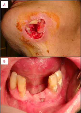
A 49-year-old female patient was referred to the Oral Surgery Unit of the Policlinic of Bari (Italy) on March 2016 for intra- and extra-oral necrotic bone exposures of the anterior mandible
(Figure 1A, 1B), associated with submandibular swelling, pus discharge, and pain.
The patient’s medical history revealed that in 2003 she was di-agnosed with Crohn’s disease and, therefore, salazopyrin (500 mg orally, three times a day) and mesalazine (800 mg oral-ly, three times a day) were administered. Subsequentoral-ly, from February 2004, due to poor response to treatments, infliximab (250 mg intravenous every six weeks) was prescribed. The pa-tient had never undergone antiresorptive, antiangiogenic, or steroid therapies, and other comorbidities or risk factors, such as smoking and alcohol abuse, were excluded.
Odontostomatological clinical history revealed the extraction of three teeth (3.2, 3.1 and 4.1) due to periodontal disease on December 2015, without infliximab drop-out. Over the next two months, the patient complained of pain and mandibular
A
B
Figure 1. Patient’s clinical features. Wide cutaneous necrotic area (A) with bone exposure and pus discharge, and (B) intraoral necrotic bone exposure on the anterior mandible in a female patient affected by Crohn’s disease, undergoing infliximab therapy. This shows the area were the teeth extractions was performed. The lesion was classified as stage 3 medication-related osteonecrosis of the jaw according to the American Association of Oral and Maxillofacial Surgeons staging system [1].
swelling. Subsequently, on February 2016, due to worsening of the symptoms and to the onset of skin ulceration with extra-oral necrotic bone exposure of the anterior mandibular area, the patient was referred to our clinic.
Clinical examination highlighted a 3 cm painful bone exposure, from the 3.2 to 4.1 at post-extractive sites, with pus discharge. The use of dental probes and the leaking of saliva from the extra-oral ulcer allowed us to diagnose an intra-extra-oral fis-tula in the area of bone exposure.
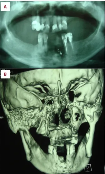
The radiological exam orthopantomography (Rx OPT) (Figure 2A) showed a poorly defined radiolucency in the area of bone ex-posure (3.2, 3.1 and 4.1 post-extraction sites) was detected, and enhanced multi-slice spiral computed tomography with 3D reconstruction (Figure 2B) showed an area of osteolysis of the alveolar process in the anterior mandible, involving the
lingual cortical plate up to the inferior margin, while the fa-cial cortical plate was preserved.
The severity of the symptoms and the clinical presentation al-lowed us to exclude the diagnosis of alveolar osteitis, while the presence of pus discharge ruled out the possibility of chronic sclerosing osteomyelitis. Also, based on routine blood tests, leukemia was ruled out, while other malignancies, such as os-teosarcoma and lymphoma were considered unlikely in view of the strict correlation between teeth extraction and the on-set of the clinical symptoms, the latter frankly pointing at the non-neoplastic origin of the process. Based on clinical exam-ination and radiological imaging, a provisional diagnosis of MRONJ was formulated, and the lesion was classified as stage 3 according to the AAOMS staging system [1].
Based on these findings, infliximab treatment was suspend-ed and the patient underwent three cycles of antibiotic ther-apy with ceftriaxone (1 g/once a day by intramuscular injec-tion) and metronidazole (500 mg/twice a day orally) for eight days, followed by 10 days of suspension.
Subsequently, the patient underwent surgical treatment under general anesthesia, consisting of anterior mandibular partial
A
B
A
B
Figure 2. Radiological exams, orthopantomography (A) and enhanced multi-slice spiral computed tomography (B) showed severe bone loss and resorption of the anterior mandible, in the region of bone exposure.
Figure 3. Surgical treatment. Surgical removal of the necrotic alveolar process (A); a iodoform gauze was put into the external infected wound after debridement of the cutaneous necrotic area (B).
resection, involving the residual alveolar process (Figure 3A) and the lingual cortical plate up to its inferior margin, with preser-vation of the facial cortical plate. After resection, the bone sur-face was treated by a piezoelectric device to remove residual infected and necrotic tissues, to possibly prevent MRONJ re-currence. Finally, a gel compound composed of sodium hyal-uronate and amino acids was put into the bone defect to re-duce the healing time. Debridement of the cutaneous necrotic area was also performed, and an iodoform gauze was put into the external infected wound (Figure 3B), which was removed three days after the surgical treatment. Also, an additional cycle of antibiotics (as previously indicated) was prescribed. The surgical specimen was fixed in neutral-buffered formalin and sent to the Pathological Anatomy Unit of the Policlinic of Bari, where it was decalcified in formic acid (5% in distilled water) for 24 hours, embedded in paraffin, sectioned at 4-μm thickness, and stained with hematoxylin-eosin. The histopath-ological analysis of the decalcified samples showed areas of
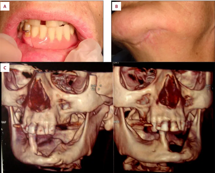
bone necrosis with inflammatory cell infiltration and several basophilic bacterial colonies, empty Haversian canals without residual osteocytes/osteoblasts, and reduction of Haversian blood vessels, thus confirming the clinical diagnosis of MRONJ. Following the surgical treatment, both intra- and extra-oral wounds healed without complications and without recur-rence after 16 months of clinical and radiological follow-up (Figure 4A–4C). Rehabilitation with a removable prosthesis was chosen to guarantee functions and aesthetics, with good stabilization of the surgical sites.
This study was performed in accordance to the principles of the Declaration of Helsinki and was approved by our institu-tion ethics committee (Study no. 4599, Prot. 1528/C.E.); the patient released informed consent for the diagnostic and ther-apeutic procedures and the possible use of the biologic sam-ples for research purposes.
A
B
Figure 4. Intra- and extra-oral wound healing. Rehabilitation with a removable prosthesis was chosen to guarantee function and aesthetics (A). Clinical (B) and radiological (C) healing of surgical wound after 12-month follow-up are shown.
Discussion
MRONJ caused by intravenous and oral BPs has been exten-sively characterized over the last 13 years. To date, inhibitors of RANKL (denosumab) [8,14], of angiogenesis (bevacizumab and rituximab) [7,15,16], of tyrosine kinase receptors (suni-tinib) [11], and of TNF (adalimumab) [10] have already been related to MRONJ in distinct case series, thus prompting the change in nomenclature from bisphosphonate-related osteo-necrosis of the jaw (BRONJ) to MRONJ.
We, hereby, report a case of infliximab-related MRONJ in a pa-tient affected by Crohn’s disease, who had been first treated by salazopirine/mesalazine and subsequently by infliximab (250 mg intravenously every six weeks for 12 years), who had never received antiresorptive/antiangiogenic therapies. A sim-ilar case of MRONJ, possibly related to infliximab administra-tion, was reported by Ebker et al. in 2013 in a patient with rheumatoid arthritis; nevertheless, the same patient had been simultaneously treated with BPs [17], thus leading the authors to stress the possible facilitating role of infliximab in the on-set of MRONJ in patients taking BPs.
In the current case, MRONJ developed after years of infliximab treatment, in the absence of BPs administration and following teeth extraction. The lesion was staged as stage 3 according to the AAOMS staging system [1], due to its severity, with in-tra-oral bone exposure and with involvement of the cutane-ous surface. Following multiple antibiotic cycles, the patient underwent surgical therapy with wide bone resection and de-bridement of the cutaneous area. The preservation of the fa-cial cortical plate prevented mandibular fracture and after 16-months of clinical and radiological follow-up, the patient completely healed without recurrence (Figure 4A–4C). The his-topathological analysis of the surgical specimen confirmed the diagnosis of MRONJ.
Infliximab is a genetically engineered chimeric human/mouse monoclonal antibody [18], which binds with high affinity to both the soluble and the transmembrane forms of human TNF-a, a key mediator of mucosal inflammation. Activities inhibited by anti-TNF-a antibodies include induction of interleukins, en-hancement of leukocyte migration, and expression of adhe-sion molecules. Increased levels of TNF-a have been reported to be involved in active Crohn’s disease, ulcerative colitis, an-kylosing spondylitis, psoriatic arthritis, plaque psoriasis, and rheumatoid arthritis. Infliximab has been increasingly used to treat all these inflammatory conditions [19]. In a recent review, Scheinfeld reported the most important side effects related
to infliximab were: lymphoma, infectious diseases, congestive heart failure, demyelinating disease, lupus-like syndromes, in-duction of auto-antibodies, reactions at the injection site, and diabetes mellitus [13]. To date, the potential implications of infliximab treatment in patients receiving oral surgery have not been fully elucidated in the literature [19]. An extended clinical trial showed that anti-TNF biologics are related to bet-ter mucosal healing but, at the same time, they may inbet-terfere with bone physiology and turnover, and with wound repair [20]. TNF-a plays an important role in systemic bone loss and turn-over, due to its ability to promote osteoclasts and osteoblasts activity [14]. Anti-TNF-a biologics possibly are responsible for bone turnover inhibition, probably by reduction of RANKL, thus resulting in osteoclast function inhibition, as already demon-strated in patients with rheumatoid arthritis [10]. Such anti-TNF-a-mediated reduction of systemic bone loss may certain-ly increase the risk of MRONJ [14], similarcertain-ly to what detected in patients taking BPs or denosumab, the latter also acting as inhibitors of osteoclast functions [21–24]. It is worth not-ing that anti-TNF-a treatments may facilitate infectious com-plications due to immunosuppression and, therefore, MRONJ occurrence in patient taking anti-TNF-a drugs may also result from “spreading of ongoing infections” [9].
Conclusions
Further studies are required to confirm the role of infliximab in MRONJ occurrence, but as recommended for patients un-dergoing treatment with BPs, denosumab, bevacizumab, or adalimumab [25], prevention of MRONJ by dental check-up be-fore and during infliximab therapy is vital to prevent MRONJ occurrence, or to detect lesion at earlier stages, thus requir-ing less invasive treatments, and possibly manifestrequir-ing lower recurrence rates.
Furthermore, in consideration of the increasing number of drugs which may facilitate MRONJ onset, the prescription of all biological drugs should require more attentive evaluation of well-known risk factors for MRONJ by all the prescribing spe-cialists. Among the latter, the role of rheumatologists is es-pecially important due to the large number of rheumatologic diseases which require the administration of new biological drugs, thus these patients are potentially at risk for MRONJ. Conflict of interest
References:
1. Ruggiero SL, Dodson TB, Fantasia J et al: American Association of Oral and Maxillofacial Surgeons position paper on medication-related osteonecrosis of the jaws – 2014 update. J Oral Maxillofac Surg, 2014; 72(10): 1938–56 2. Fleisher KE, Jolly A, Venkata UD et al: Osteonecrosis of the jaw onset times
are based on the route of bisphosphonate therapy. J Oral Maxillofac Surg, 2013; 71: 513–19
3. Marx RE, Cillo JE Jr, Ulloa JJ: Oral bisphosphonate-induced osteonecrosis: risk factors, prediction of risk using serum CTX testing, prevention, and treatment. J Oral Maxillofac Surg, 2007; 65: 2397–410
4. Kos M, Kuebler JF, Luczak K, Engelke W: Bisphosphonate-related osteonecrosis of the jaws: A review of 34 cases and evaluation of risk. J Cranio-Maxillofac Surg 2010; 38: 255–59
5. Jadu F, Lee L, Pharoah M, Reece D, Wang L: A retrospective study assessing the incidence, risk factors and comorbidities of pamidronate-re-lated necrosis of the jaws in multiple myeloma patients. Ann Oncol, 2007; 18: 2015–19
6. Ramírez L, López-Pintor RM, Casañas E et al: New non-bisphospho-nate drugs that produce osteonecrosis of the jaws. Oral Health Prev Dent, 2015; 13(5): 385–93
7. Santos-Silva AR, Belizário Rosa GA et al: Osteonecrosis of the mandible as-sociated with bevacizumab therapy. Oral Surg Oral Med Oral Pathol Oral Radiol, 2013; 115(6): e32–36
8. Pichardo SE, van Merkesteyn JP: Evaluation of a surgical treatment of de-nosumab-related osteonecrosis of the jaws. Oral Surg Oral Med Oral Pathol Oral Radiol, 2016; 122(3): 272–78
9. Preidl RHM, Ebker T, Raithel M et al: Osteonecrosis of the jaw in a Crohn’s disease patient following a course of Bisphosphonate and Adalimumab therapy: A case report. BMC Gastroenterology, 2014; 14: 6
10. Cassoni A, Romeo U, Terenzi V et al: Adalimumab: Another medication re-lated to osteonecrosis of the jaws? Case Rep Dent, 2016; 2016: 2856926 11. Fleissig Y, Regev E, Lehman H: Sunitinib related osteonecrosis of jaw: A case
report. Oral Surg Oral Med Oral Pathol Oral Radiol, 2012; 113(3): e1e3 12. Lescaille G, Coudert AE, Baaroun V et al: Clinical study evaluating the
ef-fect of bevacizumab on the severity of zoledronic acid-related osteonecro-sis of the jaw in cancer patients. Bone, 2014; 58: 103–7
13. Scheinfeld N: A comprehensive review and evaluation of the side effects of the tumor necrosis factor alpha blockers etanercept, infliximab and adali-mumab. J Dermatolog Treat, 2004; 15(5): 280–94
14. Sivolella S, Lumachi F, Stellini E, Favero L: Denosumab and anti-angioge-netic drug-related osteonecrosis of the jaw: An uncommon but potential-ly severe disease. Anticancer Res, 2013; 33(5): 1793–97
15. Pakosch D, Papadimas D, Munding J et al: Osteonecrosis of the mandible due to anti-angiogenic agent, bevacizumab. Oral Maxillofac Surg, 2013; 17: 303–6
16. Allegra A, Oteri G, Alonci A et al: Association of osteonecrosis of the jaws and POEMS syndrome in a patient assuming rituximab. J Cranio-Maxillofac Surg, 2014; 42: 279–82
17. Ebker T, Rech J, von Wilmowsky C et al: Fulminant course of osteonecrosis of the jaw in a rheumatoid arthritis patient following oral bisphosphonate intake and biologic therapy. Rheumatology, 2013; 52(1): 218–20 18. Sandborn WJ, Hanauer SB: Antitumor necrosis factor therapy for
inflam-matory bowel disease: A review of agents, pharmacology, clinical results, and safety. Inflamm Bowel Dis, 1999; 5: 119–33
19. Ciantar M, Adlam DM: Treatment with infliximab: Implications in oral sur-gery? A case report. Br J Oral Maxillofac Surg, 2007; 45(6): 507–10 20. Rutgeerts P, Van Assche G, Sandborn WJ et al: Adalimumab induces and
maintains mucosal healing in patients with Crohn’s disease: Data from the EXTEND trial. Gastroenterology, 2012; 142: 1102–11
21. Patel S, Choyee S, Uyanne J et al: Non-exposed bisphosphonate-related os-teonecrosis of the jaw: A critical assessment of current definition, staging, and treatment guidelines. Oral Diseases, 2012; 18: 625–32
22. Niibe K, Ouchi T, Iwasaki R et al: Osteonecrosis of the jaw in pa-tients with dental prostheses being treated with bisphosphonates or de-nosumab. J Prosthodont Res, 2015; 59: 3–5
23. Favia G, Tempesta A, Limongelli L et al: Medication-related osteonecrosis of the jaws: Considerations on a new antiresorptive therapy (Denosumab) and treatment outcome after a 13-year experience. Int J Dent, 2016; 2016: 1801676
24. Khan AA, Morrison A, Hanley DA et al: Diagnosis and management of osteonecrosis of the jaw: A systematic review and international consen-sus. J Bone Miner Res, 2015; 30(1): 3–23
25. Rosella D, Papi P, Giardino R et al: Medication-related osteonecrosis of the jaw: Clinical and practical guidelines. J Int Soc Prev Community Dent, 2016; 6(2): 97–104