J Oral Maxillofac Surg 60:532-540, 2002
Prevalence of p53, bcl-2, and Ki-67
Immunoreactivity and of Apoptosis in
Normal Oral Epithelium and in Premalignant
and Malignant Lesions of the Oral Cavity
Adriano Piattelli, MD, DDS,* Corrado Rubini, MD,† Massimiliano Fioroni, DDS,‡ Giovanna Iezzi, DDS,§
and Alfredo Santinelli, MD¶
Purpose: Loss of normal p53 is correlated to the progression of several preneoplastic lesions to neoplasms, and overexpression of bcl-2 determines an alteration of programmed cell death. There is an increased awareness of the importance of apoptosis in cancerogenesis, and a strong correlation of Ki-67 with high tumor grade has been reported.
Materials and Methods: The aim of our study was to investigate immunohistochemically the expres-sion and relationship of p53, bcl-2, MIB-1, and the apoptotic index (AI) in normal oral epithelium, leukoplakia, dysplasia, and oral squamous cell carcinoma.
Results: A strong correlation was found between p53 overexpression and cell proliferation (MIB-1) and the AI. An inverse relationship was found between bcl-2 expression and MIB-1 and AI. A significant inverse relationship was found between p53 and bcl-2. A good positive correlation was present between AI and MIB-1 expression.
Conclusions: Apoptosis could be important to help to understand oral carcinogenesis. © 2002 American Association of Oral and Maxillofacial Surgeons
J Oral Maxillofac Surg 60:532-540, 2002
There is an increased awareness of the importance of apoptosis in cancerogenesis.1Studies of human B-cell
lymphomas (Bcl) led to the discovery of proto-onco-gene bcl-2, which is situated on chromosome 18. This 25-kd protein is also located at the mitochondrial outer membrane and in the nuclear envelope, plasma membrane, and endoplasmic reticulum.2bcl-2
expres-sion was first described in follicular lymphomas with a chromosomal translocation; later studies also found it in lymphomas and epithelial tumors without this translocation.2 Overexpression of bcl-2 results in an
alteration of programmed cell death with persistence of cells that fail to die.3 In normal proliferating
epi-thelium, bcl-2 is expressed in stem cell zones such as the basal layers, where it acts to prevent the death of cells in the regenerative compartment.3,4 Bcl-2
pro-tein overexpression has been found in the early phase of epithelial cancerogenesis.1 Bcl-2 was mainly
ex-pressed in well-differentiated transitional cell carcino-mas, whereas it was absent in high-grade transitional cell carcinomas.5This loss may reflect a deregulation
of the mechanisms that control bcl-2 expression.5
Little is known about the presence of bcl-2 in normal, dysplastic, or neoplastic oral epithelium.6,7 Loss of
normal p53 function is correlated to the progression
*Professor, Oral Medicine and Pathology, Dental School, Univer-sity of Chieti, Chieti, Italy and Honorary Senior Lecturer, Eastman Dental Institute for Oral Health Care Sciences, London, England.
†Researcher, Institute of Pathologic Anatomy and Histopathol-ogy, University of Ancona, Ancona, Italy.
‡Research Fellow, Dental School, University of Ancona, Ancona, Italy.
§Research Fellow, Dental School, University of Chieti, Chieti, Italy.
¶Researcher, Institute of Pathologic Anatomy and Histopathol-ogy, University of Ancona, Ancona, Italy.
This work was supported in part by the National Research Council (CNR), Rome, Italy, and by the Ministry of University, Research, Science and Technology (MURST), Rome, Italy.
Address correspondence and reprint requests to Dr Piattelli: Via F. Sciucchi 63, 66100 Chieti, Italy; e-mail: [email protected]
©2002 American Association of Oral and Maxillofacial Surgeons 0278-2391/02/6005-0010$35.00/0
doi:10.1053/joms.2002.31851
of several preneoplastic lesions to neoplasms and with a shortened survival in several malignancies.5
Moreover, p53 expression showed a statistically sig-nificant correlation with tumor grade and stage.5
The detection of p53 in preinvasive areas adjacent to squamous cell carcinoma and in dysplastic oral epithelium suggests that p53 abnormalities may con-stitute an early event in the natural history of oral cancer.8-11p53 safeguards genomic stability by
block-ing the entry of a DNA-damaged cell from the G1 phase to the S phase and by activating apoptosis through a down-regulation of bcl-2.12
The p53 gene is thus involved in the apoptotic pathway.1 The majority of oral squamous cell
carci-nomas (SCCs) (up to 80%) carry p53 mutations, and this fact shows that oral mucosa is one of the most common targets for p53 mutations.13A strong
corlation of Ki-67 with high tumor grade has been re-ported,5and MIB-1 is regarded as a reliable marker of
proliferating cells.14The aims of our study were: 1) to
investigate, in normal oral epithelium, leukoplakia, dysplasia and oral SCC, the prevalence of p53, bcl-2, and Ki-67 immunoreactivity and of apoptosis; and 2) to determine the relationship between them.
Materials and Methods
A total of 70 biopsy samples were analyzed. The following formalin-fixed, paraffin-embedded tissues were used in this study: normal oral mucosa obtained during third molar removal (10 cases), leukoplakia (12 cases), epithelial dysplasia (12 cases: 6 mild and 6 severe dysplasia and carcinoma in situ), invasive car-cinoma well differentiated (G1) (12 cases), invasive carcinoma moderately differentiated (G2) (12 cases), and invasive carcinoma poorly differentiated (G3) (12 cases). The age of the patients ranged from 48 to 67 years (mean, 55 years). All selected samples had been routinely fixed in 10% neutral formalin (24 to 48 hours). dehydrated in graded alcohols, cleared in xy-lene, and embedded in paraffin. The hematoxylin-eosin slides were all reviewed, the diagnosis was confirmed, and the slides for the quantitative evalua-tion were selected. Epithelial dysplasia was diagnosed according to the criteria and the definition proposed by Pindborg et al15: mild dysplasia was considered
mild expansion of the proliferative zone, with mitotic activity, hyperchromatism, and nuclear variability;
moderate dysplasia indicated these same features were present in up to half of the thickness of the epithelium; and severe dysplasia indicated the atypi-cal proliferative zone encompassed up to three fourths of the epithelium.
Immunohistochemical staining for bcl-2 protein was performed using the following antigen retrieval system. Sections were deparaffinized in 2 changes of
xylene for 10 minutes each and then were rehydrated through graded alcohols and immersed in 0.3% hydro-gen peroxide in methanol for 30 minutes to block endogenous peroxidase activity. Sections were then washed in phosphate-buffered saline (PBS). The tissue sections were put in a microwave oven (Cooktyronic M720, 700 W; Philips, Eindhoven, The Netherlands) in a plastic Coplin jar filled with 10 mmol/L sodium citrate buffer (pH 6.0) at 5-minute intervals for a total of 10 minutes. At each 5-minute interval, the fluid in the Coplin jar was removed from the microwave oven and allowed to cool. Slides were incubated overnight in a 1:60 dilution of the primary mouse anti-human bcl-2 monoclonal antibody (124; DAKO, Glostrup, Denmark). A biotin streptavidin detection system was used with diaminobenzidine as the chromogen.
Slides were washed twice with PBS and incubated with the linking reagent (biotinylated anti-immuno-globulins) for 15 minutes at room temperature. After being rinsed in PBS, the slides were incubated with the peroxidase-conjugated streptavidin label for 15 minutes at room temperature. The sections were again rinsed in PBS and incubated with diaminoben-zidine for 10 minutes in the dark. After chromogen development, slides were washed in 2 changes of water and counterstained with a 1:10 dilution of he-matoxylin. The sections were then dehydrated, cleared in xylene, and mounted. A negative control was performed in all cases by omitting the primary antibody, which in all instances resulted in negative immunoreactivity. Sections from a lymph node with follicular lymphoma were used as positive controls. Normal lymphocytes present in the tissues repre-sented an internal positive control for bcl-2 immuno-staining. In all positive cases, immunoreactivity was restricted to the cytoplasm. The percentage of bcl-2– positive cells was evaluated from a minimum of 1000 cells in each case, and bcl-2 immunostaining was scored using a 3-tiered scale in which minus indicates less than 5% cells, plus indicates 5% to 50% cells, and double-plus indicates more than 50% cells.
Descriptive statistical analysis was performed for each group of lesions.
Immunohistochemical staining for p53 protein was performed using the following antigen retrieval sys-tem. Sections were deparaffinized in 2 changes of xylene for 10 minutes each and then were rehydrated through graded alcohols and immersed in 0.3% hydro-gen peroxide in methanol for 30 minutes to block endogenous peroxidase activity. Sections were then washed in PBS. The tissue sections were put in a microwave oven (Cooktyronic M720, 700 W; Philips) in a plastic Coplin jar filled with 10 mmol/L sodium citrate buffer (pH 6.0) at 5-minute intervals for a total of 10 minutes. Slides were incubated overnight with a 1:50 dilution of the primary mouse anti-human p53
monoclonal antibody (DO-7; DAKO). A biotin-strepta-vidin detection system was used with diaminobenzi-dine as the chromogen. Slides were washed twice with PBS and incubated with the linking reagent (bi-otinylated anti-immunoglobulins) for 15 minutes at room temperature. After being rinsed in PBS, the slides were incubated with the peroxidase-conjugated streptavidin label for 15 minutes at room tempera-ture. The sections were again rinsed in PBS and incu-bated with diaminobenzidine for 10 minutes in the dark. After chromogen development, slides were washed in 2 changes of water and counterstained with a 1:10 dilution of hematoxylin. The sections were then dehydrated, cleared in xylene, and mounted. A negative control was performed in all cases by omitting the primary antibody, which in all instances resulted in negative immunoreactivity. p53 expression and location were evaluated on histo-logic sections using a Leitz Orthoplan microscope (Wetzlar, Germany) equipped with a ⫻63 objective and an eyepiece graticule. Only nuclear staining of epithelial cells was observed, and the nuclei with a clear brown color, regardless of staining intensity, were regarded as p53 positive. The percentage of p53-positive cells was evaluated from a minimum of 1,000 cells in each case, and p53 immunostaining was scored using a 3-tiered scale, as for bcl-2 immunostain-ing. The cells were evaluated in both the basal and parabasal layers. Descriptive statistical analysis was performed for each group of lesion, and the 2 test was used to determine statistically significant differ-ences between the groups.
For MIB-1 immunostaining, the slides were pretreated with 3-aminopropyltriethoxysilane (APES; Sigma Chem-ical Co, St Louis, MO), which avoided the separation of the section from the slide during the incubation in the microwave oven. For each case, a 5-m section was cut and placed onto a pretreated slide. The stain-ing protocol of this slide consisted of the applications of a series of reagents in the following manner: over-night drying at 37°C, dewaxing and rehydration, im-mersion in a plastic box containing 0.01 mol/L citrate buffer at pH 6.0, incubation for 5 minutes in a micro-wave oven initially at 750 W until boiling began and then at 350 W for the remaining time, incubation for 5 minutes in a microwave oven at 350 W, cooling for 20 minutes at room temperature, washing in running water and then in distilled water for 5 minutes, wash-ing in Tris-buffered saline (TBS) for 5 minutes, re-moval of any excess TBS, addition of primary mono-clonal mouse anti– ki-67 antibody (Immunotech, Marseille, France) diluted 1:25 in TBS, and overnight incubation at 4°C in a humidified room. Next, the protocol consisted of washing in TBS for 5 minutes (3 times), addition of secondary prediluted biotinylated anti-mouse antibody (LSAB; DAKO) and incubation
for 10 min at room temperature, washing in TBS for 5 minutes (3 times), addition of prediluted streptavidin-peroxidase complex (LSAB; DAKO) and incubation for 10 minutes at room temperature, and washing in TBS for 5 minutes (3 times). The protocol continued with immersion in 0.05% DAB and 0.01% H2O2in TBS for 2 to 3 minutes at room temperature, washing in running water and then in distilled water for 5 min-utes, counterstaining with ethyl green for 30 minmin-utes, washing in distilled water for 30 seconds, washing in buthanol I for 5 seconds, washing in buthanol II for 30 seconds, and dehydration and mounting in Per-mount.
The positivity to MIB-1 was evaluated by counting the number of positive cells per 1000 cells, and the values were expressed in an percentage. The pres-ence of apoptotic bodies was evaluated in a random fashion in the parabasal layer in 20 high-power fields. Mann-Whitney U, Kruskal-Wallis, and2tests were used to evaluate the presence of statistically signifi-cant differences. Simple regression analysis was used to evaluate the correlation between features.
Results
p53
p53 was present in the basal layer in normal oral epithelium; in the basal and parabasal layers in leuko-plakia, dysplasia, and carcinoma in situ; and in central and peripheral regions in invasive carcinoma. In nor-mal oral epithelium in only 1 sample, the positivity was between 5% and 50%, whereas all of the other specimens showed a positivity of less than 5% (Fig 1). In leukoplakia, in 9 samples, the positivity was less than 5%, and in 3 samples, it was between 5% and 50%. In mild dysplasia, in 4 samples, the positivity was less than 5%, and in 2 samples, it was between 5% and 50%. In severe dysplasia, 5 samples had a positivity between 5% and 50% (Fig 2), and 1 sample had more than 50%. In well-differentiated carcinoma, 6 samples had a positivity of less than 5%, 5 samples had be-tween 5% and 50%, and 1 sample had more than 50%. In moderately differentiated carcinoma, 5 samples had a positivity of less than 5%, 5 samples had be-tween 5% and 50%, and 2 samples had more than 50%. In poorly differentiated carcinoma, 5 samples had a positivity of less than 5%, 4 samples had be-tween 5% and 50%, and 3 samples had more than 50%.
bcl-2
In normal oral epithelium and in leukoplakia, bcl-2 protein expression was seen in the basal layer, and in dysplasia, it was evaluated in the basal layer and rarely in the parabasal layer. In carcinoma, it was present in
the central and peripheral areas of the tumor. In normal oral epithelium, in 3 samples, the positivity was less than 5%, in 6 samples, it was between 5% and 50%, and in 1 sample, it was more than 50% (Fig 3). In leukoplakia, 8 samples showed a positivity of less than 5%, and 4 samples had between 5% and 50%. In mild and moderate dysplasia, 3 samples had a positiv-ity of less than 5% (Figs 4, 5), and 3 samples had between 5% and 50%. In severe dysplasia and carci-noma in situ, in all 6 samples, the positivity was less than 5%. In well-differentiated carcinoma, in 8 sam-ples, the positivity was less than 5%, in 3 samsam-ples, it was between 5% and 50%, and in 1 sample, it was more than 50%. In moderately differentiated carci-noma, in 9 samples, the positivity was less than 5%, in 2 samples, it was between 5% and 50% (Fig 6), and in 1 sample, it was more than 50%. In poorly differenti-ated carcinoma, in 6 samples, the positivity was less than 5%, in 2 samples, it was comprised between 5% and 50%, and in 4 samples, it was more than 50%.
MIB-1
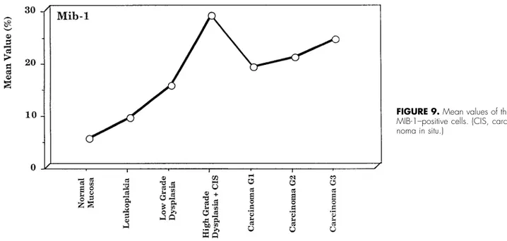
The expression of MIB-1 was very similar to that of p53. In normal oral mucosa, the value was 5.2%
(⫾2.20%); in leukoplakia, 9.0% (⫾3.89%; in mild dys-plasia, 15.3% (⫾5.61%); in severe dysplasia and carci-noma in situ, 28.6% (⫾6.21%) (Fig 7); in well-differ-entiated carcinoma, 18.8% (⫾6.20%); in moderately differentiated carcinoma, 20.8% (⫾6.06%); and in poorly differentiated carcinoma, 24.2% (⫾6.0081%) (Figs 8, 9)
The mean values for all types of carcinoma was 21.6% (⫾6.81%). With the Mann-Whitney U test, sta-tistically significant differences were found between normal oral mucosa and leukoplakia (P ⫽ .0161), normal oral mucosa and mild dysplasia (P ⫽ .0017), normal oral mucosa and severe dysplasia (P⫽ .0011), and normal oral mucosa and carcinomas (P⬍ .0001). Statistically significant differences were found, more-over, between leukoplakia and mild dysplasia (P ⫽ .039), leukoplakia and severe dysplasia (P ⫽ .0007), and leukoplakia and carcinoma (P ⬍ .0001). Statisti-cally significant differences were also found between mild and severe dysplasia (P⫽ .0065), mild dysplasia and carcinoma (P⫽ .0455), and severe dysplasia and carcinoma (P⫽ .0455). Kruskal-Wallis test showed no statistically significant differences among well-entiated, moderately differwell-entiated, and poorly differ-entiated carcinoma (P ⫽ .1209).
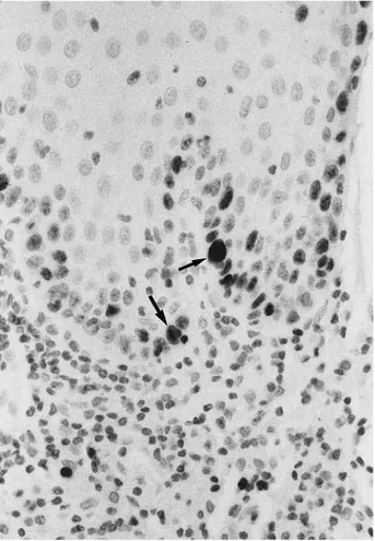
FIGURE 2. Severe dysplasia, showing strong positivity to p53 is seen in
about 5% to 10% of epithelial cells (arrows) (p53 immunostaining alkaline phosphatase antialkaline phosphatase, original magnification⫻20).
FIGURE 1. Normal oral mucosa, showing a few basal cells positive
to p53 (arrows) (p53 immunostaining alkaline phosphatase antialka-line phosphatase, original magnification⫻40).
APOPTOTIC INDEX
In normal oral mucosa, the apoptotic index (AI) was 0.09 (⫾0.07); in leukoplakia, 0.083 (⫾0.08); in
mild dysplasia, 0.133 (⫾0.10); in severe dysplasia and carcinoma in situ, 0.317 (⫾0.12); in well-differenti-ated carcinoma, 0.242 (⫾0.12); in moderately differ-entiated carcinoma, 0.367 (⫾0.23); and in poorly dif-ferentiated carcinoma, 0.433 (⫾0.25) (Fig 10). With the Mann-Whitney U test, statistically significant dif-ferences were found between normal oral mucosa and severe dysplasia (P ⫽ .0024), leukoplakia and severe dysplasia (P ⫽ .0020), and mild and severe dysplasia (P ⫽ .025). Moreover, a statistically signifi-cant difference was found between well-differenti-ated carcinoma and normal oral mucosa (P ⫽ .002), leukoplakia (P ⬍ .001), and mild dysplasia (P ⫽ .0103). Kruskal-Wallis test has been used to evalu-ate the differences of the AI of well-differentievalu-ated, moderately differentiated, and poorly differentiated carcinoma: these differences were not statistically significant (P⫽ .1286). Moreover, no statistically dif-ferences were found between normal oral mucosa and leukoplakia (P⫽ .8175), normal oral mucosa and mild dysplasia (P⫽ .4477), leukoplakia and mild dys-plasia (P ⫽ .3490), and severe dysplasia and carci-noma (P⫽ .8939).
FIGURE 3. Normal oral mucosa, showing diffuse positivity of the
basal and parabasal cell layers to bcl-2 protein (arrows) (bcl-2 immu-nostaining alkaline phosphatase antialkaline phosphatase, original magnification⫻40).
FIGURE 4. Mild dysplasia, showing negativity of the basal cell layers
to bcl-2 and positivity of the lymphocytes (arrows) (bcl-2 immunostain-ing alkaline phosphatase antialkaline phosphatase, original magnifi-cation⫻40).
FIGURE 5. Moderate dysplasia, showing complete negativity to
bcl-2 of the basal cell layer and positivity of the lymphocytes (arrows) (bcl-2 immunostaining alkaline phosphatase antialkaline phosphatase, original magnification⫻40).
CORRELATION BETWEEN FEATURES
p53 protein was negative in 16 bcl-2–negative cases (42.1%), in 16 bcl-2–positive (⫹) cases (42.1%), and in
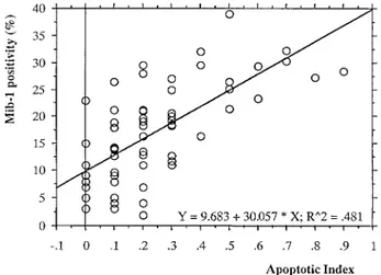
6 bcl-2–positive (⫹⫹) ones (15.8%). p53 was positive (⫹) in 20 bcl-2–negative cases (80%), in 4 bcl-2– positive (⫹) cases (16%), and in 1 bcl-2–positive (⫹⫹) case (4%). All of the 7 p53-positive (⫹⫹) cases were bcl-2 negative. There was a negative significant statis-tical correlation between p53 and bcl-2 protein (P⫽ .001). In the 43 bcl-2–negative cases, the mean value of AI was 0.314 (⫾0.217), whereas in the 27 bcl-2– positive (⫹ and ⫹⫹) cases, the mean value was 0.133 (⫾0.130); the difference between the 2 groups was statistically significant (P ⬍ .0001). A good, but neg-ative, correlation between these 2 features was present. Finally, considering all of the 70 cases, the simple regression analysis (Fig 11) showed that there was a good positive correlation (r ⫽ 0.694, P ⬍ .0001) between AI and MIB-1 expression.
Discussion
Ramsay et al3showed that compared with nevi, in
which the bcl-2 protein immunoreactivity was present in all cells, melanomas exhibited a progres-sive loss of the protein expression with increased
FIGURE 6. Moderately differentiated carcinoma, showing
nonhomo-geneous, diffuse (⬍50%) positivity of the neoplastic cells to bcl-2 (arrows) (bcl-2 immunostaining alkaline phosphatase antialkaline phos-phatase, original magnification⫻40).
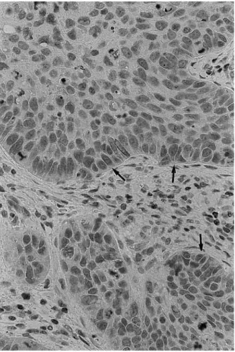
FIGURE 7. Severe dysplasia, showing strong positivity of basal,
parabasal, and superficial layers to MIB-1 (arrows) (MIB-1 immuno-staining alkaline phosphatase antialkaline phosphatase, original mag-nification⫻40).
FIGURE 8. Poorly differentiated carcinoma, showing positivity
(⬍50%) of the neoplastic cells (arrows) (MIB-1 immunostaining alkaline phosphatase antialkaline phosphatase, original magnification⫻20).
levels of malignancy. These data suggest, perhaps, that bcl-2 loss is associated with or reflects an in-creased malignant potential3. The presence of bcl-2
seems to be associated with a better prognosis in some tumors but not in others.2,3,16 High levels of
bcl-2 protein correlated with lower rates of complete remission and shorter survival in patients with acute myeloid leukemia.17,18 On the other hand, the bcl-2
expression in breast cancer seems to be predictive of a positive response to endocrine therapy and corre-lates with improved survival, whereas in prostate can-cer, bcl-2 expression is related to androgen-indepen-dent tumor growth and chemoresistance.12,16,17 In
thyroid cancer, bcl-2 expression seems to be
corre-lated with the differentiation of the tumors, and indif-ferentiated tumors do not express the protein.16,18
In neuroblastoma, bcl-2 expression does not seem to influence prognosis.16Tjalma et al2found a strong
relationship between bcl-2 expression and prognosis in patients with carcinoma of the uterine cervix. Flo-hil et al,16 in a study that compared hyperplastic
polyps, adenomas, and carcinomas of the colon, found that most carcinomas did not present a bcl-2 immunoreactivity.
The expression of bcl-2 may provide data about individual tumor cell dynamics that in the future will most probably be very important for therapy and prognosis.17 A significant inverse relationship be-FIGURE 9. Mean values of the
MIB-1–positive cells. (CIS, carci-noma in situ.)
FIGURE 10. Mean values of
the apoptotic index. (CIS, carci-noma in situ.)
tween bcl-2 and p53 was found in our specimens: similar data were reported by Harn et al1 in breast
carcinoma and by other researchers in carcinomas of the thyroid, colorectum, stomach, and esophagus and in gastric lymphoma.12Studies in human breast
can-cer and in cancan-cer cell lines have shown that p53 can down regulate bcl-2 expression4 and that apoptosis
induced by p53 can be blocked by bcl-2 in cultured cancer cells.4p53 has been shown to down-regulate
bcl-2 via binding to a negative regulatory element outside the bcl-2 gene promoter.19 A significant
in-verse relationship was found between bcl-2 expres-sion and the pathologic stage of esophageal tumors, and bcl-2 was found more frequently in early than in advanced stages.12Our results also provide evidence
of an interaction between bcl-2 and MIB-1: the highest expression of MIB-1–positive cells was observed in severe dysplasia and carcinoma in situ, where the bcl-2 protein expression was lowest or even absent. A possible influence of bcl-2 expression in the down-regulation of a proliferation marker was reported by Konstantinidouet al,20who found that bcl-2–positive
meningiomas presented a lower growth fraction rate and had a significantly higher proportion of prolifer-ating cell nuclear antigen (PCNA)– strongly positive nuclei than the bcl-2–negative subgroup.
We found, on the contrary, an inverse relationship between bcl-2 and MIB-1, and the loss of bcl-2 expres-sion in our specimens of severe dysplasia and carci-noma in situprobably reflects a deregulation of the mechanisms that control bcl-2 expression.5Our data
show that a bcl-2 down-regulation was strongly asso-ciated with peaks in MIB-1 and AI in these lesions. Ravi et al9reported an increased bcl-2 expression in
oral dysplasia and carcinoma. bcl-2 expression in-versely correlated with the degree of differentiation between CIN I/II and III in tumors of the uterine
cervix.21We found that p53 was expressed in
supra-basal layers in leukoplakia, dysplasia, and carcinoma in situ; Cruz et al13demonstrated that p53 suprabasal
expression was significantly associated with the de-velopment of carcinoma. Our results are fully in ac-cordance with the results reported by Murti et al,8
who found that the overexpression of p53 protein was significantly more common in severe than in mild epithelial dysplasia and that p53 expression peaked close to the time of transition from the precancer state to cancer rather than earlier in the natural his-tory of oral precancer. A fairly close relationship be-tween p53 and AI was found in our specimens. Bir-chall et al6 found that apoptosis increased from
normal through dysplastic epithelium to reach a max-imum in carcinoma in situ, whereas in invasive SCC AI fell to normal values6; it also increased with increasing
degrees of dysplasia. A strong relationship was found in our specimens between AI and the degree of dys-plasia and the onset of SCC; completely different results were reported by Birchall et al,6,7who found
no such association.
A diminution of apoptosis was reported in gastric carcinoma, whereas esophageal SCC, prostatic adeno-carcinoma, brain tumors, and non-Hodgkin lympho-mas showed an increased AI in relationship to less tumor differentiation and increased mitotic activ-ity.22,23Our results show a close correlation between
AI and MIB-1, whereas Birchall et al6,7found that the
AI was highly correlated with mitotic index but not with PCNA: these authors failed to demonstrate an increased expression of PCNA in dysplastic epithe-lium. Apoptosis can be important in tumor growth and prognosis; in colorectal carcinoma, Langlois et al24 found that tumors with higher apoptotic counts
seemed to have a good prognosis, and this may reflect that neoplasms with higher levels of apoptosis are slower growing. In renal cell carcinoma, Hindermann et al23found a decrease in cells undergoing apoptosis
in less-differentiated tumors with an increase in the number of tumor cells and of tumor growth. This decrease in apoptosis was correlated with an increase in the proliferative activity. The AI was much higher in the most aggressive type of prostatic carcinoma.1
The presence of Ki-67 closely coincided with p53 protein,10and a close relationship between PCNA and
p53 protein was found in some oral tumors.25 In
nasopharyngeal carcinoma, the patients with a high PCNA had a poorer disease-free survival.26
References
1. Harn HJ, Shen KL, Yueh KC, et al: Apoptosis occurs more frequently in intraductal carcinoma than in infiltrating duct carcinoma of human breast cancer and correlates with altered p53 expression detected by
terminal-deoxynucleotidyl-trans-FIGURE 11. Simple regression analysis considering apoptotic index
and MIB-1 positivity. A good, positive correlation is evident (r ⫽ 0.694, P⬍ .0001).
ferase-mediated dUTP-FITC nick end labeling (TUNEL). Histo-pathology 31:534, 1997
2. Tjalma W, Weyler J, Goovaerts G, et al: Prognostic value of bcl-2 expression in patients with operable carcinoma of the uterine cervix. J Clin Pathol 50:33, 1997
3. Ramsay JA, From L, Kahn HJ: bcl-2 protein expression in mela-nocytic neoplasms of the skin. Mod Pathol 8:150, 1995 4. Cho JH, Kim WH: Altered topographic expression of
p21WAF1/CIP1/SDI1, bcl-2 and p53 during gastric carcinogenesis. Pathol Res Pract 194:309, 1998
5. Nakopoulou L, Vourlakou C, Zervas A, et al: The prevalence of bcl-2, p53 and Ki-67 immunoreactivity in transitional cell blad-der carcinomas and their clinicopathologic correlates. Hum Pathol 29:146, 1998
6. Birchall MA, Winterford CM, Allan DJ, et al: Apoptosis in normal epithelium, premalignant and malignant lesions of the oropharynx and oral cavity: A preliminary study. Oral Oncol Eur J Cancer 31B:380, 1995
7. Birchall MA, Schock E, Harmon BV, et al: Apoptosis, mitosis, PCNA and bcl-2 in normal, leukoplakic and malignant epithelia of the human oral cavity. Oral Oncol 33:419, 1997
8. Murti PR, Warnakulasuriya KAAS, Johnson NW, et al: p53 expression in oral precancer as a marker for malignant poten-tial. J Oral Pathol Med 27:191, 1998
9. Ravi D, Nalinakumari KR, Rajaray RS, et al: Expression of programmed cell death regulatory p53 and bcl-2 proteins in oral lesions. Cancer Lett 105:139, 1996
10. Slootweg PJ, Koole R, Hordijk GJ: The presence of p53 protein in relation to the Ki-67 as cellular proliferation marker in head and neck squamous cell carcinoma and adjacent dysplastic mucosa. Oral Oncol Eur J Cancer 30B:138, 1994
11. Bongers V, Snow GB, Van der Waal I, et al: Value of p53 expression in oral cancer and adjacent normal mucosa in rela-tion to the occurrence of multiple primary carcinomas. Oral Oncol Eur J Cancer 31B:392, 1995
12. Parenti AR, Rugge M, Horng Shiao Y, et al: bcl-2 and p53 immunophenotypes in pre-invasive, early and advanced oesophageal squamous cancer. Histopathology 31:430, 1997 13. Cruz IB, Snijders PJF, Meijer CJ, et al: p53 expression above the
basal cell layer in oral mucosa is an early event of malignant transformation and has predictive value for developing oral squamous cell carcinoma. J Pathol 184:360, 1998
14. Van Diest PJ, Brugal G, Baak JPA: Proliferation markers in tumours: Interpretation and clinical value. J Clin Pathol 51:716, 1998
15. Pindborg JJ, Reichart PA, Smith CJ, et al: Histological Typing of Cancer and Precancer of the Oral Mucosa. Berlin, Germany, Springer Verlag, 1997
16. Flohil CC, Janssen PA, Bosman FT: Expression of bcl-2 protein in hyperplastic polyps, adenomas, and carcinomas of the co-lon. J Pathol 178:393, 1996
17. Sarkiss M, HsuB, El Naggar AK, et al: The clinical relevance and assessment of apoptotic cell death. Adv Anat Pathol 3:205, 1996
18. Jordan RCK, Catzavelos GC, Barrett AW, et al: Differential expression of bcl-2 and Bax in squamous cell carcinoma of the oral cavity. Oral Oncol Eur J Cancer 32:394, 1996
19. De Angelis PM, Stokke T, Thorstensen L, et al: Apoptosis and expression of Bax, Bcl-x, and Bcl-2 apoptotic regulatory pro-teins in colorectal carcinomas, and associations with p53 ge-notype/phenotype. J Clin Pathol Mol Pathol 51:254, 1998 20. Konstantinidou AE, Pavlopoulos PM, Patsouris E, et al:
Expres-sion of apoptotic and proliferation markers in meningiomas. J Pathol 186:325, 1998
21. Saegusa M, Takano Y, Hashimura M, et al: The possible role of bcl-2 expression in the progression of tumors of the uterine cervix. Cancer 76:2297, 1995
22. Drachenberg CB, Ioffe OB, PapadimitriouJC: Progressive in-crease of apoptosis in prostatic intraepithelial neoplasia and carcinoma. Arch Pathol Lab Med 121:54, 1997
23. Hindermann W, Berndt A, Wunderlich H, et al: Quantitative evaluation of apoptosis and proliferation in renal cell carci-noma: Correlation to tumor subtype, cytological grade accord-ing to Thoenes-classification and the occurrence of metastasis. Pathol Res Pract 193:1, 1997
24. Langlois NEI, Lamb J, Eremin O, et al: Apoptosis in colorectal carcinoma occurring in patients aged 45 years and under: Relationship to prognosis, mitosis, and immunohistochemical demonstration of p53, c-myc and bcl-2 protein products. J Pathol 182:392, 1997
25. Tsuji T, Minmura Y, Wen S, et al: The significance of PCNA and p53 protein in some oral tumors. Int J Oral Maxillofac Surg 24:221, 1995
26. Chan ATC, Ho S, Teo PML, et al: Assessment of proliferating nuclear antigen in nasopharyngeal carcinoma tissue and its relation to clinical findings. Oral Oncol 33:13, 1997