Graphene oxide increases
mammalian spermatozoa fertilizing
ability by extracting cholesterol
from their membranes and
promoting capacitation
Nicola Bernabò
1, Juliana Machado-simoes
1, Luca Valbonetti
1, Marina Ramal-sanchez
1,
Giulia Capacchietti
1, Antonella Fontana
2, Romina Zappacosta
2, paola palestini
3,
Laura Botto
3, Marco Marchisio
4,5, paola Lanuti
4,5, Michele Ciulla
2, Antonio Di stefano
2,
elena Fioroni
6, Michele spina
6& Barbara Barboni
1Graphene oxide (Go) is a widely used biomaterial with an amazing variety of applications in biology and medicine. Recently, we reported the ability of Go to improve the in vitro fertilization (IVF) outcomes in swine, a validated animal model with a high predictive value for human fertility. For that reason, here we characterized the mechanisms involved in this positive interaction by adopting an experimental approach combining biological methods (confocal microscopy analysis on single cell, flow cytometry on cell populations and co-incubation with epithelial oviductal cells), physical-chemical techniques (Differential Scanning Calorimetry and Thermogravimetric Analysis), and chemical methods (mass spectrometry and lipid measurement). As a result, we propose a model in which Go is able to extract cholesterol from the spermatozoa membrane without causing any detrimental effect. In this way, the cholesterol extraction promotes a change in membrane chemical-physical properties that could positively affect male gamete function, modulating sperm signalling function and increasing in this way the fertilizing potential, without losing the ability to physiologically interact with the female environment. In conclusion, these data seem to suggest new intriguing possibilities in engineering sperm membrane for improving assisted reproduction technologies outcomes, even in human medicine.
The use of biomaterials has experienced an incredible dissemination for a myriad of applications, ranging from regenerative medicine to diagnostics. In this context, one of the most promising family of materials is graphene and graphene-related materials, whose use with biomedical purposes is continuously increasing. Graphene was first isolated in 2004 by Andre Geim and Konstantin Novoselov, awarded with the Nobel Prize in Physics in 2010 for its discovery. Graphene is a single layer of carbon packed in a hexagonal honeycomb lattice with carbon–car-bon distances of 0.142 nm. Graphene possesses some interesting properties that it is interesting to note: it can be considered as an indefinitely large aromatic molecule, is the strongest material ever tested1, conducts very
efficiently heat and electricity and is nearly transparent.
Immediately after its discovery, researchers started to develop a large family of graphene-related materials, such as few layer graphene sheets (FLGS), ultrathin graphite, graphene nanosheets, graphene oxide (GO), and reduced graphene oxide (rGO), with a wide range of applications in electronic, engineering, chemistry, and
1faculty of Bioscience and technology for food, Agriculture and environment, University of teramo, Via Renato Balzarini 1, 64100, Teramo, Italy. 2Department of Pharmacy, University “G. d’Annunzio”, Via dei Vestini, 66100, Chieti, italy. 3School of Medicine and Surgery, University of Milano Bicocca, 20900, Monza, Italy. 4Department of Medicine and Aging Sciences, University “G. d’Annunzio” Chieti-Pescara, 66100, Chieti, Italy. 5center on Aging Sciences and Translational Medicine (CeSI-MeT), University “G. d’Annunzio” Chieti-Pescara, 66100, Chieti, Italy. 6Laboratorio Analisi Dr. Fioroni, Viale A. de Gasperi, 19, 63074, San Benedetto del Tronto, Italy. Correspondence and requests for materials should be addressed to n.B. (email: [email protected])
Received: 25 January 2019 Accepted: 14 May 2019 Published: xx xx xxxx
bio-medicine. In this last context, the hydrophilic forms of graphene have been used during the last years for several different applications including bio-imaging2,3, cancer theragnosis4–6, gene delivery7, tissue engineering8,9,
biosensing10, DNA sequencing11 and drug delivery12,13.
Our attention was attracted by GO for two main reasons. First, it is a relatively hydrophilic molecule able to interact with biological structures in aqueous phase (as blood or other biological fluids). Moreover, the possi-bility that GO could exert a detrimental effect on reproductive function in some animal models had been sug-gested14–16, in particular when the spermatozoa are exposed to different GO forms17–19.
In our laboratory, for the first time, we assessed the effect of GO on mammalian spermatozoa in terms of fertilizing ability20. Sperm cells are unable to fertilize the female oocyte immediately after ejaculation and they
only reach their fertilizing ability after residing for hours to days (depending on the species) within the female genital tract21. Here, and concretely in the oviduct isthmus, they complete their functional maturation (the
so called capacitation) under the guidance of endocrine signals from the surrounding female environment22.
Capacitation implies marked changes in sperm membranes, chemical composition and, as a consequence, on their physico-chemical properties. In particular, the presence of Ca2+ and bicarbonate activates a cAMP/
PKA-dependent pathway that leads to the rupture of membrane asymmetry (inner and outer membrane leaflet are characterized by a different chemical composition) with the consequent exposure of cholesterol on the outer side of plasma membrane (PM)23–33. Once exposed, cholesterol is removed from the anterior area of sperm head
by extracellular acceptors (albumin, globulins), thus promoting the increase in PM fluidity and the ability to fuse with the outer acrosome membrane (OAM) when the physiological stimulus, the oocyte zona pellucida (ZP), will be met28,34.
In our previous experiments, we found that boar spermatozoa co-incubation with GO under capacitating con-ditions was able to induce a dose-dependent effect. In particular, at relatively high concentrations (10 and 50 μg/ml), it was able to induce a toxic damage expressed as decreased viability and loss of acrosome integrity, while in a definite range of concentrations (0.5 to 1 µg/mL), surprisingly, GO seemed to promote the fertilizing ability in an
in vitro fertilization (IVF) assay20. Since this unexpected effect could be interesting either for the understanding of
the basics GO interaction with living systems as well as for the development of possible technological applications in assisted reproduction technologies (ARTs), here we carried out further experiments by combining biological, chemical and physical approaches, with the aim of exploring the molecular mechanisms of this interaction.
Results
GO affects sperm membrane chemical composition by reducing cholesterol concentration.
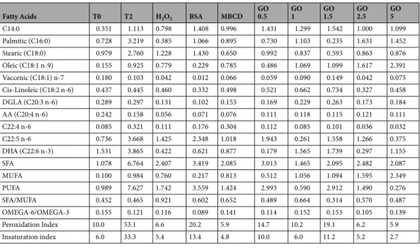
To perform a complete molecular characterization of GO effect on membrane chemical composition, we checked the potential changes in a set of lipids known to be involved in biologically relevant processes, such as oxidative metabolism.As reported in Tables 1 and 2, it is evident that incubation of spermatozoa with GO under culture conditions causes some important modifications in the lipidomic pattern (see Supplementary Information 1 for further information). To infer such information, we adopted a multivariate approach based on PCA analysis, whose results are summarized in Fig. 1.
Fatty Acids T0 T2 H2O2 BSA MBCD
GO 0.5 GO1 GO1.5 GO2.5 GO5 (A) C14:0 10,1 8,7 8,1 8,6 8,0 8,4 8,4 8,7 8,0 8,0 Palmitic (C16:0) 20,1 19,7 19,2 20,0 19,7 19,4 19,0 18,3 18,8 18,0 Stearic (C18:0) 10,0 9,7 10,0 10,1 10,6 9,8 10,2 9,5 10,2 10,2 Oleic (C18:1 n-9) 2,7 5,8 5,1 3,1 7,4 6,8 7,4 8,9 11,4 15,1 Vaccenic (C18:1) n-7 1,7 1,5 1,5 1,5 1,4 1,5 1,4 1,4 1,4 1,3 Cis-Linoleic (C18:2 n-6) 2,2 2,1 2,1 2,4 2,0 2,1 2,0 1,8 2,1 1,8 DGLA (C20:3 n-6) 1,5 1,5 1,4 1,4 1,4 1,4 1,3 1,3 1,4 1,2 AA (C20:4 n-6) 3,0 2,6 2,9 2,9 2,7 2,8 2,7 2,8 2,7 2,5 C22:4 n-6 1,1 1,1 1,1 1,2 1,2 1,1 1,1 1,1 1,0 1,0 C22:5 n-6 24,5 24,4 24,9 25,2 23,6 24,2 23,9 23,8 22,1 21,1 DHA (C22:6 n-3) 23,1 22,7 23,5 23,6 22,1 22,6 22,5 22,6 20,8 19,8 SFA 40,2 38,1 37,3 38,8 38,3 37,6 37,6 36,4 37,0 36,1 MUFA 4,4 7,4 6,7 4,6 8,8 8,3 8,8 10,3 12,9 16,4 PUFA 55,4 54,5 56,1 56,6 53,0 54,1 53,6 53,3 50,1 47,4 SFA/MUFA 9,2 5,2 5,6 8,5 4,4 4,6 4,3 3,6 2,9 2,2 OMEGA-6/OMEGA-3 1,4 1,4 1,4 1,4 1,4 1,4 1,4 1,4 1,4 1,4 Peroxidation Index 353,7 348,5 359,0 361,4 338,9 346,3 343,8 343,4 319,4 303,4 Insaturation index 235,5 235,0 241,1 240,6 230,1 234,4 233,2 234,2 221,5 214,5 Table 1. Lipidomic analysis of control (T0 and T2) and treated (different GO concentrations, BSA, MβCD)
samples. Values expressed as percentage (mean). (Panel B) Values expressed as percentage (standard deviation; SD).
In particular, it is evident that after 2 h of incubation the samples incubated under control conditions (T2) and those treated with the lower concentrations of GO (GO 1 and GO 0.5) are similar, while BSA and MβCD are characterized by more specific features. Interestingly, at higher GO concentrations, it is possible to hypothesize a dose-dependent effect in term of lipid modifications.
Looking at the PCA, it is feasible to identify the contribution of peroxidation index, unsaturation index and oleic acid as main contributors in variance, as well as the differences among the lipid components in different
Fatty Acids T0 T2 H2O2 BSA MBCD
GO 0.5 GO1 GO1.5 GO2.5 GO5 C14:0 0.351 1.113 0.798 1.408 0.996 1.431 1.299 1.542 1.000 1.099 Palmitic (C16:0) 0.728 3.219 0.385 1.066 0.895 0.730 1.103 0.235 1.631 1.452 Stearic (C18:0) 0.979 2.760 1.228 1.430 0.650 0.992 0.837 0.593 0.863 0.876 Oleic (C18:1 n-9) 0.155 0.925 0.779 0.229 0.785 0.486 1.069 1.099 1.617 2.391 Vaccenic (C18:1) n-7 0.180 0.103 0.042 0.012 0.066 0.059 0.090 0.149 0.042 0.075 Cis-Linoleic (C18:2 n-6) 0.437 0.445 0.460 0.332 0.498 0.521 0.662 0.734 0.327 0.458 DGLA (C20:3 n-6) 0.289 0.297 0.131 0.102 0.153 0.169 0.229 0.263 0.173 0.184 AA (C20:4 n-6) 0.242 0.158 0.056 0.071 0.076 0.111 0.118 0.115 0.121 0.111 C22:4 n-6 0.085 0.321 0.111 0.176 0.304 0.112 0.085 0.101 0.036 0.032 C22:5 n-6 0.736 3.668 1.425 2.348 1.018 1.943 0.261 1.558 1.266 0.375 DHA (C22:6 n-3) 1.531 3.865 0.422 0.621 0.877 0.179 1.565 1.739 0.297 1.155 SFA 1.078 6.764 2.407 3.419 2.085 3.013 1.465 2.095 2.482 2.087 MUFA 0.100 0.984 0.760 0.217 0.813 0.512 1.056 1.094 1.595 2.349 PUFA 0.989 7.627 1.742 3.559 1.424 2.993 0.590 2.912 1.490 0.276 SFA/MUFA 0.452 0.465 0.921 0.602 0.652 0.489 0.664 0.314 0.570 0.487 OMEGA-6/OMEGA-3 0.155 0.121 0.116 0.089 0.141 0.114 0.152 0.153 0.105 0.139 Peroxidation Index 10.0 53.1 6.6 20.2 5.9 14.7 10.2 19.1 6.2 5.9 Insaturation index 6.0 33.3 5.4 13.4 4.8 10.0 6.0 11.2 5.2 2.7 Table 2. Lipidomic analysis of control (T0 and T2) and treated (different GO concentrations, BSA, MβCD)
samples. Values expressed as standard deviation.
Figure 1. Effect of GO exposure on sperm membrane lipid composition. (Panel A) Effect of the exposure (in
term of lipid composition) of boar spermatozoa to different GO concentrations, BSA and MβCD, compared to the membrane composition in physiological conditions before (T0) and after 2 h of incubation under capacitating conditions (T2). (Panel B) PCA analysis realized by assessing the different experimental treatments studied (different GO concentrations, BSA, MβCD). (Panel C) Histogram showing the values of PC1 with reference to all the lipids measured. (Panel D) Histogram showing the values of PC2 with reference to all the lipids measured.
samples (see Supplementary Information 2). Oleic acid is particularly important in the spermatozoa as an energy provider for the acrosome reaction and it is associated to the induction of hypermotility35. Therefore, it is
under-standable to witness an increase in this fatty acid during capacitation. However, the relation between this and the concentration of GO is still unclear. The peroxidation index reports how susceptible the membrane is to peroxidation since only PUFA (i.e. poly-unsaturated fatty acid) can be peroxidated36. An increase in this index is
a likely indicator that the membrane is not suffering from oxidative stress. Furthermore, contrary to what could be expected, higher levels of unsaturation have been related to better quality semen in humans and, in particular, it has been associated to higher levels of motility37.
In addition, since change in cholesterol and phospholipids ratio on sperm membrane is a key controller of sperm function, we assessed the effect of GO exposure on this parameter, in comparison with the effect produced by two other molecules known to influence this balance (i.e. BSA and MβCD).
As expected, the incubation under capacitating conditions without cholesterol acceptors for 2 h did not pro-duce any statistically relevant effect on cholesterol removal and consequently, the cholesterol/phospholipids ratio remains unaffected. On the contrary, the presence of acceptors (BSA and MβCD) in the capacitation buffer caused a significant reduction of this parameter. It is very interesting that GO exhibits a similar effect, promoting the extraction of cholesterol in a dose dependent way, reaching the maximum effect at 1 μg/mL (Fig. 2).
Go exposure changes sperm membrane chemical-physical properties.
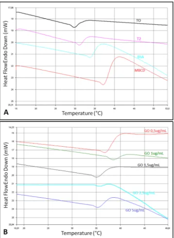
Different parameters related with sperm function that are influenced by membrane composition were assessed, in particular the effect of GO on membrane fluidity (FRAP analysis and DSC), membrane potential and ions permeability (both by flow cytometry).DSC analysis revealed substantial differences among the membrane thermograms. Figure 3 shows endo-therms corresponding to thermotropic phase transitions recorded during heating from 15 to 90 °C. A narrowing of the transition peak was observed for the sample T2 (after incubation for 2 hours) when compared to T0 (before incubation). This effect is greatest when spermatozoa are incubated with MβCD and GO 1 µg/mL. The cholesterol ratio affects also the transition peak temperature (Tm) of the samples, which increases as the concentration of
cholesterol decreases.
In particular, an increased Tm was observed for samples co-incubated with the cholesterol acceptors BSA and
MβCD, as well as in the presence of GO (Table 3). On increasing GO concentration, Tm shifted to lower
temper-atures in an approximately dose-dependent manner, indicating that the capacity to extract cholesterol is reduced when increasing the GO concentrations. The highest value of Tm, higher than that recorded for BSA and MβCD
enriched samples, was observed for the lowest concentrations of GO (0.5 and 1 µg/mL).
These results were confirmed by TGA analysis, that highlighted a behavior associated to the presence of GO in the sperm samples. In particular, the initial 4% weight loss at about 100–110 °C may be assigned to the release of CO2, CO and steam (water vapors). Since this weight loss is observed only in the GO enriched samples, except
for 0.5 GO, it is evident that this loss is connected with the presence of GO. The following step (weight loss 4%) at 200 °C was assigned to the thermal elimination of labile oxygen-containing functional groups (i.e. hydroxyl, epoxy). A further gradual weight loss was observed for GO samples above 500 °C, corresponding to the elimina-tion of more stable oxygen funcelimina-tionalities. In the temperature range from 350–500 °C there is a plateau in the GO samples curve, while the weight loss for pure spermatozoa is huge. At 350 °C it is possible to evidence a different behavior of weight loss for samples enriched at various GO concentrations. In particular, samples with a higher concentration of GO showed a reduced weight loss and an improvement in thermal stability, mainly attributable to the effect of physical barrier of GO nanosheets (Fig. 4).
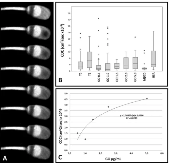
The results of FRAP analysis on one hand confirmed our previous findings20 and on the other hand evidenced
the dose-dependent effect of GO (R2 = 0.828) in the membrane fluidity increase (Fig. 5 and Supplementary
Information 3).
Figure 2. Cholesterol/Phospholipids ratio in sperm samples. Data are refered to the different experimental
Figure 3. Differential Scanning Calorimetry (DSC) analysis. Different Tm corresponding to the sperm groups
under study (treated with different GO concentrations, BSA or MβCD).
Mean Onset(°C) Peak(°C) End(°C) Peakwidth ΔH (J/g) PeakHeight Area(mJ)
T0 31,8 34,9 46,0 14,2 −11,8 −1,0 −46,1 T2 35,2 38,0 47,8 12,6 −26,3 −1,1 −56,5 BSA 34,3 37,9 53,2 18,9 −33,8 −1,5 −104,6 MβCD 34,5 37,6 50,3 15,8 −44,3 −1,5 −93,0 GO 0,5 36,1 38,9 44,2 8,1 −11,8 −1,2 −35,4 GO 1 35,1 37,4 43,5 8,4 −16,6 −0,8 −29,1 GO 1.2 36,0 38,4 48,1 12,1 −4,1 −0,6 −7,1 GO 2.5 35,7 38,2 41,9 6,1 −5,9 −0,7 −20,8 GO 5 35,5 38,7 45,8 10,2 −8,0 −1,0 −38,0
St. Dev. Onset(°C) Peak(°C) End(°C) Peakwidth ΔH (J/g) PeakHeight Area(mJ)
T0 1,6 1,3 5,5 6,9 4,1 0,5 10,0 T2 4,5 4,5 6,1 6,4 42,5 0,7 55,3 BSA 2,9 2,6 7,4 8,8 30,0 0,5 70,0 MβCD 2,5 2,5 8,5 9,4 57,7 0,6 73,7 GO 0,5 1,8 1,6 0,7 1,1 0,4 0,1 1,2 GO 1 2,9 3,0 6,2 6,5 30,1 0,6 42,4 GO 1.5 0,7 0,6 4,6 5,3 4,3 0,5 0,1 GO 2.5 0,2 0,7 3,7 4,0 7,5 0,6 23,1 GO 5 0,3 0,0 1,3 1,5 0,5 0,3 5,1
Figure 4. Thermogravimetric Analysis (TGA). Thermogram shows the differences between the sperm samples
treated with the different experimental treatments (different GO concentrations, BSA or MβCD).
Figure 5. FRAP analysis of sperm cells. (Panel A) Exemplificative gallery showing the fluorescence recovery
after photobleaching (FRAP) technique. (Panel B) Graph representing the values of Calculated Diffusion Coefficient (CDC), expressed as cm2/sec × 10−9, of sperm samples treated with the different experimental
treatments (various GO concentrations, BSA or MβCD). The box plot represent the mean, 25°–75°, 5–90° percentile and out-layers. (Panel C) Dose-dependence linking GO concentration to Calculated Diffusion Coefficient (CDC).
GO affects spermatozoa signal transduction.
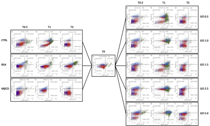
Flow cytometry analysis of spermatozoa incubated with MβCD, BSA, or GO at different concentrations displayed no effect on membrane potential (Bis-Oxonol) when compared with control groups, while GO is able to influence the intracellular calcium concentration ([Ca2+]i)
(Fig. 6A,B). Particularly, after 30 min and 1 hour of incubation an increase in the population marked positive for high [Ca2+]
i. was noticed in every sample treated with GO, independently of the concentration. This fact suggests
a possible interaction between GO and calcium channels by an unknown mechanism different to the change in the overall membrane potential, as it is evident due to the lack of signal of Bis-Oxonol.
Effect of GO on sperm-SOEC interaction.
We found no differences regarding the physical interaction of sperm cells with SOECs after pre-incubation with GO. As showed in Fig. 7, spermatozoa interact differently within the plate of cultured cells, evidencing a particular affinity to some cells compared to others. However this affinity remains unchanged in the presence of GO, indicating that preliminary incubation with GO does not interfere with the molecular mechanisms of subsequent sperm-oviduct interaction.Figure 6. Flow cytometry results. (A) CTRL, BSA and MβCD samples at different times. Spermatozoa
were stained with Fura 2-AM (x-axis) and Bis-oxonol (y-axis). (B) GO treated samples at different times. Spermatozoa were stained with Fura 2-AM (x-axis) and Bis-oxonol (y-axis).
Figure 7. Confocal image showing the interaction of live spermatozoa with swine oviductal epithelial cells.
Discussion
In a previous manuscript from our group it was demonstrated that GO, in a definite range of concentrations (0.5 to 1 µg/mL), is able to affect boar spermatozoa function leading to an increase in their fertilizing ability20. Since
this finding was potentially applicable to ART, here we carried out a chemical and functional characterization of the GO interaction with swine sperm cells to explore the molecular mechanisms involved, with the aim to pro-pose new strategies effective in control of sperm function in ART. As our previous data suggested a direct inter-action between GO and sperm membrane, without entering into the cells, the approach was mainly focused in characterizing the effect of GO on sperm membranes. Importantly, sperm membranes are not only the interface with the external environment but also the key players in biochemical signalling involved in the acquisition of fertilizing ability28,38. Indeed, male gametes are unable to synthetize new molecules and their cytosol is virtually
absent, so the main site of signal integration is the membrane, which is the key player of the capacitation process. Mammalian spermatozoa, immediately after ejaculation, are unable to fertilize the female gamete, the oocyte, while they gain the full fertilizing ability only after they reside from hours to days (depending on the species) within the female genital tract. Here, it takes place a complex dialogue between male gametes and female organ-ism (the capacitation) and spermatozoa became fertile. This process begins with the alteration of membrane physicochemical characteristics (the so-called “lipid remodelling”) (Brewis and Gadella, 2010; Gadella et al. 2008; Gadella and Luna, 2014; Bernabò et al. 2015). More in detail, the sperm plasma membrane (PM) and the outer acrosome membrane (OAM) markedly change their architecture and become able to fuse at the time of the acrosome reaction (AR), when the oocyte is reached. In lipid remodelling the cholesterol removal from PM has a pivotal role, indeed it is able to induce the increase in membrane fluidity, isotropy, and finally to allow the PM and OAM fusion (membrane fusogenicity) (Bruckbauer et al., 2010; Gadella et al. 2008; Kotwicka et al. 2011; Venditti and Bean, 2009).
The capacitation process is actively modulated by a complex functional dialogue with the oviductal envi-ronment, with the active participation of epithelial components that respond to the neuro-endocrine axis of female39,40.
Based on this biological background, we analysed the changes in the membrane chemical composition (lipi-domic analysis, measurement of cholesterol/phospholipids ratio) to explore the molecular events related to the interactions of GO and sperm membrane. In particular, the physical-chemical properties (FRAP analysis, DSC analysis and TGA), the possible effect on signalling-related biochemical events (membrane potential, intracellu-lar calcium concentration) and the interactions with swine oviductal epithelial cells (SOECs), were assessed. In all the experiments, the effects exerted by GO were compared to those exerted by serum album (bovine serum albumin, BSA), which is a physiological extracellular acceptor of cholesterol used to promote in vitro capacitation of swine spermatozoa, and methyl-β-cyclodextrin (MβCD), a stronger cholesterol extractor used for membrane engineering41–43.
First, we analysed the effect of GO during sperm incubation under capacitating conditions on sperm lipi-domic pattern. In particular, as a functional parameter, we assessed the cholesterol/phospholipids ratio, since it is a key controller of the process that leads spermatozoa to the acquisition of their fertilizing ability. As it is well known25,27,44, the incubation of spermatozoa with bicarbonate and calcium is able to activate several cAMP/
PKA-dependent pathways that lead to the activation of scramblase. That results in the translocation of cholesterol from PM of the acrosome area to the outer membrane leaflet and its consequent extraction by external acceptors. Since boar sperm membrane is characterized by a very high concentration of PUFA, the loss of cholesterol causes the increase in membrane isotropy and fluidity (collectively defined as “fusogenicity”), necessary to acquire the ability to undergo acrosome reaction when the ZP of the oocyte is met. Interestingly, in our experiment, GO was able to extract cholesterol with an efficiency similar to BSA and MβCD (p < 0.05 vs. CTRL, p > 0.05 among treat-ments), except in the case of 1 μg/ml, which showed a higher efficiency (p < 0.01 vs. CTRL). It is very suggestive that in our previous work this concentration of GO was able to promote the maximum effect in increasing the IVF efficiency, allowing us to hypothesize that cholesterol extraction mediated by GO might be one of the causes for the positive effect observed on sperm capacitation. This result confirms our previous hypothesis20 and is
sup-ported by recent in silico and in vitro studies45,46.
The effect of cholesterol extraction exerted by GO on sperm membrane physico-chemical behaviour was fur-ther characterized by physical (DSC) and biophysical (FRAP) approaches. DSC experiments gave us important information. The incubation under capacitating condition, as expected, promoted a detectable effect in terms of phase transition behaviour, when compared to T0 samples. The co-incubation with cholesterol acceptors (BSA and MβCD) was able to promote a very evident and different effect on Tm parameters, with the values shifted to
higher temperatures in relationship to a reduction of the cholesterol concentration (see Table Tm and Fig. 3), in agreement with the tendency of cholesterol containing membrane to reduce the transition temperature47. This
behaviour reflects the cholesterol-removal capacity of BSA, MβCD and GO at low concentrations. Indeed, treated spermatozoa with GO concentrations at 0.5 displayed a phase transition peak behaviour similar to that we found in MβCD-treated sperm cells, while at 1 µg/mL the peak is even higher, thus confirming the effective action of GO and strengthening the functional relevance of this treatment.
Higher concentrations of GO shift down the samples transition peak temperatures, indicating that the capac-ity of GO to extract cholesterol decrease in a dose-dependent manner (Table 3).
To better confirm this datum at the single cell level, we carried out a FRAP experiment that completed our previous analyses20. As it is evident (Fig. 5), the effect of GO in term of changes in membrane fluidity (expressed
as Calculated Diffusion Coefficient, CDC) was different from that induced by BSA and MβCD and was in a dose-dependent manner (R2 = 0.828). These findings were finally confirmed by DSC experiments, which
demon-strated the GO ability to modulate the membrane physical properties. This could likely be due to the membrane composition, since previous studies show that an increase in the membrane sterols content leads to a broad-ening of the transition peak48, evident in GO at 1 µg/mL (considering that this is the sample with the lowest
cholesterol content). Furthermore, the decrease in the transition temperature is associated with an increase in mono-unsaturated fatty acids and specifically in oleic acid content, known to reduce the phase transition temper-ature and enlarge the peak49. Once again, our data are strengthening the dose-dependency and the specificity of
GO interaction with sperm membranes.
From a functional point of view this is a remarkable finding, since the membrane rearrangement that takes place during capacitation is a key event in the spermatozoa acquisition of their full fertilizing ability. Indeed, male mammalian male gametes gain the ability to interact, recognize and fuse with the homologous oocyte only after they reside in the female genital tract or are exposed to an opportune artificial environment during in vitro procedures. In this context, the membrane is either the interface of spermatozoa with the surrounding milieu and the most important signalling system active in messages transduction.
This is the reason why we decided to study the membrane function modifications that ultimately could affect the sperm physiology. Flow cytometry experiments contribute to explain the link between the effects produced by GO on sperm membrane and cytosolic signalling. Overall, it is possible to take some important inferences. The effect on membrane potential and [Ca2+]
i exerted by BSA and MβCD is very different, either in terms of cellular
response and kinetic. In particular, as expected, BSA action is long lasting and favours cell survival, while MβCD has a rapid toxic effect. GO at higher concentrations is detrimental for cell viability and its effect is, in term of length, more similar to MβCD. Nevertheless, at lower concentrations it promotes an increase in [Ca2+]
i without
apparently affecting the membrane potential.
This is a very interesting finding, since the homeostasis of [Ca2+]
i is one of the key parameters that drives
sperm function. It is involved in the control of several function (from motility50 to cytoskeleton assembly51,52,
from lipid remodelling53 to apoptosis54) and, on the other side, it is controlled by a myriad of factors either at
intracellular and extracellular level. Nevertheless, recent reports have described a potential negative effect of GO exposure of sperm cells55, somatic stem cells56 as well as bacteria57. Here, and considering also our functional
results obtained in an earlier work, we suggest that the cause could be related to the increase on sperm fertilizing ability, as previously stated by our group20. Therefore, since calcium signalling is commonly associated to
capaci-tation, these results seem to reaffirm the role of GO as a facilitator of sperm capacitation.
Finally, as we know, sperm membrane is the interaction site of male gametes with the surrounding environ-ment and, consequently, it is involved in the information exchange that leads the spermatozoa to the acquisition of full fertilizing ability. Several reports agree in describing two different pools of male gametes within the ovi-duct: one group of spermatozoa present within the lumen, not bound to the epithelial cells and characterized by poor viability and unable to fertilize, and another group, encompassing spermatozoa attached to the OECs with potentially capacitated cells that will be able to fertilize the oocyte58. To date, the specific molecular mechanisms
involved in such interaction are not completely understood, but it has been supposed that the membrane glycoca-lyx could be involved40. Since the glycocalyx is the first interface with external molecules (such as GO) we carried
out a specific set of experiments to exclude any detrimental effect of GO at this level. In particular, we checked if spermatozoa are able to maintain their ability to interact with OECs when they are previously incubated with GO. Our results, as shown in Fig. 7, seem to confirm that the GO treatment does not affect the interaction topology, allowing to hypothesize that this molecule does not compromise the physiological pattern of sperm-epithelial cells recognition and binding, a key step on the road to fertilization.
Conclusions
We are conscious of the limitations of this study regarding the animal model and the experimental approach adopted. In particular, further experiments are needed to investigate the possible effects of GO on sperm genetic integrity56, embryo quality and implantation18 and offspring health59. Despite this, in our opinion, the present
data could have two important applications. On one hand they contribute to shed some light on the interaction between living systems and graphene (and graphene-related materials) at a molecular level. In particular, we found that their possible action in modulating membrane cholesterol content could be useful in engineering cells and/or in designing tools for diagnosis and therapy (the so-called theragnostics). On the other hand, it is possible to hypothesize the potential use of GO and GO-related materials in human and animal assisted reproduction as potential biomaterials, considering that in 2011 more than 600,000 IVF cycles have been performed only in Europe.
Methods
Chemicals.
Unless otherwise stated, all the chemicals were purchased from Sigma Aldrich (Saint Louis, Missouri). GO aqueous dispersion was purchased from Graphenea (San Sebastian, Spain). The reagents (ana-lytical grade) and HPTLC plates (Kieselgel 60) for lipid analysis were purchased from Merck KGaA (Darmstadt, Germany).preparation of graphene oxide solution.
GO aqueous dispersion was obtained by diluting a commercial sample of 4 mg/mL GO, as already described20, followed by bath ultrasonication for 10 min (Elmasonic P60H,37 kHz, 180W) and sterilization for 2 h under UV lamp (Spectronics Spectroline EF 160/C FE, 6W, 50 Hz, 0.17A). GO concentration was checked by UV-vis spectrophotometry (Varian Cary 100 BIO) at λmax 230 nm.
preparation and treatment of sperm samples.
The preparation of boar semen samples was carried out following an already standardized protocol29,60,61. In brief, sperm samples purchased from a specializedcom-pany (Suiseme s.r.l., Modena, Italy), after washing, were incubated in a capacitation medium containing TCM199 medium supplemented with 13.9 mM glucose, 1.25 mM sodium pyruvate, 2.25 mM calcium lactate and 1 mM of caffeine (300 mOsm/kg, pH 7.4), at a final concentration of 1 × 10⁷ cells/mL and up to 2 h, at 38.5 °C in 5% CO
2
(without any treatment), spermatozoa treated with different GO concentrations (5, 2.5, 1.5, 1 and 0.5 µg/mL, added to the capacitating medium), and 2 comparative conditions (0.3% BSA and 1 mM MβCD, added to the capacitating medium). Only samples that showed a motility >90% at the beginning of the incubation were con-sidered for further analysis. All the following experiments were repeated at least three times, on different boars.
evaluation of sperm membrane chemical composition after Go exposure.
Lipid analysis of sperm membrane composition by High-Performance Thin-Layer Chromatography (HPTLC). Sperm homogenates weresonicated and centrifuged at 800 g for 10 min at 4 °C to eliminate intact cells and nuclear components. Aliquots from the homogenates were used for phospholipids phosphorus determination following the Bartlett proce-dure. Then, 10 μg of phosphorus from each sample were subjected to lipid extraction according to Tettamanti’s protocol62. The lipid extracts in the organic phases were analyzed by HPTLC (High-Performance Thin-Layer
Chromatography). In particular, for phospholipid and cholesterol analysis the solvent system was chloroform/ methanol/acetic acid/water (60/45/4/2, vol/vol/vol/vol). Phospholipids and cholesterol were visualized on the same HPTLC plate with anisaldehyde reagent (0.5 mL anisaldehyde, 1 mL 97% sulfuric acid in 50 mL glacial acetic acid). After heating at 180 °C for 5 min, the plates were scanned and analyzed with ImageQuant
™
TL (GE Healthcare Life Sciences).Gas chromatography analysis of sperm glycerophospholipid fatty acid membrane composition. Sperm samples
treated as previously described were washed in DPBS and used for the extraction and transesterification of glyc-erophospholipids to fatty acid methyl esters (FAME) following a previously described protocol63. Briefly, 6 × 10⁸
sperm cells/treatment were resuspended in 200 µL of DPBS, 200 µL of distilled water and 500 µL of methanol and stored at −80 °C until extraction. After thawing, 2100 µL of methanol were added and mixed by vortex, fol-lowed by 10 min in a shaker at 250 rpm. After treatment in ultrasonic bath for 5 min, samples were centrifuged for 10 min at 4000 g to separate the cell fragments from the methanolic fraction containing the lipids. Fatty acid esterification reaction was initiated by adding 100 µL of sodium methoxide solution (25wt % in methanol). The reaction was performed at RT and stopped after 4 min by adding 300 uL of methanolic HCl (3 M). FAME were extracted with 1 mL of hexane and recovered in a new vial. The hexane recovered fraction was mixed with 2 mL of 6% potassium carbonate solution followed by 10 min centrifugation at 400 g to separate the phases. The hexane phase was recovered and FAME were dried by a steady nitrogen flow. Extracts were stored at −20 °C until quanti-fication and dissolved in 30 µL of hexane immediately before gas chromatography. FAME were quantified via gas chromatography by a standard procedure using a capillary column ZB-FAME, 30 m × 0.25 mm, film thickness 0.20 µM (Phenomenex, USA) in a 7820 A GC System (Agilent Technologies, Santa Clara, California, EUA). The data was processed using the GC7820 data System (Agilent Technologies, Santa Clara, California, EUA).
evaluation of sperm membrane physical composition after Go exposure.
Evaluation of sperm membrane physical properties by Differential Scanning Calorimetry (DSC) and Thermogravimetrical analysis (TGA). Once capacitation time was completed, 8 × 10⁸ spermatozoa (8 × 10⁸) were washed with DPBS andmembrane enriched fraction (MEF) was isolated according to an adapted protocol44. Spermatozoa were
resus-pended in hypotonic buffer (2 mM Tris, pH 7.2, 12 mM NaCl), then they were sonicated six times for 15 min (with intervals of 1 min between each sonication) and subsequently centrifuged (1600 g for 15 min). The pellet was resuspended in 1 mM potassium phosphate buffer (pH 7.4, 250 mM of sucrose and 0.1 mM of EDTA) and centrifuged (1600 g for 15 min) 3 times in the same buffer. The pooled supernatants were centrifuged at 100.000 g for 1 h and the recovered pellet was lyophilized for 24 h at −20 °C at a pressure of 0.3 hPa. For DSC analysis, MEFs were resuspended in deionized water in a 2:1 ratio. DSC heating thermograms were obtained at a scan rate of 10 °C/min in a range between 15 and 90 °C using a Differential Scanning Calorimeter DSC-7 (Perkin-Elmer Inc., Waltham MA, USA) coupled with a Thermal Analysis Controller TAC 7/DX (Perkin-Elmer Inc., Waltham MA, USA)64. The acquired data were analyzed and plotted using the PYRIS Software version 4.02 (Perkin-Elmer Inc.,
Waltham, MA).
To further characterize the effect of GO on membrane physical properties, we carried out a thermogravimetric analysis. More in detail, MEF from sperm samples, obtained as above described, have been analysed with a Pyris 1 TGA Thermogravimetric Analyzer with the following set up: heat from 30 °C to 800 °C at 20 °C/min; heat from 800 °C to 900 °C at 20 °C/min; nitrogen flow at 40 mL/min.
Membrane fluidity assessment by Fluorescence Recovery After Photobleaching (FRAP) analysis. To study the
changes in sperm membrane fluidity induced by different treatments, FRAP analysis were carried out, as already described20. In brief, washed spermatozoa were stained with the fluorescent probe DilC12(3) perchlorate
(ENZ-52206, Enzo Life Sciences, USA), at a concentration of 5 µM in PBS for 20 min (38.5 °C, 5% CO2 humidified
atmosphere). FRAP experiments were performed at T0 and T2 (after two hours in capacitating medium) using a Nikon A1r laser confocal scanning microscope. Recovery curves were realized and analyzed by using the sim-FRAP plug-in for Fiji ImageJ65, expressing the results as calculated diffusion coefficient (cm2/sec).
evaluation of sperm membrane biological properties after Go exposure.
Intracellular calcium concentration and membrane potential assessment by Flow Cytometry. To analyze the effect of GO oncal-cium intake, spermatozoa were incubated with Fura 2-AM (F1221, Invitrogen) (final concentration of 2 µM) and pluronic acid 0.25%). The effect of GO on plasma membrane potential was monitored by using Bis-oxonol (D8189, Sigma) at the final concentration of 0.02 mM, whereas 7AAD was used as a probe for vitality (50 µg/mL). Co-incubation was carried out for 30 min (38.5 °C, 5% CO2, humidified atmosphere) and samples were assessed
For each condition and time point, 10.000 events were acquired by flow cytometer (FACSVerse, BD Biosciences - three laser, eight colors configuration, or FACSCanto, BD Biosciences - three laser, eight colors configuration). Each reagent was titrated (8 points titration) under assay conditions and dilutions were estab-lished based on achieving the highest signal (Mean Fluorescence Intensity, MFI) for the positive population and the lowest signal for the negative population, and stain indexes were calculated. Instrument performances, data reproducibility and fluorescence calibrations were sustained and checked by the Cytometer Setup & Tracking Beads (BD Biosciences). In order to evaluate non-specific fluorescence, Fluorescence Minus One (FMO) controls were used. Compensation was assessed using CompBeads and FACSuite FC Beads (BD Biosciences) and single stained fluorescent samples. Data were analyzed using FACSuite v 1.0.5 (BD Biosciences) software.
evaluation of sperm membrane interaction with swine oviductal epithelial cells (soeC).
To assess the potential effects of GO on sperm interaction with the oviductal epithelium, swine oviductal epithelial cells (SOEC) were obtained from oviducts collected at a local slaughterhouse and kept to the laboratory in an isothermal contained within 1 h. Then, following a validated protocol and cultured in 3.5 cm Petri dishes with a cover glass at 104 cells/ml for 1 week (38.5 °C, 5% CO2 humidified atmosphere). Medium (α-Mem supplemented
with 10% FCS, 2% Pen/Strep and 1% ultraglutamine) was refreshed every two days. Once cells reached the con-fluence, they were coincubated with spermatozoa, previously incubated 30 min under capacitating conditions, with or without GO 1 µg/mL. After 3 washes with DPBS to remove the unbound spermatozoa, the ensemble SOEC-spermatozoa were stained with HOECHST 33342 (9 µM), 5(6)-Carboxyfluorescein diacetate (100 µM) to identify the alive sperm cells and DilC12(3) (5 µM) to delineate the membranes.
References
1. Li, Z. et al. Field and temperature dependence of intrinsic diamagnetism in graphene: Theory and experiment. Phys. Rev. B 91, 094429 (2015).
2. Yoo, J. M., Kang, J. H. & Hong, B. H. Graphene-based nanomaterials for versatile imaging studies. Chem. Soc. Rev. 44, 4835–4852 (2015).
3. Bartelmess, J., Quinn, S. J. & Giordani, S. Carbon nanomaterials: multi-functional agents for biomedical fluorescence and Raman imaging. Chem. Soc. Rev. 44, 4672–4698 (2015).
4. Miao, W. et al. Image-guided synergistic photothermal therapy using photoresponsive imaging agent-loaded graphene-based nanosheets. J. Control. Release 211, 28–36 (2015).
5. Chen, D., Dougherty, C. A., Zhu, K. & Hong, H. Theranostic applications of carbon nanomaterials in cancer: Focus on imaging and cargo delivery. J. Control. Release 210, 230–245 (2015).
6. Orecchioni, M., Cabizza, R., Bianco, A. & Delogu, L. G. Graphene as Cancer Theranostic Tool: Progress and Future Challenges.
Theranostics 5, 710–723 (2015).
7. Liu, J., Dong, J., Zhang, T. & Peng, Q. Graphene-based nanomaterials and their potentials in advanced drug delivery and cancer therapy. J. Control. Release 286, 64–73 (2018).
8. Ding, X., Liu, H. & Fan, Y. Graphene-Based Materials in Regenerative Medicine. Adv. Healthc. Mater. 4, 1451–1468 (2015). 9. Menaa, F., Abdelghani, A. & Menaa, B. Graphene nanomaterials as biocompatible and conductive scaffolds for stem cells: impact for
tissue engineering and regenerative medicine. J. Tissue Eng. Regen. Med. 9, 1321–1338 (2015).
10. Wang, Z. & Dai, Z. Carbon nanomaterial-based electrochemical biosensors: an overview. Nanoscale 7, 6420–6431 (2015). 11. Heerema, S. J. & Dekker, C. Graphene nanodevices for DNA sequencing. Nat. Nanotechnol. 11, 127–136 (2016).
12. Yang, K., Feng, L. & Liu, Z. The advancing uses of nano-graphene in drug delivery. Expert Opin. Drug Deliv. 12, 601–612 (2015). 13. Muthoosamy, K., Bai, R. & Manickam, S. Graphene and Graphene Oxide as a Docking Station for Modern Drug Delivery System.
Curr. Drug Deliv. 11, 701–718 (2014).
14. Chatterjee, N. et al. A systems toxicology approach reveals the Wnt-MAPK crosstalk pathway mediated reproductive failure in
Caenorhabditis elegans exposed to graphene oxide (GO) but not to reduced graphene oxide (rGO). Nanotoxicology 11, 76–86 (2017).
15. Nirmal, N. K., Awasthi, K. K. & John, P. J. Effects of Nano-Graphene Oxide on Testis, Epididymis and Fertility of Wistar Rats. Basic
Clin. Pharmacol. Toxicol. 121, 202–210 (2017).
16. Souza, J. P., Venturini, F. P., Santos, F. & Zucolotto, V. Chronic toxicity in Ceriodaphnia dubia induced by graphene oxide.
Chemosphere 190, 218–224 (2018).
17. Asghar, W. et al. Toxicology Study of Single-walled Carbon Nanotubes and Reduced Graphene Oxide in Human Sperm. Sci. Rep. 6, 30270 (2016).
18. Mesarič, T. et al. Sperm exposure to carbon-based nanomaterials causes abnormalities in early development of purple sea urchin (Paracentrotus lividus). Aquat. Toxicol. 163, 158–166 (2015).
19. Liang, S., Xu, S., Zhang, D., He, J. & Chu, M. Reproductive toxicity of nanoscale graphene oxide in male mice. Nanotoxicology 9, 92–105 (2015).
20. Bernabò, N. et al. Graphene oxide affects in vitro fertilization outcome by interacting with sperm membrane in an animal model.
Carbon N. Y. 129, 428–437 (2018).
21. Yanagimachi, R. Fertility of mammalian spermatozoa: its development and relativity. Zygote 2, 371–372 (1994). 22. Suarez, S. S. Regulation of sperm storage and movement in the mammalian oviduct. Int. J. Dev. Biol. 52, 455–462 (2008). 23. Romarowski, A. et al. PKA-dependent phosphorylation of LIMK1 and Cofilin is essential for mouse sperm acrosomal exocytosis.
Dev. Biol. 405, 237–249 (2015).
24. Barboni, B. et al. Type-1 cannabinoid receptors reduce membrane fluidity of capacitated boar sperm by impairing their activation by bicarbonate. PLoS One 6, e23038 (2011).
25. Boerke, A. et al. Involvement of bicarbonate-induced radical signaling in oxysterol formation and sterol depletion of capacitating mammalian sperm during in vitro fertilization. Biol. Reprod. 88, 21 (2013).
26. Bernabò, N. et al. Endocannabinoid-binding CB1 and TRPV1 receptors as modulators of sperm capacitation. Commun. Integr. Biol.
5, 68–70 (2012).
27. Gadella, B. M. & Harrison, R. A. P. Capacitation induces cyclic adenosine 3′,5′-monophosphate-dependent, but apoptosis-unrelated, exposure of aminophospholipids at the apical head plasma membrane of boar sperm cells. Biol. Reprod. 67, 340–350 (2002). 28. Gadella, B. M. & Luna, C. Cell biology and functional dynamics of the mammalian sperm surface. Theriogenology 81, 74–84 (2014). 29. Bernabò, N. et al. The role of actin in capacitation-related signaling: an in silico and in vitro study. BMC Syst. Biol. 5, 47 (2011). 30. Daniel, L. et al. Regulation of the sperm EGF receptor by ouabain leads to initiation of the acrosome reaction. Dev. Biol. 344,
650–657 (2010).
31. Cohen, G., Rubinstein, S., Gur, Y. & Breitbart, H. Crosstalk between protein kinase A and C regulates phospholipase D and F-actin formation during sperm capacitation. Dev. Biol. 267, 230–241 (2004).
33. Rajamanickam, G. D., Kastelic, J. P. & Thundathil, J. C. Na/K-ATPase regulates bovine sperm capacitation through raft- and non-raft-mediated signaling mechanisms. Mol. Reprod. Dev. 84, 1168–1182 (2017).
34. Gadella, B. M. Sperm membrane physiology and relevance for fertilization. Anim. Reprod. Sci. 107, 229–236 (2008).
35. Hossain, M. S., Afrose, S., Sawada, T., Hamano, K. & Tsujii, H. Metabolism of exogenous fatty acids, fatty acid-mediated cholesterol efflux, PKA and PKC pathways in boar sperm acrosome reaction. Reprod. Med. Biol. 9, 23–31 (2010).
36. Viviani Anselmi, C. et al. Fatty acid percentage in erythrocyte membranes of atrial flutter/fibrillation patients and controls. J. Interv.
Card. Electrophysiol. 27, 95–99 (2010).
37. Mendeluk, G. R., Cohen, M. I., Ferreri, C. & Chatgilialoglu, C. Nutrition and Reproductive Health: Sperm versus Erythrocyte Lipidomic Profile and ω −3 Intake. J. Nutr. Metab. 2015, 1–8 (2015).
38. Leahy, T. & Gadella, B. M. New insights into the regulation of cholesterol efflux from the sperm membrane. Asian J. Androl. 17, 561–7 (2015).
39. Li, S. & Winuthayanon, W. Oviduct: roles in fertilization and early embryo development. J. Endocrinol. 232, R1–R26 (2017). 40. Avilés, M., Gutiérrez-Adán, A. & Coy, P. Oviductal secretions: will they be key factors for the future ARTs? MHR Basic Sci. Reprod.
Med. 16, 896–906 (2010).
41. Oddi, S. et al. Functional characterization of putative cholesterol binding sequence (CRAC) in human type-1 cannabinoid receptor.
J. Neurochem. 116, 858–865 (2011).
42. Sanchez, S. A., Gunther, G., Tricerri, M. A. & Gratton, E. Methyl-β-cyclodextrins preferentially remove cholesterol from the liquid disordered phase in giant unilamellar vesicles. J. Membr. Biol. 241, 1–10 (2011).
43. van Gestel, R. A., Helms, J. B., Brouwers, J. F. H. M. & Gadella, B. M. Effects of methyl-β-cyclodextrin-mediated cholesterol depletion in porcine sperm compared to somatic cells. Mol. Reprod. Dev. 72, 386–395 (2005).
44. Botto, L., Bernabò, N., Palestini, P. & Barboni, B. Bicarbonate Induces Membrane Reorganization and CBR1 and TRPV1 Endocannabinoid Receptor Migration in Lipid Microdomains in Capacitating Boar Spermatozoa. J. Membr. Biol. 238, 33–41 (2010). 45. Zhang, L., Xu, B. & Wang, X. Cholesterol Extraction from Cell Membrane by Graphene Nanosheets: A Computational Study. J. Phys.
Chem. B 120, 957–964 (2016).
46. Fabbro, A. et al. Graphene-Based Interfaces Do Not Alter Target Nerve Cells. ACS Nano 10, 615–623 (2016).
47. Bach, D. & Wachtel, E. Phospholipid/cholesterol model membranes: formation of cholesterol crystallites. Biochim. Biophys. Acta -
Biomembr. 1610, 187–197 (2003).
48. Benesch, M. G. K. & McElhaney, R. N. A comparative differential scanning calorimetry study of the effects of cholesterol and various oxysterols on the thermotropic phase behavior of dipalmitoylphosphatidylcholine bilayer membranes. Chem. Phys. Lipids 195, 21–33 (2016).
49. Inoue, T., Yanagihara, S., Misono, Y. & Suzuki, M. Effect of fatty acids on phase behavior of hydrated dipalmitoylphosphatidylcholine bilayer: saturated versus unsaturated fatty acids. Chem. Phys. Lipids 109, 117–133 (2001).
50. Breitbart, H. & Naor, Z. In Male Sterility and Motility Disorders 45–53, https://doi.org/10.1007/978-1-4612-1522-6_4 (Springer New York, 1999).
51. Breitbart, H., Cohen, G. & Rubinstein, S. Role of actin cytoskeleton in mammalian sperm capacitation and the acrosome reaction.
Reproduction 129, 263–268 (2005).
52. Brener, E. et al. Remodeling of the actin cytoskeleton during mammalian sperm capacitation and acrosome reaction. Biol. Reprod.
68, 837–45 (2003).
53. Baldi, E., Luconi, M., Bonaccorsi, L., Krausz, C. & Forti, G. Human sperm activation during capacitation and acrosome reaction: role of calcium, protein phosphorylation and lipid remodelling pathways. Front. Biosci. 1, d189–205 (1996).
54. Bejarano, I. et al. In Male Infertility, https://doi.org/10.5772/32617 (InTech, 2012).
55. Hashemi, E. et al. Cyto and genotoxicities of graphene oxide and reduced graphene oxide sheets on spermatozoa. RSC Adv. 4, 27213 (2014).
56. Akhavan, O., Ghaderi, E. & Akhavan, A. Size-dependent genotoxicity of graphene nanoplatelets in human stem cells. Biomaterials
33, 8017–8025 (2012).
57. Akhavan, O. & Ghaderi, E. Toxicity of Graphene and Graphene Oxide Nanowalls Against Bacteria. ACS Nano 4, 5731–5736 (2010). 58. López-Úbeda, R., García-Vázquez, F., Gadea, J. & Matás, C. Oviductal epithelial cells selected boar sperm according to their
functional characteristics. Asian J. Androl. 19, 396 (2017).
59. Yang, X. et al. Developmental neurotoxicity and immunotoxicity induced by graphene oxide in zebrafish embryos. Environ. Toxicol. tox.22695, https://doi.org/10.1002/tox.22695 (2018).
60. Maccarrone, M. et al. Characterization of the endocannabinoid system in boar spermatozoa and implications for sperm capacitation and acrosome reaction. J. Cell Sci. 118, 4393–4404 (2005).
61. Bernabò, N., Pistilli, M. G., Mattioli, M. & Barboni, B. Role of TRPV1 channels in boar spermatozoa acquisition of fertilizing ability.
Mol. Cell. Endocrinol. 323, 224–231 (2010).
62. Tettamanti, G., Bonali, F., Marchesini, S. & Zambotti, V. A new procedure for the extraction, purification and fractionation of brain gangliosides. Biochim. Biophys. Acta 296, 160–70 (1973).
63. Klem, S., Klingler, M., Demmelmair, H. & Koletzko, B. Efficient and Specific Analysis of Red Blood Cell Glycerophospholipid Fatty Acid Composition. PLoS One 7, e33874 (2012).
64. Wolf, D. E., Maynard, V. M., McKinnon, C. A. & Melchior, D. L. Lipid domains in the ram sperm plasma membrane demonstrated by differential scanning calorimetry. Proc. Natl. Acad. Sci. USA 87, 6893–6 (1990).
65. Blumenthal, D., Goldstien, L., Edidin, M. & Gheber, L. A. Universal Approach to FRAP Analysis of Arbitrary Bleaching Patterns. Sci.
Rep. 5, 11655 (2015).
Acknowledgements
J.M.S. research is supported by Rep-Eat-H2020-MSCA-COFUND 713714. M.R.S. research is supported by MSCA-ITN REP-BIOTECH 675526. For TGA analysis, we would like to thank Auserpolimeri S.R.L. 55025 Piano di Coreglia (Lucca) Italy.
Author Contributions
N.B. designed the experiments and carried out the data analysis; J.M.S., M.R.S. and G.C. performed the experiments involving spermatozoa and somatic cells culture and staining; L.V. performed the experiment with confocal microscope and realized all the figures; A.F. and R.Z. prepared and characterized the graphene oxide solutions; M.M. and P.L. performed the flow cytometry experiments; P.P. and L.B. performed the cholesterol/ phospholipids analyses; J.M.S., M.C. and A.D.S. performed the DSC experiments; E.F. and M.S. performed the lipidomic experiments; B.B. was involved in critically revising the manuscript; N.B., J.M.S., M.R.S., L.V. wrote the manuscript with the involvement of all the others co-Authors; all Authors read, corrected and approved the final version of this manuscript.
Additional Information
Supplementary information accompanies this paper at https://doi.org/10.1038/s41598-019-44702-5.
Competing Interests: The authors declare no competing interests.
Publisher’s note: Springer Nature remains neutral with regard to jurisdictional claims in published maps and
institutional affiliations.
Open Access This article is licensed under a Creative Commons Attribution 4.0 International
License, which permits use, sharing, adaptation, distribution and reproduction in any medium or format, as long as you give appropriate credit to the original author(s) and the source, provide a link to the Cre-ative Commons license, and indicate if changes were made. The images or other third party material in this article are included in the article’s Creative Commons license, unless indicated otherwise in a credit line to the material. If material is not included in the article’s Creative Commons license and your intended use is not per-mitted by statutory regulation or exceeds the perper-mitted use, you will need to obtain permission directly from the copyright holder. To view a copy of this license, visit http://creativecommons.org/licenses/by/4.0/.