Typical Leishmaniosis in a Dog Regularly
Vaccinated with Canileish
R
Alessandra Gavazza, Anyela A. Medina Valentin, George Lubas
* AbstractThe vaccine Canileish R is distributed in Europe to reduce the risk of developing an active infection and
clinical leishmaniosis. An English Setter dog vaccinated with Canileish R and treated with anti-feeding
and repellent medications showed typical clinical signs of leishmaniosis. The dog was presented with dysorexia, weight loss, fever and forelimb lameness. The physical exam revealed moderate generalized external lymph node enlargement, sero-purulent ocular discharge, photophobia, and swollen and painful right carpal joint. Clinico-pathological findings revealed moderate microcytic-hypochromic non-regenerative anemia, mild neutropenia and thrombocytopenia, hyperglobulinemia, hematuria and mild elevation of urine protein-to-creatinine ratio, polyclonal peak in the gamma globulins, Leishmania spp. amastigotes in lymph nodes and bone marrow, and immunofluorescence antibody titer (IFAT) of 1:5120. The successful treatment included meglumine antimonate and allopurinol for 40 days, and metronidazole-spyramicin for 24 days. The dog was monitored up to 9 months and normalization of most hemato-biochemical abnormalities was achieved. The bone marrow qPCR for Leishmania infantum was negative, while IFAT was 1:160. Despite the systematic leishmaniosis prevention, the typical clinical disease can occur.
Keywords
leishmaniosis — dog — Canileish R — clinico-pathological findings — treatment
Dept Veterinary Sciences, University of Pisa, Via Livornese Lato Monte, San Piero a Grado 56122, Pisa, Italy *Corresponding author: [email protected]
Introduction
A vaccine for canine leishmaniosis (Canileish R) has been
introduced in several European countries including Italy1,
starting from 2011. Its formulation origins from LiESAp (culture supernatant of L. infantum promastigotes) and the 54-kDa excreted protein of Leishmania infantum in addition to muramyl dipeptide (MDP) as adjuvant. The vaccine is designed to stimulate an active immunity (cell-mediated) in Leishmaniasero-negative dogs in order to reduce the risk of developing an active infection (12% in vaccinated vs. 33% in control dogs) and related clinical disease (7% vs. 23%) after the exposure to L. infantum. The overall coverage is expected to reach 68% (2, 3, 7, 8, 9). Recently, a case series of sixteen dogs affected by clinical leishmaniosis pre-viously vaccinated with Canileish R have been reported as
an Abstract (10). Here, a case of a dog regularly immunized with Canileish R and treated with anti-feeding and repellent
medications presenting with a severe form of leishmaniosis, is described.
Material and Methods
“Alba”, an English setter dog, intact female of about 4 years old was referred to the authors’ facility for dysorexia, weight loss, fever and forelimb lameness (beginning of March 2015). “Alba” was living with 2 other dogs (both nega-tive on Leishmania IFAT, Indirect Fluorescence Antibody test and conjunctival PCR, Polymerase Chain Reaction) in
1Canileish; VirbacS.r.l., Milan, Italy (A.I.C., Italian marketing
autho-rization, #104281062)
Northern Tuscany (Italy), and was used for hunting. “Alba” was regularly vaccinated since the age of 3 months with vac-cines against distemper, viral hepatitis, adenovirus, and lep-tospirosis2. Furthermore, from the age of 9 months she was regularly vaccinated against Leishmania using Canileish R
according to the manufacturer’s instruction. Before the vac-cination with Canileish R, “Alba” underwent a complete
physical examination and partial hemato-biochemical anal-ysis including a Speed-Leish K R test. Following the
man-ufacturer’s instruction on Canileish R, prophylaxis against
ectoparasites was concurrently applied (repellent and anti-feeding, imidacloprid and permethrin3, heartworm preven-tion with moxidectin4). After the admission (T0) a follow-up period of 9 months at different days (T6, T10, T24, T42, T85, T148, T265) for “Alba” was performed.
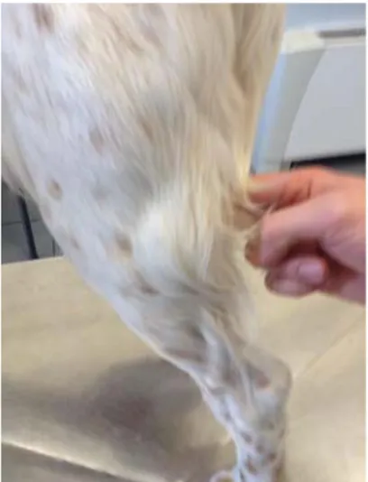
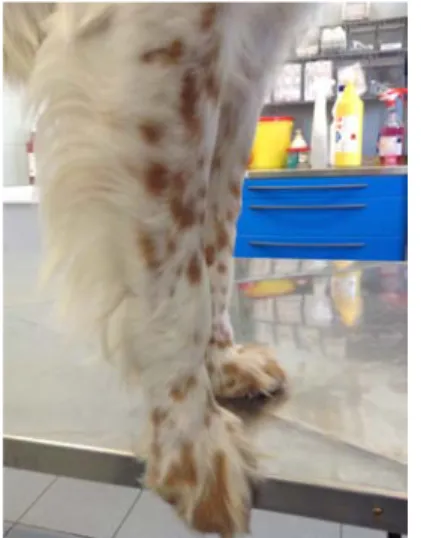
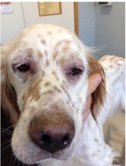
At physical examination the body condition score was 2/9 (Figure 1 and 2) and the temperature was 39.6◦C. In addition, moderate generalized peripheral lymph node en-largement (Figure 3 and 4), sero-purulent ocular discharge with photophobia (Figure 5) and swollen and painful right carpal joint (Figure 6), were recorded. Complete blood count (CBC) including the stained blood smear evalua-tion, serum biochemical profile (including total calcium, phosphates, iron, total proteins, albumin, fructosamine, C-reactive protein, urea, creatinine, total bilirubin, choles-terol, triglycerides, glucose, alkaline phosphatase,
gam-2Nobivac CEPPi+L; Intervet Italia S.r.l., Peschiera Borromeo, MI, Italy 3Advantix; Bayer Animal Health, Milan, Italy
4Guardian Sr injectable; Eli Lilly Italia S.p.a., Sesto Fiorentino, FI,
Italy
Figure 1.Body condition score 2/9 from the rear view at T0
Figure 2.Body condition score 2/9 from the front view at T0
maglutamyl transferase, aspartate amino transferase, alanine amino transferase, creatin phosphokinase, lactate dehydro-genase, amylase, sodium, potassium, chloride and bicar-bonate), coagulation profile (including prothrombin time, partial thromboplastin time, and fibrinogen), serum protein electrophoresis (separation on albumin, alpha 1 and 2 globu-lins, beta 1 and 2 globuglobu-lins, and gamma globulins), serology for L.infantum (cut-off value: negative for exposure, 1:160 for possible disease), complete urinalysis including UP/C ratio, fine needle aspirations for cytology of prescapular and popliteal lymph nodes and aspiration of bone marrow, were performed. A sample of bone marrow aspirate was also submitted for Leishmania quantitative PCR (qPCR).
Results
The presenting physical complaints resolved at different times: body weight progressively gained and normalized at T42, fever lasted for 10 days, photophobia disappeared by day 10 but blepharitis occurred at T3 (Figure 7 and 8), persisted at T10 (Figure 9), and disappeared at T24 (Figure 11), sero-purulent ocular discharge decreased over 15 days, lymph nodes were almost normal by T24, and forelimb
lameness disappeared by T3. It should be noticed that at T10 a large ulcer at the tail was evidenced (Figure 10), but almost completely recovered at T24 (Figure 12).
The CBC initially showed a moderate microcytic-hy-pochromic non-regenerative anemia (Hematocrit, Hct 23.9%, reference ranges, RR, 37.3-61.7%; Mean Corpuscular Vol-ume, MCV 54.7 fl, RR 61.6-73.5; Mean Corpuscular He-moglobin Content, MCHC 35.6 g/dL, RR 32.0-37.9; Mean Corpuscular Volume, MCH 19.5 pg, RR 21.2-26.9; reticulo-cytes 17.0 K/µL, RR 10-110), mild neutropenia (3.0 K/µL, RR 3.7-11.9), normal lymphocyte count with reactive lym-phocyte and mild thrombocytopenia (132 K/µL, RR 148-484). Starting from T3, the microcytic hypochro-mic RBCs slowly returned to normal by T85, the neutrophil count nor-malized by T10, the reactive lymphocytes disappeared from blood smears by T85 and the platelet count normalized by T10. The CBC was normal at T148 although few nucleated RBCs and Howell-Jolly bodies were observed. At T265 the CBC was within the reference ranges for all parameters.
The biochemical profile showed, initially, severe hy-perproteinemia (11.5 g/dL, RR 5.8-7.8) (normal at T85) with low albumin (1.9 g/dL, RR 2.6-4.1) (normal at T42), hyperglobulinemia (9.7 g/dL, RR 2.5-4.5) and low
albu-Figure 3.Popliteal lymph node enlargement at T0
Figure 4.Prescapular lymph node enlargement at T0
Figure 5.Photophobia at T0 Figure 6.Carpal joint swollen at T0
min/globulins ratio (0.2, RR 0.5-1.5) (both normal at T85), elevated C-reactive protein (2.3 mg/dL, RR 0.0-0.3) (nor-mal at T24), slight increase of AST (75 U/L, RR 15-40) and LDH (338 U/L, RR 20-160) (normal at T24). Then, from T42 the biochemical profile was unrewarding and all parameters were within the reference ranges.
The coagulation profile was investigated at T0, T27, T148 and T265, and was always normal during the disease course.
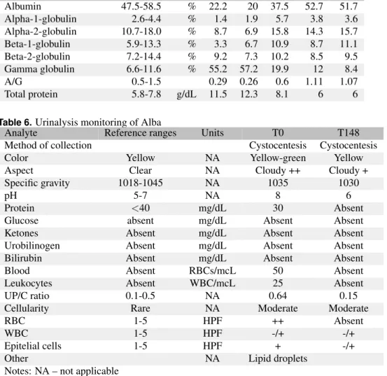
The serum protein electrophoresis at T0 (Figure 13A) showed hypoalbuminemia (22.2%) and a polyclonal peak in the gamma globulins (55.2%) that returned slowly to normal at T148 (Figure 13B). At T265 this assay was unremarkable. The urinalysis initially showed hematuria (normal at T6) and mild elevation of urine protein-to-creatinine ratio (0.64, RR 0.1-0.5) (normalized at T85). In successive samplings all the urine parameters were within the references ranges. At admission (T0) the lymph-node aspirates showed lympho-plasmacellular hyperplasia with Leishmania ama-stigotes extracellularly and in the macrophages; further aspi-rations were not performed because the lymph nodes were almost not palpable by T42. The bone marrow aspirate at T0 yielded mild hyperplasia of myeloid series (as compared to erythroid series), and slight infiltration with plasmacells and macrophages showing Leishmania amastigotes. The
bone marrow aspirate at T148 yielded mild dysplasia in the myeloid series and slight prevalence of erythroid cells (as compared to myeloid). At T265 the bone marrow evaluation was normal.
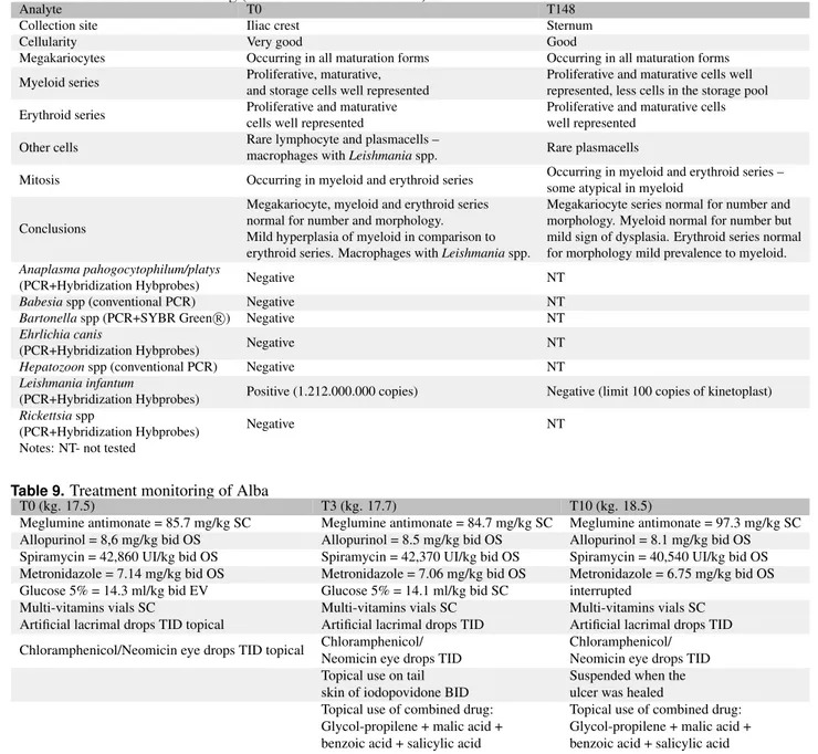
Quantitative PCR (qPCR) performed on bone marrow samples was negative for Babesia spp, Bartonella spp, Ehrlichia canis, Hepatozoon spp, and Rickettsia spp but extremely positive for L. infantum (1,212,000,000 copies). The bone marrow qPCR, repeated at T148 and T265, was negative for L. infantum. At admission (T0) IFAT titer was extremely high (1:5120) and the Speed-Leish K R assay for
Leishmaniaspp. was positive. The IFAT titer was reduced at T148 (1:320) and T265 (1:160) whereas the Speedy-Leish K R test was still positive.
“Alba” was treated with a combination of meglumine antimonate5(subcutaneous, SC) and allopurinol6(per os, PO) for 40 days; the dosage of the former was progressively increased from 85 to 94 mg/kg, q24h while the dosage of the latter was tapered from 8.6 to 7.8 mg/kg, q12h. Later, allopurinol was administered from day 42 to day 85 at 7.4 mg/kg, q12h, from day 85 to day 148 at 4.8 mg/kg, q12h, and then discontinued at T265. A course of
metronidazole-5Glucantime, Merial Italia S.p.a., Milan, Italy 6Allopurinolo generic, Sandoz S.p.a., Origgio, VA, Italy
Figure 9.Blepharitis of both eyes at T10 Figure 10.Alopecia and ulcer on the tail at T10
spyramicin7(PO) was given up until T24 (starting at 42,860 IU/kg + 7.14 mg/kg and ending at 40,540 IU/kg + 6.75 mg/kg). Fluid therapy (saline and dextrose 5%) was pro-vided for about 10 days, the topical treatment for the ocular complication based on eye-drops with a combination of
7Stomorgyl, Merial Italia S.p.a., Milan, Italy
neomycin and chloramphenicol8(q12h) was discontinued at T40, and the topical treatment for the skin lesion at the tail was started at T6 and continued up to T24 with a com-bination of iodopovidone9and ointment based on
glycol-8Antibioptal, Farmila-Thea Farmaceutici S.p.a., Settimo Milanese, MI,
Italy
9Iodopovidone, Farmacare S.r.l., San Pietro in Casale, BO, Italy
Figure 11.Blepharitis of the eyes in improvement at T24
Figure 12.Alopecia and complete recovery of the ulcer in the tail at T24
Figure 13.(A) serum protein electrophoresis showing a polyclonal peak in the gamma globulins at T0; (B) serum protein electrophoresis normalized at T148
propylene + malic acid + benzoic acid + salicylic acid10
administered both at q12h.
Discussion and conclusion
This case describes a typical clinical presentation of leish-maniosis in a dog where breakdown of Leishmania vaccine or ineffective/insufficient immunity can be documented (4, 6). The case was promptly reported to the Italian Agency for Drug Surveillance in Veterinary Medicine (5). The dis-tinction between the breakdown and ineffective/insufficient immunity could not be established as it is not common to test for ineffective/insufficient immunity as in other infec-tious disease. Indeed, all the serological assays available are directed at assessing the contact or exposure to Leishmania by a dog that could develop active infection and/or clinical disease (1).
Recently, a poster presented in an European Conference reported a case series of sixteen dogs affected by clini-cal signs of leishmaniosis, and previously vaccinated with Canileish R in Spain (10). The classical clinical presentation
of leishmaniosis was observed in all cases and the diagnosis was based on direct visualization of Leishmania amastig-otes by cytology, and indirectly by immunohistochemistry or molecular techniques. The majority of cases included were purebred, large and male dogs. Most of the dogs were diagnosed with clinical leishmaniosis prior to the first an-nual re-vaccination, and the time span between vaccination and appearance of clinical illness was 2-12 months.
The dog described in this case came from an area where leishmaniosis is known to be endemic and, therefore, at risk of developing the disease. However, the owner, a physician strictly bounded with the dog, confirmed that the use of repellent and anti-feeding agents was correctly provided to each of the three dogs living all together. All vaccination schemes were also accurately performed. Indeed, Alba was receiving the first vaccination plan in 2012 at 9 months of age, and the annual boosters in 2013 and 2014 spring-time. The time elapsed from the last vaccination to the appearance of clinical signs of leishmaniosis was about 10 months. Before each annual booster the dog was screened with Speedy-Leish K R test that resulted negative.
“Alba” showed severe and typical clinical and clinico-pathological signs of Leishmania infection, and promptly responded to standard therapy (1, 9). All the clinical signs described resolved by T27, while the clinico-pathological signs returned to normal at different times. Indeed, while CBC results progressively returned to normal at T265, it should be noticed that at T165 the appearance of few nucle-ated RBCs along evidence of Howell-Jolly bodies coupled with bone marrow aspirate results showed some signs of hemopoetic dysplasia both in erythroid and myeloid se-ries. Thereafter, the bone marrow sampling at T265 was normal. The occurrence of microcytic-hypochromic non-regenerative anemia at presentation may suggest that the clinical disease was started being present at least 3-4 months before the first admission, as RBCs half-life is 110 days. Therefore, the dog might have been infected in the summer-autumn 2014.
10Dermaflon, Zooetis Italia S.r.l, Rome, Italy
The biochemical profile was unrewarding from T42, and on the following monitoring. Surprisingly, the coagu-lation profile was always normal and particularly, the fib-rinogen was never elevated despite the clinical and clinic-pathological evidence of an inflammatory process. The hallmark of the chronic disease often used for monitoring the leishmaniosis course besides the serum protein elec-trophoresis, was strongly modified at the initial stages but returned to normal at T148. The urinalysis followed almost the serum biochemical pattern and returned to normal by T85. The immediate diagnosis of leishmaniosis was carried out with the fine needle lymph node and the bone marrow aspirations. These two cytological assays belong to the gold standard procedures in terms of specificity and sensitivity to diagnose leishmaniosis. With appropriate therapy the sensitivity of both assays to recognize the disease is decreas-ing to almost zero. Of course, parasite culture could be considered but it is not suitable for rapid diagnosis, and is currently used only for research purposes. On the contrary, the qPCR can help to detect if the pathogen is still present. Serological tests, which can be time consuming if not “in house available”, are the evidence of antibodies - a long lasting immunological response, and cannot be used for detecting the presence of the parasite (1, 6).
Additional drugs used to manage concurrent clinical signs were also successful in controlling all abnormalities in a short period of time. The initial administration of metronidazole-spyramicin was chosen in order to keep the dog under antibacterial coverage along with the specific therapy against leishmaniosis. The qPCR investigation, which was negative after the treatment, confirmed that the therapy instituted was beneficial.
Even if the claim of the Canileish R vaccine is expected
to reduce the risk of developing an active infection, the pos-sible immunity breakdown or ineffective/insufficient immu-nity and the clinical appearance of the clinico-pathological signs of disease should be kept in consideration, especially in the high risk area for Leishmania infection. The clini-cal and clinico-pathologiclini-cal signs of the dog reported here are not substantially different from the dogs not using the vaccine affected by leishmaniosis. The immunity against Leishmaniashould be evaluated by serological titers along with the qPCR performed either on lymph node or bone mar-row aspirate samples. Furthermore, as reported by Solano-Gallego (10), the use of an accurate screening with diag-nostic tests for Leishmania infection prior to vaccination in dogs living in endemic areas should be highly considered.
References
1. AAVV. (2015): Scientific Opinion on canine leish-maniosis. EFSA Journal, 13(4): 4075, 1-77.
2. Bongiorno G., Paparcone R., Foglia Manzillo V., Cuisinier A.M., Gradoni L. (2013): Vaccination with LiESPQA-21 (CaniLeish R) reduces the intensity of
infection in Phlebotomus perniciosus fed on Leish-mania infantuminfected dogs: a preliminary xenodi-agnosis study. Vet Parasitol, 197: 691–5.
3. Lemesre J.L., Holzmuller P., Goncalves R.B., Bour-doiseau G., Hugnet C., Cavaleyra M. and Papierok
ceral leishmaniasis using the LiESAp-MDP vaccine in endemic areas of France: double-blind randomised efficacy field trial. Vaccine, 25: 4223-34.
4. Paltrinieri S., Solano-Gallego L., Fondati A., Lubas G., Gradoni L., Castagnaro M., Crotti A., Maroli M., Oliva G., Roura X., Zatelli A., Zini E. (2010): Guidelines for diagnosis and clinical classification of leishmaniasis in dogs. J Am Vet Med Assoc, 236: 1184-91.
5. Report of suspected case of adverse reaction for the veterinary pharmacological surveillance (2015):http: //www.salute.gov.it/portale/ministro/p4 8 0.jsp? lin-gua =italiano&label=servizionline&idMat=MDV& idAmb =FMV&idSrv=PSK&flag=P.Accessed April 2015.
6. Solano-Gallego L., Koutinas A., Miro G., Cardoso J., Pennisi MG., Ferrer L., Bourdeau P., Oliva G., Baneth G. (2009): Directions for the diagnosis, clinical stag-ing, treatment and prevention of canine leishmaniosis. Vet Parasitol, 165: 1-18.
7. Martin V., Vouldoukis I., Moreno J., McGahie D., Gueguen S., Cuisinier A.M. (2014): The protective immune response produced in dogs after primary
vac-remains effective against an experimental challenge one year later. Vet Res, 45: 6-21.
8. Moreno J., Vouldoukis I., Schreiber P., Martin V., McGahie D., Gueguenc S., Cuisinier A.M. (2014): Primary vaccination with the LiESP/QA-21 vaccine (CaniLeish R) produces a cell-mediated immune
re-sponse which is still present 1 year later. Vet Immunol Immunop, 158: 199-207.
9. Oliva G., Nieto J., FogliaManzillo V., Cappiello S., Fiorentino E., Di Muccio T., Scalone A., Moreno J., Chicharro C., Carrillo E., Butaud T., Guegand L., Martin V., Cuisinier A.M., McGahie D., Gueguen S., Canavate C., Gradoni L. (2014): A Randomised, double-blind, controlled efficacy trial of the LiESP/ QA-21 vaccine in naive dogs exposed to two Leish-maniainfantum transmission seasons. PLOS Negl. Trop. Dis, 1-10.
10. Solano-Gallego L., Tabar M.D., Homo F., Ordeix L. (2016): A descriptive study of clinical canine leish-maniosis in dogs vaccinated with Canileish. Proc. Eur Coll Vet Internal Med - Comp Anim, Goteborg, SV, Sept 8-10, 2016, 368
Supplement
Table 1.Clinical monitoring of Alba
T0 T3 T10 T24 Weight kg 17.5 17.7 18.5 19.1 BCS 2/9 2/9 3/9 3/9 Temperature◦C 39.6 39.2 38.9 38.3 Pulse ppm 104 120 110 80 Breath bpm 36 32 30 15
Mucous membranes Light pink Pale pink Pink Pink
Eyes sero-mucous discharge
and photofobia
sero-mucous discharge and mild photofobia
Sero-mucous discharge
and blepharitis Mild blepharitis Lymphatic
lymph node enlargement swollen and bloody at
punction
lymph node enlargement more harden
lymph node enlargement more harden
Mild lymph node enlargement more harden Skeleton
forelimb lameness from bilateral carpal swollen (mostly in the right side)
Right carpal swollen Right carpal swollen
(reduced) Absent
Skin NtR NtR
Generalized furfuraceous desquamation and alopecia
and large ulcer on the tail
Only alopecia on the tail Notes: at T42, T85, T148 and T265 clinical signs were unremarkable and Alba gained her body weight of 21 kg with a
normal BCS (Body Condition Score) of 4/9 NtR -nothing to report
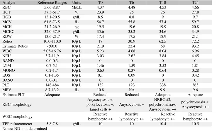
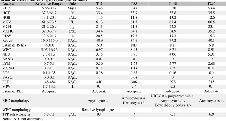
Table 2.CBC monitoring of Alba
Analyte Reference Ranges Units T0 T6 T10 T24
RBC 5.66-8.87 M/µL 4.37 4.48 4.53 4.66 HCT 37.3-61.7 % 23.9 25 26 27.8 HGB 13.1-20.5 g/dL 8.5 8.8 9 9.7 MCV 61.6-73.5 fL 54.7 55.8 57.4 59.7 MCH 21.2-26.9 pg 19.5 19.6 19.9 20.8 MCHC 32.0-37.9 g/dL 35.6 35.2 34.6 34.9 RDW 13.6-21.7 % 17.4 17.5 18.9 21.1 Retics 10.0-110.0 K/µL 17 30.9 62.5 72.2 Estimate Retics <60.0 K/µL 21.9 22.4 68 93.2 WBC 5.05-16.76 K/µL 5.23 4.68 8.04 6.96 NEU 3.7-11,9 K/µL 3.03 2.62 3.84 4.45 BAND 0.0-0.3 K/µL 0 0 0 0 LYM 0.7-5.1 K/µL 1.46 1.59 3.52 1.81 MONO 0.2-1.7 K/µL 0.63 0.37 0.64 0.28 EOS 0.1-1.35 K/µL 0.1 0.09 0 0.42 BASO 0.0-0.1 K/µL 0 0 0 0 PLT 148-484 K/µL 132 123 338 363 MPV 8.7-13.2 fL 10.8 NA 9.9 9.6
Estimate PLT Adequate Reduced Reduced Adequate Adequate
RBC morphology Anysocytosis +, poikylocytosis +, target cells + Anysocytosis + NRBC #2, polychromasia+, Anysocytosis ++ polychromasia +, Anysocytosis ++ WBC morphology Reactive lymphocyte ++ Reactive lymphocyte ++ Reactive lymphocyte ++ Reactive lymphocyte ++ TPP refractometer 5.8-7.8 g/dL 10 10 10.4 10.5
RBC 5.66-8.87 M/µL 5.45 5.49 5.78 5.64 HCT 37.3-61.7 % 33.4 33.9 37.8 35.5 HGB 13.1-20.5 g/dL 11.5 11.8 13.2 12.6 MCV 61.6-73.5 fL 61.3 61.7 65.4 66.5 MCH 21.2-26.9 pg 21.1 21.5 22.8 23.4 MCHC 32.0-37.9 g/dL 34.4 34.8 34.9 35.2 RDW 13.6-21.7 % 20.5 19.5 15.3 15.3 Retics 10.0-110.0 K/µL 40.9 34.6 79.2 40.1 Estimate Retics <60.0 K/µL ND ND ND ND WBC 5.05-16.76 K/µL 6.97 8.43 8.21 8.91 NEU 3.7-11,9 K/µL 3.15 3.96 4.06 5.31 BAND 0.0-0.3 K/µL 0.07 0 0 0 LYM 0.7-5.1 K/µL 3.36 2.53 3.77 2.68 MONO 0.2-1.7 K/µL 0.14 1.18 0.2 0.71 EOS 0.1-1.35 K/µL 0.28 0.67 0.16 0.2 BASO 0.0-0.1 K/µL 0 0.08 0 0 PLT 148-484 K/µL 410 306 278 315 MPV 8.7-13.2 fL 9.4 9.6 9.5 9.1
Estimate PLT Adequate Adeguate Adequate Adequate Adequate
RBC morphology Anysocytosis + Anysocytosis +,
Keratocyte
+/-NRBC #1, polychromasia +, Anysocytosis +, Howell-Jolly bodies
+/-Anysocytosis +,
WBC morphology Reactive lymphocyte +
TPP refractometer 5.8-7.8 g/dL 9.4 7 6.1 6.9
Notes: ND- not determined
Table 3.Serum biochemical profile monitoring of Alba
Analyte Reference ranges Units T0 T24 T42 T85 T148 T265
Total calcium 8.7-11.8 mg/dL 10.5 10.7 10.2 9.6 9.8 10.6 Phosphates 2.5-5.0 mg/dL 4.6 2.6 4.7 4 3.5 3.5 Iron 80-190 µ g/dL 95 321 NT NT 116 200 Total proteins 5.8-7.8 g/dL 11.5 12.3 9.4 7.4 6 6 Albumin 2.6-4.1 g/dL 1.9 2.5 2.6 2.9 3.2 3.1 Globulins 2.5-4.5 g/dL 9.7 9.8 6.8 4.5 2.8 2.9 A/G ratio 0.5-1.5 0,20 0.25 0.38 0.64 1.14 1.06 Fructosamine 170-430 µ mol/L 198 252 NT NT 325 329 C-reactive protein 0.0-0.30 mg/dL 2.3 0.2 0.2 NT 0.3 0.3 Urea 15-55 mg/dL 17 20 33 20 34 14 Creatinine 0.6-1.5 mg/dL 1 1 1 0.9 1.1 0.8 Total bilirubin 0.07-0.30 mg/dL 0.11 0.07 NT 0.2 0.2 0.07 Cholesterol 120-280 mg/dL 206 212 263 280 218 208 Tryglicerides 25-90 mg/dL 75 75 NT NT 52 69 Glucose 80-125 mg/dL 104 101 117 114 105 120
Alkaline phosphatase (ALP) 45-250 U/L 180 120 105 104 110 302
Gammaglutamyl transferase (GGT) 2.0-11.0 U/L 4 2 2 1 6 3
Aspartateaminotransferase (AST) 15-40 U/L 75 27 NT NT 35 21
Alanineaminotransferase (ALT) 20-70 U/L 22 22 25 69 174 44
Creatinphosphokinase (CK) 40-185 U/L 177 184 NT NT 69 56 Lactatedehydrogenase (LDH) 20-160 U/L 338 166 NT NT 57 91 Amylase 400-1500 U/L 1228 1362 NT 486 839 563 Sodium 146-156 mEq/L 152 158 157 153 146 156 Potassium 3.9-5.5 mEq/L 4 4 4.5 4 3.6 4.2 Chloride 109-122 mEq/L 115 117 118 117 116 118 Bicarbonate 21-31 mEq/L 24 26 20.5 NT 28 29
Notes: NT- not tested
Table 4.Coagulation profile monitoring of Alba
Analyte Reference Ranges Units T0 T24 T148 T265 Prothrombin time 5.4-8.1 Seconds 9 7.1 7.1 7.3 Partial thromboplastin time 10.7-17.5 Seconds 17.5 15.9 14 11.7 Fibrinogen 125-335 mg/dL 275 241 177 300
Table 5.Serum protein electrophoresis monitoring of Alba
Analyte Reference ranges Units T0 T24 T85 T148 T265 Albumin 47.5-58.5 % 22.2 20 37.5 52.7 51.7 Alpha-1-globulin 2.6-4.4 % 1.4 1.9 5.7 3.8 3.6 Alpha-2-globulin 10.7-18.0 % 8.7 6.9 15.8 14.3 15.7 Beta-1-globulin 5.9-13.3 % 3.3 6.7 10.9 8.7 11.1 Beta-2-globulin 7.2-14.4 % 9.2 7.3 10.2 8.5 9.5 Gamma globulin 6.6-11.6 % 55.2 57.2 19.9 12 8.4 A/G 0.5-1.5 0.29 0.26 0.6 1.11 1.07 Total protein 5.8-7.8 g/dL 11.5 12.3 8.1 6 6
Table 6.Urinalysis monitoring of Alba
Analyte Reference ranges Units T0 T148 Method of collection Cystocentesis Cystocentesis
Color Yellow NA Yellow-green Yellow
Aspect Clear NA Cloudy ++ Cloudy +
Specific gravity 1018-1045 NA 1035 1030
pH 5-7 NA 8 6
Protein <40 mg/dL 30 Absent
Glucose absent mg/dL Absent Absent
Ketones Absent mg/dL Absent Absent
Urobilinogen Absent mg/dL Absent Absent Bilirubin Absent mg/dL Absent Absent
Blood Absent RBCs/mcL 50 Absent
Leukocytes Absent WBC/mcL 25 Absent
UP/C ratio 0.1-0.5 NA 0.64 0.15
Cellularity Rare NA Moderate Moderate
RBC 1-5 HPF ++ Absent
WBC 1-5 HPF -/+ -/+
Epitelial cells 1-5 HPF + -/+
Other NA Lipid droplets
Notes: NA – not applicable
Table 7.Lymph node evaluation of Alba
At T0 At T265
Smear quality Good but some hemodilution Very good
General observation Polymorphic cell pattern Polymorphic cell pattern Lymphoid cells
Small lymphocytes prevalent (ca. 80%), lymphoblasts or immunoblasts (ca. 10%), plasmacells (ca. 10%)
Small lymphocytes prevalent, few immunoblasts and plasmacells Other cells A great number of macrophages
some with Leishmania spp Rare mastocytes and macrophages Mitosis Some occurring but typical Not observed
Conclusion Lympho-plasmacellular hyperplasia compatible with Leishmania infection
Normal lymphonode with a mild increase of plasmacells
Collection site Iliac crest Sternum
Cellularity Very good Good
Megakariocytes Occurring in all maturation forms Occurring in all maturation forms Myeloid series Proliferative, maturative,
and storage cells well represented
Proliferative and maturative cells well represented, less cells in the storage pool Erythroid series Proliferative and maturative
cells well represented
Proliferative and maturative cells well represented
Other cells Rare lymphocyte and plasmacells –
macrophages with Leishmania spp. Rare plasmacells
Mitosis Occurring in myeloid and erythroid series Occurring in myeloid and erythroid series – some atypical in myeloid
Conclusions
Megakariocyte, myeloid and erythroid series normal for number and morphology. Mild hyperplasia of myeloid in comparison to erythroid series. Macrophages with Leishmania spp.
Megakariocyte series normal for number and morphology. Myeloid normal for number but mild sign of dysplasia. Erythroid series normal for morphology mild prevalence to myeloid. Anaplasma pahogocytophilum/platys
(PCR+Hybridization Hybprobes) Negative NT
Babesiaspp (conventional PCR) Negative NT
Bartonellaspp (PCR+SYBR Green )R Negative NT
Ehrlichia canis
(PCR+Hybridization Hybprobes) Negative NT
Hepatozoonspp (conventional PCR) Negative NT
Leishmania infantum
(PCR+Hybridization Hybprobes) Positive (1.212.000.000 copies) Negative (limit 100 copies of kinetoplast) Rickettsiaspp
(PCR+Hybridization Hybprobes) Negative NT
Notes: NT- not tested
Table 9.Treatment monitoring of Alba
T0 (kg. 17.5) T3 (kg. 17.7) T10 (kg. 18.5)
Meglumine antimonate = 85.7 mg/kg SC Meglumine antimonate = 84.7 mg/kg SC Meglumine antimonate = 97.3 mg/kg SC Allopurinol = 8,6 mg/kg bid OS Allopurinol = 8.5 mg/kg bid OS Allopurinol = 8.1 mg/kg bid OS Spiramycin = 42,860 UI/kg bid OS Spiramycin = 42,370 UI/kg bid OS Spiramycin = 40,540 UI/kg bid OS Metronidazole = 7.14 mg/kg bid OS Metronidazole = 7.06 mg/kg bid OS Metronidazole = 6.75 mg/kg bid OS Glucose 5% = 14.3 ml/kg bid EV Glucose 5% = 14.1 ml/kg bid SC interrupted
Multi-vitamins vials SC Multi-vitamins vials SC Multi-vitamins vials SC
Artificial lacrimal drops TID topical Artificial lacrimal drops TID Artificial lacrimal drops TID Chloramphenicol/Neomicin eye drops TID topical Chloramphenicol/
Neomicin eye drops TID
Chloramphenicol/ Neomicin eye drops TID Topical use on tail
skin of iodopovidone BID
Suspended when the ulcer was healed Topical use of combined drug:
Glycol-propilene + malic acid + benzoic acid + salicylic acid
Topical use of combined drug: Glycol-propilene + malic acid + benzoic acid + salicylic acid
Table 9.Treatment monitoring of Alba (Continued)
T24 (kg. 19.1) T42 (kg. 20.3) T85 (kg. 20.8) T148 (kg. 21.3)
Meglumine antimonate = 94.2 mg/kg SC Interrupted at T40
Allopurinol = 7.8 mg/kg bid OS Allopurinol = 7.4 mg/kg bid OS Allopurinol = 4.8 mg/kg bid OS interrupted interrupted
interrupted
Multi-vitamins vials SC Multi-vitamins tablet OS Multi-vitamins tablet OS interrupted Artificial lacrimal drops TID interrupted
interrupted interrupted interrupted
Tipi ˇcna laj ˇsmanioza psa redovno vakcinisanog sa Canileish
Rvakcinom
Saˇzetak
Uvod
Vakcina protiv lajˇsmanioze pasa (Canileish R) napravljena
od LiESAp i 54-kDa proteina estrahovanih iz Leishmania infantum, a uz dodatak muramil-peptida (MDP) kao adju-vantnog sredstva, koristi se u nekoliko evropskih drˇzava.
Razvijena je da stimuliˇse ´celijski imunitet kod pasa seroloˇski negativnih na lajˇsmaniozu, kako bi se smanjio rizik od razvoja infekcije i kliniˇckih manifestacija bolesti. U ovom ˇclanku je opisan sluˇcaj psa sa teˇskim oblikom lajˇsmanioze, koji je redovno vakcinisan sa (Canileish R)
vakcinom i tretiran repelentima i drugim sredstavima protiv ektoprazita (vektora).
Materijal i metode
“Alba”, engleski seter, intaktna ˇzenka stara tri godine dove-dena je (mart 2015.) sa simptomima dizoreksije, gubitka tje-lesne mase, sa poviˇsenom temperaturom i slaboˇs´cu prednjih nogu. “Alba” je uz prevenciju ekto i endoparazitoza redovno vakcinisana protiv najˇceˇs´cih bolesti pasa, te Canileish R
vak-cinom protiv lajˇsmanioze.
Fizikalnim pregledom (T011) su zabiljeˇzeni: indeks tje-lesne kondicije 2/9, temperatura 39.6◦C, umjereno pove´ca-nje perifernih limfnih ˇcvorova, seropurulentni iscjedak iz oka sa fotofobijom i oteˇcen i bolan desni karpalni zglob.
Uradena je kompletna krvna slika (CBC), biohemijski profil, koagulacijski profil, elektroforeza serumskih pro-teina, seroloˇsko testiranje na L. infantum, analiza urina ukljuˇcuju i UP/C omjer, punkcija preskapularnih i popliteal-nih limfpopliteal-nih ˇcvorova, punkcija koˇstane srˇzi za citologiju i qPCR na L. infantum.
Rezultati
Razliˇciti kliniˇcki simptomi su se povukli sukcesivno: slabost prednjih ekstremiteta T3, temperatura i fotofobija -T10, seropurulentni iscjedak iz oka - T15, blefaritis, lim-fadenopatija i velike koˇzne promjene repa - T24, tjelesna teˇzina T42.
Kompletna krvna slika (T0) je pokazala umjerenu mi-krocitnu neregenerativnu anemiju, blagu neutropeniju i trombocitopeniju (normalni na T265). Biohemijski profil (T0) je potvrdio teˇsku hiperproteinemiju (normalne vrijed-nosti na T85), hipoalbuminemiju (normalno na T42), hiper-globulinemiju, nizak albuminsko/globulinski omjer (oboje normalni na T85), i poviˇsen C-reaktivni protein (normalan na T24). Elektroforeza serumskih proteina (T0) je pokazala hipoalbuminemiju i poliklonalnu hipergamaglobulinemiju (normalna na T148).
11T: vrijeme u danima od dana prijema (T0) pacijenta
Analizom urina (T0) ustanovljena je hematurija (nor-malno na T6) i blagi porast omjera proteina i kreatinina u urinu (normalni na T85). U aspiratima limfnih ˇcvorova i koˇstane srˇzi (T0) utvrdene su amastigote lajˇsmanije.
Uzorci limfnih ˇcvorova i koˇstane srˇzi bili su negativni qPCR tehnikom na Babesia spp, Bartonella spp, Ehrlichia canis, Hepatozoon spp, i Rickettsia spp, ali pozitivni na L. infantum(negativni na T148 i T265).
IFAT metodom (T0) je ustanovljen izrazito visok titar antitijela (1:5120), koji se smanjio na 1:320 (T148) i 1:160 (T265). Speed-Leish K R test je cijelo vrijeme bio pozitivan.
“Alba” je tretirana sa meglumin-antimonatom (SC) i alopurinolom (PO) u trajanju od 40 dana. Alopurinol je iskljuˇcen na T265. Metronidazol-spiramicin je apliciran perooralno do T24. Terapija teˇcnostima (fizioloˇski rastvor NaCl i 5% glukoze) aplicirana je deset dana, topikalna terapija repa je prekinuta na T24, a oka na T40.
Interpretacija i zaklju ˇcak
Ovaj sluˇcaj opisuje tipiˇcnu kliniˇcku i kliniˇcko-patoloˇsku sliku lajˇsmanioze nakon neuspjeha vakcine ili neefikasnog/ nedovoljnog imuniteta.
Vlasnik psa je potvrdio koriˇstenje repelenta i drugih sredstava protiv vektora te redovnu vakcinaciju. Posljednja doza Canileish R je aplicirana oko 10 mjeseci prije izbijanja
bolesti.
“Alba” je pokazala brz odgovor na standardnu terapiju. Svi opisani kliniˇcki znaci su se povukli do T27, dok su se kliniˇcko-patoloˇski znaci vra´cali u normalne granice sukce-sivno.
Kompletna krvna slika i citoloˇski pregled koˇstane srˇzi su se progresivno vratili u normalne granice na T265. S obzirom na pojavu mikrocitne hipohromne neregenerativne anemije na T0, mogu´ce da je pas zaraˇzen u ljeto/jesen 2014. godine. Biohemijski profil i analiza urina nisu bili sig-nifikantni od T42, odnosno T85. Elektroforeza serumskih proteina je znatno odstupala od normalnih vrijednosti na T0, ali se normalizirala na T148. Neposredna dijagnoza lajˇsmanioze je postavljena ciljanom punkcijom limfnog ˇcvora i biopsijom koˇstane srˇzi, za koje je poznato da imaju visoku specifiˇcnost i osjetljivost. Senzitivnost oba testa opada pod djelovanjem terapije tako da qPCR moˇze pomo´ci u otkrivanju patogena, i utvrdivanju uspjeˇsnosti terapije. Seroloˇski dokaz antitijela se ne moˇze koristiti kao potvrda prisustva parazita u organizmu jer se antitijela jako dugo zadrˇzavaju.
Opisani sluˇcaj potvrduje da i pored vakcinacije postoji mogu´cnost pojave kliniˇckih i drugih znakove lajˇsmanioze, ˇsto treba imati u vidu posebno u podruˇcjima koja su visoko-riziˇcna za pojavu lajˇsmanioze.