R E S E A R C H
Open Access
Prognostic value of morphologic and
morphometric analyses in IgA nephropathy
biopsies
Patrizia Viola
1, Lucia Centurione
2, Paolo Felaco
2, Giuseppe Lattanzio
3, Tommaso D
’Antuono
3, Marcella Liberatore
3,
Roberta Di Pietro
2, Franco Oreste Ranelletti
4, Mario Bonomini
2and Francesca Bianca Aiello
2*Abstract
Background: IgA nephropathy (IgAN) exhibits a variable course ranging from a benign condition to progressive renal failure with a high proportion of patients undergoing end-stage renal disease in the long term. It is unclear how to predict the risk of this progression. Mesangial C4d deposition has been found to be associated with a poor prognosis. Our aim was to search histological lesions with possible prognostic value in IgAN biopsies performed at the time of the diagnosis.
Methods: Clinical and laboratory records of 44 patients undergoing renal biopsy were reviewed. IgAN was diagnosed in 32 patients and minimal change disease (MCD) in 12.
C4d deposition, glomerular endothelial cell area, glomerular and interstitial macrophages were evaluated in all biopsies. Clinical and laboratory data were available in all IgAN patients at the time of diagnosis and in 21 patients at the end of follow-up (mean follow-up 48.09 ± 19.69 months).
Results: All IgAN and MCD biopsies were C4d negative. The number of glomerular macrophages and the area of glomerular endothelial cells in IgAN were greater than in MCD (P < 0.001 and P < 0.0001, respectively), indicating glomerular inflammation associated with endothelial cell enlargement. Glomerular macrophages positively correlated with diffuse IgA and fibrinogen immunoreactivity (P < 0.02 and P < 0.002, respectively).
Interstitial macrophages positively correlated with segmental glomerulosclerosis (P < 0.01) and tubular atrophy (P < 0.001), according to the Oxford classification. They correlated with hypertension (P < 0.03) and renal functional impairment both at the time of biopsy and at the end of follow-up. The receiver operating characteristic analysis curve and a cut-off value of≥19.5 macrophages per high power field showed 75.0% sensitivity and 88.9% specificity in predicting worse clinical outcome. A Poisson generalized regression fit model showed that hypertension and tubular atrophy were the most significant factors in predicting a high number of interstitial macrophages.
Conclusions: The number of interstitial macrophages correlated with hypertension, histological damage, functional impairment at the time of biopsy and worse outcome. A high number of interstitial macrophages in C4d negative biopsies may allow the identification of patients at increased risk of progression to renal failure. Keywords: IgA Nephropathy, Macrophages, C4d, Morphometry, Hypertension, Prognosis
* Correspondence:[email protected]
2Department of Medicine and Aging Sciences, University G d’Annunzio,
Chieti-Pescara, via dei Vestini, 66100 Chieti, Italy
Full list of author information is available at the end of the article
© The Author(s). 2016 Open Access This article is distributed under the terms of the Creative Commons Attribution 4.0 International License (http://creativecommons.org/licenses/by/4.0/), which permits unrestricted use, distribution, and reproduction in any medium, provided you give appropriate credit to the original author(s) and the source, provide a link to the Creative Commons license, and indicate if changes were made. The Creative Commons Public Domain Dedication waiver (http://creativecommons.org/publicdomain/zero/1.0/) applies to the data made available in this article, unless otherwise stated.
Background
IgA nephropathy (IgAN) has an incidence of approxi-mately 2.5/100000/year in adults [1]. It is characterized by the presence of glomerular IgA deposition in patients with high levels of immune complexes formed by aber-rantly glycosylated IgA and endogenous antiglycan anti-bodies. IgA deposition is predominant, and IgG, IgM and C3 deposition are frequently observed.
IgA-containing immune complexes deposited in the glomeruli are associated with inflammation, proliferation of mesangial cells, and synthesis of extracellular matrix molecules [2]. The clinical course and the histopatho-logical presentation of IgAN are highly variable [2–4]. IgAN may exist sub-clinically [3]. Although it usually initiates as a benign condition, in 15–20 years 15–40% of the patients progress to end-stage renal disease (ESRD) [3–5]. Hypertension, some renal function pa-rameters and histopathological features are known prog-nostic risk factors. These parameters, however, may only indicate the extent of the disease at a particular time, such as the stage of the disease at the time of diagnosis [3]. For instance, tubule-interstitial damage, associated with a rapid development of ESRD [2–4], indicates an advanced disease. The majority of patients present haematuria with or without proteinuria, and a slowly progressive course. A high percentage of these patients, however, will experience a decline in renal function with a rate of progression varying among patients [3, 4]. Thus, it would be important and also challenging to pre-dict the risk of progression.
In renal transplant patients the C4d fragment of the C4 complement component is released during activation of the classic complement pathway; its deposition on the surface of endothelial cells of peritubular and glomerular capillaries is a marker of antibody-mediated acute rejec-tion, and poor graft survival [6]. In IgA nephropathy complement activation typically occurs via the alterna-tive pathway, thus without C4d deposition [7]. However, C4d deposition has been reported in approximately 25% of IgAN cases, [8] probably due to activation of the mannose-binding lectin pathway [7]. As in patients with antibody-mediated renal acute rejection, this is predictive of a poor prognosis [8]. C4d deposition in antibody-mediated acute rejection biopsies is associated with a high number of interstitial macrophages and glo-merulitis [9, 10]. Acute transplant gloglo-merulitis, defined by the presence of mononuclear cells-predominantly macrophages-in the glomeruli and endothelial cell enlargement [6] in some cases progresses to chronic glomerulopathy, characterized by capillary basement membrane multilayering, proteinuria and decline of renal function [10, 11].
In IgAN the mechanism of haematuria is unknown, endothelial cell damage and alterations of the glomerular
basal membrane may contribute to its development [4, 12]. Whether endothelial cell enlargement is present in IgAN glomeruli has not been investigated. The aim of this study was to evaluate the prognostic value of histological le-sions such as glomerulitis and interstitial macrophages in IgAN biopsies. The associations between histological lesions and proteinuria, creatininemia, glomerular fil-tration rate and hypertension were evaluated, and only patients with C4d negative renal biopsies were included in the study. The patients showed a classical clinical presentation with haematuria and moderate protein-uria. Minimal change disease (MCD) biopsies were studied for comparison, considering that in this disease glomerular changes at light microscopy and activation of the complement system are not detected [13].
Methods
Patients
Clinical and laboratory records of 44 patients undergo-ing renal biopsy consecutively between January 2003 and December 2012 at our Institution were reviewed. IgAN was diagnosed in 32 patients (21 males and 11 females, mean age 35.9 ± 11.69 years) and MCD in 12 (6 males and 6 females, mean age 39.50 ± 10.81 years). Patients included in the study were more than 15 years of age and underwent biopsy before any immune-suppressive treatment. All IgAN patients showed C4d negative renal biopsies. Patients with diabetes mellitus, autoimmune diseases, abnormal hypergammaglobulinemia, and liver diseases were excluded from the study. After biopsy, 7 patients were treated with renin-angiotensin system blockers (RASB), 8 with corticosteroids, and 15 with RASB and corticosteroids. Two patients with normal renal function, proteinuria < 1 g/24 h and biopsies show-ing only mild mesangial hypercellularity received no treatment. Haematuria, proteinuria (g/24 h), serum creatinine level, (reference range obtained by our clinical laboratory: males = 0.66–1.25 mg/dl, females = 0.52– 1.04 mg/dl), blood pressure, and estimated glomerular filtration rate (eGFR) (ml/min per 1.73 m2) were re-corded at the time of biopsy. Hypertension was defined as systolic blood pressure > 135 mmHg and/or diastolic blood pressure > 85 mmHg or the use of anti-hypertensive agents. In all anti-hypertensive patients typical vascular lesions were observed in the biopsies. Evalu-ation of the clinical outcome was assessed using the Kidney Disease Outcome Quality Initiative (KDOQI) guidelines [14], which define 5 stages based on eGFR evaluation (stage 1: eGFR≥ 90 ml/min; 2: eGFR = 60– 89 ml/min; 3: eGFR = 30–59 ml/min; 4: eGFR = 15–30 ml/ min; 5: eGFR = <15 ml/min). Follow-up time was consid-ered as the interval time between renal biopsy and the last outpatient visit, haematuria, proteinuria, serum creatinine levels and eGFR data were available for 21 patients (mean
follow-up: 48.09 ± 19.69 months). They were treated with methylprednisolone (0.5–1 mg/kg/die) (n = 3), RASB (n = 7) or methylprednisolone (1.5–1 mg/kg/die) in combination with RASB (n = 9). Two patients received no treatment. To evaluate the outcome the decline of the eGFR was considered.
Histopathology and immunohistochemistry
Formalin-fixed paraffin-embedded sections were stained with hematoxylin-eosin, PAS and Masson trichrome. Direct immune-fluorescence was performed on fresh frozen tissue with FITC-conjugated polyclonal anti-bodies to detect IgG, IgM, IgA, C3 and fibrinogen (Dako, Glostrup, Denmark). IgAN biopsies were scored according to the Oxford [15] and the 3-Grade classifica-tion [5]. According to the Oxford classificaclassifica-tion [15] each glomerulus was scored for mesangial hypercellularity (M0-M1, M1 indicated hypercellularity in more than 50% of glomeruli). The other parameters were endoca-pillary hypercellularity (present/absent: E0-E1); segmen-tal glomerulosclerosis (present/absent: S0-S1) and tubular atrophy (T0 = < 25%, T1 = 26–50% and T2 = > 50% of the cortical area). According to the 3-Grade clas-sification [5] biopsies were scored as G1 when a mild in-crease in mesangial cells and no glomerulosclerosis or tubulo-interstitial changes were detected, as G2 when at least one of the following features was detected: moder-ate increase in mesangial cellularity (focal or diffuse), endocapillary proliferation, cellular crescent, (up to 50% of glomeruli), focal segmental glomerulosclerosis, tubu-lar atrophy and interstitial fibrosis (up to 1/3 of the cor-tical area), as G3, when cellular crescents were present in more than 50% of glomeruli, and/or fibrous crescents or global glomerulosclerosis were present in more than 1/3 of glomeruli, and/or segmental glomerulosclerosis involved more than 50% of glomeruli and/or tubular at-rophy/interstitial fibrosis involved more than 1/3 of the cortical area.
Sections were stained for immunohistochemistry using the Bond Polymer Refine Detection method (Leica Bio-system, Wetzlar, Germany) after high pH (pH 9.0) buff-ered pre-treatment (Leica Biosystem), using the murine monoclonal antibody (MoAb) anti-CD68 (Dako), recog-nizing macrophages. Quantification of glomerular mac-rophages was performed counting CD68+ cells in each glomerulus and dividing the sum obtained in each bi-opsy by the number of scored glomeruli (magnification 40). Interstitial macrophages in the cortical area were counted in 10 high power fields (HPF) and the mean number per HPF was reported (magnification × 40). For detection of C4d paraffin sections were stained using the XT Ultraview DAB v3 method (Ventana Medical Sys-tem, Tucson, AZ, USA) after antigen retrieval by pressure-cooking using a polyclonal anti-C4d antibody
(Biomedica, Vienna, Austria). Biopsies from 12 patients with MCD were used as negative controls, and two biop-sies from patients with lupus glomerulonephritis as posi-tive controls.
Staining of endothelial cells with anti-CD31 murine MoAb (Dako) was performed using the Ultravision LP polymer detection method (Thermo Fisher Scientific, Freemont, CA, USA) after antigen retrieval by heating the sections in Tris-EDTA buffer (pH 8.0). Primary anti-bodies were replaced by irrelevant matched MoAb or immune serum, as appropriate, as a control for non-specific staining.
Morphometric analysis
Glomerular capillary loops were examined after CD31 immunostaining of endothelial cells as previously de-scribed [16] using a Zeiss Axioskop 40 light microscope (Carl Zeiss, Gottingen, Germany) equipped with a Cool-snap Videocamera (Photometrics, Tucson, AZ, USA). MetaMorph Software System (Universal Imaging Corp Molecular Device Corp, CA, USA) was used to analyse images. Only full cross-sectioned loops were measured. Capillary loops showing endothelial cell nuclei were not considered. Leukocytes or red cells within capillaries were considered as part of the lumen. The external and the internal capillary outlines were manually delineated at × 40 magnification. The total area of the capillary (TCA), and the area of the capillary lumen (LCA) were automatically measured. The percentage of the total area of the capillary occupied by endothelial cells (ECA%) was defined as the ratio of (TCA - LCA)/TCA × 100). Analyses were conducted on 44 biopsies (32 IgAN and 12 MCD), and 3 glomeruli were examined in each bi-opsy. The area of capillary loops was measured in IgAN glomeruli (1839 loops) and in MCD glomeruli (605 loops). Glomerular areas were evaluated as previously described [17].
Statistical analyses
Results are expressed as mean ± SD. The Pearsonχ2test, Wilcoxon/Kruskal-Wallis test, Pearson’s correlation test, and one-way ANOVA were used for statistical analysis. For all analyses statistical significance was defined asP < 0.05. To estimate the performance as a prognostic marker of interstitial macrophages per HPF the receiver oper-ating characteristic (ROC) analysis curve was con-structed and the area under the ROC curve (AUC) and the corresponding 95% confidence interval (C.I. 95%) were evaluated. The most appropriate cut-off value of interstitial macrophages per HPF to predict clinical out-come was determined by the point of interstitial macro-phage number with maximum sum of sensitivity and specificity. Poisson generalized regression fit model and stepwise regression analysis were used to select the
explanatory variables for the standard least square full factorial regression model. Statistical analyses were performed by JMP 11 software (SAS Institute Inc. Cary, NC).
Results
Glomerular macrophages and immunofluorescence pattern in IgAN and MCD biopsies
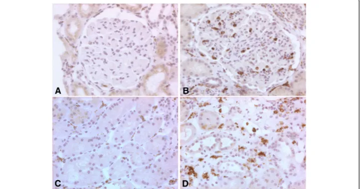
No significant differences in age and male/female ratio were present between IgAN and MCD patients. In IgAN biopsies the number of macrophages per glomerulus was significantly higher than in MCD (6.31 ± 3.51 versus 1.11 ± 0.73, P < 0.0001). CD68+ macrophages in IgAN and MCD glomeruli are shown in Fig. 1a and b. IgA, IgM, IgG, C3 and fibrinogen immune-fluorescence stain-ing was negative in all MCD biopsies. As required for the diagnosis, all IgAN patients showed predominant IgA immune-reactivity in the mesangial area of glom-eruli, with diffuse positivity in 15 biopsies. Immune-reactivities for IgG, IgM, C3 and fibrinogen were observed in 11, 22, 32 and 18 biopsies, respectively. Dif-fuse positivity was present in 3, 9, 11 and 6 biopsies, re-spectively. The number of glomerular macrophages was significantly higher in biopsies with diffuse IgA positivity (n = 15) than in biopsies with IgA focal positivity (n = 17) (7.82 ± 3.65 versus 4.97 ± 3.14, P < 0.02), and it was markedly higher in biopsies with diffuse fibrinogen posi-tivity (n = 6) than in biopsies with negative/focal
fibrinogen positivity (n = 26) (10.18 ± 2.97 versus 5.41 ± 3.19,P < 0.002).
No significant difference was observed in the presence of diffuse versus focal/negative IgG, IgM or focal C3 staining (data not shown).
This finding suggests a positive correlation between the number of glomerular macrophages and the diffuse distribution of IgA and fibrinogen.
Glomerular endothelial cell area in IgAN and MCD biopsies
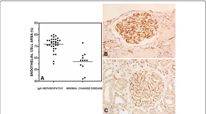
The area of the capillary loop occupied by endothelial cells (ECA%), was significantly higher in IgAN than in MCD biopsies (76.81 ± 5.89 versus 61.83 ± 8.39,P < 0.0001) (Fig. 2a). Glomerular areas in IgAN and MCD biopsies were not significantly different (18698 ± 6553 and 16895 ± 6858 μ 2, respectively P = 0.427). Representative CD31+ glomerular endothelial cells in MCD and IgAN are shown in Fig. 2b and c.
C4d deposition in IgAN and MCD biopsies
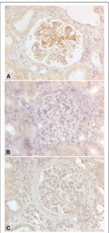
Glomerular, mesangial or peritubular C4d staining was not observed in either MCD or in IgAN biopsies (Fig. 3b and c). In two biopsies of lupus glomerulo-nephritis, included as positive controls, the glomerular staining pattern was diffuse and segmental, and peri-tubular capillaries were negative. Fig. 3a shows
Fig. 1 Glomerular and interstitial macrophages in IgAN and MCD biopsies. Immunohistochemical staining. a MCD, few glomerular CD68+ cells (original magnification × 20). b IgAN, numerous glomerular CD68+ cells (original magnification × 20). c MCD, few scattered interstitial CD68+ cells in the cortical area, (original magnification × 20). d IgAN, numerous interstitial CD68+ cells in the cortical area (original magnification × 20)
glomerular C4d positivity in a biopsy of lupus glomer-ulonephritis (class IV).
Glomerular and interstitial macrophages and histological grading
The number of interstitial macrophages per HPF in the cortical area was significantly higher in IgAN than in MCD biopsies (18.29 ± 5.01 versus 11.25 ± 4.90,P < 0.0001). CD68+ macrophages in IgAN and MCD interstitial cortical area are shown in Fig. 1c and d.
According to the Oxford Classification [15], in IgAN biopsies scored for mesangial cellularity (M0/M1) no dif-ferences in the number of glomerular or interstitial macrophages were observed (Table 1). Endocapillary hypercellularity was scored as E0 in all biopsies. In biop-sies scored for glomerulosclerosis as G1 and for tubular atrophy as T1, the number of interstitial, but not that of glomerular macrophages was significantly higher than in G0 and T0 biopsies (P < 0.01 and P < 0.001, respectively) (Table 1). Using the 3-Grade Classification [5], a low number of macrophages was found to be associated with the absence of glomerulosclerosis and tubule-interstitial changes (G1) (P < 0.005) (Table 1).
Glomerular and interstitial macrophages: clinical data and outcome
In 32 IgAN patients haematuria, proteinuria, serum cre-atinine levels, eGFR and blood pressure were examined at the time of biopsy. At the end of follow-up haema-turia, proteinuria, serum creatinine levels and eGFR data were available for 21 patients (mean follow-up: 48.09 ± 19.69 months). At the time of biopsy microscopic haematuria was present in 30 patients, gross haematuria in one, and no haematuria in one (n = 32). At the end of follow-up (n = 21), 14 patients showed microscopic haematuria The mean level of proteinuria at the time of biopsy was: 1.82 ± 1.19 g/24 h (n = 32). Proteinuria levels at the time of biopsy and at the end of follow-up did not correlate with glomerular or interstitial macrophages, or with any of the other histological lesions studied. Inter-estingly, in the 21 patients with available follow-up data the mean level of proteinuria at the end of follow-up (0.69 ± 0.78 g/24 h) was significantly lower than that ob-served at the time of biopsy (1.61 ± 1.01 g/24 h,P < 0.002) indicating a clear improvement of this parameter.
The number of interstitial, but not that of glomerular macrophages was significantly higher in patients with in-creased serum creatinine levels both at the time of
Fig. 2 Area occupied by endothelial cells in IgAN and MCD glomerular capillaries. The mean percentage of the total capillary loop area occupied by endothelial cells (ECA%) was measured in 32 IgAN and in 12 MCD biopsies as described in Methods. a ECA% distribution in the two groups of biopsies, in MCD the mean was significantly lower than in IgAN (61.83 ± 8.39 versus 76.81 ± 5.89,P < 0.0001), the standard deviation for each value was < than 20 % and was omitted. b, c Glomeruli showing CD31 positive endothelial cells in IgAN and MCD (original magnification × 20)
biopsy and at the end of follow-up (P < 0.007 and P < 0.03, respectively) (Table 2).
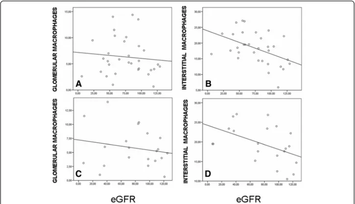
Moreover, a significant inverse correlation between the number of interstitial but not of glomerular macro-phages and the eGFR was observed at the time of
biopsy (r =−0.463, P < 0.008 and r = −0.99, P = 0.5 respectively) and at end of the follow-up (r =−0.474, P < 0.03 and r = −0.9, P = 0.3, respectively) (Fig. 4).
In patients without hypertension (n =15) the number of interstitial macrophages per HFP was significantly lower than in patients with hypertension (n = 17) (15.97 ± 4.92 versus 20.93 ± 3.72, P < 0.03), whereas there was no difference in the number of glomerular macrophages (5.80 ± 3.62 versus 6.88 ± 3.68, P = 0.40). Patients with hypertension were older than those without hyperten-sion (42.26 ± 9.46 versus 29.70 ± 10.39 years, P < 0.001), and in their biopsies one or more features of hyperten-sive nephrosclerosis were present: arteriolar hyalinosis in 5 patients, arteriolar or arterial hyperplasia of smooth muscle cells in 9, arterial proliferative intimal thickening in 3, arterial intimal fibrosis in 3, and global glomerular sclerosis in 1.
Evaluation of the clinical outcome was based on the eGFR at time of biopsy and at the end of follow-up ac-cording to the stages established by the KDOQI guide-lines for chronic kidney disease [14]. The 21 patients with follow-up data were considered. Twelve patients (group 1) had a favourable outcome: 7 were in stage 1 at the time of the biopsy and at the end of follow-up, and 5 showed an improved stage at the end of follow-up (stages 2, 3, 3, 3 and 5 at the time of diagnosis; stages 1, 2, 1, 2 and 1 at the end of follow up, respectively). The remaining 9 patients (group 2) had an unfavourable out-come: 6 had no improvement (stages 2, 2, 3, 3, 3 and 3 at the time of the diagnosis and at the end of follow-up) and 3 showed worse stages, with ESRD in two patients (stages 1, 3 and 3 at the time of diagnosis; stages 2, 5 and 5, respectively, at the end of follow-up). In group 1 biopsies the number of interstitial macrophages per HPF was significantly lower than in group 2 (17.34 ± 5.00 ver-sus 22.15 ± 3.32,P < 0.02).
ROC curve analysis showed that the number of inter-stitial macrophages predicted an unfavourable outcome. AUC was 0.80 (C.I. 95% = 0.6–1.0; P < 0.0021) and a cut-off value of ≥19.5 macrophages per HPF showed 75.0% sensitivity and 88.9% specificity in predicting a worse clinical outcome. Among 11 patients with≥ 19.5 intersti-tial macrophages per HPF, 8 (72.7%) had an unfavour-able outcome, while among 10 patients with < 19.5 interstitial macrophages, only 1 (10%) had an unfavour-able outcome (Fisher’s exact test: P < 0.0075). Therefore, the number of interstitial macrophages per HPF resulted a useful parameter to evaluate the clinical outcome.
The number of macrophages was not significantly as-sociated with different therapeutic regimens in either the favourable or unfavourable patient group (P = 0.48 and P = 0.23, respectively). To control for covariance between the factors associated with a high number of interstitial macrophages a Poisson generalized regression fit model
Fig. 3 C4d immune-reactivity in IgAN, MCD and lupus
glomerulonephritis biopsies. C4d staining was performed as described in Materials and Methods. a C4d positivity in a lupus glomerulonephritis biopsy (class IV) was observed on the surface of glomerular endothelial cells (positive control) (original magnification × 20). b, c Absence of endothelial or mesangial C4d staining in MCD (negative control) and IgAN glomeruli. (original magnification × 20)
was performed. The number of interstitial macrophages was considered as a function of 6 variables: grade of the 3-Grade classification (G), segmental glomerulosclerosis (S), tubular atrophy (T), hypertension, serum creatinine level and eGFR. These variables were chosen since uni-variate analyses showed that they were significantly cor-related with interstitial macrophage number. The whole model test yielded a significant fit (−Log Likelihood = 9.75; χ2= 19.5; P < 0.0034) in predicting the number of interstitial macrophages. We then performed variable se-lection using stepwise regression analysis for the 6 vari-ables used as explanatory effects in the Poisson generalized regression. The criterion to add or delete a variable in the model was based on F-statistics, with a critical P-value of 0.05. Hypertension and tubular atro-phy were selected as explanatory variables for the stand-ard least square full factorial regression model. The selected markers accurately validated the estimate of interstitial macrophage number (actual versus predicted number: r = 0.67, P < 0.0006). Interestingly, hyperten-sion appeared significantly nested within tubular atro-phy (P < 0.013) as its positive effect on interstitial macrophage number occurred only in the absence of tubular atrophy. As shown in Table 3 a significantly
positive effect of hypertension on the number of inter-stitial macrophages occurred only when there was no tubular atrophy. Tubular atrophy produced significantly increased interstitial macrophages independent of the hypertensive status.
Discussion
Identification of histological lesions with prognostic value at the time of diagnosis is important to identify IgAN patients with high risk of progression. We evalu-ated C4d deposition, the area of glomerular endothelial cells, glomerular and interstitial macrophages in IgAN biopsies. We showed that a high number of interstitial macrophages is associated with hypertension, unfavour-able histological grading and deterioration the renal function in C4d negative biopsy patients.
In agreement with the evidence of a good prognosis in C4d negative patients [8] only two patients underwent ESRD. Numerous glomerular macrophages and endothe-lial cell enlargement characterized the glomerulitis. Endothelial cell enlargement is one of the signs predict-ing chronic glomerulopathy in antibody-mediated renal transplant acute rejection [11]. The glomerular endothe-lial cell area in IgAN has not yet been studied. We found
Table 1 Number of interstitial macrophages correlated with Oxford and 3-Grade classification grading
Oxford Classification Glomerular macrophagesa P Interstitial macrophagesa P
M0b(n = 22) 5.98 ± 3.70 0.4 18.25 ± 5.30 0.9 M1 (n = 10) 7.03 ± 3.56 18.40 ± 4.55 S0c(n = 18) 6.13 ± 2.90 0.9 16.48 ± 4.37 < 0.01 S1 (n =14) 6.54 ± 4.51 20,65 ± 4.91 T0d(n = 19) 6.18 ± 3.60 0.8 16.08 ± 4.57 < 0.001 T1 (n = 13) 6.13 ± 2.90 21.53 ± 3.79
3-Grade Classification Glomerular macrophagesa P Interstitial macrophagesa P
G1e(n = 14) 5.95 ± 3.15 0.6 15.57 ± 4.30 < 0.005
G2 (n = 18) 6.59 ± 4,04 20.41 ± 4.55
a
Data expressed as means ± SD
b M = mesangial hypercellularity c S = segmental glomerulosclerosis d T = tubular atrophy e
G = grades of the 3-Grade classifications; none of the biopsies was scored as G3. M, S, T and G grades were scored as described in Materials and Methods
Table 2 Macrophages and creatinine levels at the time of biopsy and at the end of follow-up
Serum creatinine level (mg/dl)a Glomerular macrophages P Interstitial macrophages P
Males < 1.25, females < 1.04b 6.04 ± 3.657 0.6 16.27 ± 4.83 < 0.007
Males > 1.25, females > 1.04c 6.65 ± 3.81 20.89 ± 4.05
Males < 1.25, females < 1.04d 5.56 ± 2.81 0.6 17.85 ± 4.88 < 0.03
Males > 1.25, females > 1.04e 6.29 ± 4.80 22.62 ± 3.27
a
Data are expressed as means ± SD
b
Mean serum creatinine level at the time of biopsy in males < 1.25 and females < 1.04 mg/dl (n =18) was 0.91 ± 0.16 mg/dl
c
Mean serum creatinine level at the time of biopsy in males > 1.25 and females > 1.04 mg/dl (n = 14) was 2.08 ± 1.89 mg/dl
d
Mean serum creatinine level at the end of the follow-up in males < 1.25 and females < 1.04 mg/dl (n = 14) was 0.90 ± 0.36 mg/dl
e
that it was greater than in MCD, and similar in its extent to the area measured in acute transplant glomerulitis [16]. Endothelial cell enlargement may indicate cell dam-age and contribute to the development of haematuria, with negative prognostic implications [4, 12].
The number of macrophages in glomeruli was posi-tively correlated with diffuse deposition of IgA and fi-brinogen, a result, to the best of our knowledge, shown for the first time. This is in line with in vitro studies investigating the mechanisms underlying the recruit-ment of glomerular macrophages. Human mesangial cells cultured with polymeric IgA from IgAN patients produce RANTES and monocyte chemoattractant protein-1 (MCP-1), potent chemotactic agents for
macrophages [18]. Moreover, MCP-1 is produced by human endothelial cells cultured with fibrinogen at physiological concentrations [19].
Using two different histological classifications we found that interstitial macrophages were associated with glomerulosclerosis and tubular atrophy. In glomerular diseases, glomerular damage precedes the interstitial fi-brosis [20], thus, as also suggested by in vitro studies [21], it is possible that macrophage-mediated mecha-nisms may link these events.
Interstitial macrophages were previously correlated with poor long-term prognosis, but not with impaired renal function at the time of biopsy [22]. Other authors have reported a correlation with impaired renal function at the time of biopsy and long-term prognosis [23]. In patients with C4d negative biopsies a high number of interstitial macrophages correlated with impairment of renal function at the time of biopsy and at the end of follow-up, whereas the number of glomerular macro-phages did not have any functional or prognostic value. This is in line with the concept that macrophages in dif-ferent localizations display difdif-ferent functions [24]. For instance, in IgAN and in renal transplant rejection
Fig. 4 Interstitial macrophages and eGFR at the time of biopsy and end of follow-up. Glomerular CD68 positive cells and interstitial CD68 positive cells in the cortical area per HPF were evaluated in IgAN biopsies as described in Materials and Methods. a No correlation was observed between the number of glomerular macrophages and eGFR at the time of biopsy (r =−0.99, P = 0.5, n = 32). b A significant inverse correlation was present between the number of interstitial macrophages and eGFR at the time of biopsy (r =−0.463, P < 0.008, n = 32). c No correlation was observed between the number of glomeruli and eGFR at the end of follow-up (r =−0.9, P = 0.3, n = 21). d A significant inverse correlation was present at the end of follow-up between the number of interstitial macrophages and eGFR (r =−0.474, P < 0.03, n = 21)
Table 3 Number of interstitial macrophages according to hypertension and tubular atrophy status
Tubular atrophy
Hypertension Absent Present
Absent 14.4 ± 0,971 23.4 ± 1,56
Present 20.9 ± 1,58 21.0 ± 1,27
biopsies the receptor for the chemokine RANTES (CCRR5) was expressed by interstitial but not by glom-erular macrophages [24].
A persistent high level of proteinuria is associated with severe histological lesions, including high scores of Oxford classification parameters [4, 23, 25, 26]. In our study proteinuria levels did not correlate with histo-logical findings. This might be due to the improvement of proteinuria levels observed at the end of follow-up.
Hypertension is a well-established risk factor for IgAN progression [3, 4]. For the first time in IgAN we found a positive correlation between the number of interstitial macrophages and hypertension. The systemic angiotensin-aldosterone system and the intrarenal renin-angiotensin system contribute to the development of hypertension [27]. In renal vascular hypertension the presence of macrophages is attributed to the pro-inflammatory activity of angiotensin II (ATII) [28]. In rat models of ATII-induced hypertensive kidney injury mac-rophages were found in the interstitium, associated with MCP-1 expression in tubular cells and peritubular inter-stitial cells [29]. Macrophages in turn promote hyperten-sion [30, 31]. Infuhyperten-sion with ATII in mice was paralleled by macrophage infiltration of the vessel walls, elastin breakdown and collagen deposition [30] whereas M-CSF-deficient mice lacking mature macrophages exhib-ited a decreased hypertensive response [31]. We found that the most relevant predictive factors for a high num-ber of interstitial macrophages were hypertension and tubular atrophy. The absence of hypertension showed a protective effect, and in the absence of tubular atrophy hypertension was predictive of an increased number of interstitial macrophages. Notably, tubular atrophy was predictive of a high number of interstitial macrophages independently of the blood pressure status. RASB re-duced blood pressure, MCP-1 expression and macro-phage infiltration in some experimental hypertension models [29]. However, when biopsies of patients with hypertensive nephrosclerosis treated with RASB were compared with those of untreated patients the number of interstitial macrophages was not reduced [32]. Our data suggest that the therapeutic regimens did not influ-ence the prognostic value of the number of interstitial macrophages. In IgAN the decision to treat patients with RASB alone or to add corticosteroids depends on the presence of signs associated with poor prognosis. In the Kidney Disease Improving Global Outcome (KDIGO) practice guidelines the administration of glucocorticoids is suggested when proteinuria is higher than 1.5 g/day and the eGFR is more than 50 ml/min after 6 months of RASB therapy. The guidelines mention that histological risk factors could be a good reason to combine RASB with glucocorticoids, although no trials have been performed [33]. Glucocorticoids reduce macrophage
infiltration and tubulo-interstitial injury in murine lupus nephritis [34]. Thus, early combined treatment with RASB and glucocorticoids may improve the long-term prognosis in patients with pronounced macrophage interstitial infiltration.
A limitation in our study is the small sample size, thus, our results need to be validated with a larger cohort of patients. In addition, it will be important to assess the prognostic value of the number of interstitial macro-phages in other primary glomerulonephritides.
Conclusions
A high number of interstitial macrophages in C4d nega-tive biopsies of IgAN patients predicts an unfavourable outcome. Our data encourage the identification of novel anti-fibrotic therapies targeting macrophages.
Additional files
Additional file 1: Database 1. (XLS 37 kb) Additional file 2: Database 2. (XLS 9 kb)
Additional file 3: Reading keys for database 1. (DOC 27 kb) Additional file 4: Reading keys for database 2. (DOC 26 kb)
Abbreviations
AUC:Area under the curve; ECA%: Area of the capillary occupied by endothelial cells; eGFR: Estimated glomerular filtration rate; ESRD: End stage renal disease; HPF: High power field; IgAN: IgA nephropathy; KDOQI: Kidney Disease Outcome Quality Initiative; LCA: Area of the capillary lumen; MCD: Minimal Change Disease; MoAb: Monoclonal antibody; RASB: Renin-angiotensin system blockers; ROC: Receiver operating characteristic; TCA: Total area of the capillary
Acknowledgments
We thank Dr. Tiziana Musso for helpful discussion and Dr Chiara Cerletti for critical review of the manuscript.
Funding
Supported in part by funds from Ministero dell’Istruzione, dell’ Università e della Ricerca.
Availability of data and materials
The data sets supporting the conclusions of this article are included in its Additional files 1, 2, 3 and 4.
Authors’ contributions
All authors contributed to design the study, critically revised and approved the manuscript. PV, GL, TD’A, and ML collected and analyzed histological and immunohistochemical data. PF and MB collected and analyzed clinical data. LC and R Di P collected and analyzed morphometrical data. FOR and FBA performed statistical analyses. PV and FBA wrote the manuscript. Competing interest
The authors declare no conflicts of interest. Consent for publication
Informed consent was obtained from the patients. Ethic approval and consent to participate
The study was conducted in accordance with the Declaration of Helsinki and has been approved by the author’s Institutional Review Board.
Author details
1Department of Pathology, Whittington Health, Magdala ave, London N19
5NF, UK.2Department of Medicine and Aging Sciences, University G
d’Annunzio, Chieti-Pescara, via dei Vestini, 66100 Chieti, Italy.3Department of Pathology, ASL2, SS Annunziata Hospital, via dei Vestini, 66100 Chieti, Italy.
4Department of Histology, Università Cattolica del Sacro Cuore, Largo
Francesco Vito 1, 00168 Roma, Italy.
Received: 4 July 2016 Accepted: 3 November 2016
Referencesc
1. Mc Grogan A, Franssen CFM, de Vries CS. The incidence of primary glomerulonephritis worldwide: a systematic review of the literature. Nephrol Dial Transplant. 2011;26:414–30.
2. Wyatt R, Julian BA. IgA nephropathy. N Engl J Med. 2013;368:2402–14. 3. Huang L, Guo FL, Zhou J, Zhao YJ. IgA nephropathy factors that predict and
accelerate progression to end-stage renal disease. Cell Biochem Biophys. 2014;68:443–47.
4. Coppo R, D’Amico G. Factors predicting progression of IgA nephropathy. J Nephrol. 2005;18:503–12.
5. Manno C, Strippoli GF, D’Altri C, Torres D, Rossini M, Schena FP. A novel simpler histological classification for renal survival in IgA nephropathy. Am J of Kidney Dis. 2007;49:763–65.
6. Racusen LC, Colvin RB, Solez K, Mihatsch MJ, Halloran PF, Campbell P, et al. Antibody-mediated rejection criteria - an addition to the Banff 97 classification of renal allograft rejection. Am J Transplant. 2003;3:708–14. 7. Floege J, Moura IC, Daha MR. New insights into the pathogenesis of Ig A
nephropathy. Semin Immunopathol. 2014;36:431–42.
8. Espinosa M, Ortega R, Sánchez M, Segarra A, Salcedo M, Gonzales F, et al. Association of C4d deposition with clinical outcomes in IgA nephropathy. Clin J Am Soc Nephrol. 2014;9:897–904.
9. Magil AB. Monocytes/macrophages in renal allograft rejection. Transplant Rev. 2009;23:199–208.
10. Papadimitriou JC, Drachenberg CB, Munivenkatappa R, Ramos E, Nogueira J, Sailey C, et al. Glomerular inflammation in renal allograft biopsies after the first year: cell types and relationship with antibody-mediated rejection and graft outcome. Transplantation. 2010;90:1478–85.
11. Haas M, Mirocha J. Early ultrastructural changes in renal allografts: correlation with antibody-mediated rejection and transplant glomerulopathy. Am J Tranplant. 2011;11:2123–31.
12. Yuste C, Gutierrez E, Sevillano AM, Rubio-Navarro A, Amaro-Villalobos JM, Ortiz A, et al. Pathogenesis of glomerular haematuria. World J Nephrol. 2015;4:185–95.
13. Suzuki T, Horita S, Kadoya K, Mitsuiki K, Aita K, Harada A, et al. C4d Immunohistochemistry in glomerulonephritis with different antibodies. Clin Exp Nephrol. 2007;11:287–91.
14. National Kidney Foundation. K/DOQI clinical practice guidelines for chronic kidney disease: Evaluation, classification, and stratification. Am J Kidney Dis. 2002;9:S1–266.
15. Roberts IS, Cook HT, Troyanov S, Alpers CE, Amore A, Barrat J, et al. The Oxford classification of IgA nephropathy: pathology definitions, correlations, and reproducibility. Kidney Int. 2009;76:546–56.
16. Aiello FB, Furian L, Della B, Marino S, Severo M, Cardillo M, et al. Glomerulitis and endothelial cell enlargement in C4d + and C4d- acute rejections of renal transplant patients. Hum Pathol. 2012;43:2157–66.
17. Chihara Y, Ono H, Ishimitsu T, Ono Y, Ishikawa K, Rakugi H, et al. Roles of TGF-beta1 and apoptosis in the progression of glomerulosclerosis in human IgA nephropathy. Clin Nephrol. 2006;65:385–92.
18. Kim MJ, McDaid JP, McAdoo SP, Barrat J, Molyneux K, Masuda ES, et al. Spleen tyrosine kinase is important in the production of proinflammatory cytokines and cell proliferation in human mesangial cells following stimulation with IgA1 isolated from IgA nephropathy patients. J Immunol. 2012;89:3751–58.
19. Guo M, Sahni SK, Sahni A, Francis CW. Fibrinogen regulates the expression of inflammatory chemokines through NF-kappaB activation of endothelial cells. Thromb Haemost. 2004;92:858–66.
20. Meng XM, Nikolic-Paterson DJ, Lan HY. Inflammatory processes in renal fibrosis. Nat Rev Nephrol. 2014;10:493–503.
21. Tam KY, Leung JC, Chan LY, Lam MF, Tang SC, Lai KN. In vitro enhanced chemotaxis of CD25+ mononuclear cells in patients with familial IgAN
through glomerulotubular interactions. Am J Physiol Renal Physiol. 2010; 299:F359–68.
22. Zhu G, Wang Y, Wang J, Tay YC, Yung T, Rangan GK, Harris DC. Significance of CD25 positive cells and macrophages in noncrescentic IgA nephropathy. Ren Fail. 2006;28:229–35.
23. Silva GE, Costa RS, Ravinal RC, Zimbelli-Ramalho LN, dos Reis MA, Moyses Neto M, et al. Renal macrophage infiltration is associated with a poor outcome in IgA nephropathy. Clinics (Sao Paulo). 2012;67:697–703. 24. Wilson HM, Walbaum D, Rees AJ. Macrophages and the kidney. Curr Opin
Nephrol Hypertens. 2004;13:285–90.
25. Moriyama T, Nakayama K, Iwasaki C, Ochi A, Tsuruta Y, Itabashi M, et al. Severity of nephrotic IgA nephropathy according to the Oxford classification. Int Urol Nephrol. 2012;44:1177–84.
26. Coppo R, Troyanov S, Bellur S, Cattran D, Cook HT, Feehally J, et al. Validation of the Oxford classification of IgA nephropathy in cohorts with different presentations and treatments. Kidney Int. 2014;86:828–36. 27. Navar LG. Translational studies on augmentation of intratubular
renin-angiotensin system in hypertension. Kidney Int Supplements. 2013;3:321–5. 28. Ruiz-Ortega M, Esteban V, Rupérez M, Sanchez-Lopez E, Rodriguez-Vita J,
Carvajal G, et al. Renal and vascular hypertension-induced inflammation: role of angiotensin II. Curr Opin Nephrol Hypertens. 2006;15:159–66. 29. Hilgers KF, Hartner A, Porst M, Mai M, Wittmann CH, Ganten D, et al. Monocyte chemoattractant protein-1 and macrophage infiltration in hypertensive kidney injury. Kidney Int. 2000;58:2408–19.
30. Moore JP, Vinh A, Tuck KL, Sakkal S, Krishnan SM, Chan CT, et al. M2 macrophage accumulation in the aortic wall during angiotensin II infusion in mice is associated with fibrosis, elastin loss, and elevated blood pressure. Am J Physiol Heart Circ Physiol. 2015;309:H906–17.
31. De Ciuceis C, Amiri F, Brassard P, Endemann DH, Touyz RM, Schiffrin EL. Reduced vascular remodeling, endothelial dysfunction, and oxidative stress in resistance arteries of angiotensin II-infused macrophage colony-stimulating factor-deficient mice: evidence for a role in inflammation in angiotensin-induced vascular injury. Arterioscler Thromb Vasc Biol. 2005;25: 2106–13.
32. Imakiire T, Kikuchi Y, Yamada M, Kushiyama T, Higashi K, Hyodo N, et al. Effects of renin-angiotensin system blockade on macrophage infiltration in patients with hypertensive nephrosclerosis. Hypertens Res. 2007;30:635–42. 33. Radhakrishnan J, Cattran DC. The KDIGO practice guideline on
glomerulonephritis: reading between the (guide)lines-application to the individual patient. Kidney Int. 2012;82:840–56.
34. Yuan F, Tabor DE, Nelson RK, Yuan H, Zhang Y, Nuxoll J, et al. A dexamethasone prodrug reduces the renal macrophage response and provides enhanced resolution of established murine lupus nephritis. PLoS One. 2013;8:e81483.
• We accept pre-submission inquiries
• Our selector tool helps you to find the most relevant journal
• We provide round the clock customer support
• Convenient online submission
• Thorough peer review
• Inclusion in PubMed and all major indexing services
• Maximum visibility for your research Submit your manuscript at
www.biomedcentral.com/submit


