Original Paper
Int Arch Allergy Immunol 2015;166:91–96 DOI: 10.1159/000371350
Vitamin D Supplementation Modulates
the Immune System and Improves
Atopic Dermatitis in Children
Paola Di Filippo
a
Alessandra Scaparrotta
a
Daniele Rapino
a
Anna Cingolani
a
Marina Attanasi
a
Marianna Immacolata Petrosino
a
Kelly Chuang
b
Sabrina Di Pillo
a
Francesco Chiarelli
a
a
Department of Pediatrics, University of Chieti, Chieti , Italy; b Department of Molecular Microbiology and Immunology, Johns Hopkins University, Baltimore, Md. , USA
the SCORAD index (46.13 ± 15.68 at the first visit vs. 22.57 ± 15.28 at the second visit; p < 0.001) and of all the altered cy-tokines (IL-2, IL-4, IL-6, IFN-γ) was also found. Conclusions: This study showed vitamin D supplementation to be an ef-fective treatment in reducing AD severity in children through normalization of the Th1 and Th2 interleukin serum pattern.
© 2015 S. Karger AG, Basel
Introduction
Atopic dermatitis (AD) is one of the most common chronic inflammatory skin diseases, affecting more than 25% of children and 1–3% of adults worldwide [1] .
AD often occurs in early childhood: 45% of all cases begin within the first 6 months of life, 60% during the first year and 85% within 5 years. More than 50% of affected children do not have any evidence of IgE-mediated sen-sitization in the first 2 years of life, but sensen-sitization can occur later in life. More than 70% of these children have a spontaneous remission before adolescence [2] .
The imbalance of the Th2 and Th1 pathways and their associated cytokines is an important pathogenic mecha-nism in AD. In the skin of patients with AD, there is an
Key Words
Children · Atopic dermatitis · Vitamin D · SCORAD index · Cytokines
Abstract
Background: Vitamin D seems to influence the evolution of atopic dermatitis (AD) in children. Methods: We tested the vitamin D serum levels of 39 children with AD (AD group t 0 )
and of 20 nonallergic healthy controls (C group). AD severity was evaluated using the AD scoring system (SCORAD index). Cytokine serum levels (IL-2, IL-4, IL-6, IFN-γ, TNF-α) and atopy biomarkers were also measured. The patients were then treated with vitamin D oral supplementation of 1,000 IU/day (25 mg/day) for 3 months. We then reevaluated the vitamin D serum levels, AD severity and cytokine serum levels in all of the treated children (AD group t 1 ). Results: The
cross-sec-tional analysis on patients affected by AD (AD group t 0 )
showed that the initial levels of all the tested cytokines except for TNF-α were higher than those of the healthy con-trol group (C group), falling outside the normal range. After 3 months of supplementation the patients had significantly increased vitamin D levels (from 22.97 ± 8.03 to 29.41 ± 10.73 ng/ml; p = 0.01). A concomitant significant reduction of both
Received: July 30, 2014
Accepted after revision: December 1, 2014 Published online: March 13, 2015
Correspondence to: Dr. Alessandra Scaparrotta © 2015 S. Karger AG, Basel
increase in Th2 cells and a decrease in Th1 cells. How-ever, important changes in T-cell populations occur de-pending on whether the patient is in the acute or chronic phase of the disease. In the acute phase, Th2 cells and the associated cytokines (IL-4, IL-5, IL-13) are predominant, whereas in the chronic phase, Th1 cells, releasing mostly IFN-γ and other cytokines like IL-5 and IL-12, play a cen-tral role [2] .
The vitamin D receptor is widely distributed through-out the body. It is well represented in the skin as well as in the immune system, suggesting a central role in regu-lating these two tissues. However, it is also known to play a larger role in modulating many other functional activi-ties of the human body. In particular, the importance of vitamin D in calcium homeostasis and skeletal develop-ment in children has been well docudevelop-mented. Some au-thors have not found any correlation between vitamin D deficiency and asthma or other allergic diseases. For ex-ample, a recent study including 120 children diagnosed with asthma and 74 children with no evidence of allergic disease has suggested that vitamin D levels were not sig-nificantly different in patients with asthma, reporting vi-tamin D deficiency in the asthmatic children as well as in the control group [3] .
However, several studies have suggested a possible in-fluence of vitamin D on the development of allergic dis-eases and AD, but both positive and negative correlations have been found. The first hypothesis suggests that high-er shigh-erum vitamin D levels (vitamin D supplementation in pregnant women and neonates or high vitamin D intake during the first year of life) are responsible for the in-creased prevalence of asthma and allergy [4, 5] . In con-trast, the second hypothesis supposes that lower levels may contribute to the increased prevalence of the allergic diseases, which is supported by studies that have shown vitamin D supplementation during pregnancy prevents the development of asthma and allergic rhinitis [6] and that pregnant mothers with low vitamin D dietary intake [7] or low fish consumption [8] have a higher risk of hav-ing children who develop AD.
Peroni et al. [9] found that vitamin D serum levels were higher in children with milder dermatitis, as determined by the SCORAD index, in contrast to another study that observed that a clinical improvement of AD (evaluated using SCORAD) after 60 days was significant in the groups receiving vitamin D or E or both. In addition, Amestejani et al. [10] published a randomized, double-blind, placebo-controlled trial in which 30 patients affect-ed by AD receivaffect-ed vitamin D (1,600 IU/day) and 30 pa-tients received placebo. After 60 days, the group treated
with vitamin D improved significantly (according to SCORAD and TIS value index), while in the placebo group the improvement was not significant.
In general, taking into consideration all the support given by these studies, the second hypothesis, stating that lower vitamin D levels increase the risk of allergy-related disease, is more accepted over the first hypothesis, prob-ably because it is supported by the effects of vitamin D on the skin such as suppression of the inflammatory re-sponse, the increase of antimicrobial peptides (AMPs) and the promotion of skin barrier integrity [11] . In AD, the skin barrier function is deficient and cathelicidin lev-els are altered. An altered cytokine microenvironment may be the reason for the decreased expression of AMPs. Specifically, the Th2 cytokines such as IL-4 and IL-13 suppress the induction of AMP [12] . Vitamin D decreas-es local and systemic inflammation, thus modulating cy-tokine production and inhibiting T-helper cell (Th1) pro-liferation, as well as Th17 cells [13] . Calcitriol, the active form of vitamin D, seems to significantly decrease the se-cretion of IL-2 and IFN-γ by Th1 clones and that of IL-4 by Th2 clones, as demonstrated by some investigators [14] .
The aim of this study was to investigate the correlation between AD and vitamin D deficiency and to examine the possible effect of vitamin D oral supplementation on AD evolution in children through the modulation of the im-mune system, influencing Th1 and Th2 lymphocyte sub-populations.
Materials and Methods
Study Population and Design
This was a single-center, prospective and longitudinal study. To prevent the influence of the season on vitamin D serum values, all patients were enrolled during the winter (from November to February). We included 39 children (aged 4 ± 3.15 years) affected by AD (AD group) who were referred to the Allergy Unit of the Pediatric Department between the years 2011 and 2013.
Inclusion criteria for the AD group were the following: (1) clin-ical diagnosis of AD (erythema, edema and papulation, oozing, excoriation, lichenification, dryness, and pruritus) by a single pe-diatric allergist and (2) prepubertal stage (stage 1 of Tanner). Ex-clusion criteria for the AD group were as follows: (1) vitamin D supplementation in the previous 6 months and (2) administration of calcineurin inhibitors in the previous 2 weeks or systemic anti-inflammatory therapy in the previous 6 months.
The AD group was evaluated at the time of recruitment (t 0 ) and
after 3 months of vitamin D supplementation of 1,000 IU/day (t 1 ).
During the first visit (t 0 ) the following parameters were
SCORAD index and (3) cytokine serum concentration (IL-2, IL-4, IL-6, IFN-γ, TNF-α). Every patient in the AD group at the recruit-ment time (t 0 ) received a diary in which they documented any
ad-ministration of adjunctive therapy (topical or oral) in the case of disease exacerbation. Supplementation with oral vitamin D (1,000 IU/day or 25 mg/day) was prescribed for 3 months for all patients.
The second visit was performed after 3 months of vitamin D supplementation (t 1 ) – from February to May between the years
2012 and 2014. This visit was performed on 26 of the initial 39 patients, 22 of whom declared that they adhered to oral vitamin D supplementation (AD group t 1 ) – 4 patients were not included
in the analysis because, although they completed the second vis-it (t 1 ), they declared that they did not follow the recommended
therapy with vitamin D, and 13 patients did not complete the follow-up.
All 22 subjects in the AD group t 1 were tested for the following:
(1) vitamin D serum level after the oral supplementation, (2) AD severity using the SCORAD index and (3) cytokine serum concen-tration (IL-2, IL-4, IL-6, IFN-γ, TNF-α).
To compare the vitamin D concentration and cytokine levels of the AD group at each time point (t 0 and t 1 ), these variables were
measured in the winter period (from November to February between the years 2011 and 2013) in a healthy control group of 20 patients (C group) matched for age (4 ± 2.5 years) and sex with the AD group (online suppl. tables 1 and 2; for all online suppl. material, see www.karger.com/doi/10.1159/000371350).
Written informed consent was obtained from all parents and verbal consent was obtained from all children. The study was ap-proved by the Ethics Committee of the University of Chieti.
Assessment of AD Severity
AD severity was determined by the same operator using the SCORAD index and each patient was classified as follows: SCORAD <25: mild AD, SCORAD 25–50: moderate AD or SCORAD >50: severe AD [15, 16] .
Laboratory Tests
The cytokines serum concentration was determined by the flow cytometric method using the BD TM Cytometric Bead Array human
Th1/Th2 cytokine kit. The vitamin D status was determined through the ELISA technique LIAISON ® 25-OH Vitamin D Assay Kit (DiaSorin), with a measurement range between 7.0 and 150 ng/ ml. Blood levels of total and specific IgE to inhalant and food allergens were measured using the ImmunoCAP (Phadia, Thermo Scientific), which detects specific IgE in the range between 0 and 100 kUA/l. For eosinophils, a range from 1 to 6% was con-sidered normal.
Statistical Analysis
The results were expressed as mean ± standard deviation. p < 0.05 was considered statistically significant. A paired sample t test was used to compare the cytokine values, the SCORAD index and vitamin D levels between the two visits (AD group t 0 vs. AD group
t 1 ). The independent sample t test was used to compare cytokines
and vitamin D levels between the asthmatic patients at the two time points (AD group t 0 and AD group t 1 ) and the control group
(C group). The correlation between vitamin D change and the SCORAD change in the AD group was evaluated by Pearson’s cor-relation test. Graphs were represented as means ± SEM. SPSS 17 software (SPSS Inc.) was used for the statistical analysis.
Results
The study participants were comparable for age,
gen-der and pubertal stage. In the AD group t 0 , 15/39 children
(38.4%) had familiarity for asthma, and 34/39 children (87.1%) had familiarity for allergy. None of the children had a medical history of neonatal disease. In addition, 13/39 patients (33.3%) were affected by asthma, and 13/39 (33.3%) had a history of rhinitis.
All patients had chronic AD. In the AD group t 0 , 3
children (7.7%) had mild AD, 18 (46.1%) had moderate AD and 18 (46.1%) had severe AD. Allergy status was evaluated by the skin prick test, total IgE and specific IgE for inhalant and food allergens. Total IgE was in-creased in 35 children (about 89.7%; mean value ex-pressed in online suppl. table 3), and specific IgE was in-creased in 31 children (79.4%); 9 children were positive for food allergens only and 8 children for inhalants only, while 14 children were positive for both. The most fre-quent sensitizations were for dust mite (15/39 children) and eggs (17/39 children); the skin prick tests were posi-tive in 31/39 children, according to the results of specific IgE.
The families were strongly encouraged not to use oral or topical steroids during vitamin D supplementation to avoid this as a confounding factor in data interpretation. None of the patients used oral steroid therapy during the follow-up; 6 patients used sporadic doses of topical ste-roid.
In the AD group t 0 , only 7/39 patients (17.9%) had
suf-ficient vitamin D serum levels; 29/39 patients (74.3%) had insufficient vitamin D serum levels, and 3/39 patients (7.6%) had deficient vitamin D serum levels.
The mean value of serum vitamin D levels was
insuf-ficient and comparable between the AD group t 0 and the
C group (22.97 ± 8.03 vs. 20.08 ± 3.23), showing that this deficit is a very common condition, regardless of AD ( online suppl. table 1).
The cross-sectional analysis of the AD group t 0 showed
that the mean values of IL-2, IL-4, IL-6, and IFN-γ were higher compared to the normal values. In contrast, the mean value of TNF-α was not increased. The mean values of these cytokines in the healthy control group (C group) were within the normal range (online suppl. table 1).
After 3 months of vitamin D supplementation, it was
found that in the AD group t 1 (online suppl. table 4)
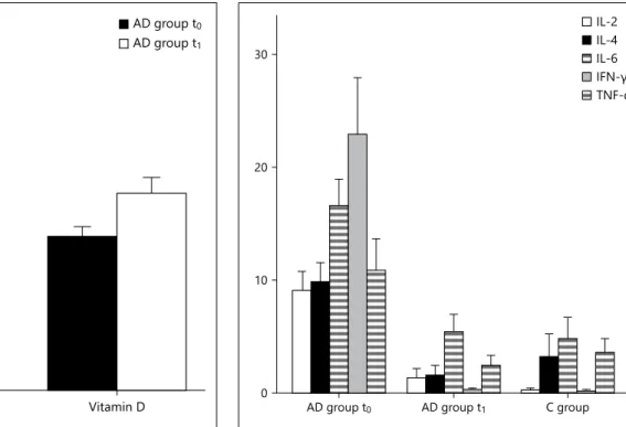
vita-min D values were significantly higher compared to the starting levels (29.41 ± 10.73 vs. 22.97 ± 8.03 ng/ml; p = 0.01). At the same time, a reduction in the SCORAD index was observed (46.13 ± 15.68 at the first visit vs. 22.57 ±
15.28 at the second; p < 0.001; fig. 1 ). A statistically sig-nificant reduction of IL-2 (8.22 ± 7.39 vs. 1.24 ± 4.06; p < 0.001), IL-4 (9.01 ± 7.05 vs. 1.36 ± 4.26; p < 0.001), IL-6 (15.11 ± 9.13 vs. 6.81 ± 9.60; p = 0.007), and IFN-γ (20.05 ± 22.84 vs. 0.19 ± 0.79; p = 0.019) was documented (online suppl. table 4). In contrast, TNF-α reduction was not sig-nificant (8.85 ± 11.09 vs. 2.33 ± 4.27). All the tested cyto-kines approached the healthy control group values back within the normal range ( fig. 2 ; online suppl. table 2).
Taking into consideration all 22 patients in the AD
group t 1 , we found no significant correlation between the
vitamin D change and the SCORAD change between the second and the first visit. Otherwise, excluding from the analysis 6 patients who declared they adhered to the min D supplementation but were unable to increase vita-min D levels between the first and the second visit (mean-ing the supplementation was not sufficient to improve their vitamin D levels), we found a significant negative cor-relation between the vitamin D change and the SCORAD change (r = –0.49; p = 0.02), strongly supporting the hy-pothesis that the positive clinical effect was mostly due to vitamin D effective supplementation (online suppl. fig. 1).
Overall, 17 individuals did not adhere to the 3 months of vitamin D supplementation, and only 4 of those at-tended the second visit. Despite the small size of the sam-ple, it was interesting to note that, without supplementa-tion, there was not a significant change in these 4 children between the first and second visit in the following: (1) vitamin D levels (22.8 ± 3.5 vs. 22.9 ± 6.6), (2) SCORAD (31.9 ± 13.2 vs. 22.8 ± 10.2) and (3) cytokine levels.
Discussion
AD is characterized by local and systemic immune dysregulation. The cutaneous inflammation displays a bi-phasic pattern of T-cell activation. In the acute phase of lesion development, there is a predominance of Th2 cy-tokines (especially IL-4 and IL-13) which promotes im-munoglobulin isotype switching to IgE production and induces the expression of vascular cell adhesion mole-cules (e.g. VCAM1), which may suggest a role for these Th2 cytokines in regulating eosinophilic infiltration in the skin [17] . Moreover, IL-4 and IL-13 inhibit the
de-0 10 20 30 40 50 SCORAD Vitamin D AD group t0 AD group t1
Fig. 1. After 3 months of vitamin D supplementation in the AD group it was found that vitamin D values were significantly higher (29.41 ± 10.73 ng/ml) compared to the starting levels (22.97 ± 8.03 ng/ml; p = 0.01). At the same time, a statistically significant reduc-tion in the SCORAD index was found (46.13 ± 15.68 at t 0 vs.
22.57 ± 15.28 at t 1 ; p < 0.001).
0 10 20 30
AD group t0 AD group t1 C group
IL-2 IL-4 IL-6 IFN-Dž TNF-į
Fig. 2. A statistically significant reduction of IL-2, IL-4, IL-6, and IFN-γ was found; no statistically significant reduction of TNF-α was found. The cytokine values decreased and ap-proached the healthy control group values back within the nor-mal range.
struction of Staphylococcus aureus by keratinocytes in pa-tients with AD. In the chronic phase of AD there is a shift to the Th1 pattern, with a predominant production of IFN-γ [18–20] .
In this study, Th1 and Th2 cytokine levels (IL-2, IL-4, IL-6, IFN-γ) were higher in patients affected by AD compared to the healthy control group. In contrast, the mean value of TNF-α was not increased compared to the controls. Our finding fits with the literature, showing immune system activation with increased levels of Th1 and Th2 cytokines. Because our population was com-posed of patients with chronic AD, the predominant lymphocyte phenotype (according to the cytokine pat-tern) was the Th1 subpopulation, with levels of IFN-γ 100 times higher than the normal values and the control group [21] .
IL-10 is a critical anti-inflammatory cytokine whose expression is induced after proinflammatory mediators. It is produced by numerous cell types, including Th2 cells. Data from studies examining IL-10 in subjects with AD are conflicting. Plasma levels of IL-10 inversely correlate with the severity of AD. In contrast, other studies have shown elevated IL-10 levels in peripheral blood mono-nuclear cells isolated from AD patients or in lesional skin. Elevated numbers of CD4+ and CD8+ cells expressing IL-10 were also observed in patients with AD. In this study, IL-10 levels were not tested due to technical prob-lems of our laboratory [22] .
In addition to its classical role in calcium homeostasis, recent studies demonstrated the influence of vitamin D in immunomodulation and cell differentiation. Although a definitive role for vitamin D in the pathogenesis of AD has not been explained, its levels seem to be related to AD severity [8] . Therefore, vitamin D is increasingly used in the management of diseases such as AD, psoriasis, vitili-go, acne, and rosacea [23, 24] .
In this study, an insufficient level of vitamin D was found in 82% of children (32/39) with AD. However, vi-tamin D deficiency has also been documented in healthy subjects despite reports of abundant solar exposure [25] . The prevalence of this deficit is very common even in the healthy population, as confirmed by the healthy control group.
Vitamin D serum levels after 3 months of supplemen-tation were significantly higher, going from insufficient to sufficient vitamin D levels. At the same time, SCORAD reduction was observed, demonstrating a clinical status improvement. Confirming that this effect was mostly due to the vitamin D supplementation, we found a negative significant correlation between the SCORAD change and
vitamin D change in the subgroup of patients in which the supplementation was able to increase vitamin D levels. In addition, the treatment with vitamin D was effective in normalizing the serum levels of all the altered cytokines (IL-2, IL-4, IL-6, IFN-γ). Their levels after 3 months of treatment were comparable to the control group and within the normal range, confirming the effect of vitamin D in modulating the immunological status of the patient.
At the skin level, vitamin D acts through the suppres-sion of the inflammatory response, increasing AMPs and promoting the integrity of the cutaneous barrier [11] . It may reduce both local and systemic inflammatory re-sponses, modulating cytokine production and reducing Toll-like receptor activation [26] . It has been shown that vitamin D also inhibits T-helper cell (Th1) proliferation (consequently the production of IL-2, TNF-α and IFN-γ decreases) [13] .
According to the literature, IFN-γ and IL-2 serum lev-els decrease in the treated group. This fact can be ex-plained by the vitamin D inhibitory effect on the acquired immunity, resulting in a reduction of Th1 cell activation [27] . Moreover, the IFN-γ reduction would lead to a de-creased expression of other cytokines such as IL-31 and IL-33 [28] and to the improvement of clinical features such as spongiosis. In fact, IFN-γ is implicated in kerati-nocyte apoptosis, which leads to eczema and spongiosis in patients with AD [29] .
Vitamin D antimicrobial activity and the negative ef-fects of its deficiency on the general well-being and on the longevity of patients have been previously demonstrated. It may reduce the risk of infection through multiple mechanisms, improving the efficiency of innate immu-nity by modulating the production of AMPs (such as cat-helicidin and β-defensin 2 [30, 31] ) and cytokine re-sponse.
AMPs are effectors of skin innate immunity (‘endog-enous antibiotics’), killing bacteria, viruses and fungi. They contribute to the formation of a chemical barrier on the skin surface and trigger a host immune response (‘alarming activity’). Therefore, from an antimicrobial viewpoint, vitamin D is able to reduce the infection sus-ceptibility in patients with AD and regulate local immune and inflammatory responses [32] .
The potential role of vitamin D in suppressing inflam-matory responses, enhancing AMP activity and promot-ing the integrity of the skin, is clear. Its supplementation has a possible therapeutic role for many skin diseases such as AD.
An important weakness in our study was the small sample size. It would be desirable to design a multicenter
case-control study that would help to decrease the bias due to many variables such as the genetic background, the environment and other confounding factors.
Conclusions
The existing literature on the influence of vitamin D on the development of allergic diseases, in general, is contradictory. It is an issue that needs to be further in-vestigated. There is growing interest in vitamin D sup-plementation in patients with AD. Many studies have
confirmed that vitamin D supplementation has a posi-tive effect on AD severity. This study highlight how the correction of the vitamin D deficit in children is able to improve the clinical evolution of AD, most likely through negative modulation of the immune system, involving Th2 but primarily Th1 lymphocyte popula-tions.
Disclosure Statement
The authors declare no conflict of interests.
References
1 Novak N, Simon D: Atopic dermatitis – from new pathophysiologic insights to individual-ized therapy. Allergy 2011; 66: 830–839. 2 Bieber T: Atopic dermatitis. New Engl J Med
2008; 358: 1483–1494.
3 Dogru M, Kirmizibekmez H, Yesiltepe Mutlu RG, Aktas A, Ozturkmen S: Clinical effects of vitamin D in children with asthma. Int Arch Allergy Immunol 2014; 164: 319–325. 4 Wjst M, Dold S: Genes, factor X, and
aller-gens: what causes allergic diseases? Allergy 1999; 54: 757–759.
5 Back O, Blomquist HK, Hernell O, et al: Does vitamin D intake during infancy promote the development of atopic allergy? Acta Derm Venereol 2009; 89: 28–32.
6 Erkkola M, Kaila M, Nwaru BI, et al: Maternal vitamin D intake during pregnancy is inverse-ly associated with asthma and allergic rhinitis in 5-year-old children. Clin Exp Allergy 2009; 39: 875–882.
7 Miyake Y, Sasaki S, Tanaka K, et al: Dairy food, calcium, and vitamin D intake in preg-nancy and wheeze and eczema in infants. Eur Respir J 2010; 35: 1228–1234.
8 Willers SM, Devereux G, Craig LC, et al: Ma-ternal food consumption during pregnancy and asthma, respiratory and atopic symptoms in 5-year-old children. Thorax 2007; 62: 773– 779.
9 Peroni DG, Piacentini E, Cametti E, et al: Cor-relation between serum 25-hydroxyvitamin D levels and severity of atopic dermatitis in chil-dren. Brit J Dermatol 2011; 164: 1078–1082. 10 Amestejani M, Salehi BS, Vasigh M, Sobhkhiz
A, Karami M, Alinia H, et al: Vitamin D sup-plementation in the treatment of atopic der-matitis: a clinical trial study. J Drugs Derma-tol 2012; 11: 327–330.
11 Searing DA, Leung DY: Vitamin D in atopic dermatitis, asthma and allergic diseases. Im-munol Allergy Clin 2010; 30: 397–409.
12 Antal S, Dombrowski Y, Koglin S, et al: Im-pact of vitamin D3 on cutaneous immunity and antimicrobial peptide expression. Der-matoendocrinol 2011; 3: 18–22.
13 Bikle DD: Vitamin D and the immune system: role in protection against bacterial infection. Curr Opin Nephrol Hypertens 2008; 17: 348– 352.
14 Rausch-Fan X, Leutmezer F, Willheim M, Spittler A, Bohle B, Ebner C, Jensen-Jarolim E, Boltz-Nitulescu G: Regulation of cytokine production in human peripheral blood mononuclear cells and allergen-specific Th cell clones by 1α,25-dihydroxyvitamin D3. Int Arch Allergy Immunol 2002; 128: 33–41. 15 Oranje AP, Glazenburg EJ, Wolkerstorfer A,
et al: Practical issues on interpretation of scor-ing atopic dermatitis: the SCORAD index, ob-jective SCORAD and the three-item severity score. Brit J Dermatol 2007; 157: 645–648. 16 Stadler JF, Taieb A: Severity scoring of atopic
dermatitis: the SCORAD index. Consensus Report of the European Task Force on Atopic Dermatitis. Dermatology 1993; 186: 23–31. 17 Leung DY, Bieber T: Atopic dermatitis.
Lan-cet 2003; 361: 151–160.
18 Eichenfield LF, Ellis CN, Mancini AJ, Paller AS, Simpson EL: Atopic dermatitis: epidemi-ology and pathogenesis update. Semin Cutan Med Surg 2012; 31:S3–S5.
19 Boguniewicz M, Leung DY: Atopic dermati-tis: a disease of altered skin barrier and im-mune dysregulation. Immunol Rev 2011; 242: 233–246.
20 Schneider L, Tilles S, Lio P, Boguniewicz M, Beck L, LeBovidge J, et al: Atopic dermatitis: a practice parameter update 2012. J Allergy Clin Immunol 2013; 131: 295–299.
21 Hamid Q, Boguniewicz M, Leung DY: Differ-ential in situ cytokine gene expression in acute versus chronic atopic dermatitis. J Clin Invest 1994; 94: 870–876.
22 Brandt EB, Sivaprasad U: Th2 cytokines and atopic dermatitis. J Clin Cell Immunol 2011; 2: 110.
23 Miller J, Gallo RL: Vitamin D and innate im-munity. Dermatol Ther 2010; 23: 13–22. 24 Mutgi K, Koo J: Update on the role of
sys-temic vitamin D in atopic dermatitis. Pediatr Dermatol 2013; 30: 303–307.
25 Brehm JM, Celedon C, Soto-Quiros ME, Avi-la L, et al: Serum vitamin D levels and markers of severity of childhood asthma in Costa Rica. Am J Respir Crit Care 2009; 179: 765–771. 26 Jeng L, Yamshchikov AV, Judd SE, Blumberg
HM, Martin GS, Ziegler TR, et al: Alterations in vitamin D status and anti-microbial pep-tide levels in patients in the intensive care unit with sepsis. J Transl Med 2009; 7: 28. 27 Abbas AK, Lichtman AH: Fondamenti di
im-munologia. Funzioni e alterazioni del sistema immunitario. Padova, Piccin Nuova Libraria, 2003, pp 295–309.
28 Seltmann J, Werfel T, Wittmann M: Evidence for a regulatory loop between IFN-γ and IL-33 in skin inflammation. Exp Dermatol 2013; 22: 102–107.
29 Rebane A, Zimmermann M, Aab A, et al: Mechanisms of IFN-γ-induced apoptosis of human skin keratinocytes in patients with atopic dermatitis. J Allergy Clin Immunol 2012; 129: 1297–1306.
30 Schwalfenberg GK: A review of the critical role of vitamin D in the functioning of the im-mune system and the clinical implications of vitamin D deficiency. Mol Nutr Food Res 2011; 55: 96–108.
31 Alitalo A: Human anti-infectious defence may be enhanced by vitamin D (in Finnish). Duodecim 2010; 126: 1127–1134.
32 Youssef DA, Miller CW, El-Abbassi AM, Cutchins DC, Cutchins C, Grant WB, Peiris AN: Antimicrobial implications of vitamin D. Dermatoendocrinol 2011; 3:220–229.