DOI
10.17219/acem/70795
Copyright
© 2018 by Wroclaw Medical University This is an article distributed under the terms of the Creative Commons Attribution Non-Commercial License (http://creativecommons.org/licenses/by-nc-nd/4.0/)
Address for correspondence
Karol Polom,
E-mail: [email protected]
Funding sources
Istituto Toscano Tumori (ITT) grant entitled “Gene expression profiles and therapy of gastric cancer” – (grant No. ITT-2007) European Union’s Seventh Framework Programme (FP7), GastricGlyco-Explorer under grant agreement No. [316929] (Karol Polom, Franco Roviello).
Conflict of interest None declared Received on September 21, 2016 Reviewed on February 13, 2017 Accepted on April 27, 2017
Abstract
Background. A better understanding of molecular gastric cancer (GC) entities may help in tailored treatments of that neoplasm. The PIK3CA mutation is one of the most important in many cancers.
Objectives. We performed a comparison of clinical and pathological data of the PIK3CA mutation in GC patients.
Material and methods. The analysis was done on 472 patients operated on in 1 center. Polymerase chain reaction (PCR) was used for the screening of PIK3CA (exon 9 and 20). For microsatellite instability (MSI) we used 5 quasi-monomorphic mononucleotide repeats − BAT-26, BAT-25, NR-24, NR-21, and NR-27. The clinical and pathological data was analyzed.
Results. PIK3CA mutation was observed in 10 out of 472 GC patients (2.1%). Nine out of 10 were MSI (9 of 111 MSI patients – 8.1%). Half of the 10 patients had mutations in exon 9 and the other half in exon 20. A majority of patients with the PIK3CA mutation had MSI (p < 0.001). The 5-year survival of MSI patients with the PIK3CA mutation was 40% and without the mutation, 70.4% (p = 0.309). For patients with the mutation in exon 9, the 5-year survival was 0%, and for those with the mutation in exon 20, 80% (p = 0.031). The Cox proportional hazards regression analysis did not show that PIK3CA is statistically correlated with a worse overall survival.
Conclusions. PIK3CA mutation in GC is a rare finding. It is strongly associated with the MSI molecular subgroup, presenting a worse outcome than other MSI patients. A completely different outcome is associated with the mutation in exon 9 compared to the mutation in exon 20, with the latter being more favorable. Key words: gastric cancer, PIK3CA, mutation, microsatellite instability, exon
PIK3CA mutation in gastric cancer and the role
of microsatellite instability status in mutations
of exons 9 and 20 of the PIK3CA gene
Karol Polom
1,6,A-F, Daniele Marrelli
1,A−F, Giandomenico Roviello
2,3,A−F, Valeria Pascale
1,B,E,F,
Costantino Voglino
1,B,C,E,F, Carla Vindigni
4,B,E,F, Daniele Generali
5,C,E,F, Franco Roviello
1,A−F1 Unit of General Surgery and Surgical Oncology, University of Siena, Italy
2 Department of Oncology, Medical Oncology Unit, San Donato Hospital, Arezzo, Italy 3 Department of Medical, Surgery and Health Sciences, University of Trieste, Italy 4 Department of Pathology, Hospital Department of University of Siena, Italy 5 Department of Medical, Surgery and Health Sciences, University of Trieste, Italy 6 Department of Surgical Oncology, Medical University of Gdansk, Poland
A – research concept and design; B – collection and/or assembly of data; C – data analysis and interpretation; D – writing the article; E – critical revision of the article; F – final approval of the article
Introduction
Gastric cancer (GC) is a disease that is characterized by multiple molecular, genetic, and epigenetic events.1
Mu-tation in a signaling pathway is one such event and the phosphoinositide 3-kinase (PI3K)/AKT/mammalian target of the rapamycin pathway (PI3K/AKT/mTOR pathway)
is one example of the mutation mechanism.2
A fundamen-tal step in this pathway is the creation of phosphatidylino-sitol-3,4,5-triphosphatate (PIP3), catalyzed by PI3K. This pathway is important in the cancer-related functions of cell proliferation, catabolism, cell adhesion, apoptosis, and au-tophagy.3 It also plays an important role in motility and
glucose homeostasis.4 The mutations of this pathway have
frequently been seen in cancers such as ovarian, breast, thyroid, and cervical. Many studies have revealed that this pathway is not of highest importance in GC patients.
The PIK3CA gene is located on chromosome 3p26.3.5
The most common mutations are seen in exon 9 and 20, representing different hotspots of mutations.4
In the current literature, PIK3CA mutations in colorectal cancer (CRC) are associated with female gender, proximal position, well-differentiated tumors, and mucinous histol-ogy, but these findings are not consistent.6 Some studies
have shown a significant coexistence of PIK3CA and KRAS mutations, while other studies have failed to show such a coexistence. Other contradictory data presented a link between the PIK3CA mutation and MSI or CpG island
methylator phenotype (CIMP).6 Another inconclusive
study showed worse outcome for early stage resectable disease, but other studies did not prove this finding.6 Such
conflicting findings can be explained by important differ-ences between cancers that have the mutation in exon 9 vs those with the mutation in exon 20.6,7 Exon 20 has a mutual
relationship with BRAF mutation, CIMP high/low and MSI-H, and exon 9 is linked with KRAS mutations.7 In the
study by Mao et al., authors showed that mutations in exon 20 are associated with resistance to anti-EGFR antibody therapy. This was not seen for mutations in exon 9.8 Tapia
et al. reported that PI3K and AKT are overexpressed in GC
with lymph node spread.9
New molecular classifications of gastric cancer have
recently been proposed.1,10 We are witnessing huge
ad-vances in our understanding of cancer from a molecu-lar, immunological, diagnostic and even bioinformatical standpoint, all contributing to better tailored treatments for patients.1,10−15 In both classifications, microsatellite
instability (MSI) is a distinct molecular subgroup of GC. In the available studies, MSI is associated with older age, female gender, intestinal histotype, non-cardia tumors, lower number of metastatic lymph nodes, and better sur-vival.16−18 It seems that the MSI subgroup is not
homog-enous and other genetic and molecular factors may play an important role for these particular patients.
The aim of the study was to compare the clinical and pathological data of PIK3CA mutation in GC patients.
We divided PIK3CA mutations into 2 categories based on the hotspot mutation sites at exon 9 and exon 20. In ad-dition, we investigated the coexistence of this mutation with MSI status and KRAS mutations.
Material and methods
Patients
The analysis was performed on a group of 472 GC pa-tients treated in the General Surgery and Surgical Oncol-ogy Department, University of Siena, Italy. We used tissue material stored in our biobank collected from patients who were operated on between 1990−2011. None of these patients received neoadjuvant treatment. We used tumoral and healthy tissues for comparative analysis. All samples were collected just after resection in the operating theatre.
PIK3CA sample preparation
Genomic DNA was extracted by tumoral and constitu-tional fresh frozen sample tissues using a standard protocol (Gentra Systems, Minneapolis, USA). The DNA concentra-tion was calculated by spectrophotometry.
Polymerase chain reaction (PCR) is used for the screen-ing of PIK3CA (exon 9 and 20).
To search for somatic alterations of the PIK3CA gene, exons 9 and 20 were sequenced according to the protocol described in detail by Velho et al.19 PCR reactions were
carried out in a volume of 20 µL containing 100 ng/µL ge-nomic DNA template, 1X Reaction Buffer, 0.5 µM of each
PCR primer, MgCl2 1.25 mM, 0.15 mM of each dNTPs, Taq
polymerase 0.5 U/µL (Euroclone, Pero, Italy). The reactions were performed in programmable thermocyclers according to the standard protocol.
A 5 µL aliquot of each PCR reaction was run on a 2% aga-rose gel to confirm the size, quantity, and purity of each PCR product. The remaining 15 µL of PCR amplified bands were extracted from the gel with the Invisorb® Spin DNA
Extrac-tion Kit (Invitek, Stratec Biomedical Systems, Birkenfeld, Germany). Samples were then purified and 2 µL aliquot of purified PCR product was cycle sequenced using a Big-Dye Terminator Kit (Applied Biosystems, Foster City, USA) in a total volume of 20 µL. Samples were then purified and sequenced using an automated DNA sequencer ABI PRISM 310 Genetic Analyzer (Applied Biosystems, Milan, Italy) ac-cording to the protocol of the manufacturer. Sequencing was performed in both strands. All sequence alterations in these genes were validated with a second independent PCR.
Pentaplex polymerase chain reaction
and microsatellite analysis
A detailed description of MSI analysis was described in our
mononucleotide repeats, namely, BAT-26, BAT-25, NR-24, NR-21, and NR-27. Following the definition of the National Cancer Institute workshop on MSI for cancer, we con-sidered a tumor as MSI when 2 or more markers showed instability on 5 loci (MSI-H).20
A detailed description of the pathological, clinical, sur-gical and follow-up data was also given in our previous publication.18
Statistical analysis
Statistical analysis was performed with the χ2 test
or Fisher exact test to compare categorical variables. The Mann-Whitney U test was used to compare continu-ous variables not normally distributed. Cumulative sur-vival was calculated by the life table method of Kaplan and Meier, and the log-rank test was used to distinguish significant differences. Statistical significance was deter-mined at p-value < 0.05.
Survival curves estimated using the Kaplan-Meier meth-od were compared using a log-rank test, considering death from cancer as the end-point (cancer-related survival).
The Kaplan-Meier estimation was used to plot survival curves, and log-rank tests were used to calculate the dif-ference of overall survival (OS) between groups.
Multivariate Cox proportional hazard regression analy-sis was used to investigate independent prognostic factors for overall survival between groups. The variables includ-ing PIK3CA status, age, sex, tumor location, MSI status, Lauren histotype, type of resection, T, N, M status, and ad-juvant therapy were used as covariates. Statistical analysis was done using commercially available statistical software (SPSS 20.0 for Windows SPSS Inc., Chicago, USA).
Results
PIK3CA mutation was observed in 10 of 472 GC patients
(2.1%). Half of the 10 patients had mutations in exon 9 and the other half in exon 20. For exon 9 mutations, we found 2 mutations of E542K, 1 mutation of E545K, 1 of N515S, and 1 of E545G. All 5 patients with a mutation in exon 20 had mutation of H1047R.
Interestingly, 9 of 10 patients were also MSI positive. In 2 patients (20%), we observed KRAS mutation and
PIK3CA mutation in exon 20. Both patient showed KRAS
mutation -12D and were associated with better prognosis. A clinicopathological comparison of PIK3CA patients and wild-type (wt) PIK3CA patients is presented in Table 1. The only statistically significant factor associated with
PIK3CA mutations was MSI status. We also performed
an analysis of PIK3CA mutation on the MSI positive subgroup. The clinicopathological analysis is presented in Table 2. Here, the only statistically significant factor was tumor position.
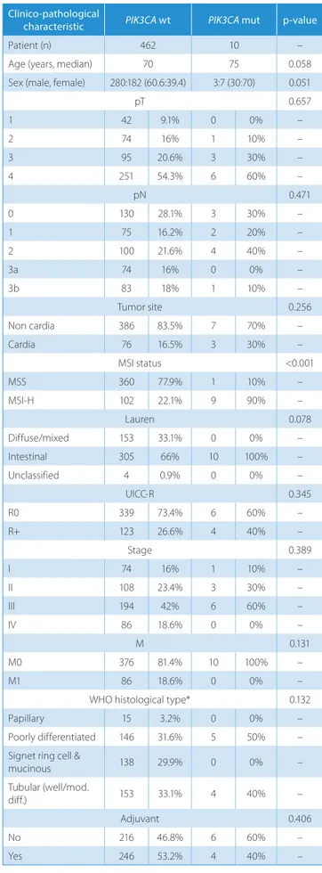
Table 1. PIK3CA wild type vs mutations in all GC group
Clinico-pathological
characteristic PIK3CA wt PIK3CA mut p-value
Patient (n) 462 10 –
Age (years, median) 70 75 0.058
Sex (male, female) 280:182 (60.6:39.4) 3:7 (30:70) 0.051
pT 0.657 1 42 9.1% 0 0% – 2 74 16% 1 10% – 3 95 20.6% 3 30% – 4 251 54.3% 6 60% – pN 0.471 0 130 28.1% 3 30% – 1 75 16.2% 2 20% – 2 100 21.6% 4 40% – 3a 74 16% 0 0% – 3b 83 18% 1 10% – Tumor site 0.256 Non cardia 386 83.5% 7 70% – Cardia 76 16.5% 3 30% – MSI status <0.001 MSS 360 77.9% 1 10% – MSI-H 102 22.1% 9 90% – Lauren 0.078 Diffuse/mixed 153 33.1% 0 0% – Intestinal 305 66% 10 100% – Unclassified 4 0.9% 0 0% – UICC-R 0.345 R0 339 73.4% 6 60% – R+ 123 26.6% 4 40% – Stage 0.389 I 74 16% 1 10% – II 108 23.4% 3 30% – III 194 42% 6 60% – IV 86 18.6% 0 0% – M 0.131 M0 376 81.4% 10 100% – M1 86 18.6% 0 0% –
WHO histological type* 0.132
Papillary 15 3.2% 0 0% –
Poorly differentiated 146 31.6% 5 50% –
Signet ring cell &
mucinous 138 29.9% 0 0% – Tubular (well/mod. diff.) 153 33.1% 4 40% – Adjuvant 0.406 No 216 46.8% 6 60% – Yes 246 53.2% 4 40% –
* 11 cases with unclassified WHO histotype are excluded ; MSS – microsatellite stable; MSI-H – microsatellite instable; M – metastases; pT – pathological tumor status; pN – pathological lymph node status.
Interestingly all ten patients showed Lauren intestinal histotype.
We also analyzed cancer-related survivals. The first comparison was performed between all patients with or without PIK3CA mutation (Fig. 1). 5-year survival for
PIK3CA wt patients was 43.2% and for PIK3CA mutation,
36% (p = 0.856). Secondly, we analyzed the group of MSI GC patients (Fig. 2). The 5-year survival of the MSI pa-tients presenting with PIK3CA mutation was 40% and Fig. 1. Cancer-related survival of patients with or without mutation in PIK3CA
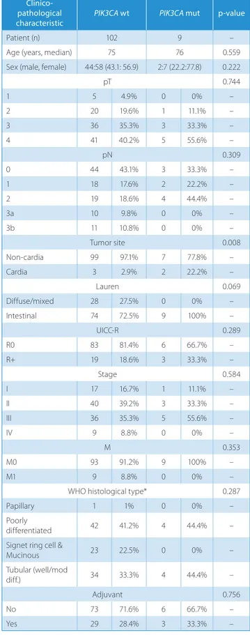
Table 2. MSI status patients and PIK3CA mutations
Clinico-pathological
characteristic PIK3CA wt PIK3CA mut p-value
Patient (n) 102 9 –
Age (years, median) 75 76 0.559
Sex (male, female) 44:58 (43.1: 56.9) 2:7 (22.2:77.8) 0.222
pT 0.744 1 5 4.9% 0 0% – 2 20 19.6% 1 11.1% – 3 36 35.3% 3 33.3% – 4 41 40.2% 5 55.6% – pN 0.309 0 44 43.1% 3 33.3% – 1 18 17.6% 2 22.2% – 2 19 18.6% 4 44.4% – 3a 10 9.8% 0 0% – 3b 11 10.8% 0 0% – Tumor site 0.008 Non-cardia 99 97.1% 7 77.8% – Cardia 3 2.9% 2 22.2% – Lauren 0.069 Diffuse/mixed 28 27.5% 0 0% – Intestinal 74 72.5% 9 100% – UICC-R 0.289 R0 83 81.4% 6 66.7% – R+ 19 18.6% 3 33.3% – Stage 0.584 I 17 16.7% 1 11.1% – II 40 39.2% 3 33.3% – III 36 35.3% 5 55.6% – IV 9 8.8% 0 0% – M 0.353 M0 93 91.2% 9 100% – M1 9 8.8% 0 0% –
WHO histological type* 0.287
Papillary 1 1% 0 0% –
Poorly
differentiated 42 41.2% 4 44.4% –
Signet ring cell &
Mucinous 23 22.5% 0 0% – Tubular (well/mod diff.) 34 33.3% 4 44.4% – Adjuvant 0.756 No 73 71.6% 6 66.7% – Yes 29 28.4% 3 33.3% –
* 3 cases with unclassified WHO histotype are excluded ; MSS – microsatellite stable; MSI-H – microsatellite instability; pT – pathological tumor status; pN – pathological lymph node status; UICC-R – Union Internationale Contre le Cancer Resection margin.
Fig. 2. Cancer-related survival of patients with MSI status with or without
PIK3CA mutation 1.0 0.8 0.6 0.4 0.2 0.0 1.0 0.8 0.6 0.4 0.2 0.0 Canc er -r el ate d s ur viv al Canc er -r el ate d s ur viv al Months Months PIK3CA wt PIK3CA mut PIK3CA wt PIK3CA mut p = 0.856 p = 0.309
without the mutation, 70.4% (p = 0.309). We also checked the difference in survival of patients with the PIK3CA mu-tation in different exons (Fig. 3). For mumu-tation in exon 9, the 5-year survival was 0% and for mutation in exon 20 – 80% (p = 0.031). For better visualization of these dif-ferences between the 2 mutation locations, we added
a survival curve for MSI patients without PIK3CA muta-tion (p = 0.026) – Fig. 3.
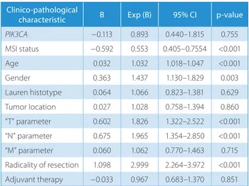
The Cox proportional hazards regression analysis showed that age, sex, MSI status, “T” parameter, “N” pa-rameter and type of “R” resection were the only prognostic factors statistically correlated with a worse overall survival (Table 3).
Discussion
In previous reports, the frequency of GC PIK3CA muta-tions varies from 4% to 13.2%.21,22 In our paper, PIK3CA
mutation occurred in 8.1% of the MSI group. In total, only a frequency of 2.1% was observed, lower in comparison with previous studies. The debate about PIK3CA muta-tion and its prognosis for different cancers is unresolved. Some authors suggest PIK3CA mutation is associated with a better prognosis in the case of breast cancer, while other authors showed worse prognosis in cancers like colorec-tal, endometrial, and lung cancers.8,23−25 Warneke et al.
presented interesting data for gastric cancer, showing worse survival in PIK3CA exon 20 mutation with intes-tinal histotype and better survival for the same mutation with diffuse histotype.26 These results were statistically
significant. In our study, it was impossible to analyze this factor because we did not observe diffuse/mixed histotype in our sample. PIK3CA mutation in exon 20 was found to be an independent prognostic factor of survival for in-testinal pathology. Also, our results show that mutation in exon 20 presented improved patient survival.
In the paper by Fang et al., they found PIK3CA mutation
in 57/432 of patients (13.2%).22 They analyzed PIK3/AKT
mutations together and found that in the intestinal his-totype, patients presenting mutation in that gene showed tumors located mostly in the lower third of the stomach. In diffuse histotype, the location of the tumor was in the upper third of the stomach and patients showed a higher rate of hematogenous metastases. The authors did not find any difference in survival between patients presenting or not presenting PIK3/AKT mutation.
The authors also searched for a link between the PIK3CA mutation and Epstein-Barr virus (EBV) infection. The rate of EBV infection was higher only in the situation where the tumor was situated in the middle part of the stomach
for GC patients with PIK3CA mutation.22
In a paper by Barbie et al., the authors also analyzed
PIK3CA mutation in GC and its association with GC and
MSI.27 Only 8 of 39 MSI GC cases harbored the H1047R
mutation. They found that this finding did not correlate with survival or any other clinical or pathological features linked with MSI GC. This is similar to our results on the exon 20 mutation in MSI patients, in which similar survival results were observed. A worse outcome was only observed in the case of exon 9 mutation, which was not observed
in the above-mentioned study.28
Table 3. Cox proportional hazards regression analysis
Clinico-pathological
characteristic B Exp (B) 95% CI p-value
PIK3CA −0.113 0.893 0.440–1.815 0.755 MSI status −0.592 0.553 0.405–0.7554 <0.001 Age 0.032 1.032 1.018–1.047 <0.001 Gender 0.363 1.437 1.130–1.829 0.003 Lauren histotype 0.064 1.066 0.823–1.381 0.629 Tumor location 0.027 1.028 0.758–1.394 0.860 “T” parameter 0.602 1.826 1.322–2.522 <0.001 “N” parameter 0.675 1.965 1.354–2.850 <0.001 “M” parameter 0.060 1.062 0.770–1.463 0.715 Radicality of resection 1.098 2.999 2.264–3.972 <0.001 Adjuvant therapy −0.033 0.967 0.683–1.370 0.851
PIK3CA status expressed as PIK3CA wt = 0 and PIK3CA mutation = 1;
MS status expressed as MSS = 0 and MSI = 1; gender expressed as F = 0 an M = 1; Lauren histotype expressed as intestinal = 0 and non-intestinal = 1; tumor location expressed as non-cardias = 0 and cardias = 1; “T” parameter expressed as T1–T2 = 0 and T3–T4 = 1; “N” parameter expressed as N− = 0 and N+ = 1; “M” parameter expressed as negative = 0 and positive = 1; radicality of resection expressed as R0 = 0 and R1–R2 = 1; adjuvant therapy expressed as yes = 1 and no = 0;
MSI status – microsatellite instability; T parameter – pathological tumor status; N parameter – pathological lymph node status; M parameter – metastases status.
Fig. 3. Comparison of cancer-related survival of patients with different
PIK3CA exon mutations together with a survival curve of MSI GC patients
without PIK3CA mutation
1.0 0.8 0.6 0.4 0.2 0.0 Canc er -r el ate d s ur viv al Months other MSI-H patients codon 9 codon 20 p = 0.026
In our study, we observed that PIK3CA patients were older and showed intestinal histotype more often but without sta-tistical significance. Analyzing the subgroup of MSI GC pa-tients, the only differences were tumor position (p = 0.008) and Lauren histotype (p = 0.069) – PIK3CA mutations were more commonly seen in the upper third of the stomach and also showed only intestinal histology. We did not observe any difference in survival between wild-type and mutated
PIK3CA GC patients; however, in the MSI GC subgroup
of patients, those with PIK3CA mutation had a worse 5-year survival rate (40%) than those without the mutation (70.4%).
PIK3CA mutations are also observed in head and neck
squamous cell carcinoma (HNSCC) for 6−21% of patients.4
Interestingly, this mutation was absent across German, Vietnamese, and Greek patients.28,29 Likely, some ethnic,
environmental, and/or other unknown factors are associ-ated with this finding. A paper by Seiwert et al. pointed out that PIK3CA mutations are more commonly associated with human papillomavirus (HPV) positive HNSCC can-cers.30 This finding did not reach statistical significance
but can be an example of a factor that may play an im-portant role in developing this mutation. In our study, we observed one of the smallest incidences of PIK3CA muta-tion in GC patients – 2.1%.
In a CRC study by Day et al., it was found that PIK3CA exon 20 mutations were associated with proximal tumors and a sessile-serrated pathway (MSI-H/CIMP high/BRAF mutations), and PIK3CA exon 9 mutations were linked with the traditional serrated pathway of tumorigenesis
(CIMP-low/KRAS mutations).6 PIK3CA mutations were
significantly associated with older age, proximal tumor
site and mucinous histology, and KRAS mutation.6
Com-parison between wt PIK3CA and either exon 9 or 20 muta-tions showed some significant results. Mucinous histology was associated with exon 20, and for exon 9, factors like older age and KRAS mutations were associated. The direct comparison of exon 9 and 20 mutations did not reach sta-tistical significance. In a study by Sukawa et al., MSI status
was observed in 50% of PIK3CA GC patients.21 Similarly,
our results show a statistically significant link between
PIK3CA mutation and MSI status. In fact, almost all of our
GC patients presenting with a PIK3CA mutation were also MSI positive. KRAS mutation was observed only in the exon 20 mutation group (2 of 5 patients – 40%).
In breast cancer, PIK3CA mutations are seen in 40% of cases.4 The presence of this mutation is associated with
better prognosis in this cancer. Also, clinicopathological factors show higher rates of small tumor size, low grade, and positive estrogen receptor much more frequently in this group of patients.24 Importantly, these patients also
showed better survival.24 In other cancers like colorectal,
endometrial, or lung cancer, PIK3CA mutation is associat-ed with worse prognosis.23,25 We did not find any difference
in survival between patients with and without PIK3CA mutations. The difference was observed when we analyzed subgroups according to the type of exon mutation.
Our study was limited by the small number of patients with PIK3CA mutations in our GC patient pool. We pre-sented a link between different PIK3CA exon mutations and MSI GC that present completely different prognoses depending on the type of mutation. The MSI subtype of GC is a relatively new molecular subgroup and requires further analysis of different mutations that may have a positive or negative impact on patient outcome.
Our research leads to some important conclusions about
PIK3CA mutations. Firstly, PIK3CA mutations in GC
is rare. It is strongly associated with the MSI molecular subgroup, presenting a worse outcome than wt PIK3CA MSI GC patients. A completely different outcome is asso-ciated with mutation in exon 9 vs exon 20, with the latter being more favorable. The role of this mutation must be further studied with larger groups of patients.
References
1. Cristescu R, Lee J, Nebozhyn M, et al. Molecular analysis of gastric can-cer identifies subtypes associated with distinct clinical outcomes.
Nat Med. 2015;21(5):449–456.
2. Wadhwa R, Song S, Lee JS, Yao Y, Wei Q, Ajani JA. Gastric cancer-molecular and clinical dimensions. Nat Rev Clin Oncol. 2013;10: 643–655.
3. Willems L, Tamburini J, Chapuis N, Lacombe C, Mayeux P, Bouscary D. PI3K and mTOR signaling pathways in cancer: New data on targeted therapies. Curr Oncol Rep. 2012;14:129–138.
4. Lai K, Killingsworth MC, Lee CS. Gene of the month: PIK3CA. J Clin
Pathol. 2015;68:253–257.
5. Volinia S, Hiles I, Ormondroyd E, et al. Molecular cloning, cDNA sequence, and chromosomal localization of the human phospha-tidylinositol 3-kinase p110 alpha (PIK3CA) gene. Genomics. 1994;24: 472–477.
6. Day FL, Jorissen RN, Lipton L, et al. PIK3CA and PTEN gene and exon mutation-specific clinicopathologic and molecular associations in colorectal cancer. Clin Cancer Res. 2013;19(12):3285–3296.
7. Whitehall VL, Rickman C, Bond CE, et al. Oncogenic PIK3CA muta-tions in colorectal cancers and polyps. Int J Cancer. 2012;131: 813–820.
8. Mao C, Yang ZY, Hu XF, Chen Q, Tang JL. PIK3CA exon 20 mutations as a potential biomarker for resistance to EGFR monoclonal anti-bodies in KRAS wild-type metastatic colorectal cancer: A systematic review and meta-analysis. Ann Oncol. 2012;23:1518–1525.
9. Tapia O, Riquelme I, Leal P, et al. The PI3K/AKT/mTOR pathway is acti-vated in gastric cancer with potential prognostic and predictive sig-nificance. Virchows Arch. 2014;465:25–33.
10. Cancer Genome Atlas Research Network. Comprehensive molecular characterization of gastric adenocarcinoma. Nature. 2014;513(7517): 202–209.
11. Isa N. Evidence based radiation oncology with existing technology.
Rep Pract Oncol Radiother. 2014;19(6):259–266.
12. Polom W, Markuszewski M, Rho YS, Matuszewski M. Usage of invis-ible near infrared light (NIR) fluorescence with indocyanine green (ICG) and methylene blue (MB) in urological oncology. Part 1. Cent
European J Urol. 2014;67(2):142–148.
13. Subhash VV, Yeo MS, Tan WL, Yong WP. Strategies and advancements in harnessing the immune system for gastric cancer immunothera-py. J Immunol Res. 2015;308574. doi: 10.1155/2015/308574
14. Alocci D, Mariethoz J, Horlacher O, Bolleman JT, Campbell MP, Lisacek F. Property Graph vs RDF triple store: A comparison on gly-can substructure search. PLoS One. 2015;10(12):e0144578. doi: 10.1371/ journal.pone.0144578
15. Mereiter S, Magalhães A, Adamczyk B, et al. Glycomic analysis of gas-tric carcinoma cells discloses glycans as modulators of RON recep-tor tyrosine kinase activation in cancer. Biochim Biophys Acta. 2016; 1860(8):1795–1808.
16. Beghelli S, de Manzoni G, Barbi S, et al. Microsatellite instability in gastric cancer is associated with better prognosis in only stage II cancers. Surgery. 2006;139:347–356.
17. Corso G, Pedrazzani C, Marrelli D, Pascale V, Pinto E, Roviello F. Correlation of microsatellite instability at multiple loci with long‐ term survival in advanced gastric carcinoma. Arch Surg. 2009;144: 722–727.
18. Marrelli D, Polom K, Pascale V, et al. Strong prognostic value of mic-rosatellite instability in intestinal type non-cardia gastric cancer.
Ann Surg Oncol. 2016;23(3):943–950.
19. Velho S, Oliveira C, Ferreira A, et al. The prevalence of PIK3CA muta-tions in gastric and colon cancer. Eur J Cancer. 2005;41:1649–1654. 20. Boland CR, Thibodeau SN, Hamilton SR, et al. A National Cancer
Insti-tute workshop on microsatellite instability for cancer detection and familial predisposition: Development of international criteria for the determination of microsatellite instability in colorectal cancer.
Cancer Res. 1998;58(22):5248–5257.
21. Sukawa Y, Yamamoto H, Nosho K, et al. Alterations in the human epidermal growth factor receptor 2-phosphatidylinositol 3-kinase-v-Akt pathway in gastric cancer. World J Gastroenterol. 2012;18(45): 6577–6586. doi: 10.3748/wjg.v18.i45.6577
22. Fang WL, Huang KH, Lan YT, et al. Mutations in PI3K/AKT pathway genes and amplifications of PIK3CA are associated with patterns of recurrence in gastric cancers. Oncotarget. 2016;7(5):6201–6220. doi: 10.18632/oncotarget.6641
23. Catasus L, Gallardo A, Cuatrecasas M, Jaime P. PIK3CA mutations in the kinase domain (exon 20) of uterine endometrial
adenocarcino-mas are associated with adverse prognostic parameters. Mod Pathol. 2008;21:131–139.
24. Kalinsky K, Jacks LM, Heguy A, Patil S, Prat J. PIK3CA mutation associ-ates with improved outcome in breast cancer. Clin Cancer Res. 2009; 15:5049–5059.
25. Janku F, Garrido-Laguna I, Petruzelka LB, Stewart DJ, Kurzrock R. Novel therapeutic targets in non-small cell lung cancer. J Thorac
Oncol. 2011;6:1601–1612.
26. Warneke VS, Behrens HM, Haag J, et al. Prognostic and putative pre-dictive biomarkers of gastric cancer for personalized medicine. Diagn
Mol Pathol. 2013;22(3):127–137. doi: 10.1097/PDM0b013e318284188e
27. Barbi S, Cataldo I, De Manzoni G, et al. The analysis of PIK3CA muta-tions in gastric carcinoma and metaanalysis of literature suggest that exon-selectivity is a signature of cancer type. J Exp Clin Cancer Res. 2010;16:29–32.
28. Kostakis GC, Papadogeorgakis N, Koumaki V, Kamakari S, Koumaki D, Alexandridis C. Absence of hotspot mutations in exons 9 and 20 of the PIK3CA gene in human oral squamous cell carcinoma in the Greek population. Oral Surg Oral Med Oral Pathol Oral Radiol Endod. 2010;109:e53–58. doi: 10.1016/j.tripleo.2010.01.015
29. Fenic I, Steger K, Gruber C, Arens C, Woenckhaus J. Analysis
of PIK-3CA and Akt/protein kinase B in head and neck squamous cell
car-cinoma. Oncol Rep. 2007;18:253–259.
30. Seiwert, TY, Zuo Z, Keck MK, et al. Integrative and comparative genomic analysis of HPV-positive and HPV-negative head and neck squamous cell carcinomas. Clin Cancer Res. 2015;21(3):632–641. doi: 10.1158/1078-0432.CCR-13-3310