University of Study of Messina
Department of Veterinary Sciences
PhD in Veterinary Sciences
Coordinator: Prof. Adriana Ferlazzo
Curriculum: Clinical Veterinary Sciences
EVALUATION OF VASCULAR PERFUSION
OF ABDOMINAL ORGANS IN THE DOG USING CEUS
(CONTRAST-ENHANCED ULTRASONOGRAPHY).
Author:
Cyndi Mangano, MVD
Tutor:
Prof. Massimo De Majo, MVD, PhD
Co-Tutor:
MVD Pavel Proks, PhD
To My Mom,
A Strong Woman
And
Always Close To Me
Thanks to all staff of:
- Veterinary Teaching Hospital of Department of Veterinary Sciences,
University of Studies of Messina, Italy, in the person of Prof.
Massimo De Majo.
- Clinica Veterinaria Camagna Spa, Reggio Calabria, Italy, in the
person of Prof. Nicola Maria Iannelli.
- Department of Diagnostic Imaging of Veterinary Hospital of
Veterinary and Pharmaceutical University of Brno, Czech Republic, in
the person of Prof. Pavel Proks.
This research project was realized in accordance with the current
legislation and only for medical purposes.
INDEX Abstract
List of abbreviations ---GENERAL SECTION--- 1 Introduction
1.1 Contrast-Enhanced ultrasound (CEUS) 1.2 Ultrasound contrast agents (USCAs) 1.3 The technique
1.4 Clinical applications 2 General Procedures
---SPECIAL SECTION ONE: RESEARCH APPLICATIONS --- 3 Spleen
3.1 CEUS knowledge 3.2 Materials and Methods 3.3 Results
3.4 Discussion 4 Bladder
4.1 CEUS knowledge 4.2 Materials and Methods 4.3 Results
4.4 Discussion 5 Kidneys
5.1 CEUS knowledge 5.2 Materials and Methods 5.3 Results
5.4 Discussion 6 Testis
6.1 CEUS knowledge 6.2 Materials and Methods 6.3 Results
6.4 Discussion
---SPECIAL SECTION TWO: OTHER APPLICATIONS --- 7 Liver
8 Gallbladder 9 Adrenal
10 Gastro-enteric tract 11 Ovary
12 Soft tissue lesion ---FINAL SECTION---
13 General Discussion and Conclusions References
Abstract
Contrast-enhanced ultrasound (CEUS) is an ultrasonographic technique that reveals the micro-vascularization of organs and is applied in veterinary medicine to study physiological and pathological conditions, focal or diffuse parenchymal lesions.
Clinical applications include the possibility to use CEUS in dogs to evaluate the perfusion of spleen, bladder, kidneys, testis, liver, gallbladder, adrenal, soft tissue lesions, pancreas, prostate and emergencies.
Aim of this study is to describe the use of CEUS in dogs for some organs, giving some new applications and analysing the literature knowledge.
SonoVue®, contrast medium agent, was used. Qualitative evaluation were done and
quantitative computerized analysis of the contrast medium blood pool phase were performed using a dedicated commercial software.
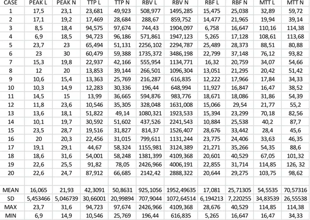
Particular results were given about Benign Nodular Hyperplasia (BNH): 20 spleens affected by BNH were studied and qualitative analysis showed simultaneous wash-in of lesion, compared to normal parenchyma, and anticipated wash-out, with hypoechoic pattern than surrounding normal spleen. Quantitative results showed the same peak of enhancement of lesions to the surrounding normal parenchyma but the contrast remain into the lesion for shorter time. In few cases, Hemangiosarcoma, Lymphoma and Histiocytic Sarcoma had early wash-in and early wash-out, prominent inner vessels characterize the lesions, associated with malignancy according to literature.
Considering previous studies, our results suggest that hypoenhancing is not a specific pattern of malignancy in splenic lesions.
Bladder wall was studied in 10 normal dogs using CEUS; wash-in began around 18 seconds and enhancing the bladder wall. Six cases of Transitional Cell Carcinoma (TCC) were evaluated, involvement of muscular layer of bladder wall with hyperenhancement of infiltrating tumor tissue was demonstrated.
About kidney perfusion, CEUS showed lower values in case ok kidney failure compared to a control group, this aspect suggested a reduction of renal perfusion during kidney disease.
Testes were studied giving referral qualitative and quantitative parameters about Interstitial Cell Tumour in n°=12 non-sedated dogs to study a single type of tumor without any influence by anesthesia: wash-in was around 25 seconds at the same time with surrounding tissue with hyperenhancement of lesion, heterogeneity and inner vessels.
CEUS was used also in other organs (Special Section two) including: Liver, Gallbladder, Adrenal, Gastro-enteric tract, Ovary, Soft tissue lesions with few cases. In conclusion, the evidence of hypoenhancement of BNH opens new considerations about CEUS characterization of malignant lesions that was demonstrated by previous studies. Based on our preliminary results about bladder lesions, a larger number of cases of TCC in comparison with inflammatory hyperplastic bladder lesions are needed to conclude if CEUS may be a feasible tool to study bladder wall lesions. Lower CEUS parameters of contrast medium diffusion in kidney of dogs affected by renal failure than normal dogs suggest a reduction of kidney perfusion in case of renal failure. CEUS is a technique that gives important information about perfusion, even if previous data have produced variable results; CEUS may be used expanding the knowledge given by basic ultrasonography.
List of abbreviations
AIFA Italian Agency of Drugs BNH Benign Nodular Hyperplasia BPH Benign Prostatic Hyperplasia
CECT Contrast-Enhanced Computed Tomography CEUS Contrast-Enhanced Ultrasound
CVC Caudal Vena Cava CT Computed Tomography
EFSUMB European Federation of Societies for Ultrasound in Medicine and Biology EMA European Medicine Agency
FDA Food and Drug Administration GB Gallbladder
GE Gastro-Enteric
HCC Hepato-Cellular-Carcinoma ICT Interstitial Cell Tumour IFI Indirect Immunofluorescence
IRIS International Renal Interest Society Leish Leishmaniasis – Leishmania MI Mechanical Index
MR Magnetic Resonance MTT average Time Transit P Peak
PCR Polymerase Chain Reaction PI Peak Intensity
PV Porta Vein
RBF Regional Blood Flow RBV Regional Blood Volume RES Reticle Endothelial System RI Resistivity Index
ROI Region Of Interest SCT Sertoli Cell Tumour SD Standard Deviation SEM Seminoma
TCC Transitional Cell Carcinoma TIC Time Intensity Curve
TTP Time To Peak
UCA Ultrasound Contrast Agent US Ultrasonography
---GENERAL SECTION--- 1 Introduction
1.1 Contrast-enhanced ultrasound (CEUS)
Contrast-enhanced ultrasound (CEUS) is an ultrasonographic technique applied most widespread in human medicine at the beginning of 2000. (Piscaglia, et al., 2012)
In 2004, the European Federation of Societies for Ultrasound in Medicine and Biology (EFSUMB) released the first guidelines about use of CEUS for liver applications in human patients. (Albrecht, et al., 2004)
EFSUMB, between 2008 and 2011, enlarged CEUS guidelines for non-hepatic applications.
CEUS, based on safety and efficacy, can be performed everywhere and without losing time for preliminary laboratory testing and it operates in real time so that rapid changes can be captured.
The procedure provides that CEUS examination should be preceded by careful assessment of the target with conventional B-mode US and, when appropriate, with Doppler (Spectral and/or Colour - Power). SonoVue® (sulphur hexafluoride with a phospholipid shell) produced by Bracco International, Imaging Milan S.p.A®, introduced in 2001, is the UCA mostly used in Europe. Use of UCA is finalized to improve B-mode and Doppler information: the underlying conventional B-mode image is seen, Doppler shows the bigger vascularization and CEUS, in addition of colour Doppler information, shows micro-vascularization. The description of the behaviour of tissue or lesion under examination should be in terms of its enhancement, taking into consideration its temporal behaviour, degree of enhancement, and contrast distribution. (Piscaglia, et al., 2012)
The most important clarification about the use of UCAs regards the safety of use firstly in human medicine and then in veterinary medicine.
The UCAs are very safe with a low incidence of side effects.
Between December 2001 to December 2004, it was the period of data collection in 28 centres for a total number of 23188 investigations mostly for liver: total number of adverse events was 29 but just 2 seriously needed treatment and 27 not serious, not needed treatment. (Piscaglia & Bolondi, 2006). Cardiac evaluation, using MI 0,4 effects, appears to increase rapidly: studied effects include premature ventricular contractions, micro-vascular rupture and petechial haemorrhage;, these damages may be reparable but good practice suggests caution when using UCAs. (Haar ter, 2009)
Despite FDA in 2007 announced a stop for using UCAs after death of 4 patients, that was not directly related to UCA’s administration, confirmed by the following study conducted for human patients and in veterinary as confirmed in Sailer et al, 2013. (Seiler, et al., 2013)
The interaction of diagnostic US with UCA could produce bio-effects, in vitro interactions between gas, bodies and cells; in fact, were observed alteration and cell death were observed. (EMA: European Medicines Agency, 2017)
In human medicine, there are important considerations are about using of CEUS in paediatric. Only the UCA Levovist® was approved for use in children and only for vesical-ureteral reflux; in fact, manufacturer’s instructions declare that the safety and effectiveness of SonoVue® in patients under 18 years old has not been established and the product should not be used in these patients. (EMA: European Medicines Agency,
2017). However for CEUS paediatric applications remain of critical importance. (Piscaglia, et al., 2012)
In veterinary medicine some hypersensitivity or allergic events occur rarely and there is no evidence of nephrotoxicity or cardio-toxicity. However, there is the possibility of bio-effects such as micro-vascular rupture with the insonation of microbubbles. (Ohlerth & Obrien, 2007)
No side effects have been reported with the use of Definity®, Imagent®, SonoVue® and Levovist®, the most used UCAs, in none dogs and cats studied (O’Brien, et al., 2004 - Rademacher, et al., 2008).
Immune reactions have not been associated with the use of Optison® in humans. Although there are reports of uncomplicated use of Optison® in dogs and other small animals, anaphylactic response related to the human albumin component has been reported in two dogs. (Yamaya, et al., 2004)
In 2007 Ohlerth & OBrien, wrote about CEUS in veterinary medicine about basic concepts.
They defined that the ultrasound contrast agent is an exogenous substance, consisting of gas or air microbubbles encapsulated in a shell of different composition, that can be administered intravenously or into a body cavity to enhance ultrasonic signals and wrote about use of CEUS in veterinary medicine. (Ohlerth & OBrien, 2007)
Recently, another guideline on non-hepatic application in human medicine was published. In this review, they present updated knowledge on CEUS application in the following fields: urogenital system (kidney, prostate and testis), thyroid, small bowel and lymph nodes.(Cantisani, et al., 2015)
1.2 Ultrasound contrast agents (UCAs)
It is possible to classify different UCAs for different composition and diffusion, generation and utility (Table 1.1: UCAs).
Most UCAs do not diffuse across the endothelium and therefore, they basically are blood pool agents. Vascular enhancement usually lasts a few minutes.
Typically, the gas content in the contrast agent is eliminated by lungs, while the shell components are filtered by the kidneys and eliminated by liver (Quaia, 2005, p. 3-14). This has led to the development of the first generation of trans-pulmonary contrast agents using air as the gas in the microbubbles covered by a shell. Third generation agents use the stabilization of a hard shell (polymer shells) and contain either air or perfluorocarbons, resulting in a much longer persistence time but are not still approved according to AIFA.
For example the Levovist® has been shown to have a late hepato-splenic-specific parenchymal phase with accumulation in human liver and spleen up to 20 min after intravenous (IV) bolus injection: accumulation may be mediated by the reticuloendothelial system, the Kupffer cells, or microbubbles are entrapped in the liver sinusoids (Quaia, 2005). Therefore, the late phase is also called sinusoidal phase.
SonoVue® or Imavist®, blood pool agents, were also subsequently found to have the
sinusoidal phase: maybe the agents are trapped or slowed in the hepatic sinusoids, during the study of liver.
The microbubble behaviour is depending on the local acoustic power (Quaia, 2005, p. 15-30) and the output power is reflected by the mechanical index (MI) which originally measures the potential for mechanical damage to tissues exposed to intense ultrasound pulses: the higher is the MI, the greater are its destructive properties (Quaia, 2005, p. 15-30).
The linear effects of ultrasound contrast agents are utilized in the clinical setting by Doppler technologies. If the acoustic power of the ultrasound beam at the resonance frequency increases (intermediate MI: 0.1 < MI > 0.5), microbubbles will show a longer expansion than contraction phase and begin nonlinear (not-sigmoidal shaped) oscillation (Quaia, 2005, p. 15-30). They start to emit the harmonics of the resonance frequency. The frequency of sub-harmonics is at half and that of higher harmonics is at multiples of the fundamental frequency. The second harmonic response is found at twice the fundamental frequency and usually has the highest intensity among the harmonic responses. The theoretical advantage of the harmonic over the fundamental frequency is that only microbubbles resonate with harmonics while adjacent tissues do not resonate or their harmonic resonation is very little. The increase of intensity of ultrasound beam disrupted the microbubbles: stimulated acoustic emission or loss of correlation.
Microbubble persistence and stability were increased by two principal ways: bubble encapsulation with or without surfactants and selection of gases with low diffusion coefficient. (Quaia, 2005, p. 3-14 - Ohlerth & OBrien, 2007)
The advantages to prefer sulphur hexafluoride-filled microbubbles, in comparison to the other UCAs are the prolonged stability in the vial (up to 6 h) and the peripheral blood (half-life of 6 min), and the uniformity of their size, which improves backscattering and harmonic behaviour at low acoustic power (Quaia, 2005, p. 3-14). Similar to Levovist®, SonoVue® has also been shown to have a late hepatosplenic-specific parenchymal phase in humans. SonoVue® provides a non-linear response with production of a clinically useful signal at low acoustic pressures, and destruction of the microbubbles is limited. (Ohlerth & OBrien, 2007)
Table 1.1: UCAs. UCAs that are/ have been clinically approved and some characteristics. The table include UCAs differentiating according to name, generation, first approval for clinical use, shell material, gas, half-life, countries and distributor. (Paefgen, et al. 2015)
NAME GENER ATION
FIRST APROVED FOR CLINICAL USE SHELL MATERIAL GAS HALF-LIFE COUNTRIES PROCEDURE/ DISTRIBUTOR
ALBUNEX 1 1993 WITHDRAW SONICATED SERUM ALBUMIN
AIR SHORT JAPAN, US MOLECULAR BIOSYSTEMS INC. SAN
DIEGO, CA,USA
ECHOVIST 1 1991 WITHDRAW AIFA W
GALACTOSE MICROPARTICLES
AIR SHORT GERMANY, UK
SCHERING AG, BERLIN, DE LEVOVIST 1 1995 WITHDRAW AIFA W GALACTOSE MICROPARTICLES, PALMITIC ACID
AIR SHORT CANAD, EU, CHINA, JAPAN
SCHERING AG, BERLIN, DE OPTISON 2 1998 AIFA APPROVED CROSS-LINKED SERUM ALBUMIN OCTAFLUOROPR OPANE
LONG US, EU GE HEALTCARE, BUCKINGHAMSHIRE, UK LUMASON / SONOVUE 2 2001/2004 AIFA APPROVED PHOSPHOLIPID SULPHURHEXAFL UORIDE
LONG US, EU, CHINA
BRACCO INTERNATIONAL IMAGING MILAN S.p.A.,
ITALY
SONAZOID 2 2007 PHOSPHOLIPID PERFLUOROBUT ANE LONG JAPAN, SOUTH KOREA GE HEALTCARE, BUCKINGHAMSHIRE, UK DEFINITY/ LUMINITY 2 2001/2006 AIFA APPROVED PHOSPHOLIPID OCTAFLUOROPR OPANE LONG NORTH AMERICA, EU LANTHEUS MEDICAL IMAGING, NORTH BILLORICA, MA IMAGENT/ IMAVIST
2 2002,WITHDRAW PHOSPHOLIPID PERFLUOROHEX ANE, NITROGEN
LONG US SCHERING AG, BERLIN, DE
ECHOGEN 3 2000, WITHDRAW COLLOID PERFLENAPENT
DODECAFLUORO PENTANE
LONG EU SONUS PHARMACEUTICALS
SonoVue® is composed by 8 microliters/ml of sulphur hexafluoride microbubbles in a
kit that includes also the solvent for dispersion for injection: 1 ml of the resulting dispersion contains 8 μl sulphur hexafluoride in the microbubbles, equivalent to 45 micrograms. This kit is composed with 1 vial containing 25 mg of lyophilized powder, 1 pre-filled syringe containing 5 ml sodium chloride and 1 Mini-Spike transfer system. According to the manufacturer’s instructions, SonoVue® is
1. for use with ultrasound imaging to enhance the echogenicity of the blood, which results in an improved signal to noise ratio and should only be used in patients where study without contrast enhancement is not-conclusive.
2. is a trans pulmonary echocardiographic contrast agent for use in patients with suspected or established cardiovascular disease to provide opacification of cardiac chambers and enhance left ventricular endocardial border delineation.
3. increases the accuracy in detection or exclusion of abnormalities in cerebral arteries and extra cranial carotid or peripheral arteries by improving the Doppler signal to noise ratio.
4. increases the quality of the Doppler flow image and the duration of clinically-useful signal enhancement in portal vein assessment.
5. improves display of the vascularity of liver and breast lesions during Doppler sonography, leading to more specific lesion characterization.
In human medicine, the recommended doses of SonoVue® are: B-mode imaging of cardiac chambers, at rest or with stress: 2 ml and for vascular Doppler imaging: 2.4 ml. During a single examination, a second injection of the recommended dose can be made. (EMA: European Medicines Agency, 2017)
The microbubbles dispersion is prepared before use by injecting through the septum 5 ml of sodium chloride 9 mg/ml (0.9%) solution for injection to the contents of the vial. The vial is then shaken vigorously for a few seconds until the lyophilizate is completely dissolved. The desired volume of the dispersion can be drawn into a syringe any time up to six hours after reconstitution. Just before drawing into the syringe, the vial should be agitated to re-suspend the microbubbles. SonoVue® should be administered immediately after drawing into the syringe by injection into a peripheral vein. Every injection should be followed by a flush with 5 ml of sodium chloride 9 mg/ml (0.9%) solution for injection. (EMA: European Medicines Agency, 2017)
Some contraindications have been seen, such us hypersensitivity to the active substance(s) or to any of the excipients. SonoVue® is contraindicated in patients known to have right-to-left shunts, severe pulmonary hypertension (pulmonary artery pressure >90 mmHg), uncontrolled systemic hypertension, and in patients with adult respiratory distress syndrome.
SonoVue® should not be used in combination with dobutamine in patients with
conditions suggesting cardiovascular instability where dobutamine is contraindicated. ECG monitoring should be performed in high-risk patients as clinically indicated. Use extreme caution when considering the administration of SonoVue® in patients with recent acute coronary syndrome or clinically unstable ischemic cardiac disease, including: evolving or ongoing myocardial infarction, typical angina at rest within last 7 days, significant worsening of cardiac symptoms within last 7 days, recent coronary artery intervention or other factors suggesting clinical instability (for example, recent deterioration of ECG, laboratory or clinical findings), acute cardiac failure, Class III/IV cardiac failure, or severe rhythm disorders because in these patients allergy like and/or vasodilatory reactions may lead to life threatening conditions.
SonoVue® should only be administered to such patients after careful risk/benefit
assessment and a closely monitoring of vital signs should be performed during and after administration.
Emergency equipment and personnel trained in its use must be readily available.
In the event of an anaphylactic reaction, beta blockers (including eye drop preparations) may aggravate the reaction. Patients may be unresponsive to the usual doses of adrenaline used to treat the allergic reactions.
Caution is advised when SonoVue® is administered to patients with clinically significant pulmonary disease, including severe chronic obstructive pulmonary disease. It is recommended to keep the patient under close medical supervision during and for at least 30 minutes following the administration of SonoVue®.
The number of patients with the following conditions who were exposed to SonoVue® in the clinical trials has been limited, and therefore, caution is advisable when administering the product to patients with: acute endocarditis, prosthetic valves, acute systemic inflammation and/or sepsis, hyperactive coagulation states and/or recent thromboembolism, and end-stage renal or hepatic disease.
SonoVue® is not suitable for use in ventilated patients, and those with unstable
neurological diseases.
In animal studies, the application of echo-contrast agents revealed biological side effects (e.g. endothelial cell injury, capillary rupture) by interaction with the ultrasound beam. Although these biological side effects have not been reported in humans, the use of a low mechanical index is recommended.
No interaction studies have been performed.
No clinical data on exposed pregnancies are available about use in pregnancy cases. The safety of SonoVue® was evaluated in 4653 adult patients who participated in 58 clinical trials.
The adverse reactions are classified by System Organ Class and frequency, using the following convention: Very common (≥ 1/10), Common (≥ 1/100 to < 1/10), Uncommon (≥ 1/1,000 to < /100), Rare (≥ 1/10,000 to < 1/1,000), Very rare (< 1/10,000), not known (cannot be estimated from the available data)
In very rare cases, fatal outcomes have been reported in temporal association with the use of SonoVue®.
In all these patients, there was a high underlying risk for major cardiac complications, which could have lied to the fatal outcome.
Reporting suspected adverse reactions after authorization of the medicinal product is important. It allows continued monitoring of the benefit/risk balance of the medicinal product.
The addition of sodium chloride 9 mg/ml (0.9%) solution for injection to the lyophilised powder followed by vigorous shaking results in the production of the microbubbles of sulphur hexafluoride.
The microbubbles have a mean diameter of about 2.5 μm, with 90% having a diameter less than 6 μm and 99% having a diameter less than 11 μm. Each millilitre of
SonoVue® contains 8 μl of the microbubbles. The interface between the sulphur
hexafluoride bubble and the aqueous medium acts as a reflector of the ultrasound beam thus enhancing blood echogenicity and increasing contrast between the blood and the surrounding tissues.
The intensity of the reflected signal is dependent on concentration of the microbubbles and frequency of the ultrasound beam. At the proposed clinical doses, SonoVue® has been shown to provide marked increase of more than 2 minutes in signal intensity for B-mode imaging in echocardiography and of 3 to 8 minutes for Doppler imaging of the macrovasculature and microvasculature.
Sulphur hexafluoride is an inert, innocuous gas, poorly soluble in aqueous solutions. There are literature reports of the use of the gas in the study of respiratory physiology and in pneumatic retinopexy.
The total amount of sulphur hexafluoride administered in a clinical dose is extremely small, (in a 2 ml dose the microbubbles contain 16 μl of gas). The sulphur hexafluoride dissolves in the blood and is subsequently exhaled.
After a single intravenous injection of 0.03 or 0.3 ml of SonoVue®/kg (approximately 1 and 10 times the maximum clinical dose) to human volunteers, the sulphur hexafluoride was cleared rapidly. The mean terminal half-life was 12 minutes (range 2 to 33 minutes). More than 80% of the administered sulphur hexafluoride was recovered in exhaled air within 2 minutes after injection and almost 100% after 15 minutes.
In patients with diffuse interstitial pulmonary fibrosis, the percentage of dose recovered in expired air averaged 100% and the terminal half-life was similar to that measured in healthy volunteers.
Non-clinical data reveal no special hazard for humans based on conventional studies of safety pharmacology, genotoxicity and toxicity to reproduction.
Shelf life is 2 years.
Once reconstituted, chemical and physical stability has been demonstrated for 6 hours. From a microbiological point of view, the product should be used immediately. If not used immediately, in use storage times and conditions prior to use are the responsibility of the user.
Before use examine the product to ensure that the container and closure have not been damaged.
Do not use if the liquid obtained is clear and/or if solid parts of the lyophilisate are seen in the suspension.
SonoVue® should be administered immediately by injection into a peripheral vein.
If SonoVue® is not used immediately after reconstitution the microbubble dispersion should be shaken again before being drawn up into a syringe. Chemical and physical stability of the microbubble dispersion has been demonstrated for 6 hours.
The vial is for a single examination only. Any unused medicinal product or waste material must be discarded in accordance with local requirements. (EMA: European Medicines Agency, 2017)
1.3 The technique
Contrast-specific US modes are required and are generally based on the cancellation and/or separation of linear US signals from tissue and the use of the non-linear response from microbubbles. The non-linear response from microbubbles arises from non-linear response from microbubbles oscillations at low acoustic pressure, chosen to minimize the disruption of the microbubbles and high-energy broadband non-linear response arising from microbubbles disruption.
Non-linear harmonic US signals also arise in tissues themselves from distortion of the sound wave during its propagation through the tissue. The extent of this tissue harmonic response increases with the acoustic pressure, which is proportional to the MI. The precise unit of measurement for acoustic pressure is the Pascal, but the most common reference unit is the MI.
Minimization of bubble disruption is the main reason for using low MIs for real time imaging but it also reduces tissue harmonics and artefacts, thus facilitating the separation of signals from UCAs from those of tissue. Low MI is typically below 0.3. CEUS is defined as low MI real time contrast specific imaging, unless otherwise specified. This is in agreement with the terminology of the vast majority of the literature.
EFSUMB characterized the quality of the equipment used for CEUS examinations. Sensitivity reflects the ability of a system to detect extremely small amounts of microbubbles. Good sensitivity extends the duration of useful enhancement. The ability to image small differences in local contrast concentration is a component of sensitivity. Tissue suppression is mandatory to differentiate contrast enhancement from tissue echoes. Strongly reflective structures such as vessel walls, the abdominal wall and gas-filled structures can break through and appear on the CEUS part of the screen.
Temporal resolution is defined by the frame rate at a given line density, depth and width. A high frame rate allows visualization of the flow direction in arteries. It may also produce more rapid destruction of bubbles within the acoustic field. Spatial resolution mainly refers to the ability to display bubble echoes with optimal detail. Additionally, the image should be homogeneous throughout. The MI is the most important setting for CEUS and gain is the second one.
Competence ensured by adequate training is a prerequisite to achieve correct diagnoses when using ultrasonography and especially CEUS. EFSUMB has defined three levels of training. (Piscaglia, et al., 2012)
CEUS has some artefacts, mainly caused by incorrect machine settings or UCA dosages, particularly: acoustic power (mechanical index) and other aspects resulting in micro bubble destruction; the possibility of false positive contrast signals in non-vascularized areas; attenuation caused by too high contrast agent dose; influence of the frame rate on the spatial resolution; dealing with deep located lesions; differences in focus positioning in detection and characterization studies; advantages and disadvantages of replenishment studies; reliability of contrast enhanced spectral Doppler measurements. (Dietrich, et al., 2011)
The CEUS interpretation of results is done from a qualitative and a quantitative analysis of the video clips images.
Significant variations exist in the imaging results, and the lack of understanding regarding their origin of acquisition data when performing CEUS. This is related to potential sources of variability in the quantification of tissue perfusion based on microbubbles contrast-enhanced ultrasound images. These depends from factors relating
to the scanner setting, which include transmission power, transmission focal depth, dynamic range, signal gain and transmission frequency; factors relating to the patient, which include body physical differences, physiological interaction of body with bubbles, propagation and attenuation through tissue, and tissue motion, and factors relating to the microbubbles, which include the type of bubbles and their stability, preparation and injection and dosage. It has been shown that the factors can significantly affect the imaging results and contribute to the variations observed. (Tang, et al., 2011)
The bubble contrast agents are especially sensitive to the local deposition of sound energy. Sufficient energy (sound amplitude) must be provided to generate an adequate signal-to noise ratio for agent detection, but not so high that bubble destruction occurs, preventing real-time display of perfusion. It is necessary one energy balance that includes adjustment of MI (output power), frame rate, number and location of focal zones, and transmitted frequency. Included in the pulse sequence adjustments are specific optimization of grey scale maps, frame averaging and edge enhancement. Technologies now exist to image contrast agents in both the fundamental (1st harmonic) and harmonic (2nd harmonic) frequencies by differing applications and manufacturers. Ultrasound contrast agents can be used with conventional B-mode and Doppler sonography. Contrast agents cause significant enhancement of Doppler signals and have been used for many years for vascular studies.
Before the introduction of UCAs some studies were based on Doppler use but Conventional contrast-enhanced power Doppler ultrasound has been shown to be more sensitive for detecting low velocities and small parenchymal vessels than power Doppler alone or colour Doppler with or without the use of a microbubbles contrast agent (Bude, et al., 1994) (Eriksson, et al., 1991).
To avoid interpretative errors, during performing CEUS, artefacts need to be recognized and the following settings should be adapted; reduction of colour gain, persistence and MI, slower infusion of contrast agent, and increasing the wall filter and pulse repetition frequency (Quaia, 2005, p. 15-30)
By varying the phase and amplitude of multiple pulse interactions the contrast agent signal can be separated from the tissue signals.
The system should be optimized for both low and high MI studies, as clinically indicated. Contrast enhancement in most parenchymal organs is divided into early and late phases. The late phase is approximately up to 20 min after introduction of the contrast material and after elimination of the majority of the blood pool portion of contrast agent.
The remaining contrast agent is described variably within the sinusoids or RES of the liver and spleen (Quaia, 2005, p. 3-14).
Low MI studies allow a real-time display of arteriolar perfusion and venous portions of early phase contrast imaging. As discussed later in the clinical applications section, the perfusion portion of the early phase imaging is especially important for determining the characteristic of liver nodules in the differential between malignant and benign nodules. Contrast-enhanced colour and power Doppler ultrasound images may be subjectively evaluated for vascularity (number of vessels per unit tissue volume), distribution of vessels within a lesion (vascular pattern) and vessel morphology.
Subjective quantification of vascularity can be performed by using a score. Altered vessel morphology such as stenosis, occlusion, trifurcations, abnormal branching patterns and loop formation may also be assessed.
For previously identified nodules, masses or abnormal organs (especially lymph nodes), characterization of the size, shape, number and location (vascular pattern) of afferent vessels provides important information.
During the perfusion portion of the early phase, regional hypo-perfusion can be detected and, when compared to timing of perfusion in the surrounding normal tissue, it is vital for characterization of malignancy. All of this is performed in real-time and assessed subjectively. Similarly, occult malignant nodules can be detected by methodical scanning through an organ during the peak of the normal tissue perfusion.
Regional hypo-perfusion can be detected in cases of infarction, thrombosis or necrosis by comparison to more normal regions.
Important considerations have to be done about the influence of the pharmacological agents used for sedation or anesthesia on contrast medium diffusion.
In a study about kidney perfusion, was clearly explained that use of Propofol and Butorphanol did not influenced UCA diffusion (Stock, et al., 2014).
Also for kidney was confirmed that tiletamine-zolazepam don’t give changes (Choi, et al., 2016).
The action of dexmedetomidine implies a decrease of the PI in the renal cortex but not in the spleen, liver and intestines (Restitutti, et al., 2013); a significant reduction of splenic enhancement was observed with dexmedetomidine and contraindicate the use as sedative for splenic CEUS procedures in the dog (Rossi et al., 2016).
Most manufacturers provide online image analysis packages for post-imaging quantitative analysis. This is especially important for research applications. Contrast-enhanced colour and power Doppler ultrasound.
Basically, a region of interest (ROI) is applied to the image, and the percentage of coloured pixels within the ROI determines a vascularity index indicating the percentage area of the tumour occupied by blood vessels. Taking the colour level of each pixel, determined by the hue, saturation and brightness values, into account, a measure of blood volume or perfusion within the tissue may be derived.
With the onset of injection of an ultrasound contrast agent, time intensity curves can be generated over an appropriate time by applications of ROIs to assess perfusion within tissue volumes or individual vessels. The following quantitative haemodynamic indices are most commonly obtained: peak enhancement, time to peak enhancement, up-slope, down-slope and area under the curve (Nyman, et al., 2005 - Ziegler, et al., 2003). By this method, baseline data for normal organ perfusion but also disease processes can be obtained. One caveat for determination of these indices is accounting for respiratory motion. Regardless of the MI, all imaging leads to destruction of contrast agent.
Movement of the liver with respiration presents previously non- imaged liver with naive bubbles for analysis.
Estimations of baseline and peak values are affected by patient motion. Time to absolute peak may introduce too many motion and bubble integrity variables in the analysis, falsely prolonging time-to-peak and lowering wash-in rate indices. A better method may be to calculate values of time to Peak or indices of washout based on the times associated with 20% above baseline (as a better baseline value) and 90% (or other objective suboptimal value) of peak for the various indices, as appropriate for the organ and species (Ziegler, et al., 2003 - Ohlerth & Obrien, 2007).
If Levovist® is used, after blood pool clearance, is visible an underlying mechanism is not fully understood consisting in a late hepatosplenic-specific parenchymal phase with accumulation that may be mediated by the reticuloendothelial system (RES), e.g. the
Kupffer cells, or microbubbles are entrapped in the liver sinusoids. (Quaia, 2005, p. 3-14)
Differently, SonoVue® was also subsequently found to have the “sinusoidal phase” because it is believed to be trapped or slowed in the hepatic sinusoids.
1.4 Clinical applications
According to literature, CEUS can be utilized for Clinical applications.
The first organ mostly studied by this technique was liver (Albrecht, et al., 2004) and after some years, the applications in other organs were showed (Claudon, et al., 2008 - Piscaglia, et al., 2012 - Cantisani, et al., 2015).
An excursus of literature for spleen includes the use of CEUS to evaluate the physiological parenchyma (Nakamura, et al., 2009 - Ohlerth, et al., 2007), and to differentiate between malignant and benign parenchymal lesions, both in humans (Piscaglia, et al., 2012) and in veterinary medicine (Nakamura, et al., 2010 - Ohlerth, et al., 2008 - Rossi, et al., 2008 - Ivančić, et al., 2009 - Taeymans & Penninck, 2011). For Bladder CEUS studies were finalized to evaluate the bladder wall alteration, including TCC (Piscaglia, et al., 2012 - Caruso, et al., 2010 - Wang, et al., 2011 - Nicolau, et al., 2011 - Drudi, et al., 2014 - Drudi, et al., 2012 - Pollard, et al., 2017) Referring to human applications of CEUS in kidneys (Piscaglia, et al., 2012 - Cantisani, et al., 2015 - Oh, et al., 2014 - Xue, et al., 2014 - Schneider, et al., 2014 - Girometti, et al., 2017), some studies were discussed for dogs with final objective to study kidney failure and kidneys tumour (Haers, et al., 2010 - Kinns, et al., 2010 - Waller, et al., 2007 - Dong, et al., 2013 - Stock, et al., 2014 - Haers, et al., 2013 - Stock, et al., 2016 - Tsuruoka, et al., 2010 - Choi, et al., 2016)
Particular relevance was given to canine testis affected by tumours studied before in human (Piscaglia, et al., 2012 - Cantisani, et al., 2015) and veterinary medicine (Volta, et al., 2014).
Liver applications (Albrecht, et al., 2004) are finalized to evaluate the parenchyma perfusion (Ziegler, et al., 2003) and lesions differentiating between benign and malignant (Jang, et al., 2009 - O’Brien, et al., 2004 - Nyman, et al., 2005 - Trillaud, et al., 2009 - Nakamura, et al., 2010 - Yang, et al., 2015)
About gallbladder, in our knowledge, CEUS was used for surgical planning in dogs with suspected gallbladder necrosis/ rupture. (Bargellini, et al., 2016). Can be use also to detect lesions(Wang, et al., 2016 - Tsuji, et al., 2012). Possible diagnosis on CEUS of wall correspond to biliary lithiasis, gallbladder polyp, tumours. (Liu, et al, 2015 - Si, et al., 2013 - Tang, et al., 2013 - Spârchez & Radu, 2012 - Liu, et al., 2012).
Adrenal glands were studied with CEUS in human medicine (Piscaglia, et al., 2012) and in case of tumours use of CEUS was considered a big tool for malignant differentiation (Friedrich-Rust, et al., 2011). Bargellini et al. (2013) studied CEUS characteristics of adrenal glands in dogs with pituitary dependent hyperadrenocorticism giving an important contribute to literature. (Bargellini, et al., 2013). Important considerations about use of CEUS in case of adrenal masses were established by Bargellini, et al. (2016).
For the gastro-enteric tract the guidelines have been given only in human medicine (Piscaglia, et al., 2012 - Cantisani, et al., 2015) and one study has been done in veterinary medicine to study duodenal perfusion in 8 beagle (Nisa, et al., 2017): it could be a big diagnostic tool in dogs.
The ovary can be studied with CEUS in dogs where it should be increased the vascularization during ovulatory phase (Barbosa, et al., 2013 - Polisca, et al., 2013). In human medicine, since 2010, they used CEUS to detect ovarian lesions. (Sconfienza, et al., 2010 - Wang, et al., 2011 - Qiao, et al., 2015). All these applications could be a good objective of research.
A soft tissue lesion can be evaluated by CEUS according to human literature. (Piscaglia, et al., 2012 - Coran, et al., 2015).
Important CEUS applications, for pancreas, in human medicine, were established from EFSUMB.
Focal pancreatic lesions identified with US can be studied by CEUS in order to improve: characterisation of ductal adenocarcinoma, differential diagnosis between pseudocysts and cystic tumours, differentiation of vascular (solid) from avascular (liquid/necrotic) components of a lesion, defining dimensions and margins of a lesion, including its relationship with adjacent vessels, management of the lesion with a better distinction between solid and cystic lesions, thus providing information for the choice of the next imaging modality , diagnosis in cases that are indeterminate on CT (vascularization of solid pancreatic lesions; differential diagnosis between pseudocysts and pancreatic cystic tumours, especially mucinous cystic tumour). (Piscaglia, et al., 2012 - Recaldini, et al., 2008 - D’Onofrio, et al., 2010). CEUS using bolus injection and continuous infusion can be used in dogs to image the pancreas and duodenum (Lim, et al., 2013). In case of acute pancreatitis using quantitative CEUS should be helpful to detect pancreatic perfusional changes (Lim, et al., 2014).
Using contrast-enhanced ultrasonography (CEUS) were obtained different images of insulinoma (Nakamura, et al., 2015). Recently was studied using CEUS the pattern of ultrasonographic contrast enhancement of the pancreatic body and left lobe of 10 clinically healthy cats using SonoVue®: this perfusion pattern of normal pancreatic parenchyma may be useful for characterizing cats with exocrine pancreatic disorders (Diana, et al., 2015). Pancreatitis was studied using CEUS in eight dogs in comparison to healthy dogs. CEUS can be utilized to diagnose pancreatitis, pancreatic necrosis, and disease monitoring following therapy in dogs. (Rademacher, et al., 2016)
In human medicine, several studies investigated the potential of CEUS to identify prostate cancer.
Preliminary investigations show that CEUS can also be useful in evaluating prostate hemodynamics in response to medical treatment in patients with BPH. (Cantisani, et al., 2015)
CEUS, in dogs, through quantitative hemodynamic parameters, is useful in distinguishing between physiological and pathologic prostates. The quantitative study showed that through both techniques, TTP and MTT values were significantly lower in pathologic prostates than those in normal prostates (Troisi, et al., 2015)
Useful application of CEUS should be to evaluate lymph-nodes.
CEUS can be used for detecting sentinel lymph nodes in cancer patients: CEUS appears to be capable of discriminating benign from malignant superficial lymph nodes only in special clinical settings and CEUS with subcutaneous injection of contrast agent to identify the sentinel lymph node is a field of ongoing research and can therefore not be recommended for clinical practice to date (Piscaglia, et al., 2012). Some veterinary information is given in 2007 (Salwei, et al., 2005 - Ohlerth & OBrien, 2007).
New prospectives include use of CEUS to evaluate Articular changes: CEUS can be utilized for further assessment of the degree of vascularisation in joints of patients with rheumatoid arthritis and repeat contrast enhanced Doppler US may provide useful information on the response to treatment, to guide therapeutic strategy. The technique has the potential to be utilized within dedicated centres using standardized methodology. (Piscaglia, et al., 2012 - Löffler, et al., 2016 - Zhao, et al., 2017).
CEUS can be employed consistently in trauma imaging, considering the wide spectrum of scenarios encountered in clinical practice (Piscaglia, et al., 2012 - Miele, et al., 2016) also in veterinary medicine (Zhou, et al., 2013).
2 General Procedures
All medical procedures were performed after signature of Informed Consent by owners, in accordance with the official laws on animal welfare and only for medical necessity, in the period between 2015 and 2017.
The devices used were ultrasound Esaote Mylab40Vet®, Esaote Mylab60® and
Esaote7®; the scanner was equipped with a contrast-tuned imaging technology. These
facilities were supplied and the CEUS was performed respectively at Veterinary Teaching Hospital of the University of the Study of Messina - Department of Veterinary Sciences, Italy, at Clinica Veterinaria Camagna in Reggio Calabria, Italy and at Department of Diagnostic Imaging of Veterinary and Pharmaceutical University of Brno, Czech Republic.
At the beginning, for all exams, B-mode and Doppler US were performed.
The UCAs used was sulphur hexafluoride (SonoVue®, Bracco International Imaging, Milan, Italy).
The organs were examined in B-mode with linear probe (7.5 to 12 MHz) and Micro-convex transducer (5-8 MHz) for different scans. Immediately after B-mode ultrasonography, was performed Doppler study of the organs, and the study with ultrasound contrast (CEUS).
The contrast, prepared before use and shaken vigorously for 20 seconds, was rapidly infused at a dose of 0.03-0.05 ml/kg bodyweight through a three-way valve connected to an intravenous catheter of 18/20 G, inserted at the level of the cephalic vein, followed by 5 ml of physiological saline (NaCl 0.9%), injected immediately after contrast as "push" in to the venous circulation.
The dogs received two bolus injections, with a minimum interval between each injection at least 7-10 minutes, even if the residual bubbles were destroyed by a scan of the organ and the aorta in fundamental mode with an output power adjusted to 100%. The administration was carried out in a standardized way by the same person during the course of the study. All studies were carried out by two operators. The first operator injected the contrast through the cephalic vein, while the second will perform the ultrasound exam.
The second-one video clips obtained (not the first one because maybe needed adjustment of the settings to better perform the CEUS), were analysed for qualitative and quantitative analysis using a dedicated software (Qontrast®, Bracco International, Imaging Milan S.p.A®), supplied Veterinary Teaching Hospital, in Messina.
For each organ, were established some criteria to interpret the video clips, after excursus of literature.
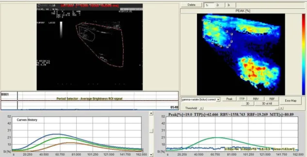
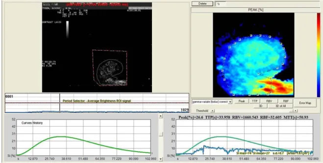
The time-intensity curve (TIC), generated by two regions of interest (ROI), as large as possible, was analysed.
The software for analysis of TIC inside the ROI produced Peak perfusion intensity (%) that was defined as the percentage increase in SI, from baseline intensity to maximal SI, Time To Peak (TTP, sec)perfusion that was defined as the time of arrival of UCA to its maximum SI, Mean transit time (MTT, sec) was defined as the time interval between half of the maximum SI of the UCA in the ascending phase of the curve (wash in) and the same value of SI in the descending phase (wash out), Regional Blood Volume (RBV) that was defined as the integral of the video SI (%) changes during the the extrapolated transition time without recirculation, Regional Blood Flow (RBF) that was
defined as the ratio between regional blood volume and mean transit time, and the Signal Intensity (average).
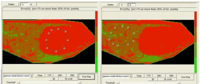
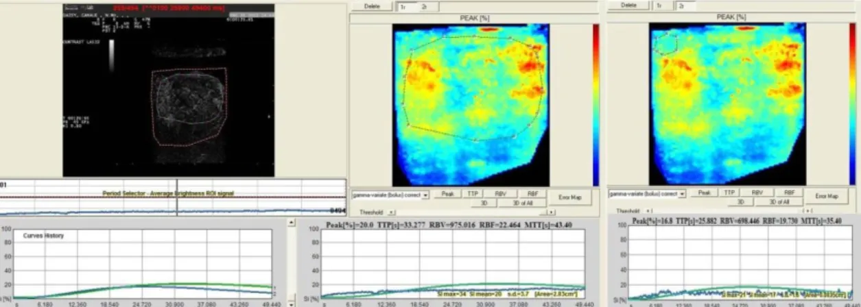
Parametric Maps and Error Maps were evaluated. Calculated curves are then fitted to parametric curves and, as a result, parametric maps are obtained. Parametric maps are visualized as easy-to-interpret colour coded images describing different aspects of perfusion. Error Map is a new map will be than displayed where the pixels are depicted as a 4 levels iso-intensity gradient map and where the colour coded bar represents respectively high fitting errors (red areas), medium fitting errors (dark red areas), low fitting errors (dark green areas) and very low fitting errors (light green areas).
All results obtained were analyzed with a statistical commercial software (GraphPad
software, InStat®)
As regards the timing of enhancement, should be recognized two phases: the arterial phase, starting from the first arrival of contrast (usually in 10 – 20 seconds) until around 30 – 45 seconds, during which the degree of enhancement increases progressively, and the venous phase, which starts from approximately 30 – 45 seconds after contrast injection, during which the degree of enhancement shows a plateau and then decreases progressively. Most organs have a single blood supply with a single inflow (arterial) phase, the exceptions being the liver, which is supplied by its artery and the portal vein, leading to two distinct inflow phases (arterial and portal venous) and the lungs, which are supplied by pulmonary and bronchial arteries with different arrival times. Liver and spleen are also exceptions since they tend to retain microbubbles longer than other organs, probably due to the trapping of microbubbles in their unique microcirculations after clearance from the remainder of the macro-vasculature.
Consequently, the wash-out phase in most organs is shorter than in liver and spleen, whose prolonged retention is termed the late phase. There is no precisely definable event that allows an exact distinction between the arterial and venous and late phases. The time of contrast arrival is usually 10 to 20 seconds after intravenous injection, but factors such as a slow injection of microbubbles in to very peripheral small veins or cardiac diseases may prolong it, whereas intra-cardiac or pulmonary shunting or a hyper-dynamic circulation may shorten it.
The degree of enhancement is difficult to assess. Generally, when the target of the study is a focal region in a parenchymal organ, the degree of enhancement should be compared to the surrounding parenchyma or to the paired organ when available. The lesion might be relatively hyper-enhancing, iso-enhancing, hypo-enhancing or non-enhancing and the pattern should be described separately for the arterial and venous phases. The transition from hyper- or iso-enhancement to hypo-enhancement is commonly referred to as “wash-out”.
Contrast distribution regards the description of whether the enhancement is homogeneous or heterogeneous and, in the latter case, if non-perfused regions exist, should be included. In general terms, the CEUS depiction of non-perfused (potentially necrotic or liquid) areas might be relevant prior to any US-guided biopsy in order to better identify the target.(Piscaglia, et al., 2012)
The subjects of all groups should have an assessment of the appearance of any side effects due to the CEUS up to 2 months after the procedure.
---SPECIAL SECTION ONE: RESEARCH APPLICATIONS--- 3 Spleen
3.1 CEUS knowledge
The spleen is one parenchymal organ of the left hypo-gastric region composed by a white and a red pulp (the real blood reservoir) (Evans & De Lahunta, 2013, p. 558-559), and it is studied mostly with US, in B-mode and Doppler, that are used to detect, also like incidental findings, diffuse splenic abnormalities, focal or multifocal lesions of spleen parenchyma. (Mattoon & Nyland, 2015, p. 400-405 - Penninck & D'Anjou, 2015, p. 242)
The spleen is the second organ (first the liver) most studied with Contrast-enhanced Ultrasonography (CEUS) in human medicine (Piscaglia, et al., 2012).
To understand the application of CEUS in canine spleen it is important to explain splenic physiological parenchymal vascularization. The splenic artery derives from celiac artery while the splenic vein drains into the gastrosplenic vein. The blood enters by up to 25 splenic branches: rami lienales that pass through the long hilus. The branches continue in the trabeculae, branching repeatedly and becoming smaller when they go surrounded by the white pulp and continue entering in the red pulp, where they branch in to penicilli. The venous side begins in the venous sinuses (sinus lienis) playing an active role for the RES. The sinuses coalesce into veins of the red pulp, and these finally merge to become the trabecular veins. Most of the erythrocytes are contained in large, thin-walled splenic sinusoids, that are supplied by the penicillar arterioles of the adjacent splenic cords and continue into the pulpar and trabecular veins. The time interval for each cycle of passage of blood in spleen varies from a few minutes to as long 10 hours. (Evans & De Lahunta, 2013, p. 558-559).
Benign Nodular Hyperplasia (BNH) was identified in the group of the most frequent benign lesions of the spleen of dogs in case of splenomegaly. (Corbin, et al., 2017). In case of BNH, it is identified with nodules typically seen on B-mode ultrasonography like hypoechoic to essentially isoechoic or sometimes the nodules are not always observed ultrasonographically and in such cases, it can be suspected when the border of the spleen is smoothly irregular if parenchymal abnormalities are not detected. (Mattoon & Nyland, 2015, p. 409)
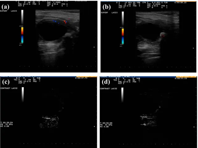
Colour and power Doppler evaluation of blood flow to splenic masses in dogs are not helpful to distinguish benign from malignant splenic masses in dogs (Sharpley, et al., 2012).
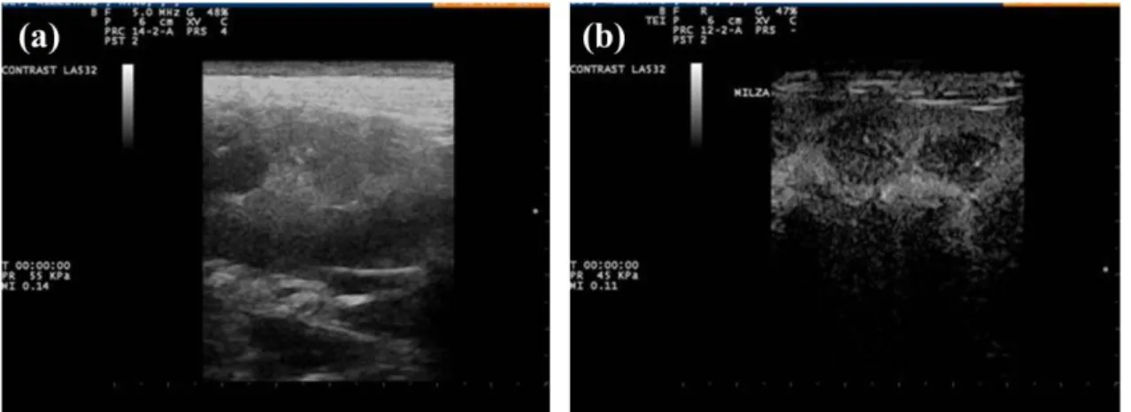
Using CEUS, it is possible to show this splenic perfusion, in fact, the splenic enhancement after injection of contrast medium should derive from the presence of such a large vascular space with slow blood passage. (Rossi, et al.,2008)
Using SonoVue®, normal perfusion values were determined for the canine spleen and it was observed that the small splenic arteries radiating from the splenic hilus opacified rapidly after injection of the contrast. There was heterogeneous enhancement of the splenic parenchyma, the splenic parenchyma became homogeneously enhanced before a gradual homogeneous decrease of opacification and persistent enhancement was not seen during the late phase: there was no evidence of tissue specificity in the canine spleen.(Ohlerth, et al., 2007)
Compared to SonoVue®, Sonazoid® had long-lasting contrast enhancement of canine spleen parenchyma: the optimal time for splenic parenchymal imaging in the dog would be from 7 to 30 min after injection. In splenic arteries, the time to upslope from injection (5.2-0.4 s) and time to peak from initial upslope (2.7-1.0 s) afforded only a narrow window.
In beagles injected with Sonazoid®, the appropriate timing for examining the arterial phase of the spleen ranges from 5 to 22 s after injection. (Nakamura, et al., 2009)
BNH (lymphoid hyperplasia) is one of the most frequent benign lesions of spleen of dogs, particularly old ones, in case of splenomegaly. (Corbin, et al., 2017). Ultrasonography is the method of choice to detect splenic lesions, but it is not specific in their diagnosis. BNH is identified with nodules typically seen on B-mode ultrasonography like hypoechoic to essentially isoechoic lesions, sometimes the nodules are not immediately detected in normal parenchyma and, in such cases, can be suspected only as solid nodule bulging from a regular border of the capsule. (Mattoon & Nyland, 2015, p. 409).
Colour and Power Doppler study of splenic focal lesions in dogs showed that detection of tortuous or aberrant vessels, entering or within them, is nearly significant to distinguish benign from malignant lesions (Sharpley, et al., 2012).
Different studies were finalized to characterize benign and malignant lesions in spleen using Contrast Enhanced Ultrasonography (CEUS) and a hypoechoic pattern to the surrounding parenchyma during wash-in, peak enhancement and wash-out phases was significantly associated with malignancy (Rossi, et al., 2008 - Ohlerth, et al., 2008 - Nakamura, et al., 2010). CEUS pattern in BNH had predominantly similar wash-in and wash-out to the surrounding spleen, so that, after a few seconds the nodules were isoechoic and diffusely homogeneous with the remainder; a normal architecture of the vascular network in benign hyperplastic conditions is supposed to explain this pattern (Rossi, et al., 2008). Histologic examination had showed a common relationship between splenic hyperplastic nodules and hematomas and this is suggested as a consequence of alteration of circulation in and around hyperplastic nodules with failure of marginal zone flow and accumulation of blood (Spangler & Culbertson, 1992 - Cole, 2012).
CEUS was used to differentiate benign from malignant lesions: it was clear that the accuracy of contrast-enhanced ultrasonography in differentiating benign from malignant splenic lesions in dogs is conflicting but the results from early washin-/early washout-based interpretation reported were characterized by poor sensitivity and specificity. (Ivančić, et al. 2009 - Nakamura, et al., 2009 - Nakamura, et al., 2010 - Taeymans & Penninck, 2011).
Qualitative assessment of tortuous feeding vessels and persistence of visualization of the vessels throughout all perfusion phases may improve the accuracy in characterizing benign vs. malignant splenic lesions (Taeymans & Penninck, 2011).
CEUS was a highly accurate method of characterizing nodule images as malignant or benign in dogs with hemangiosarcoma, with an accuracy of 100%. (Ivančić, et al., 2009)
General principles, that referring to cytological or histological final diagnosis, give to CEUS a not invasive role for a not invasive differentiation of malignant splenic tumours from haematomas or benign nodules like a valuable method since fine needle aspirates of splenic lesions are often not diagnostic and tissue core biopsies may cause further bleeding. (Ohlerth & OBrien, 2007)
Hemangiosarcomas are included in the group of canine splenic malignant neoplasm. Detected in B-mode, CEUS was applied giving in all phases by homogeneous anechoic (not perfused) areas with highly vascularized surrounding parenchyma (peripheral irregular perfusion pattern), with presence of thin septae and tortuous vessels were visible on the periphery of the lesion ; based on the time–intensity curves, there was no enhancement in the large anechoic lesions, and pixel intensity did not change from the baseline. (Rossi, et al., 2008)
Hemangiosarcoma showed characteristic hypoechoic pattern during the early vascular phase using Sonazoid® in (Nakamura, et al., 2010). This hypoechoic area may correspond to the haemorrhagic or necrotic areas commonly associated with hemangiosarcoma. However, differentiation between hemangiosarcoma and hematoma is referred to the presence of aberrant wide vessels and none of the other malignant or benign lesions, including hematoma, had such vessels. Hematoma derives from BNH like evolution of disease and its always more rejected the traumatic nature, becoming hematoma an evolution of benign diseases: the presence of lymphoid hyperplasia is supportive of a diagnosis of hematoma, although not diagnostic in itself and only this appeared to be useful for increasing confidence that a lesion is truly a hematoma and that hemangiosarcoma had not been overlooked. (Cole, 2012)
Considering the presence of tortuous feeding vessels in the arterial phase, together with persistence of these feeding vessels in the parenchymal phase as an indicator of malignancy resulted in an accuracy of 100%. (Taeymans & Penninck, 2011)
In one examination using Sonazoid® on 7 of the 8 malignant nodules other than hemangiosarcoma were isoechoic in the early phase. However, CEUS in these 7 nodules rapidly decreased, and they became hypoechoic in the late vascular phase. Finally, all 8 lesions became hypoechoic in the parenchymal phase. It means that there is no significant difference between benign and malignant lesions in the parenchymal phase instead on the liver the parenchymal phase is suggestive of malignant tumours. The splenic nodular hyperplasia became hypoechoic during the parenchymal phase in this study. The contrast defect during the parenchymal phase created by splenic nodular hyperplasia might be because of a decrease of splenic macrophages. Therefore, the parenchymal phase imaging was not useful for differentiation between benign and malignant splenic lesions. In some cases, however, nodules that could not be visualized with conventional ultrasonography became clearly hypoechoic in the parenchymal phase: the parenchymal phase imaging could be useful for the detection of focal splenic lesions.
They speculate that the lack of normal sinusoids combined with neoplastic angiogenesis might be one of the causes of a malignant hypoechoic pattern during the late vascular phase.
The early vascular phase could also differentiate malignant and benign lesions with high specificity: hemangiosarcoma showed characteristic hypoechoic pattern during the early vascular phase that may correspond to the haemorrhagic or necrotic areas commonly associated with hemangiosarcoma. (Nakamura, et al., 2010)
In dogs, however, it was demonstrated that sulphur hexafluoride microbubbles allowed for vascular phase imaging but not for parenchymal phase imaging. (Ohlerth, et al., 2007) The vascular phase imaging with sulphur hexafluoride microbubbles could differentiate benign and malignant focal splenic lesions based on the finding that malignant tumours were hypoechoic to the surrounding normal spleen parenchyma in the wash-out phase (30 seconds after injection of sulphur hexafluoride microbubbles). (Rossi, et al., 2008)
CEUS allows detection of abnormal perfusion patterns associated with splenic
malignancies. In some cases of lymphosarcoma, CEUS had early wash-in and wash-out phases, and this possibly reflects the lack of normal sinusoidal vessels of the red pulp combined with neoplastic angiogenesis. (Rossi, et al., 2008)
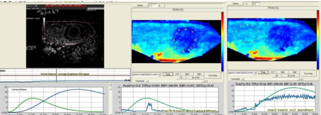
Aim of our study is to evaluate diffuse and nodular canine spleen lesions, differentiating between benign and malignant lesions. A focus was done about BNH, particularly on qualitative and quantitative analysis and according to final diagnosis done with cytology and histology: the hypothesis was that benign hyperplastic nodules, having an altered vascular pattern, would have, consequently, a different microbubbles distribution related to normal spleen parenchyma.
3.2 Materials and Methods
Equipment staff was composed according to general procedures established and summarized in the following table. (chapter 2 and Table: General Procedures).
Patients included all dogs with focal or diffuse splenic lesions detected by Basic ultrasonography.
This procedure was used to study 20 cases of BNH and cases of malignant lesions including Hemangiosarcoma, Lymphoma and Histiocytic Sarcoma.
Qualitative criteria for interpretation of the results were:
Doppler: The presence of a wide or tortuous vessel within the mass is suggestive of malignancy. (Note: dogs) (Sharpley, et al., 2012)
Differentiation between benign and malignant lesions: qualitative assessment of tortuous feeding vessels and persistence of visualization of these vessels throughout all perfusion phases may improve the accuracy in characterizing benign vs. malignant splenic lesions. (Note: SonoVue® - dogs) (Taeymans & Penninck, 2011)
It is suggested the use of early vascular phase to differentiate malignant and benign lesions with high specificity. (Note: Sonazoid® - dogs) (Nakamura, et al., 2010)
Hemangiosarcoma: hypoechoic pattern during the early vascular phase that may correspond to the haemorrhagic or necrotic areas. (Note: Sonazoid® - dogs) (Nakamura, et al., 2010)
Benign lesions: no contrast enhancement or rapid wash-in, followed by persistent enhancement in the late phase.
Malignant lesions (metastases or lymphoma): the combination of contrast enhancement (diffuse or peripheral) in the arterial phase followed by rapid and marked wash-out.
(Note: EFSUMB recommendation - SonoVue® - human) (Piscaglia, et al., 2012)
BNH in B-mode, included: size and shape, echo pattern (homogeneous or heterogeneous) and location. Spleen masses were recorded as “homogeneous” or “heterogeneous” according to echogenicity. Doppler evaluation was assessed comparing perfusion Colour appearance of lesion to surrounding tissue in homogeneous or heterogeneous, with prominent inner vessels and with rim enhancement. (Penninck & D'Anjou, 2015) (Mattoon & Nyland, 2015)
For lesions with CEUS: hyperenhancing (brighter than surrounding tissue), isoenhancing (no more visible during contrast ultrasound) or hypoenhancing (hypoechoic to the surrounding tissue) compared to the surrounding tissue. Presence of heterogeneity, rim enhancement or prominent inner vessels was also evaluated. Important consideration is done for individuation of wash-in (arrival time of contrast medium) and wash-out (outgoing time of contrast medium) in comparison between lesion and surrounding tissue. (Rossi, et al., 2008)
Quantitative evaluation of results using Qontrast®, software analysis, included:
Creation of two ROI as similar as possible dimensions: one ROI for lesion and one for surrounding parenchyma
Evaluation of following parameters generated from TIC: P, TTP, RBF, RBV, MTT, Parametric maps and Error Maps.
Between January, 2015 until February, 2017, at the Veterinary Teaching Hospital of the University of Messina (Italy), a CEUS prospective study was performed in dogs with focal or multifocal splenic lesions detected during routine abdominal ultrasonography. In this part of study, dogs were included only if a diagnostic ultrasound-guided aspiration or histo-pathologic samples taken during surgery for splenectomy were performed after ultrasonography contrast enhanced exams and conclusive diagnosis was benign nodular hyperplasia. For the dogs that did not undergo surgery, a ultrasound follow up exam was scheduled 1 month after CEUS exam. Each dog underwent a physical examination and blood analysis.
B-mode and Colour Doppler Ultrasonography of all abdominal cavity was performed by the same operator B-mode and Doppler ultrasonography of the spleen was performed with microconvex (5.0 to 8.0-MHz) and linear (10 to 12-MHz) transducers. Lesions of the spleen were assessed for their size, number and echogenicity in comparison with the normal spleen (hypoechoic, hyperechoic, mixed), and presence of cavitary areas (anechoic areas surrounded by irregular parenchyma with distal acoustic enhancement). Colour and Power Doppler evaluation was performed to describe vascularisation, evaluating the path of blood vessels in and around the lesions.


![[3] G K Batchelor: Axial flow in trailing line vortices, Journal of Fluid Mechanics, Vol. 20, 645-658 (1964).](data:image/gif;base64,R0lGODlhAQABAIAAAP///wAAACH5BAEAAAAALAAAAAABAAEAAAICRAEAOw==)