O R I G I N A L S C I E N T I F I C R E P O R T
IPOD Study: Management of Acute Left Colonic Diverticulitis
in Italian Surgical Departments
Massimo Sartelli1• Gian Andrea Binda2•Francesco Brandara3•Andrea Borasi4•
Francesco Feroci5•Salvatore Vadala`6•Francesco M. Labricciosa7•
Arianna Birindelli8•Gianluigi Luridiana9•Federico Coccolini10•Salomone Di Saverio11•
Fausto Catena12• Luca Ansaloni13• Fabio Cesare Campanile14•Ferdinando Agresta15•
Diego Piazza16•IPOD study Collaborative Working Group
Published online: 10 November 2016 Ó Socie´te´ Internationale de Chirurgie 2016
Abstract
Background In recent years, the emergency management of acute left colonic diverticulitis (ALCD) has evolved dramatically despite lack of strong evidence. As a consequence, management strategies are frequently guided by surgeon’s personal preference, rather than by scientific evidence. The primary aim of IPOD study (Italian Prospective Observational Diverticulitis study) is to describe both the diagnostic and treatment profiles of patients with ALCD in the Italian surgical departments.
Methods IPOD study is a prospective observational study performed during a 6-month period (from April 1 2015 to September 1 2015) and including 89 Italian surgical departments. All consecutive patients with suspected clinical diagnosis of ALCD confirmed by imaging and seen by a surgeon were included in the study. The study was promoted by the Italian Society of Hospital Surgeons and the World Society of Emergency Surgery Italian chapter.
Results Eleven hundred and twenty-five patients with a median age of 62 years [interquartile range (IQR), 51–74] were enrolled in the IPOD study. One thousand and fifty-four (93.7%) patients were hospitalized with a median duration of hospitalization of 7 days (IQR 5–10). Eight hundred and twenty-eight patients (73.6%) underwent medical treatment alone, 13 patients had percutaneous drainage (1.2%), and the other 284 (25.2%) patients underwent surgery as first treatment. Among 121 patients having diffuse peritonitis, 71 (58.7%) underwent Hartmann’s resection. However, the Hartmann’s resection was used even in patients with lower stages of ALCD (36/479; 7.5%) where other treatment options could be more adequate.
Conclusions The IPOD study demonstrates that in the Italian surgical departments treatment strategies for ALCD are often guided by the surgeon’s personal preference.
& Salomone Di Saverio
[email protected]; [email protected] 1 Department of Surgery, Macerata Hospital, Macerata, Italy 2 Department of Surgery, Galliera Hospital, Genoa, Italy 3 Department of General Surgery, ULSS 3,
Bassano Del Grappa (VI), Italy
4 Department of General Surgery, Humanitas Gradenigo, Turin, Italy
5 Department of Surgery, Santo Stefano Hospital, Prato, Italy 6 Department of Surgery, Cannizzaro Hospital, Catania, Italy
7 Department of Biomedical Sciences and Public Health, Unit of Hygiene, Preventive Medicine and Public Health, UNIVPM, Ancona, Italy
8 Department of General Surgery, University of Bologna, Bologna, Italy
9 Department of Surgery, Brotzu Hospital, Cagliari, Italy 10 Department of Surgery, Rimini Hospital, Rimini, Italy 11 General Surgery, Emergency and Trauma Surgery Unit,
Maggiore Hospital, Bologna, Italy
12 Department of Emergency Surgery, Maggiore Hospital, Parma, Italy
Introduction
Diverticulitis is the most usual complication of diverticu-losis, affecting 15–25% of patients [1]. It does include a variety of conditions, ranging from localized diverticular inflammation to fecal peritonitis. It is usually classified in uncomplicated and complicated according to the extension of the infection process to the peritoneum [2].
In recent years, the emergency management of acute left colon diverticulitis (ALCD) has evolved dramatically despite the lack of strong evidence [3]. As a consequence, management strategies are frequently guided by the sur-geon’s personal preference [4], rather than by scientific evidence.
The aim of the IPOD study (Italian Prospective Obser-vational Diverticulitis study) is to describe the management profiles of patients with ALCD based on data collected over a 6-month period (from April 1 2015 to October 1 2015) from 89 Italian surgical departments. The study was promoted by the Italian Society of Hospital Surgeons (ACOI) and the World Society of Emergency Surgery (WSES) Italian chapter.
Given the broad distribution of the participating medical centers, the study may give a description of the manage-ment profiles of ALCD in Italy.
Method
Aim
The primary aim of the IPOD study is to describe the diagnostic and treatment profiles of patients with ALCD in Italian surgical departments.
Study design
This prospective multicenter observational study was per-formed in 89 Italian surgical departments over a 6-month period (April 1 2015–October 1 2015). All consecutive patients with imaging diagnosis of ALCD were included in the study.
The center coordinator of each participating medical institution collected and compiled clinical data in an online case report database.
The collected data included the following: age, sex, previous episodes of diverticulitis (no episodes, one epi-sode, two or more episodes), comorbidities (immunosup-pression, severe cardiovascular disease), sepsis at admission, radiological diagnosis (ultrasound and com-puter tomography findings), type of management (no treatment, antimicrobial therapy, percutaneous drainage or surgical procedures, admission to Intensive Care Unit (ICU), duration of hospitalization, re-operation and mor-tality. All patients were monitored until they were dis-charged or transferred to another ward.
Staging according to WSES classification [2] was requested for all patients undergoing CT scan at admission:
Uncomplicated
Stage 0 Diverticula, thickening of the wall or increased density of the pericolic fat.
Complicated
Stage 1 A Pericolic air bubbles or little pericolic fluid without abscess (within 5 cm from inflamed bowel segment)
Stage 1 B Abscess B4 cm Stage 2 A Abscess [4 cm
Stage 2 B Distant air ([5 cm from inflamed bowel segment)
Stage 3 Diffuse fluid without distant free air (no hole in colon)
Stage 4 Diffuse fluid with distant free air (persistent hole in colon)
Staging according to the Hinchey classification was requested for all patients undergoing surgical intervention [5]:
Stage 1 Pericolic abscess
Stage 2 Pelvic, intra-abdominal, or retroperitoneal abscess
Stage 3 Generalized purulent peritonitis Stage 4 Generalized fecal peritonitis
The study met the standards outlined in the Declaration of Helsinki and Good Epidemiological Practices.
Differences in daily surgical practice of each center were kept as such. Each center followed its ethical stan-dards. In each center, the coordinator collected and filled in the data in an online case report form. The study was monitored by a coordinating center, which processed and 13 Unit of General Surgery I, Papa Giovanni XXIII Hospital,
Bergamo, Italy
14 Unit of General Surgery, AUSL VT, San Giovanni Decollato-Andosilla Hospital, Civita Castellana, Italy
15 Department of General Surgery, ULSS19 del Veneto, Adria, RO, Italy
16 Department of Surgery, Vittorio Emanuele Hospital, Catania, Italy
verified missing or unclear data submitted to the central database.
Bivariate analyses were performed to analyze the asso-ciation between risk factors and in-hospital mortality using a two-sided Chi-square test or a two-sided Fisher’s exact test, if the expected value of a cell was \5. The level of significance was set at P \ 0.01. Data were analyzed using Epi Info version 7.2.0.1 software package.
The study protocol was approved by the board of the Italian Society of Hospital Surgeons (ACOI) and the World Society of Emergency Surgery (WSES) Italian chapter, and the study was conducted under their supervision. The board of the Italian Society of Hospital Surgeons (ACOI) and the World Society of Emergency Surgery (WSES) Italian chapter grant the proper ethical conduct of the study.
Inclusion criteria
All patients with suspected clinical diagnosis of ALCD confirmed by imaging and seen by a surgeon were included in the study.
Study protocol provided that all patients with a clinical suspicion of ALCD performed CT scan. Intravenous con-trast-enhanced multislice CT scan diagnosis was requested. However, in patients with contraindications to CT scanning such as renal insufficiency, contrast allergy or hemody-namic instability needing emergency surgery abdominal ultrasound was considered sufficient to enroll the patients in the study. A requirement for inclusion was that patients needed to be seen by a surgeon to be considered eligible for the study.
Results
Patients and diagnosis
During the study, 1135 cases were collected, 10 cases did not meet the inclusion criteria because of incomplete submission. A total of 1125 patients were enrolled in the IPOD study; they included 553 (49.2%) women and 572 (50.8%) men, with a median age of 62 years [interquartile range (IQR), 51–74]. One thousand and fifty-four (93.7%) patients were admitted to the hospital, with a median duration of hospitalization of 7 days (IQR 5–10). Seventy-one patients (6.3%) were treated as outpatients.
Six hundred and seventy-six (60.1%) patients had no previous episodes of ALCD, 230 (20.4%) patients had one previous episode of ALCD and 219 (19.5%) patients 2 or more previous episodes. Sixty-one (5.4%) patients were immunosuppressed, and 127 (11.3%) patients suffered from a severe cardiovascular disease.
Radiological examinations performed by patients are illustrated in Table 1. Nine hundred and twenty-seven patients (82.4%) underwent an abdominal CT scan.
In all patients who underwent CT scan, the WSES staging was recorded: Three hundred and twenty-seven (35.3%) were uncomplicated, while 263 (28.4%) had Stage 1a, 94 (10.1%) Stage 1b, 75 (8.1%) Stage 2a, 47 (5.1%) Stage 2b, 43 (4.6%) Stage 3 and 78 (8.4%) Stage 4.
Among all 284 patients undergoing surgical treatment, Hinchey staging was also recorded in 267 patients: Fifty-six (19.7%) patients had Stage 1, 50 (17.6%) Stage 2, Stage 3 82 (28.9%) and 79 (27.8%) Stage 4. In 17 patients, (6.0%) Hinchey stage was not reported.
Management
Among all patients enrolled in the IPOD study, 828 (73.6%) underwent medical treatment alone, 13 (1.2%) patients had percutaneous drainage, and the other 284 (25.2%) patients underwent surgery as first treatment.
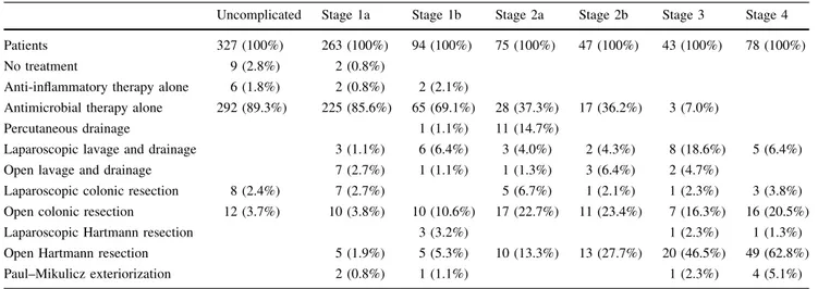
Initial treatment according to the WSES staging is described in Table2.
Sixty patients underwent a second procedure during the same hospitalization because of a postoperative compli-cation or a worsening of the initial stage after conservative treatment.
Six patients performed a damage control surgery by an ‘‘open abdomen procedure’’ and underwent abdominal re-explorations.
The median interval of time between the first and the second procedure was 8 days (IQR 5–14).
Elective sigmoid resection during a second hospitaliza-tion was planned at discharge in 162 patients with acute diverticulitis treated without resection (18.7%, 162/868).
A total of 1017 (1017/1125, 90.4%) patients received antimicrobial therapy during the hospitalization, which was in 796 (796/1017, 78.3%) patients a monotherapy. One hundred and one patients (101/1125, 9.0%) were treated Table 1 Radiological diagnosis
Radiological diagnosis Patients no 1125 (100%) Abdominal X-ray 42 (3.7%)
Abdominal X-ray, CT 189 (16.8%) Abdominal X-ray, US 52 (4.6%) Abdominal X-ray, US, CT 123 (10.9%)
CT 470 (41.8%)
US 75 (6.7%)
US, CT 145 (12.9%)
Not reported 29 (2.6%)
with carbapenems and 13 (13/101, 12.9%) of them had an uncomplicated diverticulitis, according to the WSES Staging (Table3).
Outcome
Among all patients, 71 (6.3%) were managed as outpa-tients, 1054 (93.7%) patients were admitted to the hospital, and the median duration of hospitalization was 7 days (IQR 5–10). In the early postoperative phase, 152 (13.5%) patients were admitted to ICU.
The overall mortality rate was 1.4%. Characteristics of patients who died during the hospital stay are reported in Table4.
Bivariate analyses were performed to analyze the asso-ciation between risk factors and in-hospital mortality using a two-sided Chi-square test or a two-sided Fisher’s exact test.
Distribution of predictive variables of in-hospital mor-tality is reported in Table5.
Independent variables associated with mortality according to the multinomial logistic regression are reported in Table6.
Discussion
Some interesting aspects have emerged from the results of the IPOD study about the management of ALCD in the Italian surgical departments.
CT abdomen has been the most used radiological examination in diagnosing acute diverticulitis (82.4%). This is in keeping with the literature evidence, because CT imaging has become the standard radiological examination
in patients with ALCD. In fact, CT imaging with intra-venous contrast has sensitivity and specificity reported as high as 98 and 99% [6].
It is well known that the utility of CT imaging may go beyond accurate diagnosis of diverticulitis and the grade of severity on CT may drive treatment planning of patients with acute diverticulitis.
For the past three decades, the Hinchey’s classification has been the most commonly used in the international Table 2 Initial treatment according to WSES staging
Uncomplicated Stage 1a Stage 1b Stage 2a Stage 2b Stage 3 Stage 4 Patients 327 (100%) 263 (100%) 94 (100%) 75 (100%) 47 (100%) 43 (100%) 78 (100%)
No treatment 9 (2.8%) 2 (0.8%)
Anti-inflammatory therapy alone 6 (1.8%) 2 (0.8%) 2 (2.1%)
Antimicrobial therapy alone 292 (89.3%) 225 (85.6%) 65 (69.1%) 28 (37.3%) 17 (36.2%) 3 (7.0%)
Percutaneous drainage 1 (1.1%) 11 (14.7%)
Laparoscopic lavage and drainage 3 (1.1%) 6 (6.4%) 3 (4.0%) 2 (4.3%) 8 (18.6%) 5 (6.4%) Open lavage and drainage 7 (2.7%) 1 (1.1%) 1 (1.3%) 3 (6.4%) 2 (4.7%)
Laparoscopic colonic resection 8 (2.4%) 7 (2.7%) 5 (6.7%) 1 (2.1%) 1 (2.3%) 3 (3.8%) Open colonic resection 12 (3.7%) 10 (3.8%) 10 (10.6%) 17 (22.7%) 11 (23.4%) 7 (16.3%) 16 (20.5%)
Laparoscopic Hartmann resection 3 (3.2%) 1 (2.3%) 1 (1.3%)
Open Hartmann resection 5 (1.9%) 5 (5.3%) 10 (13.3%) 13 (27.7%) 20 (46.5%) 49 (62.8%) Paul–Mikulicz exteriorization 2 (0.8%) 1 (1.1%) 1 (2.3%) 4 (5.1%)
Table 3 Antimicrobial therapy administered during hospitalization in 1017 patients
Patients receiving antibiotics n 1017
Metronidazole 586 (57.6%) Piperacillin/tazobactam 318 (31.3%) Ciprofloxacin 144 (14.2%) Amoxicillin/clavulanic acid 117 (11.5%) Ampicillin/sulbactam 81 (8.0%) Ceftriaxone 70 (6.9%) Meropenem 44 (4.3%) Imipenem/cilastatin 34 (3.3%) Ertapenem 23 (2.3%) Rifamixin 17 (1.7%) Gentamicin 13 (1.3%) Amikacin 12 (1.2%) Levofloxacin 11 (1.1%) Ceftazidime 5 (0.5%) Tigecycline 5 (0.5%) Cefepime 1 (0.1%)
The overall number of administered antibiotics (1481) is different from the number of patients receiving antibiotics (1017) since 221 patients received a combined antimicrobial therapy
literature [5]. However, Hinchey’s classification is based on surgical findings and therefore can only be applied to patients who have already been operated. As CT imaging has become a primary diagnostic tool in the diagnosis, staging and decision-making of patients with ALCD, there is a clear need for a CT-based classification being well related to the disease stage and the further therapy. The increasing information provided by CT scans led to several modifications of the Hinchey’s classification based on CT preoperative findings [7–10].
A new proposal for a CT-guided classification of left colon acute diverticulitis was published in 2015 [2] by the WSES acute diverticulitis working group.
Outpatient treatment of acute diverticulitis has been highly debated within the medical community [11–14]. The DIVER multicenter randomized clinical trial [14] recently demonstrated that outpatient treatment may be safe and effective in selected patients with uncomplicated ALCD, allowing significant cost savings for the health systems without negatively influencing the quality of life of patients. Data from the IPOD study showed that, among 1125 observed patients, 1054 patients (93.7%) were hos-pitalized, while only 71 patients (6.3%) were treated as out-patient.
The efficacy of antibiotic use in acute uncomplicated diverticulitis is another controversial issue within the med-ical community [15–17]. Chabok et al. [16] in a randomized clinical trial demonstrated that antibiotic treatment for acute uncomplicated diverticulitis neither accelerates recovery nor prevents complications or recurrence. In our study, among the 327 patients with WSES stage of uncomplicated diver-ticulitis, 292 (89.3%) received an antimicrobial therapy.
Approximately 15–20% of patients admitted with acute diverticulitis have an abscess on CT scan [18]. The size of 3–6 cm has been generally accepted (all of low level of evidence) to be a limit between antimicrobial therapy alone versus percutaneous drainage and antimicrobial in the management of diverticular abscesses [18–22].
Percutaneous drainage has the advantage of avoiding urgent operation in patients with large abscesses. It may be used as a ‘‘bridge’’ to elective resection. IPOD study highlights a very low use of percutaneous drainage even for larger abscesses where it should be the first-line treatment [23–25]. Only eleven (14.7%) out of 75 patients having WSES stage 2a (CT findings of abscess larger than 4 cm) underwent percutaneous drainage.
The optimal treatment for ALCD with CT finding of distant extra-luminal air without diffuse fluid is still Table 4 Characteristics of patients who died during hospitalization
Pt. Age Sex Previous episodes
Comorbidities Clinical conditions at admission WSES stage Hinchey stage First treatment
Time of death post-hospital admission (days)
Cause of death
1 84 F No SCD Severe sepsis Stage 4 4 OHR 25 SRM
2 79 M No Septic shock Stage 4 4 PME 3 SRM
3 81 F No SCD, IS Severe sepsis Stage 4 3 OHR 2 SRM
4 84 F 1 SCD Stable Stage
2b
AMT 2 SRM
5 65 M [1 SCD, IS Septic shock Stage 4 3 OCRS 50 SRM
6 84 M No SCD Stable Stage
2b
AMT 9 SRM
7 82 F 1 IS Severe sepsis Stage 4 4 OHR 6 SRM
8 83 F No Severe sepsis Stage 4 4 OHR 14 SRM
9 84 F No SCD Stable Stage 4 4 OCRS 2 SRM
10 69 M No Septic shock Stage 4 4 OCRS 40 SRM
11 78 F No SCD,IS Septic shock Stage 4 4 OCRS 15 SRM
12 51 F [1 Stable Stage 0 4 OCRS 25 SRM
13 84 M No SCD Septic shock Stage 4 4 OCR 7 SRM
14 85 F [1 SCD Severe sepsis Stage 2b
3 OHR 4 SRM
15 75 M No Stable Stage 4 3 OHR 10 SRM
16 56 M [1 SCD, IS Septic shock Stage 3 4 OHR 2 SRM
M male, F female, SCD severe cardiovascular disease, IS immunosuppression, OHR open Hartmann resection, PME Paul–Mikulicz exterior-ization, AMT antimicrobial therapy (alone), OCRS open colonic resection with stoma, OCR open colonic resection without stoma, SRM sepsis related mortality
controversial. Free air on CT has already been reported to be a predictor of failure of non-operative management of ALCD [26], Some authors reported that patients with dis-tant air may be treated by conservative treatment alone in
selected cases because it may be associated with failure and may need immediate surgical operation [27, 28]. Among 47 patients having WSES stage 2b (CT findings of distant air without diffuse fluid), 17 (17/47, 36.2%) were treated at the beginning by antimicrobial therapy alone and 6 (6/47, 8.5%) needed a surgical treatment because of deterioration of the clinical conditions. Two of these patients died during hospitalization.
Hartmann’s resection is still useful in managing diffuse peritonitis with signs of diverticular perforation. Common use of Hartmann’s resection in treating diverticular perfo-ration worldwide is confirmed by a recent Australian study, performed in eight tertiary referral centers with specialized colorectal services [29] and by a population-based retro-spective cohort study using administrative discharge data, conducted in Ontario (Canada) [30].
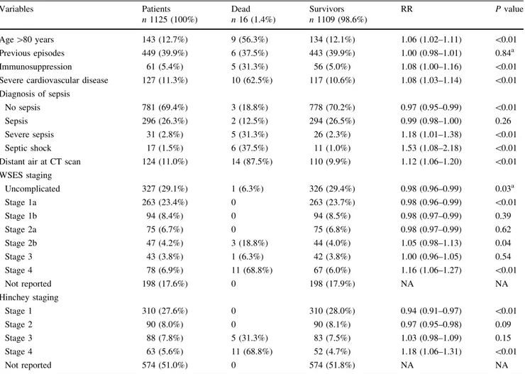
In the IPOD study among 121 patients having WSES stages 3 or 4 (CT findings of diffuse peritonitis with or Table 5 Distribution of predictive variables and mortality
Variables Patients n 1125 (100%) Dead n 16 (1.4%) Survivors n 1109 (98.6%) RR P value Age [80 years 143 (12.7%) 9 (56.3%) 134 (12.1%) 1.06 (1.02–1.11) \0.01 Previous episodes 449 (39.9%) 6 (37.5%) 443 (39.9%) 1.00 (0.98–1.01) 0.84a Immunosuppression 61 (5.4%) 5 (31.3%) 56 (5.0%) 1.08 (1.00–1.16) \0.01 Severe cardiovascular disease 127 (11.3%) 10 (62.5%) 117 (10.6%) 1.08 (1.03–1.14) \0.01 Diagnosis of sepsis
No sepsis 781 (69.4%) 3 (18.8%) 778 (70.2%) 0.97 (0.95–0.99) \0.01
Sepsis 296 (26.3%) 2 (12.5%) 294 (26.5%) 0.99 (0.98–1.00) 0.26
Severe sepsis 31 (2.8%) 5 (31.3%) 26 (2.3%) 1.18 (1.01–1.38) \0.01 Septic shock 17 (1.5%) 6 (37.5%) 11 (1.0%) 1.53 (1.08–2.18) \0.01 Distant air at CT scan 124 (11.0%) 14 (87.5%) 110 (9.9%) 1.12 (1.06–1.20) \0.01 WSES staging Uncomplicated 327 (29.1%) 1 (6.3%) 326 (29.4%) 0.98 (0.96–0.99) 0.03a Stage 1a 263 (23.4%) 0 263 (23.7%) 0.98 (0.96–0.99) \0.01 Stage 1b 94 (8.4%) 0 94 (8.5%) 0.98 (0.97–0.99) 0.39 Stage 2a 75 (6.7%) 0 75 (6.8%) 0.98 (0.97–0.99) 0.62 Stage 2b 47 (4.2%) 3 (18.8%) 44 (4.0%) 1.05 (0.98–1.13) 0.04 Stage 3 43 (3.8%) 1 (6.3%) 42 (3.8%) 1.00 (0.96–1.05) 0.54 Stage 4 78 (6.9%) 11 (68.8%) 67 (6.0%) 1.16 (1.06–1.27) \0.01 Not reported 198 (17.6%) 0 198 (17.9%) NA NA Hinchey staging Stage 1 310 (27.6%) 0 310 (28.0%) 0.94 (0.91–0.97) \0.01 Stage 2 90 (8.0%) 0 90 (8.1%) 0.97 (0.95–0.98) 0.09 Stage 3 88 (7.8%) 5 (31.3%) 83 (7.5%) 1.03 (0.98–1.09) 0.15 Stage 4 63 (5.6%) 11 (68.8%) 52 (4.7%) 1.18 (1.06–1.31) \0.01 Not reported 574 (51.0%) 0 574 (51.8%) NA NA
All P values calculated using two-sided Fisher’s exact test unless otherwise noted RR risk ratio, NA not applicable
a Two-sided Chi-square test
Table 6 Results of multinomial logistic regression for the analysis of variables associated with mortality
Variables OR 95% CI P value
Age [80 2.40 0.51–11.18 0.27
Severe cardiovascular disease 7.55 1.73–33.05 \0.05
Sepsis 0.29 -1.24–1.09 0.26
Severe sepsis 2.00 0.31–13.02 0.47 Septic shock 4.58 0.63–33.33 0.13 Distant free air at CT scan 10.58 1.79–62.42 \0.05 Hinchey stage 4 4.89 1.12–21.26 \0.05 CI confidence interval, OR odds ratio
without distant air), 71 (58.7%) underwent Hartmann’s resection. However, Hartmann’s resection was used even for lower stages of ALCD where other treatment options could be more adequate.
In the past years, primary colonic anastomosis, with or without defunctioning stoma, has been debated. In clini-cally stable patients with no comorbidities, primary resection and anastomosis with or without diverting stoma has been considered also in case of diffuse peritonitis [31], even if it may not be defined as the treatment of choice in diffuse peritonitis [32].
In the IPOD study, 27 patients out of 121 (22.3%) having WSES stages 3 or 4 (CT findings of diffuse peri-tonitis with or without distant air) underwent sigmoidec-tomy (23 by open and 4 by laparoscopic approach).
In the past years, some prospective trials have been conducted on laparoscopic lavage and drainage with con-flicting results [33–36]. In our study, among the 284 patients undergoing initial surgical treatment, 27 (9.5%) underwent laparoscopic lavage and drainage. There was no mortality related to this procedure. The small number of patients submitted to laparoscopic lavage may be justified by the great debate that is still open on this topic, mainly due to the discrepancy and sometime disappointing results of the latest prospective trials such as SCANDIV, Ladies, and DILALA trials [33–37].
In critical ill patients, damage control surgery with open abdomen (OA) procedure may be helpful in managing them. The OA allows to control any persistent source of infection, preventing abdominal compartment syndrome and deferring definitive intervention and anastomosis until the patient is appropriately resuscitated and hemodynami-cally stable [38]. In our study, six critically ill patients (12.5% of patients with either severe sepsis or septic shock) were treated with an open abdomen procedure and they all were Hinchey stage 4.
The IPOD study underlines a critical issue in antimi-crobial treatment for patients with ALCD because of the high number of patients treated by anti-Pseudomonas car-bapenems (imipenem, meropenem) although they almost all have a community-acquired infection.
In this study, among 1017 (90.4%) patients who received antimicrobial therapy, one hundred and one patients (9.9%) were treated with carbapenems and 13 (12.9%) of them had an uncomplicated diverticulitis, according to the WSES Staging.
Carbapenems have been widely used in many countries due to the increasing rate of ESBL-producing Enterobac-teriaceae with a consequent impact on the emergence of resistance to these antimicrobials, especially in K. pneu-moniae [39]. The recent and rapid spread of carbapenem-resistant K. pneumoniae [40–42] should pose a serious challenge for clinicians and a preserving
carbapenems-approach should always be mandatory in treating ALCD that are generally community-acquired infections. There-fore, the use of carbapenems should be optimized in terms of indication and exposure.
In the last decade, indications for elective sigmoid resection after recovery from uncomplicated acute diver-ticulitis have changed [23–25]. Some authors have shown that more episodes of uncomplicated AD do not increase the risk of complicated recurrences and the need for emergency operative management and that the highest risk of free perforation is at the time of the first episode of disease [43,44]. Therefore, routine ‘‘prophylactic’’ elective resection should be no longer recommended after an acute episode of uncomplicated ALCD [45]. Despite current recommendations of more restrictive indications for elec-tive surgery, in the past years there has been a trend toward an increased use of elective operations for ALCD [26]. In IPOD study, an elective resection, during a second hospi-talization, was planned in 162 patients (18.7%) with acute diverticulitis treated without resection. One hundred and nine (67.3%) of these patients had a mild disease, within stage 1b, at low risk of severe recurrence. Furthermore, 64 patients (64/162, 39.5%) reported no previous episodes of ALCD. Elective surgery was planned at patient discharge and therefore as prophylactic surgery, apparently not related to persistent symptoms. These data on the elective surgery of the present study show the most significant deviation of the Italian surgical policy from the actual guidelines.
Given the broad distribution of the participating medical centers, IPOD study may give a detailed description of the management profiles of acute diverticulitis in Italian sur-gical departments.
Nonetheless, we must acknowledge several potential limitations of this study. In fact, the study design, even if data were carefully collected, includes only cases managed in surgical departments. In some hospitals, acute left colon diverticulitis, especially of the early stages, is also man-aged in medical departments and for some participating centers the reported cases did not represent all cases of left acute diverticulitis.
The IPOD study demonstrated that in Italy ALCD treatment strategy is often guided by the surgeon’s personal preference.
IPOD study Collaborative Working Group Collaborators (IPOD study Group) Gabriele Anania, Emanuele Caproli, Marcello Gas-parrini, Pierpaolo Bordoni, Andrea Lucchi, Stefano Scabini, Biagio Picardi, Giuliano Sarro, Alice Piccinini, Natalino Bedin, Alessandro Bussotti, Renato De Angelis, Gian Luca Baiocchi, Antonella Andreotti, Nicola Cillara, Barbara Petronio, Sergio Grimaldi, Alessia Biancafarina, Dario Somenzi, Andrea Costanzi, Alberto Marvaso, Alfonso Canfora, Giorgio Vasquez, Carlo Chiodo, Mario Nano, Angelo Cavicchi, Alberto Ruffato, Paolo Baccari, Roberto Polastri, Patrizia Marsanic, Giuseppe Portale, Luca Gordini, Hariscine K
Abongwa, Michela Pili, Luca Turati, Vittoria Nusca, Gianluca Guercioni, Leonardo Andrea Delogu, Umberto Robustelli, Danilo Piras, Fernando Serventi, Daniela Prando, Antonio Brunelli, Bruno Zani, Salvatore Pintaldi, Augusto Verzelli, Silvia Mulas, Gianmaria Confalonieri, Giuditta Spagni, Antonio Crucitti, Andrea Sagnotta, Stefania Fiume, Francesco Balestra, Matteo Gatti, Emilio Eugeni, Amedeo Carraro, Michele Genna, Lucio Taglietti, Antonio Azzin-naro, Stefano Ferfoglia, Giuseppe Miranda, Giuseppe Tirone, Pietro Luparello, Stefano Berti, Roberta Tutino, Andrea De Manzoni Gar-berini, Francesco Roscio, Valeria Maglione, Mauro Podda, Giovanna Ioia, Fabrizio Cantore, Franco Mazzalai, Francesco Cortesi, Giacomo Arcuri, Giovanni Bellanova, Massimo Beltramo, Antonella Chessa, Massimiliano Coppola, Davide Gozzo, Asaf Harbi, Edoardo Min-ciotti, Francesco Pata, Giovanni Pinna, Mario Testini, Serafino Vanella.
References
1. Lame´ris W, Van Randen A, Van Gulik TM et al (2010) A clinical decision rule to establish the diagnosis of acute diverticulitis at the emergency department. Dis Colon Rectum 53:896–904 2. Sartelli M, Moore FA, Ansaloni L et al (2015) A proposal for a
CT driven classification of left colon acute diverticulitis. World J Emerg Surg 10:3
3. Morris AM, Regenbogen SE, Hardiman KM et al (2014) Sigmoid diverticulitis: a systematic review. JAMA 311:287–297 4. Andeweg CS, Mulder IM, Felt-Bersma RJ et al (2013) Guidelines
of diagnostics and treatment of acute left-sided colonic divertic-ulitis. Dig Surg 30:278–292
5. Hinchey EJ, Schaal PH, Richards MB (1978) Treatment of per-forated diverticular disease of the colon. Adv Surg 12:85–109 6. Lame´ris W, van Randen A, Bossuyt PMM et al (2008) Graded
compression ultrasonography and computed tomography in acute colonic diverticulitis: meta-analysis of test accuracy. Eur Radiol 18:2498–2511
7. Kaiser AM, Jiang JK, Lake JP et al (2005) The management of complicated diverticulitis and the role of computed tomography. Am J Gastroenterol 100:910–917
8. Neff CC, van Sonnenberg E (1989) CT of diverticulitis. Diag-nosis and treatment. Radiol Clin North Am 27:743–752 9. Ambrosetti P, Becker C, Terrier F (2002) Colonic diverticulitis:
impact of imaging on surgical management—a prospective study of 542 patients. Eur Radiol 12:1145–1149
10. Mora Lopez L, Serra Pla S, Serra-Aracil X et al (2013) Appli-cation of a modified Neff classifiAppli-cation to patients with uncom-plicated diverticulitis. Colorectal Dis 15:1442–1447
11. Etzioni DA, Chiu VY, Cannom RR et al (2010) Outpatient treatment of acute diverticulitis: rates and predictors of failure. Dis Colon Rectum 53(6):861–865. doi:10.1007/DCR.0b013e31 81cdb243
12. Jackson JD, Hammond T (2014) Systematic review: outpatient management of acute uncomplicated diverticulitis. Int J Colorectal Dis 29:775–781
13. Rodrı´guez-Cerrillo M (2013) Treatment of elderly patients with uncomplicated diverticulitis, even with comorbidity, at home. Eur J Intern Med 24:430–432
14. Biondo S et al (2014) Outpatient versus hospitalization man-agement for uncomplicated diverticulitis: a prospective, multi-center randomized clinical trial (DIVER Trial). Ann Surg 259:38–44
15. De Korte N, Kuyvenhoven JP, van der Peet DL et al (2012) Mild colonic diverticulitis can be treated without antibiotics. A case– control study. Colorectal Dis 14:325–330
16. Chabok A, Pa˚hlman L, Hjern F et al (2012) Randomized clinical trial of antibiotics in acute uncomplicated diverticulitis. Br J Surg 99:532–539
17. Shabanzadeh DM, Wille-Jørgensen P (2012) Antibiotics for uncomplicated diverticulitis. Cochrane Database Syst Rev 11:CD009092
18. Ambrosetti P, Chautems R, Soravia C et al (2005) Long-term outcome of mesocolic and pelvic diverticular abscesses of the left colon: a prospective study of 73 cases. Dis Colon Rectum 48:787–791
19. Brandt D, Gervaz P, Durmishi Y et al (2006) Percutaneous CT scan guided drainage versus antibiotherapy alone for Hinchey II diverticulitis: a case–control study. Dis Colon Rectum 49:1533–1538
20. Siewert B, Tye G, Kruskal J et al (2006) Impact of CT-guided drainage in the treatment of diverticular abscesses: size matters. AJR Am J Roentgenol 186:680–686
21. Singh B, May K, Coltart I et al (2008) The long-term results of percutaneous drainage of diverticular abscess. Ann R Coll Surg Engl 90:297–301
22. Kumar RR, Kim JT, Haukoos JS et al (2006) Factors affecting the successful management of intra-abdominal abscesses with antibiotics and the need for percutaneous drainage. Dis Colon Rectum 49:183–189
23. Andersen JC, Bundgaard L, Elbrønd H et al (2012) Danish national guidelines for treatment of diverticular disease. Dan Med J 59:C4453
24. Feingold D, Steele SR, Lee S et al (2014) Practice parameters for the treatment of sigmoid diverticulitis. Dis Colon Rectum 57:284–294
25. Binda GA, Cuomo R, Laghi A et al (2015) Practice parameters for the treatment of colonic diverticular disease: Italian Society of Colon and Rectal Surgery (SICCR) guidelines. Tech Coloproctol 19:615–626
26. Etzioni DA, Mack TM, Beart RW et al (2009) Diverticulitis in the United States: 1998–2005. Changing patterns of disease and treatment. Ann Surg 249:210–217
27. Ambrosetti P, Jenny A, Becker C et al (2000) Acute left colonic diverticulitis compared performance of computed tomography and water soluble contrast enema: prospective evaluation of 420 patients. Dis Colon Rectum 43:1363–1367
28. Sallinen VJ, Mentula PJ, Leppa¨niemi AK (2014) Nonoperative management of perforated diverticulitis with extraluminal air is safe and effective in selected patients. Dis Colon Rectum 57:875–881
29. Hong MK, Tomlin AM, Hayes IP et al (2015) Operative inter-vention rates for acute diverticulitis: a multicentre state-wide study. ANZ J Surg 85:734–738
30. Li D, Baxter NN, McLeod RS et al (2014) Evolving practice patterns in the management of acute colonic diverticulitis: a population-based analysis. Dis Colon Rectum 57:1397–1405 31. Oberkofler CE, Rickenbacher A, Raptis DA et al (2012) A
multicenter randomized clinical trial of primary anastomosis or Hartmann’s procedure for perforated left colonic diverticulitis with purulent or fecal peritonitis. Ann Surg 256:819–826 32. Binda GA, Serventi A, Puntoni M et al (2015) Primary
anasto-mosis versus Hartmann’s procedure for perforated diverticulitis with peritonitis: an impracticable trial. Ann Surg 261:116–117 33. Schultz JK, Yaqub S, Wallon C et al (2015) Laparoscopic lavage
vs primary resection for acute perforated diverticulitis: the SCANDIV randomized clinical trial. JAMA 314:1364–1375 34. Vennix S, Musters GD, Mulder IM et al (2015) Laparoscopic
peritoneal lavage or sigmoidectomy for perforated diverticulitis with purulent peritonitis: a multicentre, parallel-group, ran-domised, open-label trial. Lancet 386:1269–1277
35. Morris AM (2015) Laparoscopic peritoneal lavage for perforated diverticulitis: in search of evidence. Lancet 386:1219–1221 36. Angenete E, Thornell A, Burcharth J et al (2016) Laparoscopic
lavage is feasible and safe for the treatment of perforated diver-ticulitis with purulent peritonitis: the first results from the ran-domized controlled trial DILALA. Ann Surg 263:117–122 37. Di Saverio S, Birindelli A, Catena F et al (2016) The ladies trial:
premature termination of the LOLA arm and increased adverse events incidence after laparoscopic lavage may be influenced by inter-hospital and inter-operator variability? Take-home mes-sages from a center with laparoscopic colorectal expertise. Int J Surg 36(Pt A):118–120. doi:10.1016/j.ijsu.2016.10.016 38. Sartelli M, Abu-Zidan FM, Ansaloni L et al (2015) The role of
the open abdomen procedure in managing severe abdominal sepsis: WSES position paper. World J Emerg Surg 10:35 39. Hawser SP, Bouchillon SK, Hoban DJ et al (2010) Incidence and
antimicrobial susceptibility of Escherichia coli and Klebsiella pneumoniae with extended-spectrum beta-lactamases in com-munity- and hospital-associated intra-abdominal infections in Europe: results of the 2008 study for monitoring antimicrobial
resistance trends (SMART). Antimicrob Agents Chemother 54:3043–3046
40. Yigit H, Queenan AM, Anderson GJ et al (2001) Novel bapenem-hydrolyzing beta-lactamase, KPC-1, from a car-bapenem-resistant strain of Klebsiella pneumoniae. Antimicrob Agents Chemother 45:1151–1161
41. Nordmann P, Poirel L (2014) The difficult-to-control spread of carbapenemase producers among enterobacteriaceae worldwide. Clin Microbiol Infect 20:821–830
42. Munoz-Price LS, Poirel L, Bonomo RA et al (2013) Clinical epidemiology of the global expansion of Klebsiella pneumoniae carbapenemases. Lancet Infect Dis 13:785–796
43. Broderick-Villa G, Burchette RJ, Collins JC et al (2005) Hospi-talization for acute diverticulitis does not mandate routine elec-tive colectomy. Arch Surg 140:576–583
44. Ritz JP, Lehmann KS, Frericks B et al (2011) Outcome of patients with acute diverticulitis: multivariate analysis of risk factors for free perforation. Surgery 149:606–613
45. Wieghard N, Geltzeiler CB, Tsikitis VL (2015) Trends in the sur-gical management of diverticulitis. Ann Gastroenterol 28:25–30