SCUOLA DOTTORALE IN SCIENZE BIOMOLECOLARI E CELLULARI
XXIII CICLO DELLA SCUOLA DI DOTTORATO
“Constructing a Minimal Cell”
Paolo Carrara
A.A. 2010/2011
Docente Guida: Prof. Pier Luigi Luisi Coordinatore: Prof. Paolo Mariottini
Index
Abstract 4
Riassunto 7
Abbreviations 10
Chapter 1. Introduction 11
1.1 The Origin of life 11 1.2 Origin and definition of life 11 1.3 Autopoieis and the Logic of Cellular Life 13 1.4 The “Minimal Cell” project 15 1.5 Lipid vesicles (liposomes) 17 1.6 Giant Vesicles (GVs):
Preparation and application 18 1.7 GVs: main preparation methods 20
1.7.1 The Natural Swelling method 22 1.7.2 The Electroswelling method 21 1.7.3 The Emulsion Inversion
(or droplet transfer) method 22
1.8 The emulsion inversion method as a convenient way to prepare GVs
for minimal cell research 24
Chapter 2. Aim of the work 31
Chapter 3. Results and discussion 34
3.1 Optimization of GVs production formed
from a lipid-stabilized water in oil emulsion 34
3.1.1. Effect of the lipids in w/o emulsion and interface:
lipid mixture and "asymmetric" design 38 3.1.2. Effect of the outer aqueous volume 39 3.1.3. Effect of the w/o emulsion volume
and of the interface 44
3.1.4. Effect of the inner buffer volume 49 3.1.5. Summary of the first three
optimization steps 53 3.1.6. Effect of the buffer
concentration of the internal
and external aqueous phase 55 3.1.7. Further investigation
on the POPC:oleate system 60 3.1.8. Direct observation of GVs formation 62
3.2 Relevance of the emulsion inversion (droplet transfer) method and its optimization for studying cell models 64 3.3 GVs colonies as a model of primitive
cell colonies: formation and characterization 66 3.3.1 Effect of PLA and GVs concentration 68
3.3.2 GVs fusion inside colony 70 3.3.3 Advantage to the colony
with respect to individual GVs 74
Chapter 4. Conclusion and prospective 77
Chapter 5. Bibliography 82
Publications 86
Abstract
One of the open questions in origin of life is about the formation of primitive cells from separated molecular components. In recent years, this subject has been approached in the “Minimal Cell” project, namely the laboratory study on cell-like compartments with the minimal and sufficient number of components that may allow cellular life.
The theoretical framework of this research is the theory of
autopoiesis, that elegantly defines what minimal life is in terms of
self-bounded biochemical network capable of reconstructing itself (boundary included) thanks to its own reactions.
Current conceptual and experimental studies are focused on the study of cell-models created from lipid compartments, i.e., lipid vesicles (or liposomes). Until now, great attention has been devoted to consider the emergence of cellular life as an event that is bases on individual compartments, e.g., a single cell within a population that coentrap simple solutes that later internally develop a minimal metabolism, or complex solutes that are already part of an external metabolism. This view can be however not realistic, due to the low probability of such events. An alternative view explicitly considers the interaction between compartments as a way of stepwise increase of metabolic molecular complexity thanks to fusion or solute exchange processes.
The aim of this work is to put forward this new view, developing experimental strategies for its study and possibly revealing key aspects of this novel approach. No previous experimental reports on the subject can be found in the literature.
This can be done by achieving two main goals. Firstly, define and develop cell-model systems, and then propose a novel view about the possible important steps in primitive pre-cellular evolution, from the viewpoint of cell communities.
Giant vesicles (GVs) are used as cell model, because of their large size (1–100 um) which allows direct observation by light and fluorescent microscopy.
GVs have been produced with emulsion inversion method. This was selected because of its versatility and for the high solute entrapment that characterize it. However, this method was just discovered when this PhD work started and essentially no standard protocol was available. We have therefore, as a first step, outlined an experimental protocol that allows the highly reproducible production of GVs at high yield (10000 GVs/ul). It was possible to obtain GVs with an internal environment different from that outside, and entrap a wide range of water-soluble solutes as calcein (a small soluble molecule), FITC and RITC-dextran (sugars), allophycocyanin (APC), phycoerythrin (PE) (proteins) in their aqueous core. Preliminary results have extended this method to the entrapment of nucleic acids and enzymes.
In order to create a model of primitive colonies we exploited the electrostatic attraction, a very basic and long-range physical force that had to be present also in ancient time. As model cationic peptide we employed poly(arginine) (PLA) to trigger the aggregation of negatively charged GVs (POPC:oleate 1:1). Immediately after addition of PLA, GVs move towards each other, stick together and form GVs colonies of various size.
The process of the formation of colonies has been characterized by changing the variables affecting its occurrence. The first step was to test the threshold concentration of PLA. The second step was to verify whether the formation of colonies depended on the number of GVs initially present in the slide well. Thanks to these experiments we are able to reproduce the process of vesicle aggregation into colonies in a reproducible and controlled way.
Regardless of the amount of PLA added or the number of GVs initially present in the slide well, it was always possible to detect 1-5% GVs that derive, without any doubts, from the fusion of two or more GVs. This first important result confirms that the colony formation brings about an increase of complexity, giving an advantage to the colony with respect to individual GVs. Additional advantages of the GVs colonies with respect to individual (free) GVs lies in their resistance to flow, and against osmotic stress. Moreover, GVs colonies can grow by incorporating new GVs.
In conclusion, this work has opened a new vision with regard to phenomena at the base of the origin of life. It is the first time that from a vision of a single compartment it is passed to that of a community. A colony of individuals that work together is able to have selective advantages in respect to the individual one and thus evolve and achieve a greater level of complexity. We think that this model of primordial cell community is able to simulate a more likely the reality of cellular life.
Riassunto
Una delle questioni aperte nell’origine della vita riguarda la formazione di cellule primitive a partire dai componenti molecolari separati. Negli ultimi anni, questo tema è stato affrontato nel progetto “Cellula Minima”, che prevede lo studio sperimentale di compartimenti cellulari, contenenti il numero minimo e sufficiente di componenti molecolari tali che possano consentire la vita cellulare.
Il quadro teorico di questa ricerca risiede nella teoria dell’autopoiesi, che definisce elegantemente ciò che è la vita minima in termini di network biochimici auto-organizzati e auto-confinati, in grado di ricostruire se stessi (compreso il proprio “confine” con l’ambiente) grazie alle loro reazioni.
Gli attuali studi, sia dal punto di vista concettuale che sperimentale, sono concentrati sullo studio di modelli cellulari creati dai compartimenti lipidici (vescicole lipidiche (o liposomi). Fino ad oggi, l’attenzione è stata dedicata all'emergere della vita cellulare come un evento che si basa su singoli compartimenti, per esempio, una singola cellula, all'interno di una popolazione, che cointrappoli semplici soluti che poi sviluppano, al suo interno, un metabolismo minimo, o complessi soluti che sono già parte di un metabolismo sviluppatosi all’esterno. Questo punto di vista può essere, tuttavia, non realistico, a causa della bassa probabilità di tali eventi di intrappolamento. Un punto di vista alternativo considera esplicitamente l'interazione tra compartimenti come un modo per aumento graduale della complessità molecolare metabolica, grazie a processi di scambio di fusione o di soluto.
Lo scopo di questo lavoro è di presentare questa nuova visione, e quindi di sviluppare strategie sperimentali per il suo studio, rivelando aspetti chiave di questo nuovo approccio. In letteratura non si trovano approcci sperimentali simili.
Questo goal può essere raggiunto mediante la realizzazione di due obiettivi principali. In primo luogo, definire e sviluppare sistemi cellulari modello, e quindi proporre una nuova visione sui possibili importanti passaggi nello scenario della evoluzione primitiva pre-cellulare. Questo deve essere fatto tenendo presente il nuovo punto
di vista delle comunità cellulari piuttosto che quello di cellule isolate.
Come modelli cellulari, sono state usate le vescicole “giganti” (GVs), le quali, grazie alle loro grandi dimensioni (1-100 micron), permettono la loro osservazione diretta mediante microscopia ottica e a fluorescenza.
Le vescicole giganti sono state prodotte con il metodo dell’inversione di una emulsione. Questo metodo è stato selezionato per la sua versatilità e per il fatto che permette di raggiungere alte rese di intrappolamento di vari soluti. Tuttavia, questo metodo era stato appena scoperto quando questo lavoro di dottorato di ricerca fu avviato, e sostanzialmente non era disponibile alcun protocollo standard. Abbiamo quindi, come primo passo, delineato un protocollo sperimentale che permettesse la produzione di vescicole giganti in modo altamente riproducibile e con alte rese (10000 GVS/ul). E’ stato possibile ottenere GVs con una soluzione interna diversa da quella esterna, e intrappolare una vasta gamma di soluti idrosolubili nel loro nucleo acquoso, come calceina (una piccola molecola solubile), e RITC o FITC-destrano (polisaccaridi), alloficocianina (APC), ficoeritrina (PE) (proteine). Risultati preliminari hanno esteso questo metodo per l'intrappolamento degli acidi nucleici e degli enzimi.
Al fine di creare un modello di colonie primitive abbiamo sfruttato l'attrazione elettrostatica, una forza fisica molto semplice e a lungo raggio, che deve aver avuto un ruolo chiave anche nei tempi primitivi. Come modello di peptide abbiamo impiegato la poli(arginina) (PLA), un policatione, per attivare la aggregazione di GVs anioniche (POPC:oleato 1:1). Immediatamente dopo l'aggiunta di PLA, si osserva che le GVs si spostano le une verso le altre, e si legano formando “colonie” di GVs di varie dimensioni.
Il processo di formazione di colonie è stato caratterizzato cambiando le variabili che ne influenzano la formazione. Il primo passo è stato quello di testare la concentrazione soglia di PLA capace di indurre aggregazione. Il secondo passo è stato quello di verificare se la formazione di colonie dipendeva dal numero di GVs inizialmente presenti. Grazie a questi esperimenti, siamo in grado di osservare il
processo di aggregazione delle vescicole in colonie in modo riproducibile e controllata.
Indipendentemente dalla quantità di PLA aggiunto o dal numero di GVs inizialmente presenti, è stato sempre possibile rilevare nuove GVs, in ragione del 1-5% rispetto al numero totale, che ne derivano senza dubbio dalla fusione di due o più GVs. Questo primo importante risultato conferma che la formazione di colonie determina un aumento di complessità, e fornisce alla colonia un vantaggio rispetto alla GVs individuali. Tale vantaggio è dato dall’aumento della complessità molecolare che si ottiene dal mescolamento di più soluti inizialmente separati, rendendo possibile quindi la loro interazione. Ulteriori vantaggi delle colonie GVs in relazione a singole GVs libere risiede nella loro resistenza al flusso, e nei confronti di stress osmotici. Inoltre, le colonie GVs può crescere integrando nuove GVs.
In conclusione, questo lavoro ha cercato di proporre una nuova visione per quanto riguarda i fenomeni alla base dell'origine della vita. E’ la prima volta che si passa da uno scenario focalizzato su una unica cellula a quello di una comunità cellulare. Una colonia di individui che cooperano in modo sinergico è in grado di acquisire vantaggi selettivi rispetto ad individui isolati, e quindi evolversi e raggiungere un maggiore livello di complessità attraverso lo scambio di soluti, anche a causa di una maggiore stabilità fisica. E’ verosimile pensare che questo nuovo modello di cellule primordiali, caratterizzate da una visione comunitaria, sia in grado di simulare più realisticamente i primi passaggi dell’evoluzione cellulare.
Abbreviations
ADF-cofilin cofilin derivatized with actin depolarizing factor Alexa Fluor 350 Alexa Fluor probe (excitation 346 nm, emission 442 nm)
APC allophicocyanin BSA bovine serum albumine BSA-RITC Rhodamine labeled BSA
BSA-TRITC Tetramethylrhodamine labeled BSA DABCO 1,4-diazabicyclo[2.2.2]octane
DDAB didodecyl-methyl-ammonium-bromid
DMPC 1,2-dimyristoyl-sn-glycero-3-phosphatidylcholine
DOGS-NTA-Ni 1,2-dioleoyl-sn-glycero-3-[(N-(5-amino-1-carboxypentyl)imidodiacetic acid)succinyl, nickel salt
DOPC 1,2-dioleoyl-sn-glycero-3-phosphatidylcholine DOPS 1,2-dioleoyl-sn-glycero-3-phosphatidylserine DOTAP dioleoyl trimethylammonium propane
DPPC 1,2-dipalmitoyl-sn-glycero-3-phosphatidylcholine DTT dithiothreitol
EggPC egg yolk phosphatidylcholine EGTA ethylene glycol tetraacetic acid
FITC-dex Fluorescein isothio-cyanate-Dextran GVs Giant Vesicles
HEPES 4-(2-hydroxyethyl)-1-piperazineethanesulfonic acid POPC 1-palmitoyl-2-oleoyl-sn-glycero-3-phosphatidylcholine POPG 1-palmitoyl-2-oleoyl-sn-glycero-3-phosphatidylglycerol POPS 1-palmitoyl-2-oleoyl-sn-glycero-3-phosphatidylserine PUCE 1-palmitoyl-2-undecylcarnitine-ester chloride salt RITC-dex Rhodamine B isothio-cyanate-Dextran TBS tris buffered saline
Tris HCl Trizma base, minimum
Chapter 1. Introduction
1.1 The Origin of life
Human beings, during the course of history, has always placed the existential questions like where do we come from? What is the purpose of our lives? What comes after death? And – above all, “what is life?” Which are the laws that govern nature? How life originated on Earth?
During the course of centuries, the human curiosity and the desire to provide answers to those questions, has stimulated the work of philosophers and scientists, but to the present day, we do not have yet a clear understanding of what is life and how it originated on the Earth. What is the scientific view of the origin and definition of life?
1.2 Origin and definition of life
In primitive times, till the born of experimental scientific approach, the idea of spontaneous generation was generally accepted. We have to wait the XVIII and XIX century, when many scientists performed experiments to refute the spontaneous generation of microorganisms in solutions of organic substances. Only thanks to Louis Pasteur, who in 1862 published his investigations on spontaneous generation and in a series of brilliantly executed experiments, which left no room for scepticism, that it was demonstrated the utter impossibility of the formation of microorganisms in various infusions and solutions of organic substances.
No new notable research or theory on the subject appeared until 1924, when the Russian scientist Alexander Oparin reasoned that atmospheric oxygen prevents the synthesis of certain organic compounds that are necessary building blocks for the evolution of life. In his “The Origin of Life”, Oparin proposed that the “spontaneous generation of life” that had been attacked by Louis Pasteur, did in fact occur once – in the primitive Earth, but it was later impossible because the conditions found in the early Earth had changed, and the presence of living organisms would immediately consume any spontaneously generated organism. Oparin argued that a “primeval soup” of organic molecules could be created in an oxygen-less atmosphere through the action of sunlight.
Many modern theories of the origin of life still take Oparin’s ideas as a starting point. Around the same time, J. B. S. Haldane suggested that the Earth's pre-biotic oceans (very different from their modern counterparts) would have formed a “hot dilute soup” in which organic compounds could have formed.
The current scientific views on the origin of life on Earth take as starting point the Oparin-Haldane hypothesis, and most research has been carried out in the twenty century along this direction.
Under this umbrella, there have been found a wide array of disparate discoveries and conjectures such as the following, listed in a rough order of postulated emergence:
1. Some theorists suggest that the atmosphere of the early Earth may have been chemically reducing in nature, composed primarily of methane (CH4), ammonia (NH3),
water (H2O), hydrogen sulfide (H2S), carbon dioxide (CO2)
or carbon monoxide (CO), and phosphate (PO43-), with
molecular oxygen (O2) and ozone (O3) either rare or absent.
2. In such a reducing atmosphere, electrical activity can catalyze the creation of certain basic small molecules (monomers) of life, such as amino acids. This was demonstrated in the Miller-Urey experiment by Stanley L. Miller and Harold C. Urey in 1953.
3. Fatty acids as well as phospholipids (of an appropriate length) can spontaneously form closed shells surrounded by lipid bilayers, i.e., lipid vesicles (also called liposomes), which are structurally similar to living cells.
4. A fundamental question is about the nature of the first self-replicating molecule(s). Since replication is accomplished in modern cells through the cooperative action of proteins and nucleic acids, the major schools of thought about how the process originated can be broadly classified as “proteins first” and “nucleic acids first”. Here the views become less convincing, even if the standard paradigm of the RNA-first systems (the so-called RNA world) is currently presented in every biology textbook. This view, however, contrast with the experimental evidence of the quite difficult “prebiotic” synthesis of long RNA molecules (requested for catalysis, i.e., ribozyme species), and there is still no agreement on this aspect.
One of the consequences of the RNA-centred view of origin of life is that according to such view, the emergence of the first self-replicating molecule would correspond to the origin of life.
However, it is also well known that a single isolated molecule cannot be alive. Instead, life can be defined as a property of molecular
systems, where reciprocal dynamic relationships and confinement in
space are the two main ingredients.
In conclusion, the basic assumption is that life on Earth originated from inanimate matter by prebiotic molecular evolution, throughout a very long and lengthy series of steps of increasing complexity, often dominated by contingency. This leads up to the formation of self-reproducing protocells that were finally able to display biological autonomy, and start the ascent towards biological evolution and the construction of all living entities. The basic idea is namely that life developed by itself, without any transcendental help. It is fair to say that this is a hypothesis, as it has not been demonstrated yet.
The general concept of “life” becomes therefore associated to that one, more specific and realistic, of cellular life, where the compartmentation nature of the cell is one of the key features that allow the emergence of the living. This view finds the most elegant and powerful representation in the concept of autopoiesis.
1.3 Autopoieis and the Logic of Cellular Life
Cell’s life is the starting point for the development of autopoiesis (from Greek auto = self, poiesis = production), introduced in the Seventies by the two Chilean biologists Humberto R. Maturana and Francisco J. Varela (Maturana and Varela, 1980). Autopoiesis does not concern the origins of life, it answer, instead, to the question “what is life”, analyzing the living organism as it is – here and now – without questions about how it originates. In this sense, autopoiesis is a descriptive theory, and founds its strength on the analysis of organization of the processes that underlie the biochemistry of cell. Moreover, it can be applied as well to different hierarchical levels, from cells to societies, and also abstract concepts as cognition can be approached by the autopoietic theory; this advanced aspects, however, will be not discussed here (Luisi, 2003).
It is well known that even the simplest cell, such as a bacterium, actually consists in a maze of metabolic pathways (circular, controlled, regulated, etc.) involving interplay of nucleic acids, enzymes, and several chemical building blocks. When a cell is alive, the molecular components are continuously built and degraded, nutrients are taken up from the environment, and waste products are discarded. All components of a cell are subjected to turnover. The semi-permeable boundary is a key element of autopoietic view. It allows selective passage of elements in and out the cell, without releasing all components in the environment. However, despite these continuous transformations, which act on the molecular level, the cell – at an higher hierarchical level – maintains its identity. Under steady-state conditions, i.e., homeostasis, the cell regenerates, within its own boundary, all those chemicals that are being destroyed or transformed (boundary molecules included!). The cell, being a dissipative, thermodynamically open system, utilizes available nutrients from the environment and transforms them into components of the cell itself. To do so, cells do not need external commands, orders, or programs. All what is needed is already stored inside in form of organized reaction network. Therefore, a cell is a thermodynamically open but operationally closed system, and an autopoietic unit is a system that sustains itself thanks to an inner reaction network that generates the system’s components (Fig.1).
It is clear, therefore, how autopoiesis is so strongly connected with the definition of life at the cellular level. In fact, the property of being alive belongs to the cell as a whole and not to single parts. In this respect, autopoiesis is clearly related to the compartment approach that is so important in origin of life scenarios.
The theory of autopoiesis stimulated some experimental research programs, like those related to the self-reproduction of vesicles, micelles and reverse micelles (Stano and Luisi, 2010).
Fig. 1: The cyclic logic of cellular life. The cell – an autopoietic unit – is a self-organized bounded system that determines a network of reactions, that in turn produces molecular components, that assemble into the organized system, that determines the reactions network, that …
Ultimately, the whole approach to the construction of synthetic or semi-synthetic cell can be ascribed to the theory of autopoiesis.
1.4 The “Minimal Cell” project
Thanks to the concepts in autopoiesis, we have a theoretical framework for exploring, experimentally, the issue of the origin of cellular life from separated components. In particular, it is evident that we are not interested in create models of modern cells, due to the fact that they are firstly very complex, and second we need to capture the essence of life, the minimal requirements for life, also considering that primitive cells could not have been as complex as modern ones.
In fact, even the simplest living cells existing on Earth have several hundred genes, with hundreds of expressed proteins, which, more or less simultaneously, catalyse hundreds of reactions within the same tiny compartment, an amazing enormous complexity. This is the result of billions of years of evolution in which a series of defence and control mechanisms, redundancies and metabolic loops was developed.
The definition of life is notoriously a very controversial subject, however most scientists would be satisfied with a general, cellular-level, and very comprehensive definition of this type: life corresponds to the simultaneous implementation of three basic properties: reproduction, metabolism (or more precisely self-maintaining), and evolvability. All these considerations contribute to define an ambitious experimental project, called “The Minimal Cell” project which aims to build a synthetic or semi-synthetic cell in the laboratory, which is living but with a much lower degree of complexity than our modern cells, and having the minimum number of components and sufficient to define “alive”, or a cell capable of self-maintenance (metabolism homeostasis), self-reproduction, and having the ability to evolve.
In the top-down approach we would like to build a minimal cell model by let molecules and lipids self-assemble in form of a compartment and study of the occurrence of the molecular reactions therein. By molecular reactions we mean all these transformations that are required for sustain a minimal metabolism and self-reproduction. We can therefore imagine an experimental roadmap that starts from the control of compartment formation, to the study of
simple and later complex enzyme-catalyzed reactions inside lipid vesicles, to the more complex protein synthesis, DNA/RNA synthesis, and lipid synthesis. Finally, it will be required the concerted, simultaneous reproduction of internal components (proteins, DNA, RNA) and of the lipid shell, in order to achieve a self-reproductive dynamics.
In the research group that hosted my PhD work, the Minimal Cell project is approached from several complementary directions. The EU-FP6 Synthcells project deals with the engineering of synthetic and semi-synthetic “modules” aimed at the final construction of minimal cells (Luisi et al., 2006); whereas in the Human Frontier project much emphasis is given to the biomolecular approach and protein synthesis by purified components entrapped into liposomes (the “pure-system” kit, developed in Japan by Prof. Takuya Ueda and co-workers (Shimizu et al., 2001)).
In both cases, the central ingredient for the study of minimal cell is the notion of compartment and the formation of a structure that contains all necessary components for functioning.
Here we see the first important connection with origin of life scenarios. When real systems are considered, what is the probability of the formation of a structure containing all needed components? A recent work calculated that the probability of co-entrapping tens of different compounds in the same compartment is vanishing small, especially for solutes at low concentration and small vesicle size (Souza et al., 2009). It follows that additional mechanisms, like fusion or solute exchange, could have played a role in origin of life scenarios, so that the interaction among different compartments – very often neglected – could have had a key role in the developments of early protocells.
As it will be discussed later, this theme is the central idea discussed in this thesis: how can we depict realistic mechanisms that would lead to the achievement of a functional (yet minimal) protocell starting from simpler compartments and taking into account the adverse probability of solutes co-entrapment in a single vesicle? In other words, when we consider the study of minimal cell and their role in the origin of life, it is correct to consider them individually, looking at the “single-compartment” level, or is it more realistic to consider how vesicles in a population, thanks their mutual interactions can cooperate and collectively bring about to jumps along the route from simple to complex systems?
In the next paragraph, the liposome model will be introduced, by shortly discussing their properties, formation and relevance in origin of life. More in particular, since this thesis deals with giant vesicles (GVs), i.e., vesicles having micrometric dimensions, the final paragraph will provide a short overview of current methods of GVs formation, with particular attention to one of the newest method, the “emulsion inversion” one, also known as “droplet transfer”, which is very promising in Minimal Cell research.
1.5 Lipid vesicles (liposomes)
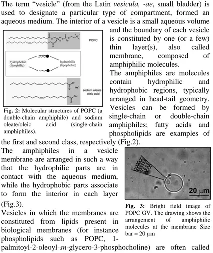
The term “vesicle” (from the Latin vesicula, -ae, small bladder) is used to designate a particular type of compartment, formed an aqueous medium. The interior of a vesicle is a small aqueous volume
and the boundary of each vesicle is constituted by one (or a few) thin layer(s), also called membrane, composed of amphiphilic molecules.
The amphiphiles are molecules contain hydrophilic and hydrophobic regions, typically arranged in head-tail geometry. Vesicles can be formed by single-chain or double-chain amphiphiles; fatty acids and phospholipids are examples of the first and second class, respectively (Fig.2).
Fig. 2: Molecular structures of POPC (a double-chain amphiphile) and sodium oleate/oleic acid (single-chain amphiphiles).
The amphiphiles in a vesicle membrane are arranged in such a way that the hydrophilic parts are in contact with the aqueous medium, while the hydrophobic parts associate to form the interior in each layer (Fig.3).
Vesicles in which the membranes are constituted from lipids present in biological membranes (for instance phospholipids such as POPC,
1-palmitoyl-2-oleoyl-sn-glycero-3-phosphocholine) are often called
Fig. 3: Bright field image of POPC GV. The drawing shows the arrangement of amphiphilic molecules at the membrane Size bar = 20 µm
lipid vesicles or more frequently liposomes, or also artificial vesicles. Finally, vesicles prepared from synthetic surfactants, which are chemically different from naturally occurring amphiphilic lipids, are also called (synthetic) surfactant vesicles
Vesicles are classified according to their size. There are small unilamellar vesicles (SUVs) with a size around 30-40 nanometers; large unilamellar vesicles (LUVs) with a size of hundreds nanometers and also Giant Vesicles (GVs) which have a size more than one micrometer, and therefore can be directly visualized by optical microscopy (Fig.4). For the latter reason, their use in Minimal Cell research is quite interesting, because it allows the observation of these lipid compartments, as well as their morphological transformations and internalised chemical reactions.
Fig. 4: SUVs, LUVs and GVs along a logarithmic size axis (vesicles drawn not to scale).
GUVs, as well as LUVs and SUVs are aggregates that are usually not at a “true” thermodynamic equilibrium, but rather in a kinetically trapped state. The energy barrier often is so high that the “true” thermodynamic equilibrium of the system cannot be reached easily. Once formed, the vesicles might remain as a kinetically stable system for an extended period of time, which can be hours, days or even weeks.
1.6 Giant Vesicles (GVs): Preparation and application
Giant vesicles (GVs) are considered a valid model for building minimal cell because they have similar size of living cells, in the range of about 1–100 um, and they can be studied with conventional light microscopic techniques. The limited resolution of light microscopy, however, does not allow to readily distinguish unilamellar from oligolamellar vesicles, except if unilamellarity is confirmed by corresponding bending elasticity measurements, or determined with fluorescence assays (Akashi et al., 1996). Depending on the experimental conditions, such as the osmotic pressure difference between the vesicle’s interior and exterior, giant vesicles can be spherical or non-spherical.
A giant unilamellar vesicle (GUV) resembles the basic compartment structure of all biological cells, in the sense that the vesicle membrane is a mimic of the self-closed lipid matrix of the plasma membrane. To illustrate the geometric properties, a spherical unilamellar giant vesicle with a diameter of 50 um and a membrane thickness of 4 nm is first considered; 4 nm is the approximate thickness of a hydrated POPC bilayer. It is astounding that a membrane that is only 4 nm thin can separate the inside of such a large object from the external aqueous solution. If extrapolated to sizes with which humans are more familiar, the relationship between vesicle diameter and membrane thickness becomes more obvious. Taking instead of a 50 um giant vesicle a balloon with a diameter of 50 m, this balloon would have a skin with a thickness of only 4 mm. Due to their cell-mimicking characteristics, GUVs currently are intensively studied in different areas of biomimetic chemistry, biomembrane physics and in the field of artificial cell synthesis. In many studies that focus on mimicking biological membranes, for example by reconstituting membrane components in giant vesicle bilayers. One of the most obvious applications of giant vesicles is their use as simple model systems for studying certain physicochemical properties of biological membranes. Examples include mechanical properties of the entire vesicle or of the membrane, lipid domain formation, lipid dynamics, membrane growth, budding, fission and membrane fusion.
A further application of giant vesicles is in the field of membrane protein research. The prerequisite for such studies is that the membrane protein of interest can be reconstituted in a giant vesicle membrane. Classical procedures allow the reconstitution of membrane proteins in submicrometer-sized vesicle membranes, typically by the detergent depletion method. However, this method is not directly applicable to the formation of giant vesicles. Girard et al. (Girard et al., 2004) described a procedure for the successful reconstitution of the sarcoplasmic reticulum CaII-ATPase and the H+-pump bacteriorhodopsin in giant vesicle membranes.
There are several advantages of using giant vesicles instead of submicrometer-sized vesicles, for example in studies of the effect of antimicrobial substances (for example, antimicrobial peptides) on phospholipid membranes. If a suspension of SUVs is used, only average observations are possible. Reactions occurring within or on the surface of single vesicles cannot be measured with SUVs.
Studies with SUVs and GUVs give complementary information. By using GUVs it was for example possible to study details of vesicle membrane fusion and fission; this provided unique insights into the two processes. Furthermore, the use of GVs allowed a direct visualization and quantitative analysis of the pore formation in phospholipid membranes by the peptide magainin-2.
Perhaps one of the most fascinating and challenging fields of application of giant vesicles is the research that aims at preparing systems that have at least some of the basic features of living cells. Currently, the expression of proteins inside vesicles is the state-of-the-art in this field of research. From a theoretical point of view, this is of relevance, because the activity of a synthesized protein might lead to complex reaction dynamics in the system, with consequences at the macroscopic level, such as internal enzyme-mediated lipid synthesis with consequent vesicle growth, or vesicle-vesicle interaction mediated by membrane proteins, or uptake of externally added nutrient or signals by membrane receptors, and many other examples. In general terms, the expression of functional proteins inside GVs allows the experimenter to build “from inside” a complex bioreactor that mimics in great detail the basic behavior of cells. This approach, that can be defined as “learning by constructing” is typical of the new discipline of synthetic or constructive biology, which aims to construct novel biological parts, devices and systems to perform useful functions or to understand at the most basic level the mechanism of certain biochemical patterns (the frequently cited motto is “What I cannot create, I do not understand” by Richard Feynman).
Due to their large size, giant vesicles are not suitable for drug delivery applications, but they might find applications in the field of biosensors, for example to detect permeabilizing toxins or xenobiotics.
1.7 GVs: main preparation methods
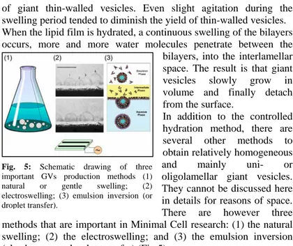
Historically, one of the first methods for the preparation of giant vesicles was described by Reeves and Dowben in 1969. It was a controlled hydration of a thin dry film of egg yolk phosphatidylcholines (egg PC) deposited on the glass surface on the bottom of a flat-bottomed 2 L Erlenmeyer flask. The lipid film was allowed to swell for two hours or more, and this led to the formation
of giant thin-walled vesicles. Even slight agitation during the swelling period tended to diminish the yield of thin-walled vesicles. When the lipid film is hydrated, a continuous swelling of the bilayers occurs, more and more water molecules penetrate between the
bilayers, into the interlamellar space. The result is that giant vesicles slowly grow in volume and finally detach from the surface.
In addition to the controlled hydration method, there are several other methods to obtain relatively homogeneous and mainly uni- or oligolamellar giant vesicles. They cannot be discussed here in details for reasons of space. There are however three methods that are important in Minimal Cell research: (1) the natural swelling; (2) the electroswelling; and (3) the emulsion inversion (also known as droplet transfer) (Fig.5).
Fig. 5: Schematic drawing of three important GVs production methods (1) natural or gentle swelling; (2) electroswelling; (3) emulsion inversion (or droplet transfer).
1.7.1 The Natural Swelling method (Fig. 5.1)
The principle of the method is the one just described. It consists of the controlled hydration of (dry) lipids deposited on a solid surface, mainly glass, as originally developed by Reeves and Dowben (1969). This method is also known as spontaneous swelling, natural swelling or gentle hydration method. The method is usually particularly successful for the preparation of GUVs from samples containing charged lipids. Furthermore, it is important to note that the hydration has to be carried out in the liquid-disordered state of the bilayers, that is, at a temperature above Tm, corresponding to the so-called
solid-ordered (so)/liquid-disordered (ld) or gel/liquid-crystalline main
phase transition temperature.
1.7.2 The Electroswelling method (Fig. 5.2)
In 1986, Angelova and Dimitrov published a study concerning the effect of externally applied electric fields on the hydration of lipids deposited from an organic solution on a conductive glass surface (indium tin oxide (ITO) coated glass), or on platinum wires.
Depending on the experimental conditions, that is, type, composition and amount of lipids used, applied electric field parameters, hydration medium, rather homogeneous giant unilamellar vesicles can be obtained in aqueous solution or buffer solutions of low ionic strength. The method is known as electroformation or electroswelling method (Angelova and Dimitrov, 1986).
Electroformed vesicles remain connected to the residual lipid film on the electrode on which they are formed. This might be an undesired property or an advantage, particularly if individual vesicles shall be punctured with a micro-needle. The electrode provides a back pressure and the connected vesicles do not move away easily during micromanipulation.
The spontaneous swelling and electroformation methods generally do not allow efficient encapsulation of large water-soluble molecules (e.g., enzymes) or charged compounds during the formation process because the molecules to be encapsulated have to somehow move below the outermost layer of the deposited lipid film; this is difficult due to slow transbilayer movements of large or charged molecules. Encapsulation is, however, possible after vesicle formation by microinjection, puncturing individual vesicles with micro-needles.
1.7.3 The Emulsion Inversion (or droplet transfer) method (Fig. 5.3)
If a (stable) w/o emulsion can be prepared from an oil and a bilayer-forming lipid, this w/o emulsion can be used as a starting system for the preparation of giant vesicles. The w/o emulsion is poured onto a two-phase system consisting of a lower aqueous phase and an upper oil phase containing a bilayer-forming lipid. If the two phase system is first preincubated for a while prior to the addition of the w/o emulsion, the interfacial region between the two phases will become saturated with lipids, and form a monolayer in which the hydrophilic head groups of the lipids are in contact with the lower aqueous phase and the lipophilic chains protrude into the upper oil phase containing excess lipid. Because water is heavier than the oil used, the water droplets of the upper w/o emulsion tend to move from the w/o emulsion toward the oil/water interface and finally through it into the lower aqueous phase where giant vesicles are formed. If spontaneous water droplet migration does not occur, or only slowly proceeds, centrifugal forces can be applied to force the water droplets to move from the upper w/o emulsion through the interface into the lower aqueous solution.
In another approach, again starting with a lipid-stabilized w/o emulsion, Tan et al. reported that giant vesicles can be formed from an initial w/o emulsion stabilized with a phospholipid by simple addition of this w/o emulsion to an ethanol/water mixture in a microfluidic device (Tan et al., 2006).
With classical preparation methods (as the gentle hydration) it is possible to prepare GVs with various types of lipids, unilamellar and in large numbers, but the entrapment of large amounts of solutes within the aqueous core is not easily done, especially for macromolecules. In contrary, by the method of w/o emulsion, it is possible to entrap solutes with high efficiency. Therefore, this formation method could be very useful when a large number of valuable solutes such as DNA, RNA and proteins must be co-entrapped in GVs.
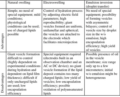
Advantage and disadvantage of the three methods are summarized in Table 1.
Tab. 1: Some of the advantages and disadvantages of the different methods for giant vesicle preparation.
Natural swelling Electroswelling Emulsion inversion
(droplet transfer)
Advantages
Simple; no need of special equipment; mild conditions;
physiological conditions can be used; use of charged lipids possible
Control of hydration process by adjusting electric field parameters; high reproducibility; giant vesicles formed are uniform, unilamellar and spherical; the vesicles are attached to the electrode which facilitates microinjection No need of special equipment; possibility of forming vesicles with asymmetric bilayers; control of vesicle size by droplet size in the w/o emulsion; high encapsulation efficiency; high yield of vesicles
Disadvantages
Giant vesicle formation difficult to control (highly dependent on experimental conditions during hydration and dependent on lipid film thickness); difficult if only uncharged lipids are used; low encapsulation efficiency
Special equipment required (electrodes built in an observation chamber and an AC or DC device); no giant vesicle formation if the lipid deposit contains too many charged lipids; low yield of vesicles; low encapsulation efficiency; possible oxidation of polyunsaturated lipids
Possible presence of oil in the vesicle
membrane; size range only up to a few micrometers; initial w/o emulsion might be heterogeneous
1.8 The emulsion inversion method as a convenient way to prepare GVs for minimal cell research (Table 2)
The method of GVs formation from water-in-oil emulsion, for the first time proposed by Weitz and coworkers in 2003, although conceptually very simple, not always gives good results, in terms of the number of GVs obtained and for the morphology of vesicles. The authors describe a number of technical limitations that restrict the use of this method. This technique relies on the formation of a stable inverted emulsion and on the passage of the emulsion droplets through a second interface to make the bilayer. Unfortunately, both of these steps create problems when lipids are used and limit the generality and practicality of this technique. The problems originate from the use of lipids in the role of surfactants. First, lipids form large aggregate structures in both oil and water and hence do not stabilize the interface of the emulsion as well as a traditional surfactant can; this severely limits the effectiveness of the emulsification and restricts the concentration of water that can be emulsified. Second, lipids adsorb very slowly to the interface and require extended periods of time to fully cover the surface. Third, lipids at the interface between oil and water apparently undergo spontaneous emulsification; this seems to result in a strongly preferred drop size, making it exceedingly difficult to produce drops of arbitrarily controlled sizes. Fourth, the slow equilibration of the interface makes it difficult to replenish the second interface as the emulsion drops pass through it; this severely limits the efficiency of the formation of the vesicles, particularly as the size of the droplets increases (Pautot et al., 2003a).
Despite these limitations, the authors were able to entrap proteins, such as G-actin, and to obtain the polymerization reaction inside GVs of phospholipid vesicles. In another work, they report the formation of GVs with an asymmetric membrane (inner leaflet different than outer leaflet) by using POPC and POPS (Pautot et al., 2003b).
The following year (2004), Noireaux and Libchaber at the Rockefeller University, constructed a cell-like model capable of express the enhanced green fluorescent protein (EGFP), by co-entrapping a cell extract of E. coli and the plasmid codifying for the EGFP inside GVs made of egg-lecithin (Noireaux and Libchaber, 2004).
Between 2006 and 2009, a group of Japanese researchers coordinated by Yoshikawa, deeply investigated the method of droplet transfer, and produced 5 scientific articles. They have studied in detail what happens at the water/oil interface, using a special microscope slide, consisting of a micro-chamber where they have reconstituted the two-phases system characteristic of the method. Using small volumes, they observe the crossing of water drops in oil through the interface and the spontaneous formation of giant vesicles in the aqueous phase driven only by gravity (Yamada et al., 2006). They showed, in their first articles, that water droplets spontaneously transfer from the oil phase to the water phase, and that, stable liposomes with controlled size and composition can be prepared in large quantity (Yamada et al., 2007). Thereafter, they were able to obtain GVs with an asymmetric membrane and lipid raft micro-domain (liquid-ordered phase) (Hamada et al., 2008). Finally, they have entrapped proteins at high concentration inside the GVs (Takiguchi et al., 2008), and studied the effect of osmotic stress using sucrose and KCl solutions at various concentrations as outer buffers (Ohno et al., 2009).
In the articles cited above, the authors use the same slide with microchamber, and although many GVs are formed, they remain adhering at the water/oil interface, without being released in solution.
In 2008, Whittenton et al., have been able to generate GVs with an asymmetric membrane and to entrap inside fluorescent oligoDNA, and other fluorescent markers. Vesicles produced have been found in the solution like in the early articles of Pautot et al., and Noireaux and Libchaber (2004). The authors use the centrifugation to accelerate the crossing of water droplets through the interface, obtaining less then 20% yield in terms of solutes entrapment (Whittenton et al., 2008).
Cecile Sykes's group, in 2009, has been able to obtain GVs containing a concentrated salt solution containing the protein G-actin. The external environment is a concentrated salt solution where the nutrients are present. After the formation of vesicles alpha-hemolysin has been added, an extracellular protein secreted by
Staphylococcus aureus, which assembles into a ring structure on
membranes and forms transmembrane pores to render liposomes selectively permeable for nutrients. Only at this point the authors
observed the reaction of polymerization of G-actin (Pontani et al., 2009).
Also in 2009, Nishimura et al., used fluorescence flow cytometry, comparing the characteristics of GVs obtained by different methods of formation. GVs obtained with the method of water in oil emulsion have a thin lipid membrane and a more regular morphology than other formation methods (Nishimura et al., 2009).
Finally, Kubatta and Rehage have studied the effect of different oil phases and of different kinds of lipids or surfactants. Changing these parameters, it has been possible to get GVs of various sizes and with different stability. The authors manage to prepare GVs with diameters in the order of magnitude of millimeters (Kubatta and Rehage, 2009).
In table 2 there is a list of the experimental conditions described in above-cited articles in which GVs have been produced by the method of water in oil emulsion.
Chapter 2. Aim of the work
As mentioned in the Introduction, investigations on the origin of early cells are currently performed by several group. In particular, one of the open questions concerns the steps that are required to achieve functional primitive cells starting from separated compartments. This aspect is discussed in terms of encapsulation of several biomolecules in primitive compartments; lipid vesicles, and more in particular fatty acid vesicles are the most plausible candidates of primitive compartments. This has stimulated some experimental research projects, as the “Minimal Cell” one, which aims at the laboratory construction of cell models containing the minimal and sufficient number of molecules and capable of displaying the most basic features of living cells.
However, from the conceptual viewpoint, the current approach is based on the idea that a single primitive cell forms directly from separated components or that a complex metabolism developed within a single primitive cell (starting from a simpler encapsulated molecular mixtures). However, alternatively, and perhaps more realistically, it is also possible that cellular or pre-cellular systems are not the product of individual events, but as the outcome of a series of multiple interactions within a community of cell-like structures, resulting cumulative benefits. We may rephrase these views as an open question: did cellular life originate from individual compartments or from a community of compartments. Such questions has been recently debated in a Workshop dedicated to Origin of Life (San Sebastian, Spain, June 2009).
From these considerations comes the question of how to create and to study a primordial protocell community.
The aim of my PhD work is the experimental study of minimal cell-like systems, and to understand the nature of the processes that led to the development and evolution of cellular life.
There are two main goals of this work. Firstly, define and develop cell-model systems, and then propose a novel view about the possible important steps in primitive pre-cellular evolution, from the viewpoint of cell communities.
GVs are useful models for the study of cells. They are closed compartments with a lipid membrane boundary, which separates an
interior content that can be very different from the external environment. Thanks to their semi-permeable membrane, GVs can exchange solutes with the environment. Most importantly, GVs have size between 1 and 100 um, which allow their direct observation by light microscopy. Thanks to all these features, that make them comparable to the actual living cells, GVs are very valuable models for developing cell models for ambitious projects as the “Minimal Cell” one. The most promising method for GVs production is the recently reported “emulsion inversion” (or “droplet transfer”) method, which allows the preparation of GVs with high efficiency of solute entrapment. This method is therefore essential for the construction of Minimal Cells. However, this method is quite new and – as evident by the analysis of literature – is not easy to reproduce. When this work was started, not only the know-how for producing GVs with this method was not available in my hosting group, but also a very limited number of reports was published. Actually, our group started the development of this GVs technology simultaneously to other international groups.
The first goal in this study was to create GVs in large quantities, in a reproducible and controlled manner, and with several kinds of solutes entrapped inside.
This achievement is a major step forward in developing new methods for studying minimal cells, not limited to the current work – which would address more basic aspects, but more in general for constructing bio-reactors capable of sustaining minimal metabolism or be able to protein expression.
The second goal will then be the development of innovative systems for understanding whether and how GVs communities are formed, if they have an advantage in respect of single GVs, and if it is so, to know how to create and manipulate them in a controlled and reproducible way. This would represent an extension and an evolution about the studies on the origin of life and on the primordial cells, shifting the focus from individual compartments to GVs colonies in order to simulate a more likely reality of cellular life. If this community model works, one could then expand it, and give a new vista on the study of GVs-based systems as model for primitive cell “ecology”, that is also a quite new concept in current research.
Although being a very ambitious goal, the work carried out in this thesis, might certainly help to advance in constructing cell model systems for studying the mechanisms underlying the development of cellular life.
Chapter 3. Results and discussion
3.1 Optimization of GVs production formed from a lipid-stabilized water in oil emulsion.
As stated before, the main goal of my PhD work is the experimental study of cell models at the aim of understanding in greater details the early steps of primitive cell development, within the so-called Minimal Cell project. At this aim, GVs will be used as standard cell model and – for the first time in the literature – it will be investigated the importance of intervesicle interaction as a way to progress along the path of functional cell formation, starting from simpler cellular or
protocellular systems.
We have chosen a preparation method that allows the formation of GVs with high entrapped yield. As noticed in the Introduction, Pautot et al. (2003), as well as Noireaux and Libchaber (2004) have reported on the conversion of w/o droplets into GVs. The method of preparing GVs starting from w/o emulsion droplets is extremely advantageous when bioreactors must be constructed, and more in general in our top-down synthetic biology approach. In fact, thanks to the total entrapment of water-soluble solutes in w/o droplets, it becomes feasible the construction of lipid based compartments containing a high number of diverse macromolecules and therefore build complex cell models, that we may call semi-synthetic minimal cells.
Fig. 6: Scheme of the emulsion inversion method. From Pautot et al 2003a.
The method was introduced by Pautot et al. (2003) Pre-formed w/o droplets are centrifuged in a two-phase medium, so that they convert in GVs by crossing the macroscopic interface (see Fig.6).
The first step is the preparation of a water-in-oil (w/o) emulsion, which can be obtained by emulsifying a small aqueous volume in a lipid-containing apolar solvent mineral oil by means of mechanical forces (stirring, vortexing, shaking). This step can be describe in the following terms: the macroscopic water droplets, possibly containing solutes, is broken in millions of small droplets which are stabilized by lipids at the w/o microinterface, presumably forming a lipid
monolayer. In the absence of lipids, the emulsion is unstable and quickly returns to the state of two macroscopically separated phases.
The hydrophilic solutes which were present in the aqueous drop are therefore segregated into the w/o droplets, becoming entrapped – in the next step, into the GVs. Ideally, each w/o droplet is transformed into a GV. It is convenient to dissolve fluorescent probes so that the w/o droplets first and the GVs later can be observed, also
quantitatively, by fluorescence microscopy. In order to collect the GVs after passage through the macroscopic oil/water interface, the solution used for preparing the emulsion must be denser that the aqueous solution placed below. This is achieved by using sucrose- vs
glucose-containing buffers. Glucose and sucrose are two sugars that are compatible with all biomolecular systems we are interested in. Therefore, different densities are obtained by using sucrose in the “inner solution” (the aqueous solution used to make the emulsion, that will Saccharose Saccharose b bufferuffer+ + fluorescent fluorescent marker marker Solution of lipids in Solution of lipids in
Mineral Oil plus aqueous
Mineral Oil plus aqueous
solution 0.5%
solution 0.5%
Droplets
Droplets’’sizesize(1(1--100 100 µµm)m)
~5,000,000
~5,000,000 dropletsdroplets/ml/ml
Vortex
Vortex
all the molecules into the water solution all the molecules into the water solution are entrapped into the droplets. are entrapped into the droplets. Saccharose Saccharose b bufferuffer+ + fluorescent fluorescent marker marker Saccharose Saccharose b bufferuffer+ + fluorescent fluorescent marker marker Solution of lipids in Solution of lipids in
Mineral Oil plus aqueous
Mineral Oil plus aqueous
solution 0.5%
solution 0.5%
Droplets
Droplets’’sizesize(1(1--100 100 µµm)m)
~5,000,000
~5,000,000 dropletsdroplets/ml/ml
Vortex
Vortex
all the molecules into the water solution all the molecules into the water solution are entrapped into the droplets. are entrapped into the droplets. Fig. 7: Preparation of a water in oil emulsion. A big drop of water solution (it is indicated from the arrow) is broken in millions of small droplets which are stabilized by lipids that formed a monolayer.
Glucose buffer
Glucose buffer
Water in oil
Water in oil emulsionemulsion Emulsion W/O Interface Before Before Centrifugation Centrifugation Glucose buffer Glucose buffer Water in oil
Water in oil emulsionemulsion Emulsion W/O Emulsion W/O Interface Interface Interface Before Before Centrifugation Centrifugation After Centrifugation After Centrifugation 1 Buffer Mineral Oil Interface GVs W/O After Centrifugation After Centrifugation 1 Buffer Mineral Oil Interface GVs W/O 1 Buffer Mineral Oil Interface GVs W/O
Fig. 8: Scheme of the emulsion inversion method. Before centrifugation, in the eppendorf there are three phases: water in oil emulsion, oil-water interface and an aqueous solution. After centrifugation, the water in oil droplets are transformed into GVs when they cross the oil-water interface.
become the internal GVs solution) and glucose in the “outer solution” (the aqueous solution used to collect GVs, that is the external GVs solution). In order to avoid osmotic stress, isotonicity is obtained by using the same sugar concentration. For example, 100 mM sucrose has a density of about 1.011 g/l, whereas this value is 1.005 in the case of 100 mM glucose. From the practical viewpoint, the w/o emulsion, prepared by emulsifying an aqueous solution of a fluorescent dye is poured above a lipid-containing oil layer that represent the “interface” phase. Below them, there is the aqueous phase where the GVs will be collected (Fig. 8). At the interface between the oil and water phases, the lipids have the polar head in the water solution and hydrophobic tail in the mineral oil. In order to collect GVs from w/o droplets, the eppendorf tube is centrifuged. The droplets are then converted into GVs when they cross the oil/water interface.
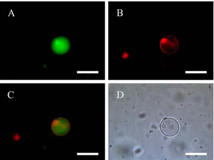
After some preliminary testing, we have firstly shown that method works also when the zwitterionic phospholipid POPC is used. GVs were heterogeneous with respect to size, namely between 1 to 50 micrometers (diameter), and in their aqueous core there was a fluorescent marker. However, despite the large amount of water in w/o droplets (summed up to 1 ul), a small number of GVs was initially observed, and – at the same time – the aqueous phase (the “outer phase”) contained some of the fluorescent marker that was initially present in the w/o droplets. When a hydrophobic marker (rhodamine 6G) was added to the mineral oil solutions, and this solution was used to prepare a w/o emulsion from a calcein solution, it was possible to produce double-labeled GVs (Fig.9). The green fluorescence from the aqueous GV core and the red fluorescence from the GV membrane confirm the correct position of both fluorophores.
Initial experiment, although successful, showed that even if the “emulsion inversion” method is clearly extremely advantageous for minimal cell research, it is also very difficult to achieve. Indeed the authors of the articles do not explain in detail the experimental conditions, and, due to the novelty of this method, in literature there are only a very limited number of reports (see Introduction). Therefore, the first part of the work has been devoted to optimize the method of GVs production via “emulsion inversion”. The goal was to obtain in reproducible way the maximal number of solute-filled GVs, by using different markers.
Fig. 9: Double-labeled POPC GV. (A) Calcein in the aqueous core; (B) rhodamine 6G on the membrane; (C) overlapping of channels (A-B); (D) bright field image. Size bar = 20 µm.
To quantify the effectiveness of GVs production (expressed in number of GVs/mL, i.e. numerical density) we used a hemocytometer, which generally used in cell biology to count cells (Fig. 10). The hemocytometer is loaded by a definite volume of GVs dispersion, and thanks to its calibrated grid it is possible to estimate the number of GVs by direct visualization at the microscope. GVs are very difficult to identify in bright field. They are almost invisible because they are not colored. So it is virtually impossible to count them directly under the microscope as it does in cell biology for the cells. But when GVs are produced by employing a fluorescent marker (trapped inside) it
Fig. 10: Hemocytometer microscopic slide. This Particular kind of microscope slides has a grid with a defined size. It is usually used in Cell Biology to cell counting. In our case it was used to count GVs.
become possible to see them by fluorescent microscopy, still using the hemocytometer. In our approach, we have collected and analyzed by a image analysis software (Image J) the number of GVs in each micrographs, and from this value the numerical density is easily calculated by taking into account the sample volume.
With this strategy, we were able to assess what were the best experimental conditions for the emulsion inversion method.
3.1.1. Effect of the lipids in w/o emulsion and interface: lipid mixture and "asymmetric" design
Firstly, GVs production was attempted by using the same lipid or surfactant in the w/o emulsion phase, and in the interface phase. We employed phospholipids like POPC, POPS, POPG, fatty acids like sodium oleate, and surfactant like DDAB and PUCE. In all cases, we got a very limited number of GVs (less than 100/ml). When POPC, POPG and POPS were used a few vesicles with sizes between 1 and 50 um, regular in shape were obtained. These GVs effectively entrapped the fluorescent markers and appeared to be uni- or oligo-lamellar. When DDAB and PUCE, two cationic surfactant were used, a slightly larger number of GVs was obtained. When, on the other hand, oleate was used, no vesicles were found in the aqueous phase.
After these attempts, the experimental strategy was changed. Instead of using one lipid, mixtures of two lipids were employed. To form the emulsion and the interface we mixed a POPC solution (in mineral oil) in a 1:1 v/v ratio with another lipid solutions of the same concentration, in order to have 50% of negative charge (POPS, POPG and oleate) or positive (PUCE or DDAB). In all cases, significant increase in GVs production was observed.
Finally, we also tested the case when POPC only was used for the preparation of the w/o emulsion, whereas another (different) surfactant was used to create the macroscopic oil/water interface. Surprisingly, a greater number of GVs was obtained. It is clear that the charged lipid at the interface helps the formation of GVs, and that this condition is the optimal one for producing a large number of GVs.
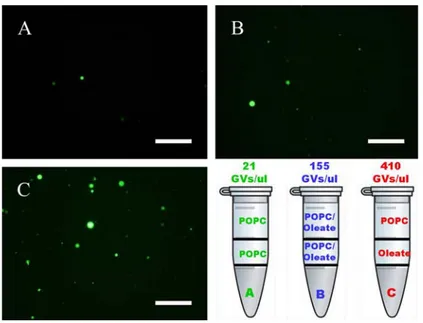
In figure 11 it is reported the results of the case POPC/oleate. Whereas when POPC only was used the number of GVs per ml was around 20, the use of a POPC/oleate 1:1 mixture increased such value to ca. 150. If, however, oleate was used to form the interfase
whereas POPC was used for the emulsion, the GVs production yield was around 400/ml. Similar results have been obtained with other kinds of charged lipids (data not shown). In other words, separating two lipid species in the two phases (w/o emulsion and interface) brings about a significant increase of GVs production. Notice that this procedure possibly produces asymmetric membranes.
Fig. 11: Counts were performed using 10 photos of vesicles within the hemocytometer by the fluorescence microscopy, using the 10x magnification. For image analysis was used Image J software. A) GVs obtained with only POPC; B) GVs obtained with POPC and oleate mixed; C) GVs obtained with POPC and oleate unmixed. In the bottom right is shown the number per ul of GVs obtained and the phase patterns in the eppendorf in three cases. Size bar = 250um.
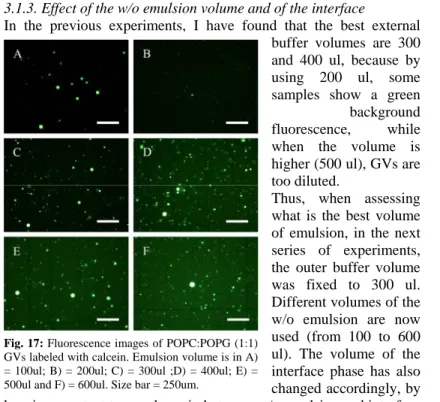
3.1.2. Effect of the outer aqueous volume
The next optimization step involved the exploration of other parameters such as the volume of the external buffer (“outer phase”), the volume of the internal buffer (“inner phase”), the volume of the w/o emulsion and the concentration of both aqueous buffers. All experiments were performed by using POPC-based w/o emulsion and one kind of charged lipids for the interface.
Lipid combinations used are as follows: • POPC:POPG 1:1; • POPC:POPS 1:1; • POPC:oleate 1:1; • POPC:DDAB 1:1; • POPC:PUCE 1:1.
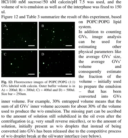
In the first series of experiments, we varied the volume of the external buffer, from 200 to 500 ul (10 mM Tris HCl/100 mM glucose/pH 7.5 was used). As the internal buffer, 10 mM Tris HCl/100 mM sucrose//50 uM calcein/pH 7.5 was used, and the volume of w/o emulsion as well as of the interphase was fixed to 150 ul.
Figure 12 and Table 3 summarize the result of this experiment, based on POPC:POPG lipid system.
In addition to counting GVs, image analysis can be used for estimating other physical parameters like the average GVs’ size, the average GVs’
volume and consequently estimate
the fraction of the volume – initially used to prepare the emulsion – that has been converted into GVs inner volume. For example, 30% entrapped volume means that the sum of all GVs’ inner volume accounts for about 30% of the volume used to produce the w/o emulsion. The missing part can be ascribed to the amount of solution still solubilized in the oil even after the centrifugation (e.g. very small reverse micelles), or to the amount of solution, initially present as w/o droplets that instead of being converted into GVs has been released due to the competitive process of w/o droplet break at the oil/water interface (see below).
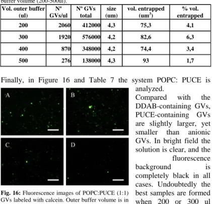
Fig. 12: Fluorescence images of POPC:POPG (1:1) GVs labeled with calcein. Outer buffer volume is in A) = 200ul; B) = 300ul; C) = 400ul and D) = 500ul. Size bar = 250um.
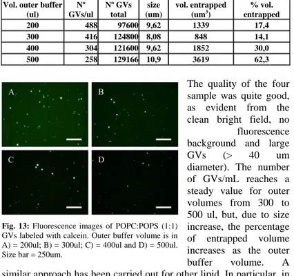
Tab. 3: Image analysis results about POPC:POPG GVs obtained with different outer buffer volume (200-500ul).
Vol. outer buffer (ul) Nº GVs/ul Nº GVs total size (um) vol. entrapped (um3) % vol. entrapped 200 488 97600 9,62 1339 17,4 300 416 124800 8,08 848 14,1 400 304 121600 9,62 1852 30,0 500 258 129166 10,9 3619 62,3
The quality of the four sample was quite good, as evident from the clean bright field, no
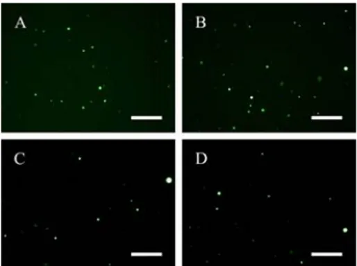
fluorescence background and large GVs (> 40 um diameter). The number of GVs/mL reaches a steady value for outer volumes from 300 to 500 ul, but, due to size increase, the percentage of entrapped volume increases as the outer buffer volume. A similar approach has been carried out for other lipid. In particular, in Figure 13 and Table 4 results from the system POPC:POPS are reported. Here we can observe that the number of GVs/ml is similar to that one of the previous case, but their different sizes imply a minor entrapped volume.
Fig. 13: Fluorescence images of POPC:POPS (1:1) GVs labeled with calcein. Outer buffer volume is in A) = 200ul; B) = 300ul; C) = 400ul and D) = 500ul. Size bar = 250um.
Tab. 4: Image analysis results about POPC:POPS GVs obtained with different outer buffer volume (200-500ul).
Vol. outer buffer (ul) Nº GVs/ul Nº GVs total size (um) vol. entrapped (um3) % vol. entrapped 200 614 122800 7,4 469,7 7,7 300 630 189000 6,8 434,1 11,0 400 614 245600 7,4 550,4 18,0 500 498 249000 7 349,5 11,6