UNIVERSITÀ DEGLI STUDI DI ROMA
"TOR VERGATA"
FACOLTA' DI SCIENZE M. F. N.
DOTTORATO DI RICERCA INIMMUNOLOGIA
XX
CICLO DEL CORSO DI DOTTORATO
Invasive Haemophilus influenzae type b disease after
widespread use of Hib conjugate vaccines in chil dren
Dottoranda: Maria Giufre'
Tutor: Dott.ssa Marina Cerquetti
Coordinatore: Prof. Paolo Rossi
INDEX
INTRODUCTION
1. HAEMOPHILUS INFLUENZAE INVASIVE DISEASE 4
1.1 Infection and disease 4
1.2 Haemophilus influenzae : the microrganism 4
1.2.1 General features 4
1.2.2 Encapsulated strains and nonencapsulated strains 5
1.2.3 H. influenzae type b (Hib) 6
1.2.4 capb locus 8
1.2.4.1 Duplication of capb locus 9
2. INTRODUCTION OF ANTI -HIB CONJUGATE VACCINES 11
2.1 Pre-vaccine situation 11
2.2 Introduction of Hib vaccine 11
2.3 Decrease in incidence of invasive Hib disease 13
3. INVASIVE DISEASE DUE TO HIB IN CHILDREN PREVIOUSLY VACCINATED WITH HIB CONJUGATES (TRUE VACCINE FAILURES) 15 3.1 The UK experience 15
3.2 The Netherlands experience 18
3.3 The Italian experience 18
3.4. Factors involved in TVF 20
3.4.1 Host risk factors 20
3.4.2 Factors due to the Hib vaccine 21
3.4.3 Factors due to particular virulence of the microrganism 22 3.5 Aim of the study 23
MATERIALS AND METHODS
1. UK study 24
1.1 Characteristic of cas es 24
1.2 PCR 25
1.3 Immunological assays 25
1.4 Determination of the copy number of the capb locus 26
1.5 Statistics 27
2. Italian study 28 2.1 Bacterial strains 28
2.2 Immunological assays 28
2.3 Determination of the copy number of the capb locus 29
2.4 Statistics 29
RESULTS
1. UK study 30 1.1 TVF epidemiology data 30 1.2 Analysis of copy number of capb locus 332. Italian study 39
2.1 Epidemiology data 39
2.2 Analysis of copy number of capb locus 3 9
DISCUSSION
42CONCLUSIONS
47ACKNOWLEDGMENTS 49
INTRODUCTION
1. HAEMOPHILUS INF LUENZAE INVASIVE DISEASE 1.1 Infection and disease
Haemophilus influenzae (Hi), a facultative anaerobe bacterium, was first
described in 1892 by Richard Pfeiffer during an influenza pandemic and mistakenly considered to be the cause of the common flu until 1933, when the viral etiology of the flu became apparent. Probably, H. influenzae was an important secondary invader to the influenza virus in the 1892 pandemic. Naturally-acquired disease caused by H. influenzae occurs only in humans:
H. influenzae is highly adapted to its human host and it has not been
detected in any other animal species. H. influenzae organisms are carried in the nose and the throat of both children and adults who may be healthy or have mild symptoms, but when other factors (such as a viral infection or reduced immune function) create an opportunity they can cause serious disease. H. influenzae can be transmitted by direct contact with respiratory droplets and discharges from the nose and throat of infected or colonized persons.
1.2 Haemophilus influenzae : the microrganism 1.2.1 General features
H. influenzae is a small non-motile Gram-negative coccobacillus, which can
be grown on chocolate agar (heated blood) added with hemin (factor X) and nicotinamide adenine dinucleotide (NAD+: factor V) (Campos J. M ., 6thed.). The bacterium grows best at 35-37 degrees at high CO2 concentration (5%)
requirement can be used to distinguish between H. influenzae, which requires both, H. parainfluenzae, which requires factor V only, and H.
ducreyi which requires factor X only. The organism is also catalase and
oxidase positive. It shows small, con vex, smooth, pale, grey or transparent colonies. In specimens from acute infections, the organisms are short (1.5 m) coccoid bacilli, sometimes occurring in pairs or short chains. In cultures, the morphology depends on age of the medium: at 6 –8 hours in rich medium, the small coccobacillary forms predominate; later , there are longer rods, lysed bacteria, and very pleomorphic forms.
1.2.2 Encapsulated strains (typeable) and nonencapsulated strains (non-typeable).
Two major categories of H. influenzae isolates have been defined: the encapsulated strains and the nonencapsulated strains, also called non -typeable (NTHi). Encapsulated isolates express one of six chemically distinct capsular polysaccharides (types a trought f), while non -typeable strains have no capsule (Pittman, 1931). Serotyping by slide agglutination test using specific antisera is the most commonly method used in identifying the capsular type of H. influenzae isolates. However, this method has been shown to be defective, so a PCR -based method for identifying the capsular types of H. influenzae has been developed (Falla T.J. et al., 1994). Encapsulated strains and nonencapsulated strains cause different spectrum of diseases. Generally, encapsulated strains are mainly associated with severe infections. Their capsule allows them to resist phagocytosis and complement-mediated lysis in the non-immune host. The capsule material is antiphagocytic, and it is ineffective in inducing the alternative complement
pathway, so that the bacterium can invade the blood or cerebrospinal fluid without attracting phagocytes or provoking an inflammatory response and complement-mediated bacteriolysis. Among encapsulated strains,
Haemophilus influenzae type b (Hib) causes almost all systemic infections.
Nonencapsulated strains are less invasive, but they are able to induce a strong inflammatory response that causes disease, such as respiratory tract infections and otitis media (fig.1).
Figure 1. Infectious diseases caused by Hib and NTHi strains (Todar K.,
2004)
1.2.3 H. influenzae type b (Hib)
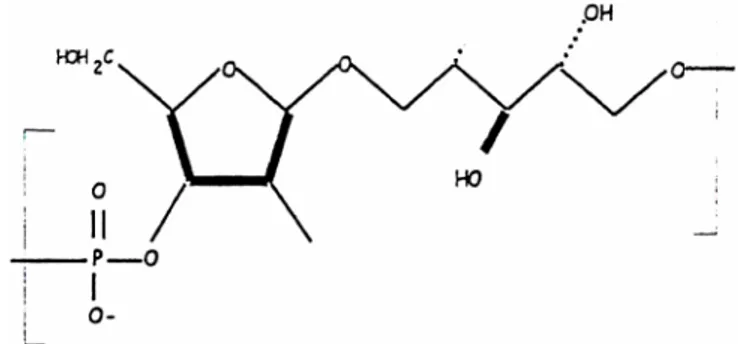
Before introduction of conjugate vaccines against Haemophilus influenzae type b (Hib), Hib was one of the leading causes of invasive bacterial infection in young children in industrialized c ountries. Hib causes meningitis, bacteraemia and, occasionally, cellulitis, osteomyelitis, epiglottitis and joint infections. The type b polysaccharide capsule, a polymer of D-ribose-ribitol-phosphate (PRP), is the major virulence factor
of Hib (fig. 2). The polysaccharide capsule may provide a survival advantage during transmission and colonization (Moxon et al., 1990), but it also facilitates survival of the microrganism in the blood through resistance to complement mediated killing and phagocytosis (Anderson et al., 1972; Weller et al., 1978). In natural immunity, acquisition of antibodies directed against the polysaccharide capsule of the microrganism occurs during childhood, presumably as a result of the asympto matic carriage of the microorganism in the nasopharynx. Such antibodies confer protection so that Hib disease is much less common after the age of 4 years. Age-specific profiles of anti-PRP antibodies show that relatively high levels of transplacentally acquired anti-PRP antibodies fall over the first months of life to very low levels by around 6 months of age. Subsequently, antibody titres rise again during the second year of life, presumably as a result of exposure to Hib in the nasopharynx or other organ isms with cross-reactive antigens (Anderson et al., 1977).
Figure 2. H. influenzae type b D-ribose-ribitol-phosphate (PRP)
1.2.4 capb locus
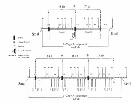
In H. influenzae, all the genes involved in capsulation are localized in a DNA segment of about 18 kb, defined cap locus. The cap loci of all H.
influenzae serotypes share an identical organization consisting of
functionally unique regions I, II, and III (f ig. 3A). Regions I and III are common to all six capsular types and contain genes necessary for the processing and the export of the capsular material, while region II is serotype-specific (Aubrey R. et al., 2003). In particular, Region I genes (bexDCBA) code for an ATP-driven capsule export apparatus ( Kroll J. S. et al, 1990). Region II contains serotype-specific biosynthesis genes that appear to be unique to each of the six capsule types ( van Eldere J., L. et al, 1995). Region III genes appear to be involved in capsule post -polymerization steps (Frosch M., et al, 1993).
Figure 3. cap locus
A. Common organization of H. influenzae cap locus.
B. Partially duplicated cap locus of Hib showing the truncated region I with the 1.2-kb deletion between IS 1016 and bexA (Satola S.W. et al., 2003)
1.2.4.1 Duplication of capb locus
Most invasive Hib strains contain a partial duplication of the cap locus that consists of 1 intact copy of the locus and a second copy with a 1.2 -kb deletion within the bexA gene (which is necessary for polysaccharide export) and the IS1016 insertion element that fl anks the locus (Kroll et al., 1988) (fig. 3B). The presence of an IS 1016-bexA deletion on the 5’ end of the duplicated Hib cap locus is thought to stabilize capsule production by reducing chances of recombination events that could result in loss of cap genes. Moreover, duplication of capb locus may serve as template for further amplification of capsule gene sequences (number of copies >2 repeats, fig. 4), thereby providing a mechanism for enhanced capsule expression (Kroll et al., 1991). Since there is a di rect relationship between capsule production and virulence, amplification of capsule genes could enhance Hib potential to cause disease: amplification of the cap locus occurs frequently in clinical isolates and has been proposed to be a mechanism by which this microrganism evades host defense. Amplification of the capb locus is associated with decreased susceptibility to complement -mediated lysis and decreased complement -mediated opsonization and suggest that amplification is used by these pathogens to incr ease their resistance to complement-dependent host defense mechanisms.
2. INTRODUCTION OF ANTI -HIB CONJUGATE VACCIN ES: IMPACT OF VACCINATIO N ON INVASIVE DISEAS ES CAUSED BY HIB
2.1 Pre-vaccine situation
As above mentioned, individuals can carry Hib bacteria in their nose and throat without showing signs of the disease. Before Hib vaccine was introduced, about four in every 100 preschool children carried the Hib organism (McVernon et al., 2004). The risk of disease is highest for children between six months and two years of age. Before 1985, when the first Hib polysaccharide vaccines were licensed for use in the United States, Hib was the most common cause of bacterial meningitis in children under 5 years of age in the U. S. (about 12,000 cases per year, most in children younger than 18 months). An additional estimated 7,500 cases of other invasive Hib infections also occurred annu ally in young children (Bisgard K.M. et al., 1998). During 1980-1990, incidence was 40 -100/100,000 children < 5 years old in the U.S.A.. Approximately 5% of affected children died, and neurological sequelae developed in 15% to 30% of the surviving children. In European countries, the risk for infection by Hib in the early 1990s varied from 12 to 52 cases/100,000 persons, depending on the countries. In Italy, national incidence of meningitis in 1994 was 0.15/100,000, in particular
5.7/100,000 in children < 2 years old (D’Alessandro D. et al., 1995).
2.2 Introduction of Hib vaccine
The first vaccine to be developed against Hib was composed of the organism's polysaccharide capsule, polyribosylribitol phosphate (PRP), alone. An early trial of this vaccine showed >90% efficacy in children who
received vaccine aged 18 months or older, but no protection was demonstrated in younger children (Peltola H. et al., 1977). In fact, although Hib polysaccharide induces production of bactericidal antibodies in older children and adults, it does not reliably elicit protective le vels of antibodies in children less than 18 months of age, since PRP does not induce immunological memory. It is well known that bacterial polysaccharides capsules are T cell-independent antigens not capable to induce immunological memory or to yield a boo ster response with repeated immunization, in children <18 months of age (Kaythy, H. et al., 1984). Pure PRP polysaccharide is not processed by antigen presenting cells (APC) nor presented in context of MHC II to T cells. Conjugation of capsular PRP to a carrier protein renders them immunogenic in infants and capable of eliciting memory, booster responses, and isotype switching of anti -PRP antibodies to IgG1 (Black S. B. et al., 1991). The enhanced immunogenicity of glycoconjugate vaccines compared with nati ve bacterial PS is thought to be due to processing of carrier protein by the APC (such as dendritic cells) and presentation of carrier protein-derived peptides in the context of MHC class II molecules to T helper cells, followed by induction of cytokine production. The cytokines produced by activated T helper cells are presumed to stimulate PRP-specific B cells to undergo clonal expansion, differentiation to memory cells, isotype switching to predominantly IgG1, and increased production of PRP-specific antibodies (Stein K. E. et al., 1992; Guttormsen H. K. et al., 1999).
For these reasons, a new generation of Hib vaccines was developed by conjugating a T-cell dependent protein antigen, such as diphtheria and tetanus toxoids, to the PRP polysaccharide (Kell y D.F. et al., 2004). This covalent linkage induce presentation of carrier protein in conjunction with
MHC II to the T cell, leading to germinal centre formation with antibody class switching, avidity maturation and memory B -cell production (Arpin C. et al., 1995). These Hib conjugate vaccines not only induce protective circulating antibodies and immunological memory in infants, but also result in decreased nasopharyngeal colonization of Hib (Anderson P. et al., 1985). Thus, a herd effect is achieved throug h reduced transmission of the microorganism. The efficacy of Hib conjugate vaccines was demonstrated in trials performed in Finland and the USA and it was estimated to be >95% (Heath P.T. et al., 1998).
The first country to use Hib conjugate vaccines in infancy was Finland in 1986, followed by Iceland and the U.S.A.
2.3 Decrease in incidence of invasive Hib disease due to routine infant vaccination
Introduction of Hib protein –polysaccharide conjugate vaccines in almost all industrialized countries over the past 15 years has resulted in the virtual elimination of invasive Hib disease. These vaccines have shown protective efficacy in early infancy. Hib vaccines was introduced in 1 08 countries by the end of 2006. Global Hib vaccine coverage is estimated at 22% in 2006, reaching 92% in the Americas, but only 24% in Africa. Following introduction of Hib conjugate vaccines into routine childhood immunization programmes in the 1990s, Hi b disease has largely disappeared in Western Europe, Canada, the United States, Australia and New Zealand. However, Hib remains a major cause of invasive disease in infants and children in developing countries where vaccine is not widely used, being bacteriemic pneumonia the most common clinical presentation in children with Hib
invasive disease (an estimated 173,000 deaths per year). The cost of Hib conjugate vaccines has limited their use in developing countries even though Hib is a major cause of morbidi ty and mortality. In 1998, WHO recommended that Hib vaccine should be included in routine infant immunization.
Currently, several different Hib vaccines, all conjugate vaccines, are on the market. The vaccines currently licensed for use against Hib diseas e are based on Hib-polysaccharide conjugated to a protein carrier, such as diphtheria toxoid (PRP -D), a diphtheria toxoid-like protein (PRP-CRM197), tetanus toxoid (PRPT), or meningococcal outer membrane protein (PRP -OMP) (table1). The Hib vaccine is usua lly given in infancy as repeated doses following vaccination schedule including also diphtheria/tetanus/ pertussis (DTP) and other vaccines. Recently, in several industrialized countries, combined vaccines including several different antigens, such as Diptheria-Tetanus-Acellular-Pertussis, Hepatitis B, Inactivated Polio and Hib conjugate, have been introduced. A booster dose is recommended in most countries at 12-18 months of age. In adults and children over 18 months of age a single dose is sufficient to i nduce immunity. One of the vaccines (PRP-D) has been demonstrated to perform less well than the others in children below 18 months of age, and it is therefore not licensed for use in infants in many countries.
Table1. Hib conjugate Vaccines (Watt P.J. et al., 2003)
Vaccine Coniugate protein
PRP-T Tetanus toxoid
PRP-OMP Neisseria meningitidis outer membrane protein complex
PRP-CRM 197 CRM 197, a cross-reactive mutant of diphtheria toxoid
3. INVASIVE DISEASE DUE TO HIB IN CHILDREN PREVIOUSLY VACCINATED WITH HIB CONJUGATES (TRUE VACCINE
FAILURES)
The incidence of Hib invasive disease among children aged 4 years or younger has declined by 98% since the introduction of Hib conjugate vaccines. However, since 1999, some E uropean countries (United Kingdom and The Netherlands) have experienced an unexpected resurgence of cases of invasive Hib disease even in fully vaccinated children. This increase involved all age classes including adults, but the rise was particularly striking in children <5 years old. True vaccine failure (TVF) is defined as an invasive Hib disease case occurring in a previously vaccinated child (Heath PT et al., 2000). Several studies have been conducted to evaluate the possible factors responsible for su ch vaccine failures and some hypothesis have been formulated, as detailed following.
3.1 The UK experience
Before routine vaccination was introduced, Hib was a notable cause of paediatric morbidity in UK, but the inclusion of Hib conjugate vaccines in the routine immunisation schedule resulted in a rapid decline in reports of invasive Hib disease. In particular, the incidence of invasive Hib disease fell from 22.9/100,000 in 1990 to 0.65/100,000 in 1998 in children <5 years old (McVernon J. et al., 2003). Routine immunisation with Hib conjugate vaccines was introduced in the UK in October 1992, according to the primary schedule of three doses at 2, 3, and 4 months of age. No booster dose of Hib vaccine was included in the original schedule. During the first
children up to 48 months of age, with the aim to reduce carriage and thereby transmission of Hib in the childhood population. Compared with Hib programmes elsewhere, three unique features characterized the UK approach: i. primary vaccination was given earlier and completed at 4 months of age; ii. a fourth (booster) dose of Hib vaccine was not given; iii. vaccination was offered to all children up to 48 months of age in a nationwide “catch up” programme. In order to monitor the impact of this programme, a surveillance study was initiated under the auspices of the British Paediatric Surveillance Unit (BPSU) in collaboration with the Haemophilus Reference Unit (HRU) of the Public Health Laboratory Service.
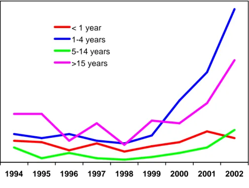
Since, differently to that observed in other industrialized countries, in UK pre-vaccination incidence of invasive Hib disease was higher in infants than in children, the rationale for the use of an accelerated schedule was to protect infants earlier, believing that immunological memory after three doses of vaccine would be able to protect even through the first years of childhood. However, in spite of the high coverage reached in infants, several cases of TVF occurred during the yea rs 1993-1999 (Heath and McVernon, 2002). Remarkably, a steady rise up to 4.6/100,000 was observed in cases of invasive Hib disease in fully immunized children under 5 years in years between 1999 and 2003 (fig. 5). Of note, during this period there was no significant change in vaccine coverage (Ramsay M.E. et al., 2003), but an acellular pertussis component present in the combined vaccine including Hib was introduced during 2000 -1.
Figure 5. Invasive Hib disease in UK, according to age group, 1994 -2002
Combined PHLS HRU/CDSC data (McVernon J. et al., 2004)
As result of resurgence of cases of invasive Hib disease in vaccinated children, the UK Department of Health implemented a booster campaign in 2003, whereby children over 6 months and less than 4 years of age were offered an extra dose of Hib vaccine. The campaign had a marked effect on the incidence of invasive Hib disease in 1 -4 years olds, with an 87% reduction in the number of cases in 2004. Following , a booster dose in the second year of life was introduced in 2006, to improve the persistence of population immunity against Hib.
1994 1995 1996 1997 1998 1999 2000 2001 2002
< 1 year 1-4 years 5-14 years >15 years
3.2 The Netherlands experience
Before the nationwide introduction of Hib vaccination in The Netherlands, the incidence of Hib meningitis was 22/100,000 per year for children less than 5 years old, with a maximal incidence of 250/100,000 per year for 11 -month-old infants (van Alphen L. et al., 1990). Vaccination with Hib conjugate vaccine was incorporated into the National Vacci nation Program in 1993. At the time of introduction, the Hib vaccine was given at the ages of 3, 4, 5, and 11 months, in a combined vaccine with the diphtheria, tetanus,
pertussis and the poliomyelitis antigens. There was no “catch -up” campaign
when the vaccine was introduced and a whole cell pertussis vaccine is still used in the Netherlands (Rijkers GT et al., 2003). Since January 1999, infants have been vaccinated at 2, 3, 4, and 11 months of age. In The Netherlands, the Hib conjugate vaccine was proved to be successful as in UK, with an efficacy of 99.4% (van Alphen L. et al., 1997): after the introduction of Hib vaccination in 1993, the number of invasive Hib infections decreased steadily (the incidence of invasive Hib disease dropped from 22 per 100,000 in 1992 to 0.8 in 2002 among children younger than 5 years of age (Van der Ende A. et al., 2003). However, since 2002, 9 years after the introduction of the Hib vaccination program for infants in The Netherlands, a small number of TVF were reported (the number increased by a factor of 3 compared to the years 1996 to 2001) (Rijkers GT et al., 2003).
3.3 The Italian experience
In Italy, Hib conjugate vaccines was introduced in February 1995 on a voluntary basis. Subsequently, Hib vaccination (consisting of 3 doses at 3, 5,
and 11 months of age) has been included in the national vaccination program since 1999. Hib vaccine coverage by 24 months of age was estimated to be 20% in 1998, 55% in 2000, 84% in 2002, 87% in 2003 and
95% in 2005 (Italian Ministry of He alth
http://www.ministerosalute.it/promozione/malattie/dati_statistici.jsp). To evaluate the burden of H. influenzae invasive disease and to monitor the impact of the vaccination program, a laboratory -based surveillance study has been conducted in a sample of Italian regions since 1997. The impact of to e
To evaluate the burden of H. influenzae invasive disease and to monitor the impact of the vaccination program, a la boratory-based surveillance study has been conducted in a sample of Italian regions since 1997. The impact of vaccination on the incidence of invasive Hib disease in Italy was comparable with that of other industrialized countries, leading to a decrease in the annual incidence from 0.27 cases/100,000 persons to 0.02/100,000 persons in the total population and from 4.78/100,000 persons to 0.44/100,000 persons among children aged <5 years (Cerquetti M. et al., 2006, fig.6). Until now, 8 years after the inclusion of the Hib vaccination in the national vaccination program, a small amount (<10) of TVF cases has been reported.
0 1 2 3 4 5 6 1997 1998 1999 2000 2001 2002 2003 2004 in c id e n z a p e r 1 0 0 .0 0 0 0 10 20 30 40 50 60 70 80 90 100 C o p . v a c c in a le e n tr o 2 a n n i incidenza cop. Vaccinale
Figure 6. Incidence of invasive Hib disease in children <5 years and vaccine coverage (1997-2004). (Ciofi Degli Atti M.L. et al. , 2004).
3.4. Factors involved in TVF
It is well known that several factors may interfere with the effectiveness of a vaccine included in a routine vaccination program. Such factors may comprise clinical host risk factors, host immunological deficiencies, interferences of the vaccine in use with other concomitant vaccines, waning immunity in the absence of booster doses of vaccine, and reduced natural boosting as a result of decreased transmission of the organism. Prior to discuss the factors responsible for TVF, is needed mentioning the relationship between antibody to PRP capsule and clinical protection against invasive Hib disease, which has been inferred from studies of natural immunity and passive immunisation. A serum anti -PRP antibody concentration of at least 0.15 µg/ml has been established as a correlate of short term protection against disease, and 1.0 µg/ml as a correlate of long term protection (Kayhty H. et al., 1983).
3.4.1 Host risk factors
In a study conducted on TVF cases in UK, several host risk factors were
identified: prematurity, Down’s syndrome, malignancy and immunoglobulin
subclass deficiency (Heath P.T. et al., 2000). Of the clinical conditions listed, a history of premature delivery is the most frequent (12.3%). It has been shown that vaccinated premature infants generally have lower anti -PRP antibody concentrations than vaccinated term infants. Infants with Down's syndrome have been shown to have a number of immunological abnormalities that may affect their response to vaccination (Loh KS et al., 1990). Children with malignancy have an increased risk of invasive Hib disease and a lesser response to Hib conjugate vaccines (Feldman S. et al.,
1990). As far as immunoglobulin subclass deficiency is concerned, the most commonly found is associa ted with subnormal levels of antibodies of the IgG2 subclass. Indeed, a recent study conducted on a small cohort of TVF cases in the Netherlands, concluded that a few of such patients not only had a quantitative defect in the production of anti -Hib antibodies, but also they showed a qualitative defect, in anti -PRP antibody avidity maturation which particularly involved the IgG2 sub -class (Breukels M.A. et al., 2002). This may be linked to a common developmental pathway involving isotype switching and avidit y maturation. Interestingly, a recent investigation, conducted on patients with TVF in UK, seemed to confirm these data since children with TVF had significantly lower anti -PRP antibody avidity than did healthy control subjects (Lee Y. C. et al., 2008).
However, according to Heath’s study, only 44% of the children who experienced Hib TVF presented a clinical risk factor or some immunological defects. Therefore, more than half of TVF remained unexplained (Heath P.T. et al., 2000).
3.4.2 Factors due to the Hi b vaccine
Specific features of the UK vaccination program (accelerated primary schedule including three doses at 2, 3, and 4 months of age, with no booster dose in the second year of life and use of less immunogenic, combination vaccines containing an acel lular pertussis component) appear to have contributed to the increase in vaccine failures. In fact, preceding the increase in vaccine failures, there was a deficit of combined Diphtheria/Tetanus/ whole-cell Pertussis/Hib (DTwP-Hib) conjugate vaccine. As a result there was widespread use of a DTaP -Hib vaccine from late 1999 onwards.
Reduced antibody responses to Hib conjugates component have been well documented using acellular pertussis/Hib combinations, especially in children aged <6 months (Eskola J. et a l., 1999).
3.4.3 Factors due to particular virulence of the microrganism
Although predisposing host risk factors and the use of less immunogenic vaccines have been considered (Heath P.T. et al., 2000; McVernon J. et al., 2003) the possibility that particular virulent traits of the bacterium may contribute to vaccine failure cannot be ruled out. In particular, amplification of the capb locus (number of copies >2) might be involved in TVF. In fact, as above mentioned, previous studies s uggest that amplification is used by Hib strains to increase resistance to complement -dependent host defence mechanisms (Noel G.J. et al., 1996).
3.5 Aim of the study
For the present thesis, we conducted two different studies: i. UK study
To study TVF cases, we took advantage of the large collection of serum samples and Hib strains obtained from cases detected through the population-based national surveillance of invasive H. influenzae disease conducted in the United Kingdom. We examined the anti -PRP antibodies level in children with TVF from the UK. Moreover, we investigated whether amplification of the capb locus might play a role in TVF cases. To this aim, the number of copies of the capb locus was determined in Hib strains isolated from children with TVF and from control children (unvaccinated children with invasive Hib disease) of the same age class in the UK.
ii. Italian study
To investigate possible changes in capsule gene structure in Hib strains circulating in Italy, as result of the widespread use of Hi b conjugate vaccines, we determined the number of copy of the capb locus in invasive Hib strains collected before and after Hib vaccination had been included in the national vaccination program (1997 -2003).
MATERIALS AND METHODS
1. UK STUDY1.1 Characteristic of cases
A total of 281 cases of children <60 months old with invasive Hib disease were analyzed. Cases were detected through population -based national surveillance of invasive H. influenzae disease conducted in the United Kingdom during the study period. A case of invasive H. influenzae disease was defined as the isolation of H. influenzae from normally sterile site, such as blood or cerebrospinal fluid, combined with a clinical picture compatible with invasive H. influenzae disease, regardless of vaccination status. Surveillance system involved sending strains to a reference laboratory (Haemophilus Reference Unit, Oxford), where strains were identified as Hib by serotyping and polymerase chain reaction (PCR) capsular genotyping. True vaccine failure (TVF) was defined as invasive Hib disease occurring either >2 weeks after the administration of a single dose of vaccine to a child >1 year old or at least 1 week after the administration of 2 or more doses to an infant ≤1 year of age (Heath P.T. et al., 2000). Of the 281 cases analyzed in the present study, 142 were from children with TVF between 1993 and 1999, 50 were from unvaccinated children during the same years (the 1993-1999 control group), and 89 were from unvaccinated children (±1 month old vs. the TVF group) during the years 1991-1992, before routine immunization was introduced (the 1991-1992 control group).
1.2 PCR
Capsular genotype was identified by PCR ( Falla T.J.et al., 1994). Briefly, in a first round of PCR, primers to the ompP2 gene were used to confirm the
H. influenzae species, while primers directed to the bex region confirmed
capsulation. A second round of PCR with primers directed to the capb– specific region (Falla T.J.et al., 1994) generated an expected product of 480 bp, confirming the presence of type b capsule.
1.3 Immunological assays
Serum samples (acute and/or convalescent serum) obtained from 142 children with TVF were analyzed for assessing anti -PRP antibody levels. Acute serum specimens were obtained within 48 hours of hospital admission while convalescent samples were collected at least 10 days after hospital admission. The in-vitro measurement of specific IgG antibodies against Hib PRP capsular polysaccharide in serum sample s was determined by ELISA, by using the BINDAZYME Human Anti Haemophilus Influenzae Enzyme Immunoassay Kit (The Binding Site, Birmingham, UK),
according to the manufacturer’s instruction . Briefly, microwells pre-coated
with the Hib capsular polysaccharide antigen conjugated to human serum albumin were used. Samples were diluted (1:100), dispensed into the wells, as well as each calibrator (diluted human serum with the following concentrations of anti-Hib antibody: 9, 3, 1, 0.33, 0.11 mg/L) and controls (low control: <0.35; high control: 2.4 -3.6 mg/L), and incubated at room temperature for 30 minutes. After the incubation, microplates were washed 3 times with wash buffer to remove all unbound proteins. The anti -human IgG antibody conjugate (purified peroxida se labelled rabbit anti-human IgG
antibody, chain specific) was dispensed into each well (100 l) and the plates were incubated at room temperature for 30 minutes. After washing to remove the excess unbound conjugate, the TMB (3, 3', 5, 5' tetramethylbenzidine) substrate was dispensed into each we ll (100 l) and the plates were incubated at room temperature in the dark for 30 minutes. The stop solution (3M phosphoric acid) was then added into each well (100 l) to stop the reaction. The optical density (OD) of each well was then read at 450 nm on a microplate reader, within 30 minutes. The level of the anti -Hib IgG antibody in the diluted samples were inferred directly from the calibration curve.
1.4 Determination of the copy number of the capb locus
The number of copies of the capb locus was determined in 229 invasive Hib strains isolated from children who had or had not previously received conjugate Hib vaccine: 90 were from children with TVF between 1993 and 1999, 50 were from unvaccinated children during the same years (the 1993-1999 control group), and 89 were from unvaccinated children (the 1991-1992 control group). The strains had been passaged 3 times before study. The Hib strain Eagan was also included in the study as a reference strain (Cerquetti M. et al., 2003). Because KpnI and SmaI restriction sites flank the
cap locus of the encapsulated H. influenzae strains (Corn PG et al., 1993),
the number of copies of the capb locus was determined by Southern-blot analysis based on the size of the fragments after digestion of the chromosome with these restriction enzymes and by using a 480-bp amplicon, which was obtained by PCR from the reference Hib strain Eagan, as a probe (capsule type b-specific) (Cerquetti M. et al., 2003). Genomic
DNA agarose plugs for pulsed-field gel electrophoresis (PFGE) were prepared as previously described (Cerquetti M. et al., 2003). Briefly, the bacteria were suspended in 100 mM EDTA buffer (pH 8.0) at a concentration of 109 cells/ml. The suspension was mixed with melted low -melting-point agarose (Wako Pure Chemical In dustries Ltd., Osaka, Japan). After solidification the plugs were incubated for 5 h at 37°C in 100 mM EDTA buffer containing lysozyme (1 mg/ml). Then the plugs were incubated in 250 mM EDTA containing proteinase K (0.5 mg/ml) and 1% sodium dodecyl sulfate overnight at 50°C. The DNA plugs were then digested with KpnI and SmaI enzymes (20 U of each). PFGE was performed in 0.5x TBE (Tris-borate-EDTA) buffer in a contour -clamped homogeneous electric field apparatus (CHEF -DR II apparatus; Bio-Rad, Richmond, California). Portions of the agarose plugs containing KpnI/SmaI-digested DNA were loaded directly into the wells of a 1% agarose gel. Electrophoresis was performed for 21 h at 12°C at 6.0 V/cm with a ramped pulse time of 1 to 20 s. After restriction fragments were separated by PFGE, they were transferred to a nylon membrane, hybridized with the probe, and visualized by autoradiograph. The KpnI/SmaI fragment for a 2-copy strain was expected to be ~45 kb (Corn PG et al., 1993). Three -, 4-, and 5-copy strains featured KpnI/SmaI fragments of increased size, with additional DNA in 18-kb increments for each further copy (63, 81, and 99 kb, respectively).
1.5 Statistics
Statistical analysis was performed by using Epi Info for Windows (version 3.3; Centers for Disease Control and Prevention) and StatXact (version 6.2; Cytel Software). Medians were compared by use of the Mann -Whitney U test. Proportions were compared by use of the χ2 test or, when appropriate,
Fisher's exact test. A stratified analysis was conducted to evaluate the potential confounding effect of geographical location (United Kingdom or Republic of Ireland), age ( ≤12 or >12 months), and site of isolation of the strain (blood or cerebrospinal fluid [CSF]). The Cochran -Armitage exact test for linear trend was used to compare the proportion of strains with multiple copies observed in unvaccinated children over time (1991 –1992, 1993–1994, 1995–1996, 1997–1998, and 1999) and the proportion of strains by number of capb locus copies (i.e., 1 copy or 2, 3, 4, and 5 copies) in the TVF group versus unvaccinated children.
2. ITALIAN STUDY
2.1 Bacterial strains
Ninety-five Hib strains isolated during the period of June 1997 through December 2003 from patients with invasive H. influenzae disease detected through the active surveillance on invasive disease were sent to the National Reference Laboratory at Istituto Superiore di Sanita` (ISS), Rome, Italy, and were included in this study. All isolates were confirmed as serotype b by PCR capsular genotyping following procedures reported above for the UK study.
2.2 Immunological assays
In patient harbouring the Hib strain 237, the serum concentration of IgG antibodies against Hib capsular polysaccharide PRP was determined using
the BINDAZYME Human Anti Haemophilus Influenzae Enzyme Immunoassay Kit as described above.
2.3 Determination of the copy number of the capb locus
For each isolate, the copy number of the cap b locus was determined by Southern blot analysis following procedures reported above.
2.4 Statistics
Proportions were compared using the Χ2 test or Fisher’s exact test. The Cochran- Armitage exact test for linear trend was used to compare the proportion of strains with multiple copies over time (1997 –1998, 1999– 2000, 2001–2002, and 2003).
RESULTS
1. UK STUDY1.1 TVF epidemiology data
A total of 142 children with TVF were reported to the surveillance study during the 7 years from 1 January 1993 to 31 December 1999. The clinical presentations were available for 133 (93.7%) patients (table 2). The most common diagnosis was meningitis (78 pati ents, 58.6%), followed by epiglottitis (26 patients, 19.6%) and bacteremia (16 patients, 12%) . The mean and median ages of the children with TVF were 26.3 and 23.1 months respectively (range, 3.8–58.5 months).
Table 2 Clinical presentations of 133 child ren with true vaccine failure
who were vaccinated against Haemophilus influenzae type b
Clinical presentation Patients, no. (%) Meningitis 78 (58.6) Epiglottitis 26 (19.6) Bacteremia 16 (12) Pneumonia 4 (3)
Septic arthritis or osteomyelitis 3 (2.3)
Cellulitis 2 (1.5)
Serum specimens drawn during the acute phase of illness were available for 111 (78.2%) children: 63 (56.7%) had an anti -PRP antibody concentration
<1.0 μg/mL. Out of these 63, 27 (24.3%) had a concentration <0.15 μg/mL.
Therefore, the 24.3% of children had an anti -PRP antibody concentration below that accepted as a minimum level required for protection. The remaining 48 (43.3%) children showed a geometric mean concentration (GMC) of anti-PRP antibody of 12.78 μg/mL (95% CI, 10.23-15.32 μg/mL). Considering all the 111 children for whom the acute -phase sera where available, the GMC of anti -PRP antibodies was 1.21 μg/mL (95% CI, 0.91-1.51 μg/mL). Serum specimens obtained during the convalescent phase of illness (median of 28 days after hospital admission) were available for 71 (53.4%) children: 27 (38%) had convalescent -phase anti-PRP antibody
responses that were <1 μg/mL. Out of these 27, nine chi ldren (12.7%) had
an anti-PRP antibody concentration <0.15 μg/mL (table 3). The remaining 38 children showed a geometric mean concentration (GMC) of anti -PRP
antibody of 9.13 μg/mL (95% CI, 7.61-10.61 μg/mL).
Table 3 Proportions of children with TVF show ing convalescent-phase
serum anti–PRP concentrations <0.15 μg/mL or <1.0 μg/mL, according to age group.
No. (%) of patients with a concentration Age group (n) <0.15 μg/mL <1.0 μg/mL <12 months (14) 3 (21.4) 11 (78.6) 12–23 months (33) 4 (12.1) 10 (30.3) 24–35 months (14) 0 2 (14.3) 36–47 months (3) 0 1 (33.3) >48 months (7) 2 (28.5) 3 (42.8)
Considering all the 71 children for whom the convalescent -phase sera where available, the GMC of anti -PRP antibodies was 1.85 μg/mL (95% CI, 0.65 –
3.85 μg/mL). In table 4, the anti-PRP antibody levels are detailed, according
to age group at onset of Hib disease.
Table 4 Convalescent-phase anti-PRP antibody concentration according to
age group.
Paired acute-phase and convalescent -phase sera were available for 63 (56.7%) children. The median increase in antibody between paired sera was ~10-fold (range, 0.10–2275-fold increase).
Age group (n) GMC of anti-PRP antibody,
μg/mL (95% CI, μg/mL) <12 months (14) 0.61 (0.32-0.90) 12–23 months (33) 1.95 (1.19-2.71) 24–35 months (14) 4.87 (3.97-5.77) 36–47 months (3) 11.98 (7.94-16.02) >48 months (7) 1.92 (0.91-2.93)
1.2 Analysis of copy number of capb locus
The copy number of the capb locus was determined in 229 Hib strains isolated from both TVF children an d controls (unvaccinated children with invasive Hib disease). Of the 229 Hib strains, 90 were from children with TVF between 1993 and 1999, 50 were from unvaccinated children during the same years (the 1993-1999 control group), and 89 were from age-matched unvaccinated children (±1 month old vs. the TVF group) during the years 1991-1992, before routine immunization was introduced (the 1991-1992 control group).
The mean and median ages of the children belonging to 3 groups were: i. 23.7 and 22.4 months (ran ge, 3.8–58.5 months) for the TVF group; ii. 14.0 and 9.2 months (range, 1.4 –58.8 months;) for the 1993 –1999 control group (P< .0001 vs. the TVF group and the 1991 –1992 control group); iii. 23.0 and 21.5 months (range, 3.5 –59.5 months) for the 1991 –1992 control group.
Considering all the 229 Hib strains analyzed, irrespective of the vaccination status, most strains (187/229) exhibited hybridization bands at the expected position for a 2-copy arrangement of the capb locus (45 kb) (figure 7). Of the remaining 42 strains, 6 showed hybridizing bands at <30 kb, which suggests that they harbored a single copy of the locus, whereas 36 showed bands at the expected positions for 3 -copy (23 strains), 4-copy (10 strains), or 5-copy (3 strains) arrangements, respective ly. Because the amplified state is unstable in vitro and bacteria in this state are virtually impossible to maintain as a pure culture (Corn PG. et al., 1993), our strains harboring multiple copies often showed, in Southern blotting, a mixed population tha t contained different numbers of the capb locus at the same time (figure 7).
Figure 7. Examples of Southern-blot analysis of DNA from Haemophilus influenzae type b strains digested with KpnI/SmaI, separated by pulsed-field
gel electrophoresis, and probed with the 480 -bp capb probe. Lanes 1–4, strains from unvaccinated children; lanes 5–9, strains from children with TVF. The strains in lanes 3, 4, 5, 7, and 9 showed exclusively the 2 -copy arrangement of the capb locus. The strain in lane 2 had a si ngle copy of the locus. The strain in lane 1 contained a mixed population of 2 and 3 copies, and the strain in lane 6 was exclusively a 4 -copy strain. Finally, the strain in lane 8 was a predominantly 5 -copy strain, but the 3-copy arrangement was also visible.
According to the vaccination status, the proportion of strains that contained multiple copies was significantly higher in the TVF group (22/90 [24.4%]) than in the 1993–1999 control group (5/50 [10.0%]) (odds ratio [OR], 2.9 [95% confidence interval {CI}, 1.03–8.25]; P=.0379) (table 5).
Table 5. Amplification status of the capb locus in 229 invasive Hib strains
isolated from children with TVF and from unvaccinated children, by years of isolation.
The percentage of multiple -copy strains in unvaccinated children did not vary by time period, being ~10% in both t he 1991–1992 and 1993–1999 (9/89 vs. 5/50; OR, 1.0 [95% CI, 0.32 –3.21]; P= 0.9831) control groups. Moreover, the proportion of multiple -copy strains isolated from both groups of unvaccinated children during the years 1991 –1999 did not show any significant variation over time (P=1.0000, Cochran -Armitage exact test for trend).
When the proportion of multiple -copy strains from TVFs was compared with that found in strains from all 139 unvaccinated children, irrespective of the year of isolation, again a signifi cantly greater proportion of TVF strains contained multiple copies (22/90 strains from the TVF group vs. 14/139
No of copies TVFs (1993-1999) 1993-1999 control group 1991-1992 control group Total unvaccinated children 1 3 1 2 3 2 65 44 78 122 3 13 3 7 10 4 8 1 1 2 5 1 1 1 2 Total 90 50 89 139
from unvaccinated children; OR, 2.9 [95% CI, 1.39 –6.01]; P = 0.0035). The OR of multiple-copy strains in the TVF group, compared with that found in unvaccinated children, increased along with the number of capb locus copies (3 vs. ≤2 copies, OR, 2.39 [95% CI, 0.99 –5.74]; P=0.0462; ≥4 vs. ≤2 copies, OR, 4.14 [95% CI, 1.23 –13.93]; P= 0.0169). This dose -response effect was confirmed by the Cochran -Armitage exact test for trend (P=0.0119).
No significant difference in mean age at th e time of disease onset by copy number of the capb locus was found (TVF group, 23.3 months for 1 or 2 copies and 25.1 months for multiple -copy strains, P= 0.6122; 1993 –1999 control group, 14.3 months for 1 or 2 copies and 12.0 months for multiple -copy strains, P= 0.9227; 1991–1992 control group, 23.7 months for 1 or 2 copies and 16.3 months for multiple -copy strains, P= 0.1359). When the proportion of multiple-copy strains from the TVF group was compared with that found in strains from all unvaccinated chil dren, the stratified analysis by age group, geographical location, and site of isolation of the strain did not reveal any confounding effect due to these variables.
Clinical presentation data were available for all but 8 children in the 1991 – 1992 control group. The proportion of children presenting with meningitis was similar in all groups: 55 in the TVF group (61.1%), 30 in the 1993 – 1999 control group (60.0%), and 56 in the 1991 –1992 control group (69.1%) (P= 0.4509). In all groups, there was no significan t difference in age at onset of disease between children with meningitis and those with other diagnoses (TVF group, 22.4 months for meningitis and 25.9 months for other diagnosis, P= 0.3825; 1993 –1999 control group, 11.6 months for
meningitis and 17.7 mont hs for other diagnosis, P= 0.0684; 1991 –1992 control group, 21.3 months for meningitis and 26.7 months for other diagnosis, P= 0.0819).
Meningitis was more frequently associated with 1 - or 2-copy strains than were other clinical presentations in all 3 grou ps of patients (TVF group, 46 cases of meningitis due to 1 - or 2-copy strains/55 cases of meningitis vs. 22 other clinical presentation due to 1 - or 2 copy-strains/35 other clinical presentations, OR, 0.33 [95% CI, 0.12 –0.89], P= 0.0253; 1993 –1999 control group, 29/30 vs. 16/20; OR, 0.14 [95% CI, 0.01 –1.34], P= 0.0759; 1991 – 1992 control group, 53/56 vs. 20/25; OR, 0.23 [95% CI, 0.05 –1.04], P= 0.0551). When all 221 cases with known clinical presentation were considered, irrespective of vaccination status and year of isolation, the proportion of meningitis due to 1 - or 2-copy strains was 90.8% (128/141), compared with 72.5% (58/80) of other clinical presentations (OR, 0.27 [95% CI, 0.13–0.57]; P= 0.0003). The ORs adjusted for vaccination status, age group, geographical location, and site of isolation were, respectively, 0.27 (95% CI, 0.12–0.58), 0.28 (95% CI, 0.13 –0.58), 0.27 (95% CI, 0.13 – 0.57), and 0.21 (95% CI, 0.07 –0.65).
Clinical outcome was known for 193 of 229 cases. Overall, 5 children died (2.6%); the case-fatality ratio was not significantly associated with the number of copies of the capb locus (4/160 1- or 2-copy strains vs. 1/33 multiple-copy strains; P= 1.0000).
Finally, the anti-PRP antibody levels in children with TVF in relationship with the capb locus copy number of the Hib strains isolated from the same children were analyzed. Paired acute-phase and convalescent -phase serum specimens were available for 39 children harboring 1 - or 2-copy strains and for 13 children harboring multiple -copy strains. As shown in table 6, no significant difference in acute and/or convalescent -phase sera antibody levels were observed among children with strain exhibiting different amplification status of capb locus (P=0.0003).
Table 6 Anti-PRP antibody levels in children with TVF in relationship with
the capb locus copy number of the Hib strains isolated from the same children.
GMC of anti-PRP antibody, μg/mL (95% CI, μg/mL)
capb locus copy
number
Acute Convalescent
Up to 2 copies 0.56 (0.33-0.79) 1.81 (1.47-2.11)
2. ITALIAN STUDY
2.1 Epidemiology data
Of the 95 Hib strains analyzed, 60 were obtained during the years 1997– 1998, before Hib vaccination was included in the national program, and 35 strains were isolated during the years from 1999 –2003, after Hib immunization had become routine. Strains had been isolated from various clinical specimens, as follows: CSF, 61 isolates; blood, 33 isolates; and synovial fluid, 1 isolate. The age of patients was available for all but 5 patients. Seventy-eight strains were recovered from children aged <5 years (median age, 11.0 months; range, 2.0–54.0 months), and 12 were recovered from patients aged >5 years (median age, 51.5 years; range, 6.0 –89.0 years). Of the 78 strains obtained from children, 2 were collected from those who had previously received 1 dose of conjugate Hib vaccine, and 2 were collected from patients who had expe rienced TVF.
2.2 Analysis of copy number of capb locus
When the copy number of the capb locus was determined, 56 strains (58.9%) exhibited hybridization signals at the expected position for the 2-copy arrangement of the locus, and 39 strains (41.1%) showe d hybridization bands at the expected position for 3-copy (20 strains), 4-copy (9 strains), or 5-copy (10 strains) arrangements. The proportion of multiple -copy isolates, harboring ≥3 repeats, steadily increased during the study period, ranging from 33.3% in 1997–1998 to 75% in 2003 (table 7). Despite the small
number of isolates collected during 2001–2003, the observed temporal variation in proportion of multiple-copy strains was significant ( P=0.03). Moreover, grouping isolates into 2 periods —before (1997–1998) and after (1999–2003) Hib vaccination was included in the national program—the proportion of multiple-copy isolates was significantly higher in the years 1999–2003 (19 [54.3%] of 35) than in the years 1997 –1998 (20 [33.3%] of 60; P=0.046). Multiple-copy strains were found more frequently among isolates from blood (16 [48.5%] of 33) than among those from CSF (22 [36.0%] of 61), although this finding was not statistically significant. This result was in agreement with previous observations showing that presence of multiple-copy strains was associated with disease other than meningitis . Four TVF cases were detected during the study period. Interestingly, 3 of the 4 Hib strains isolated from vaccinated children with invasive disease harboured multiple copies of the capb locus. In particular, 2 strains had 3 copies, and 1 strain contained as many as 5 copies. Considering the clinical data for the patients, the most common risk factor, prematurity, was present in only 1 child (harbouring a Hib strain with 3 copies). For the patient from whom the 5-copy strain had been isolated, both acute - and convalescent-phase serum samples had an anti -PRP antibody concentration >0.15 μg/mL but <1 μg/mL (0.55 μg/mL and 0.31 μg/mL, respectively), suggesting that both factors—suboptimal antibody response and amplification of the cap b locus—might have played a role in the failure.
Table 7 Amplification status of the capb locus in 95 invasive Hib strains
isolated before (1997–1998) and after (1999–2003) Hib vaccination was included in the National Program.
No. (%) of strains, by period
After widespread vaccination No. of copies Before widespread vaccination: 1997-1998 1999-2000 2001-2002 2003 Total 1999-2003 2 40 (66.7) 11 (50) 4 (44.4) 1 (25) 16 3–5 20 (33.3) 11 (50) 5 (55.6) 3 (75) 19 Total 60 22 9 4 35
DISCUSSION
The UK experience is almost unique in the occurrence of cases of vaccine failure observed since introduction o f Hib immunization. Although conjugate Hib vaccines are highly protective and despite consistently high coverage in infants, there have been several cases of TVF in children of all age classes, but especially in those <4 years old (Heath and McVernon, 2002). To further analyze this phenomenon, in this study, we examined all TVF cases which occurred in the UK during the years 1993 -1999, and characterized the Hib strains isolated from such cases.
As far as TVF epidemiology is concerned, our data showed that c hildren with TVF presented at an older age than children with natural Hib disease, with a peak age of presentation occurring in the second year of life. The distribution of clinical presentations is slightly different from that observed in unvaccinated children with Hib disease: there is a predominance of meningitis, but the proportion is not as high as in unvaccinated children, and, according to the shift in age at onset of disease, there is a greater proportion of presentations due to epiglottitis.
Because Hib conjugate vaccines confer protection by eliciting serum antibodies against the capsular polysaccharide, the most likely reason for the development of disease is an inadequate serum concentration of anti -PRP antibody. Actually, in this study, we found that the majority (56.7%) of children with TVF had a low anti -PRP antibody concentration in acute serum collected at the onset of disease (a concentration < 1.0 μg/mL, the correlate of long term protection) . Since, when antibody levels are below a
protective threshold, several days will elapse after exposure to an antigen before antibody levels in serum rise, this window allows for the possibility of invasive Hib disease, if invasion occurs too quickly after nasopharyngeal acquisition. It has been demonstrated that, following primary immunization, antibody levels wane rapidly over the next years to a point where, if not boosted, a significant proportion of the children population has titres below conventional measures of protection (Booy R. et al., 1997). In addition, a recent study has demonstrated that Hib conjugate vaccine effectiveness falls at 2 years post vaccination, correlating with the time of lowest an ti-PRP titres (Ramsay M.E. et al., 2003). The low titres observed in children may
have been exacerbated by the loss of ‘natural boosting’ associated with the
reduced transmission of the microorganism, since Hib conjugate vaccines have been demonstrated to produce a remarkable decrease in carriage (Rushdy A. et al., 1999).
However, in this study, we observed that the majority (62%) of children with TVF had a satisfactory convalescent -phase anti-PRP antibody response
(>1.0 μg/mL), even if the corresponding acute -phase anti-PRP concentration
was below the optimum protective level. This is in agreement with the presence of immunological memory, so that an increase in anti -PRP antibody concentration may be achieved in response to exposure to the microrganism, despite a low background antibody concentration (even if not so rapid to attain protection against the microrganism). Our data confirm a previous study demonstrating priming for Hib conjugates even when antibody titres have fallen to low levels (Goldblatt D. et al., 1998). Actually, in UK, the resurgence of invasive Hib cases in vaccinated children was
controlled by offering a booster do se of Hib conjugate vaccine in 2003 to all children aged <4 years.
Recently, the focus of investigations on Hib has moved from host to bacterial properties. Several reports have emphasized the need to monitor the impact of vaccines on the circulating Hib population in which particular clones might successfully evade host immune response (Campos J . et al., 2003; Schouls LM. Et al., 2005). In the era of Hib conjugate vaccines, careful analysis of circulating Hib strains is essential for prompt detection of any change in the properties of bacteria, enabling particular clones to overcome the host's immune response. For these reasons, in the present study, we determined the number of copies of the capb locus in 229 invasive Hib strains isolated from children, wh o had or had not previously received conjugate Hib vaccine, and we found that the number of copies of the capb locus ranged from 1 to 5, irrespective of the vaccination status. Considering the number of copies of the capb locus according to the vaccination status of children, we observed a significantly greater proportion of multiple-copy strains from patients with TVF, compared with unvaccinated children during the same time period (1993 –1999 control group). Actually, because of the obvious difficulty in collecting invasive strains from unvaccinated children after routine Hib immunization was introduced, children in the 1993 –1999 control group were less numerous than those in the TVF group and also were not of exactly the same age. Because no variation in the proportion of multiple -copy strains occurred during 1991–1999, in the analysis we considered all strains from unvaccinated children (the 1991-1992 control group and the 1993 -1999 control group) as a unique group; the results further confirmed that mult
iple-copy strains were more frequent in the TVF group than in the control groups. Of note, as a result of the increased size of the control sample, the statistical significance of the difference strongly increased, compared with that found for the TVF grou p versus the 1993–1999 control group. Overall, the probability of being infected with a multiple -copy strain was ~3 times higher in children with TVF than in unvaccinated children. Interestingly, when strains were stratified into categories on the basis of numbers of copies of the capb locus, ORs increased with increasing number of copies, which suggests a dose-response effect. Stratified analysis did not reveal any confounding effect due to age, geographic location, or site of isolation.
It is noteworthy that, in the present study, the clinical presentation of meningitis was more frequently associated with 1 - or 2-copy strains than with multiple-copy strains in all the groups of children. This result does not seem to be related to differences in age groups, site of isolation (CSF or blood), vaccination status, or geographic location, because no confounding effect due to those variables was revealed by our analysis. Recent studies of the invasion of the meninges by extracellular pathogens have shown that this occurs as consequence of both a high degree of bacteremia and the pathogen's ability to directly interact with the blood -brain barrier endothelial cells (Nassif X. et al., 2002; Kim KS. Et al., 2002). Therefore, a possible explanation for our results is t hat, although the presence of a thicker capsule on the cell surface of multiple -copy strains is an advantage in terms of survival in the bloodstream, other surface components may be involved in the direct interaction between bacteria and brain endothelial cells. In this case, a thicker capsule could even be a disadvantage, in that it may interfere with this interaction. Further studies are needed to address this point.
Finally, when we looked at the anti -PRP concentrations in relation to the number of copies of capb locus in Hib strains isolated from the same
children, we didn’t observe a significant difference in acute -phase sera
between children harbouring up to 2 copies and children with multiple copy strains, suggesting that the immunological conditions of children were similar at the moment of invasion by Hib strain.
As far as the Italian experience is concerned, until now, a very few TVF cases have been detected. However, when we investigated the number of copies of capb locus in invasive Hib strains collected before and after introduction of Hib conjugate vaccine , a statistically significant temporal variation in the proportion of multiple -copy isolates was observed during the study period. In fact, the proportion of multiple -copy isolates steadily increased, ranging from 33.3% in 1997 –1998 (before Hib vaccination was included in the national program) to 75% in 2003 (5 years after Hib immunization had become routine). Although the small number of isolates collected in recent years can be considered a l imit, the steady trend we observed requires great attention and circulation of strains with multiple repeat of the locus should be regarded with concern.
CONCLUSIONS
Cases of invasive Hib disease in vaccinated children have been related to associated medical conditions of the host or to both quantitative and qualitative defects in specific anti -Hib antibody production or, recently, to the use of less immunogenic combined vaccines that contain the acellular pertussis component (Heath PT. et al., 2000; McVe rnon J. et al., 2003). Several other factors such as changes in the immunization schedule, the inability of the conjugate vaccine to induce pathogen -specific helper T cells, and a decline in natural boosting due to reduced circulation of Hib as result of vaccination-have been suggested to contribute to the recent increase in incidence of invasive Hib disease within the vaccinated population in the UK (McVernon J. et al., 2004; Kelly DF. Et al., 2004). Given the importance of factors related to both host and vaccine, the results of the present study suggest that a particular virulent trait of the microorganism — amplification of the capb locus—may also play a role in vaccine failures, at least in some cases. In fact the strains examined in this study containing amplified capb sequences were quite common among our Hib isolates and were significantly more frequent among strains from children with TVF than among those from unvaccinated children, suggesting that the amplification of the capb locus may contribute to failure. It may be supposed that strains with an amplified capb locus are more capable of overcoming host defense and invading the bloodstream of immunized patients (especially those with a suboptimal anti -PRP antibody concentration), in whom the presence of serum rich in antibodies to the capsule polysaccharide may play a role in maintenance of the amplified
state. This finding suggests that having multiple copies of the capb locus might be a means of evading the immune response in blood.
Even if the actual role of the number of copies of capb in TVF has to be further investigated, our results illustrate the need to closely monitor Hib strains from children with TVF to verify whether, following widespread use of Hib conjugate vaccines in children , selection of Hib strains harboring multiple copies of the capb locus has occurred. Actually, the results of the study conducted in Italy on Hib strains (collected before and after introduction of Hib conjugate vaccine) seems to suggest that vaccine pressure may be positively selecting for strains that harbor amplified cap b sequences. Overall, our results underline the importance of investigating the number of copies of capb locus in each future case of invasive Hib disease.
ACKNOWLEDGMENTS
I am very grateful to Marina Cerquetti1 for helpful discussion and comments. I thank Rita Cardines1 for technical assistance. I thank Tonino Sofia1 for editorial assistance. I am very grateful to Marta Luisa Ciofi degli Atti2, Antonino Bella2 and Marco Massari2 for the help provided in performing statistical analysis. I am grateful to Mary Slack3 for providing strains from the UK.
1
Department of Infectious, Parasitic and Immune -Mediated Diseases and
2
National Centre of Epidemiology Surveillance and Health Promotion, Istituto Superiore di Sanità, Rome, Italy;
3
Health Protection Agency Haemophilus Reference Unit, Respiratory and Systemic Infection Laboratory, Centre for Infections, London, United Kingdom
REFERENCES
Anderson P, Johnston RB Jr, Smith DH. Human serum activities ag ainst Hemophilus influenzae, type b. J Clin Invest 1972; 51: 31.
Anderson P, Smith DH, Ingram DL, Wilkins J, Wehrle PF, Howie VM. Antibody of polyribophate of Haemophilus influenzae type b in infants and children: effect of immunization with polyribophosp hate. J Infect Dis 1977; 136 ( Suppl.): S57.
Anderson P, Pichichero ME, Insel RA. Immunogens consisting of oligosaccharides from the capsule of Haemophilus influenzae type b coupled to diphtheria toxoid or the toxin protein CRM197. J Clin Invest 1985; 76: 52.
Arpin C, Dechanet J, Van Kooten C et al. Generation of memory B cells and plasma cells in vitro. Science 1995; 268: 720.
Aubrey R., and C. Tang. The pathogenesis of disease due to type b
Haemophilus influenzae. Methods in Molec Med 2003, 71(2):29-50.
Black, S. B., H. R. Shinefield, B. Fireman, M. Hiatt, M. Polen, E. Vittinghoff. Efficacy in infancy of oligosaccharide Haemophilus influenzae type b (HbOC) vaccine in a United States population of 61,080 children.
Pediatr. Infect. Dis. 1991: 10:97.
Bisgard, K.M., A. Kao, J. Leake, P.M. Strebel, B.A. Perkins, and M.Wharton. Haemophilus influenzae invasive disease in the United States,