U
NIVERSITÀ DEGLIS
TUDI DELM
OLISED
IPARTIMENTO DIM
EDICINA ES
CIENZE DELLAS
ALUTED
OTTORATO DIR
ICERCA INM
EDICINAT
RASLAZIONALE EC
LINICAXXX
CICLO
SETTORE SCIENTIFICO DISCIPLINARE BIO/09
MicroRNAs Profiling in Dopaminergic Neurons
COORDINATORE CH.MO PROF. CIRO COSTAGLIOLA TUTOR CH.MO PROF. DAVIDE VIGGIANO CO-TUTOR CH.MO DOTT.
GIAN CARLO BELLENCHI
CANDIDATO
CLAUDIA DE SANCTIS
MATRICOLA 153778
INDEX
1 - INTRODUCTION ... 3
1.1 - Midbrain dopaminergic neurons ... 3
1.2 - Dopamine metabolism ... 6
1.3 - Development of midbrain DA neurons ... 8
1.3.1 - Early midbrain patterning: regionalization ... 9
1.3.2 - Induction and specification of DA precursors ... 11
1.3.3 - Post-mitotic maturation of mDA neurons ... 12
1.3.4 - Functional maturation and survival of mDA neurons ... 14
1.4 - In vitro generation of mDA neurons ... 15
1.5 – MicroRNAs ... 16
1.5.1 - miRNAs biogenesis ... 17
1.5.2 - miRNAs: Mechanism of action and bioinformatics identification ... 19
1.5.3 - miRNAs & diseases ... 21
1.6 - Aim of PhD thesis work ... 23
2 - MATERIALS AND METHODS ... 24
2.1 - Cell cultures ... 24
2.1.1 - Animals and dissections ... 24
2.1.2 - Mesencephalic primary cultures (mE12.5-PCs) ... 24
2.1.3 - Embryonic stem cells (ESCs) ... 25
2.1.4 - Epiblast Stem Cells (EpiSCs) ... 25
2.1.5 - HEK293T cells ... 27
2.1.6 - HeLa cells ... 27
2.2 - Lentiviral production ... 27
2.2.2 - Transfection and lentiviral production ... 29
2.3 - Dual fluorescent reporter sensor ... 31
2.4 - Molecular biology methods ... 32
2.4.1 - miRVana RNA extraction and TaqMan® MicroRNA Assays ... 32
2.4.2 - High-Content imaging ... 33
2.5 - Luciferase Assay ... 34
2.6 - In situ Hybridization and Immunohistochemistry ... 34
2.6.1- In situ Hybridization on Paraffin-embedded brain section ... 34
2.6.2 - Immunohistochemistry on Paraffin-embedded brain section ... 36
2.6.3 – Microscopy ... 36
2.7 - FACS Analysis ... 37
2.8 - Generation of miR-218-2 conditional knock-out mouse model ... 37
2.9 - Statistical Analisys ... 39
3 – RESULTS ... 40
3.1 - miRNAs upregulated in dopaminergic cells in vitro ... 40
3.2 - Identification of miRNAs involved in dopaminergic development ... 43
3.2.1 - miR-34b/c suppresses Wnt1 expression via targeting at the 3′UTR ... 44
3.2.2 - miR-204-5p and miR-34b/c-5p inhibit Nurr1 expression by targeting 3’UTR ... 46
3.3 - Wnt1 is a target of miR-34b/c and is expressed during DA neurons differentiation ... 47
3.4 - miRNAs enriched in Midbrain during development ... 49
3.5 - miR-218 is expressed in the Midbrain ... 50
3.6 - miR-218-2 conditional KO mouse model ... 53
5 – APPENDIX ... 59
6 – BIBLIOGRAPHY ... 60
7 - LIST OF ABBREVIATIONS ... 73
1 – INTRODUCTION
1.1 Midbrain dopaminergic neurons
Neurons producing dopamine (DA, member of catecholamine) as neurotransmitter represent a heterogeneous group of cells involved in the control of different behaviors and physiological aspects of the mammal organisms.
In the mammalian central nervous system (CNS) dopaminergic nuclei have a broad distribution from the mesencephalon to the olfactory bulb described by Dahlstrom and Fuxe in 1964 (Dahlstroem et al., 1964) (fig. 1.1).
They are located in the area A16 of the olfactory bulbs (Gudelsky et al. 1976), area A17 of the retina (Djamgoz et al. 1992), areas A11-A15 of the diencephalon [e.g. hypothalamic arcuate nucleus (A12; Kizer et al. 1976) and sub-parafascicular thalamic nucleus (A13; Takada 1993)]. The areas identified as A8, A9 and A10 nuclei are usually indicated as midbrain dopaminergic neurons (mDA).
Figure 1.1 Schematic representation of anatomical localization of dopaminergic neurons in the adult brain of rodent. Dopaminergic neurons are distributed in 10 groups (A8 - A17) from the mesencephalon to the olfactory bulb described by Dahlstrom and Fuxe in 1964 (Dahlstroem et al., 1964). A8, A9 and A10 nuclei are usually indicated as midbrain dopaminergic neurons. Blu arrows represent nigro-striatal pathway; green arrows represents meso-cortico-limbic pathway. A8, Retrorubral field; A9, Substantia Nigra; A10, Ventral Tegmenta Area. Amyg, amygdala; DA, dopaminergic neurons; GP, globus pallidus; Hp, hippocampus; N Acc, nucleus accumbens; OB, olfactory bulb; O. Tub, olfactory tubercle; Pit, pituitary; Str, striatum; SVZ, subventricular zone; Thal, thalamus. (Rodríguez-Traver et al., 2015).
The mDA neurons in ventral midbrain (fig. 1.2) have been quantified, in rodents, as 20000 - 40000 neurons, while in human 400000 - 60000 (Björklund et al., 2007). However, it has been shown that environmental stimuli can modify their number and distribution (Tomas et al., 2015).
Figure 1.2 Midbrain Dopaminergic Nuclei. a) Immunostaining for Tyrosine hydroxilase (TH) on adult mouse ventral midbrain showing Substantia nigra (SN; laterally) and ventral tegmental area (VTA; medially). b) SN, enlargement. It is possible to distinguish SN pars compacta (SNc), where locate DA somata, and SN pars reticulata (SNr) with DA dendrites (di Porzio et al., 1990).
In detail the mDA neurons in ventral midbrain are located as described hereafter:
1) Retrorubral field (RRF, A8) is involved in the modulation of orofacial movements. The retrorubral DA neurons project mainly to Substantia nigra and ventral tegmental area (fig. 1.1) and probably coordinate the action of these two nuclei (Arts et al., 1996). Others projections of RRF are involved in the arousal (Simmons et al., 2011).
2) Substantia Nigra (SN, A9), the mDA fibers depart from SN pars compacta (SNc) that contains
DA cell bodies, while the dendritic extensions are located in the pars reticulata (SNr) where they
connect to the intrinsic GABAergic neurons and reach the striatum (corresponding to nuclei
caudate-putamen in humans) to give rise to the Nigrostriatal pathway (NSp; fig. 1.1). NSp is
responsible for decision and control of extrapyramidal motor functions, movement velocity and postural position (Kandel et al., 2000). Moreover, along with the other dopaminergic pathways, it is also partially involved in reward and in the memory consolidation (Wise, 2009).
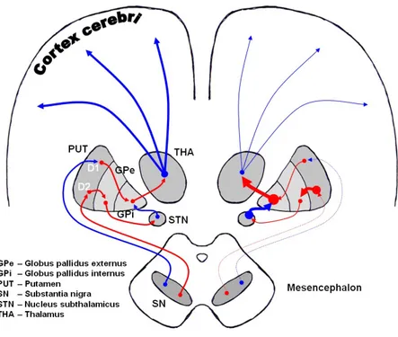
The extrapyramidal pathway shown in Figure 1.3, imply the strict collaboration between the nuclei of basal ganglia and the cortex. Two different pathways called direct and indirect have been described. Both pathways reach the thalamus and go back to the cortex.
In the direct pathway (fig. 1.3), neurons from the cortex projects to the striatum. Here they remove inhibition of internal globus pallidus (GPi) and SNr on the thalamus, promoting the
feedback signals of thalamus to the cortex. In the indirect pathway (fig. 1.3) neurons from the striatum project to the thalamus via the external globus pallidus (GPe) the subthalamic nucleus
and the globus pallidus again.
Figure 1.3 Extrapyramidal system neural networks in normal condition (left) and Parkinsons Disease (right). It is shown the modulatory action of substantia nigra pars compacta (SNc) dopaminergic neurons on the nuclei caudate-putamen (striatum in rodents). Excitatory pathways are in blu; inhibitory pathways are in red. SNr, Substantia Nigra pars reticulata; GPe, external Globus pallidus; GPi, internal Globus pallidus; STN, Subthalamic nucleus; D1 and D2 are the different types of DA receptor in the striatum (image from Dorland's Illustrated Medical Dictionary).
The NSp has a critic role in modulating this circuit so to determine, basing on experience, which are the most suitable adaptive behaviors. The loss of SN DA neurons causes Parkinson Disease (PD; Thomas et al., 2007), commonly a disorder with a late-onset. The degeneration of the NSp is responsible for an over-activation of GABAergic inhibitory neurons in the internal globus pallidus, with a subsequent inactivation of thalamic glutamatergic excitatory neurons direct to the cortex. This causes all the typical motor symptoms of PD, such as progressive loss of muscle control, tremor, loss of facial expressivity, rigidity and difficulties in completing simple tasks. It
was first described in 1817 by the famous neurologist James Parkinson in his “Essay on the shaking palsy” (Jenner et al., 1992).
3) Ventral Tegmental Area (VTA, A10), gives rise to a Meso-corticolimbic pathway (MCLp; 1.1), that is implicated in superior cognitive abilities such as reward, attention, and emotions, essential for social behaviors (sex, sociality, and aggression). MCLp neurons project to the nucleus accumbens, olfactory tubercle, cortical areas (prefrontal, cingulate and perirhinal cortex) as well as septum, amygdala, and hippocampus. The alteration of MCLp is the main cause of mood disorders (Zacharko et al., 1991, Martin-Soelch 2009), schizophrenia (Laviolette 2007), attention deficit hyperactivity disorder (ADHD; Ohno 2003), drug addiction and hallucinations (Morales et al., 2012).
Despite SN and MCL pathways are independent their connections overlap each other to promote higher cognitive processes. Indeed different studies have shown that the dysfunction of one can alter the function of the other (Jellinger 1991, Péron et al., 2012).
Moreover, DA neurons are also involved in the working memory formation by connecting basal ganglia with the prefrontal cortex. This specific kind of memory is well described in humans and is necessary to keep “active” in mind the acquired information for several seconds, to allow processes like reasoning, comprehension, problems resolution, planning and other complex cognitive functions (Lieberman 2009).
1.2 Dopamine metabolism
Dopamine (DA, a contraction of 3,4-dihydroxyphenethylamine) is neurotransmitter of the catecholamine family. Until of the 1950s, DA was considered only the intermediate for the synthesis of noradrenaline and adrenaline, but the studies conducted by of Von Euler and Lishajko first, and subsequently Bertler and Rosengren, demonstrated that DA is active by itself too. About ten years later, was discovered that the degeneration of DA neurons, in striatum, is a main cause of PD (Carlsson 1959, Ehringer and Hornykiewicz 1960) and the unique treatment for the symptoms of this disorder is use of DA precursor L-3,4-dihydroxyphenylalanine (L-DOPA; Carlsson 1959, Ehringer et al., 1960). Then Carlsson and his collaborators identified in the ventral midbrain (Mb) the origins of the DA found in the striatum and also in the limbic
In the soma and in the presynaptic terminal of dopaminergic neurons, tyrosine is transformed into L-DOPA (fig. 1.4) by the action of tyrosine hydroxylase (TH), the limiting enzyme in the biosynthesis of catecholamines (dopamine, noradrenaline, and adrenaline). TH activity is modulated by its phosphorylation via the protein kinase cyclic adenosine monophosphate (cAMP)-dependent (PKA).
Then, L-DOPA is subsequently transformed into DA by the action of the Aromatic L-amino acid decarboxylase enzyme (AADC). In turn, DA is transferred in vesicles by the vesicular monoamine transporter 2 (VMAT-2), a protein consisting of 12 transmembrane domain. VMAT2 is coupled to the vesicular H+-ATPases (V-ATPases), which functions as ATP-driven proton pump keeping the internal milieu in synaptic vesicles acidic since DA oxidizes rapidly.
Figure 1.4 Schematic representation of dopamine metabolism and dopaminergic synapse. DA, dopamine; TH, tyrosine hydroxilase; L-DOPA, L–3,4–dihydroxyphenylalanine; AADC, Aromatic L-amino
acid decarboxylase; VMAT2, vesicular monoamine transporter; MAO, monoamine oxidase; COMT, catechol-O-methyltransferase; DAT, dopamine active transporter; D2-like dopaminergic inhibitory autoreceptors
(DRD2); D1-like dopaminergic postsynaptic excitatory receptors (DRD1); AC, adenyl cyclase; PNMT,
Phenylethanolamine N-methyltransferase; DβH, Dopamine beta-hydroxylase (adapted from Sharples et al., 2014).
After exocytosis of the DA vesicles, DA binds to DA receptors on the postsynaptic membrane (D1-5), leading to the transduction of the signal in the postsynaptic neuron. There are two types of DA receptors (G-protein-coupled), constituted by 7 transmembrane domain receptors: DRD1 (D1 and D5) and the DRD2 (D2 - D4), that act in different and opposite manner. In detail: both types of DRDs modulate the cAMP/PKA transduction cascade and the intracellular Ca2+ levels, however, the DRD1 type promotes an increase in cAMP, conversely, DRD2 determines cAMP decrease (fig. 1.4). These receptors have selective agonists (Vallone et al., 1999) and specific anatomical and cellular distribution that can be pre-synaptic or post-synaptic. The D2 receptors are also localized on the DA neurons membranes (autoreceptors), regulating DA release as feedback inhibition of DA transmission (Bello et al., 2011).
The DA released into the synaptic cleft in part is recaptured by the dopamine transporter (DAT or SLC6A3) a protein of 12 transmembrane domain Na+/Cl- dependent transporter that is target
of several drugs (cocaine, amphetamine, etc.) and mice knock-out show hyperactivity and insensibility to treatment by these drugs (Amara et al., 1993).
In the alternative, DA is catabolized by cathechol-O-methyltransferase (COMT) in homovanillic acid (HVA) or degraded to 3,4-dihydroxyphenylacetic acid (DOPAC) bys the action of the extracellular or mitochondrial monoamine oxidases (MAOs; fig. 1.4).
1.3 Development of midbrain DA neurons
Dopaminergic neurons in the brain are generated through the action of many transcription factors and endogenous molecules involved in their development in accurate spatiotemporal sequence. Intrinsic and extrinsic stimuli, such as environmental or soluble factors, electric activity, and cell-cell interactions coordinate specific developmental programs.
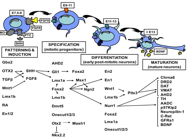
The complete generation of mDA neurons (fig. 1.5; Abeliovich et al., 2007; Perrone-Capano et al., 2008) is divided into four different phases: early Mb patterning, induction and specification of dopaminergic precursors, differentiation of post-mitotic mDA neurons, functional maturation of mDA neurons.
Figure 1.5 Schematic model of mDA neurons development. Diagram showing the main transcription or inductive factors involved in dopaminergic neurons development, divided into phases of the developmental stage in which they are involved (adapted from Perrone-Capano et al., 2008).
1.3.1 Early midbrain patterning: regionalization
During the neural tube development, many inductive factors are released from specific zones, named "organizers" to give rise to different early brain structures: telencephalon, diencephalon, mesencephalon, metencephalon, and myelencephalon. The midbrain generation is guided by signalling of the floor plate (FP) and the mid-hindbrain boundary (MHB or IsO, isthmic organizer). In this process are involved several transcription factors (FTs) such as: Otx2 and Gbx2, which are responsible for MHB formation and their expression is mutually exclusive (Hidalgo- Sánchez et al., 1999). In particular, Gbx2 is expressed more caudally and it is essential for the correct development of hindbrain and cerebellum. Conversely, Otx2 is crucial in the specification and regionalization of telencephalon and mesencephalon (Simeone et al., 2002). Otx2 is also involved in the regulation of some proneural genes like Ascl1 (or Mash1) and Neurogenin2 (Ngn2), implicated into proliferation phases of the mesencephalic progenitors (Vernay et al., 2005). Gbx2 OTX2 TGFβ Wnt1 Lmx1b RA En1/2 SHH FGF8 AHD2 Gli1 Lmx1a Foxa2 Lmx1b Dmrt5 Onecut1/2/3 Otx2 Nkx2.2 Foxa2 Msx1 Ngn2 Mash1 En2 En1 Wnt1 Lmx1b Nurr1 Pitx3 Chrna6 DRD2 DAT VMAT AHD2 TH AADC p57Kip2 Neuropilin-1 C-Ret GFRa1 BDNF PATTERNING & INDUCTION SPECIFICATION (mitotic progenitors) DIFFERENTIATION
(early post-mitotic neurons) MATURATION (mature neurons) FGF8 PTC SHH FGFR SMO E7.5-9 > E13 E9-11 E11-13 BDNF Foxa2 Lmx1a Onecut1/2/3
After MHB formation (E8 in mice) other factors are secreted, indeed it promotes the expression of engrailed transcription factors (En1/2), releases the fibroblast growth factor 8 (FGF8) and guides the correct regionalization along the anteroposterior axis of the developing CNS (1.6). Gene expression analysis and in vitro studies shown that different factors are implicated to correct positioning of the MHB such as the transforming growth factor β (TGFβ; Farkas et al., 2003), the LIM-homeodomain factor Lmx1b (Smidt et al., 2000) and the morphogenetic factor Wnt1 (Schulte et al., 2005).
In detail, Wnt1 is a member of Wnt protein family (19 members), a class of secreted glycoproteins, associated with the transmembrane G-protein-coupled receptors frizzled (Fz) determining the activation of the cytoplasmic protein Dishevelled (Dsh) that regulates transcription of Wnt target genes through its intracellular transducer β-catenin. Numerous members of the Wnt/β-catenin pathway seem to be involved in specification, proliferation, and neurogenesis in the ventral Mb (Prakash et al., 2006). In fact, null mutations for frizzled3 (Fz3) and frizzled6 (Fz6) result in a reduction of mDA neurons (Sousa et al., 2010, Stuebner et al., 2010). Importantly, the activation of Wnt pathway is stronger in the hindbrain.
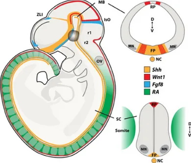
Figure 1.6 Morphogen signaling during neural tube development. Schematic representation of sagittal (left side) and coronal (right side) views of the midbrain (top) and spinal cord (bottom) with the expression pattern for the morphogenes SHH, Wnt1, FGF8 and Retinoic Acid (RA) at E9.5. FP, floor plate; IsO, isthmic organizer; MB, midbrain; NC, notochord; OV, otic vesicle; RP, roof plate; MN, Spinal motor neurons; SC, spinal cord; ZLI, zona limitans intermedia; r1, rhombomer 1; r2, rhombomer 2; D, dorsal; V, ventral. Image from Allodi et al., 2014.
Moreover, Lmx1b is expressed in the MHB at the early stage (E7.5) of mouse development (Adams et al., 2000, Guo et al., 2007), but it is not detectable until E10.5 in mDA precursor (Smidt et al., 2000). Studies conducted on mutant mice for Lmx1b reveal defects in MHB structure, therefore mDA neurons deficiency and altered genes regulation, such as: Fgf8, Engrailed 1 and 2 (En1/2), Pax2, Gbx2 and Wnt1. (Guo et al., 2007). Indeed specific inactivation of Lmx1b in mDA progenitors and not in the MHB does not alter the differentiation of these neurons (Yan et al., 2011). Surprisingly, Lmx1b expression disappears around E11.5 but reappears again at E16 in postmitotic mDA progenitors to be maintained until the adulthood in co-expression with Pitx3 and TH (Dai et al., 2008). Although the loss of Lmx1b leads to a reduction of mDA neurons (Smidt et al., 2000), in Lmx1b null mice neural precursors express Nurr1 (Nuclear Receptor-Related 1 protein or Nr4a2) and TH+ dopaminergic markers but fail to
express Pitx3. These TH+ neurons lacking Pitx3 expression are lost around birth suggesting a role
for Lmx1b in Pitx3 regulation and in mDA neurons survival. Interestingly, a similar phenotype is observed for the Wnt1 null mutant. In this case, the few TH+ neurons generated lack Pitx3
expression and are lost by E12.5 (Prakash et al., 2006).
The similar effects caused by the loss of these two factors suggest the existence of a regulatory loop between Wnt1 and Lmx1b (Adams et al., 2000, Guo et al., 2007), in addition Lmx1b and Lmx1a cooperation contributes to Wnt1 regulation in MHB and mDA progenitors proliferation (Panhuysen et al., 2004).
1.3.2 Induction and specification of DA precursors
While MHB is essential to determinate the anteroposterior axis, at the same time the notochord and then the floor plate (FP) determinate the dorsoventral axis by releasing the morphogen sonic hedgehog (SHH; before E9.5 in mice). SHH binds patched 1 receptor (Ptc1) preventing its inhibition on Smoothened protein (Smo) and triggering the activation of the Gli family transcription factors (Gli-1/3; Stone et al., 1996, Taipale et al., 2002). FGF8 and SHH are necessary for induction and proliferation of mDA precursors, establishing the proper inductive signals for the correct differentiation (Hynes et al., 1999). The ectopic expression of SHH and FGF8 generate mDA neurons and for that reason, they are usually used for the in vitro strategies of mDA differentiation (Lee et al., 2000).
Lmx1a, as well as Lmx1b, is a LIM homeobox conserved transcription factor (TF). LIM TFs have two zinc finger motifs specialized in the interaction with cofactors in order to form transcriptional-regulator complexes (Doucet-Beaupré et al., 2015). Lmx1a is expressed early in mice brain (E8.5) in the dorsal midline (roof plate) of the neural tube and then in progenitor zone of ventral midbrain and in optic vesicles (Failli et al., 2002, Millen et al., 2004, Andersson et al., 2006). Loss of Lmx1a (Dreher mice) completely abolishes roof plate induction in the spinal cord (Millonig et al., 2000), the specification of dopaminergic neurons (Andersson et al., 2006) and retina formation in Drosophila (Wang et al., 2016). Thank Wnt1/SHH and FoxA1/FoxA2 pathways are possible to express Lmx1a, the former is regulated by Lmx1a generating an autoregulatory loop (Chung et al., 2012). Lmx1a is co-expressed with Lmx1b also during the post-mitotic maturation (Zou et al., 2009). The loss of one can be compensated from the other one as confirmed by single KO mice (Ono et al., 2007). Both can ectopically generate mDA neurons (Nakatani et al., 2010) and their structures are homologs at 80%. In a recent work, it has been hypothesized that during the specification stage, Lmx1b is required and influences the differentiation of several neuronal subtypes in the Mb, including ocular motor and red nucleus neurons, while Lmx1a functions seem to be more restricted to the mDA fate (Deng et al., 2011). FoxA1 and FoxA2 are two additional TFs involved in embryonic development and in tissue specification of mDA by regulating Ngn2 and later Nurr1 and TH (Ferri et al., 2007, Lin et al., 2009).
Dmrt5 (doublesex and mab-3-related transcription factor 5) is a zinc-finger TF, identified from a differential expression screening within the ventral Mb cell populations, for this reason, it is considered crucial in mDA fate specification (Gennet et al., 2011).
1.3.3 Post-mitotic maturation of mDA neurons
In mice, during the post-mitotic stage (E9.5 - E13.5), immature neurons exit of the cell cycle and migrate radially from the ventricular surface (Bayer et al., 1995, Abeliovich et al., 2007). As previously observed, starting from E9.5 radially migrating cells express TH. The first TH+ cells
and fibers appear close to the ventricular ependymal layer (di Porzio et al., 1990). Many TFs are involved in post-mitotic mDA, such as: En1/2, Nurr1, Pitx3, and Lmx1a, but none of these transcription factors are sufficient to induce the mDA phenotype in Embryonic Stem Cells (ESc)
in vitro, suggesting that a more intricate regulatory network is required for proper DA
development (Abeliovich et al., 2007).
Nurr1 (Nr4a2) is a member of orphan receptor subfamily of the steroid nuclear hormone receptors, with Nur77 (Nr4a1) and Nor-1 (Nr4a3) (Zhao et al., 2010). It is considered an orphan nuclear receptor since its ligands have not been identified yet (Mangelsdorf et al., 1995). Nurr1 is largely expressed in the SN, VTA, limbic system and olfactory bulbs (Zetterström et al., 1997). In mice, its starts being expressed at E10.5, before TH (Volpicelli et al., 2004, Jankovic et al., 2005). Mice Nurr1-/- born with a normal frequency but they die within two days; they are
deprived of TH+ cells in the Mb (SN and VTA) and are characterized by loss of striatal
innervations and DA markers (Zetterström et al., 1997). Nurr1 overexpression is typically used to induce different cell type toward DA phenotype (Wagner et al., 1999, Chung et al., 2002). Indeed, it is involved in the regulation of several DA markers, such as: TH (Zhou et al., 1995), Dat (Giros et al., 1996), Vmat2 (Colebrooke et al., 2006), p57Kip2 (Joseph et al., 2003) and c-Ret (Jain et al., 2006, Kramer et al., 2007), Bdnf (Volpicelli et al., 2007). Furthermore, genetic studies prove that mutations in Nurr1 are a cause of the rare familial form of PD (Le et al., 2003, Sleiman et al., 2009, Decressac et al., 2013).
Pitx3 (Pituitary homeobox 3) is expressed in several tissues during embryonic development, but after the birth, it is detectable only in mDA (Smidt et al., 1997). Aphakia mutant mice (ak), harboring a spontaneous mutation in the gene coding for Pitx3, shown motor impairment due to specific loss of mDA neurons in SN while VTA appears unaffected (Smidt et al., 2007). Pitx3 expression in SN precedes that of TH while in VTA it occurs simultaneously (Maxwell et al., 2005). These data strongly suggest that specific differentiation programs must take place in the two DA subpopulations, thus explaining their different susceptibility to the loss of Pitx3.
A recent study demonstrated that Nurr1, Pitx3, and Lmx1a are key of mDA generation (Hong et al., 2014). Both Lmx1a and Lmx1b persist during maturation of mDA. Lmx1a inhibits non-dopaminergic destinies (Deng et al., 2011) and regulates axonal guidance promoting Slit2 expression (Yan et al., 2011); while Lmx1b is involved in the maturation of post-mitotic cells by regulating Pitx3. Indeed, Lmx1b KO mice show the specific loss of mature mDA neurons (Simeone 2005).
TFs En1 and En2, like Lmx1b, are important during the first phase of MHB formation (Liu et al., 2001) but their expression is not detectable anymore till E11.5 when En1 and En2 start to be expressed again in ventral mDA differentiating neurons. This expression is maintained into and throughout the adulthood (Simon et al., 2001, Albéri et al., 2004). En1 and En2 are required to prevent apoptosis suggesting a role in maintenance and survival of ventral mDA (Albéri et al., 2004).
1.3.4 Functional maturation and survival of mDA neurons
The last stage of mDA neurons generation takes place in the striatum (E15.5 in mice), where various FTs and molecules are involved in neurons maturation, maintenance, and guidance. EphrinB2 and its receptor EphB1, respectively express in the striatum and mDA neurons, enrich SN DA striatal innervation (Yue et al., 1999). In vitro EphrinB2 overexpression in mesencephalic primary cultures, increases Nurr1 transcript (Calò et al., 2005). mDA circuits formation are also regulated by the semaphorins and netrin (Torre et al., 2010, Xu et al., 2010). mDA projections undergo also axonal growth inhibitions by the diffusible chemorepellents. For e.g. Slit2 and its receptors Robo, play a major role in guiding developing axons towards their correct targets by preventing them from entering or steering them away from certain regions (Lin et al., 2005, Dugan et al., 2011).
After correct formation of dopaminergic circuits, there are two moments around postnatal day 2 (P2) and around P14, when mDA neurons undergo towards naturally-occurring cell-death (Burke 2003). In this moment, mDA need to receive growth and neurotrophic factors by post-synaptic cells to survive. The most well-established target-derived neurotrophic factor for VM DA neurons is the Glial cell line-derived neurotrophic factor (GDNF; Lin et al., 1993, Beck et al., 1995, Akerud et al., 1999). Other neurotrophic factors identified for VM DA neurons include the brain-derived neurotrophic factor (BDNF; Hyman et al., 1991), and the more recently identified dopamine neurotrophic factor (CDNF; Krieglstein 2004, Lindholm et al., 2007). Both GDNF and BDNF show a protective role on mDA neurons, following a number of experimental lesions. They also promote neuronal survival and differentiation in vitro (Hyman et al., 1991, Feng et al., 1999, Consales et al., 2007).
Many neurodegenerative diseases depend on the absence of neurotrophic signals, for this reason, these molecules could be therapeutic drugs for the treatment of DA-associated neurological disorders.
1.4 In vitro generation of mDA neurons
In the last years, several studies have been dedicated to the identification of mechanisms underlying development and function of dopaminergic neurons. The final aim is being able to generate, in vitro, mDA neurons useful for both transplantation approaches and modeling DA-related pathologies.
Several studies have shown that Embryonic Stem Cell (ESC) and Neural Stem Cell (NSC) cultures can generate TH+ cells expressing mDA phenotypic markers and that the in vitro
developmental program appears to recapitulate the temporal course of normal mDA development (Kim et al., 2002, Barberi et al., 2003, Martinat et al., 2004, Sonntag et al., 2004, Andersson et al., 2006). The embryonic stem cells (ESC) are pluripotency and generate every cell type. Conversely, multipotent and pluripotent stem cell lines can generate fewer cell types but can be isolated also from adult tissues and for these reasons, they are useful for the generation of cellular models from affected patients.
A useful alternative for modeling DA neurons in vivo are Epiblast Stem Cells (epiSCs), pluripotent cells isolated from mouse post-implantation epiblasts (around E9, E10). An interesting feature of epiSCs is that they show patterns of gene expression and signaling responses more similar to human ESC (hESC) then to mouse ESC (mESC) (Chenoweth et al., 2010). Previous works showed mESC and hESC are distinct in their epigenetic state and in the signalling guiding their differentiation. Moreover, mESC and hESC use also different signalling pathways to maintain their pluripotent status.
EpiSCs maintain OCT4 and SOX2 expression, but they downregulate expression of most of the other pluripotency factors, including NANOG, ESRRβ, KLF2 and KLF4 (Hackett et al., 2014). Moreover, EpiSCs have not undergone differentiation, but they upregulate lineage commitment factors such as homeobox protein OTX2, Brachyury and zinc-finger protein ZIC2 (Buecker et al., 2014).
In classical growth condition, epiSCs are able to proliferate and self-renew in presence of Activin and bFGF, to form teratomas following in vivo injection (Guo et al., 2009).
The main advantages in using epiSC rather than ESC are that epiSC are more homogenous in the undifferentiated state and they seem to be primed to differentiate, as demonstrated they are a more rapid and efficient tool for the obtainment of DA neurons in vitro (Jaeger et al., 2011). As described following in Materials and Methods, it is possible to differentiate epiSCs toward the neuronal phenotype, removing Activin and bFGF. With this differentiation protocol, both neuroectodermal and neuronal markers appear earlier when compared to ESC. Interestingly, by a short treatment with the FGF/ERK pathway inhibitor PD0325901 (PD03), we could induce an earlier expression of Lmx1a and Foxa2, two of the main TFs implied in the early phases of mDA development. Addition of SHH and FGF8 is necessary to address differentiation toward the DA phenotype. Using this protocol, nearly 40% of TH+ neurons generated from epiSCs co-express
Pitx3 while in the case of ESC derived neuronal populations, Pitx3+ neurons were rarely observed
(Jaeger et al., 2011).
Starting from epiSCs differentiated toward DA phenotype, the goal of this thesis was to discover microRNAs (miRNAs) expressed in mDA neuron and potentially involved in their differentiation.
1.5 MicroRNAs
MicroRNAs (miRNAs) are a class of small non-coding single-strand RNA (~22 nucleotides) evolutionary conserved and encoded within the genomes of almost all eukaryotes (Fabian et al., 2012). Organisms express hundreds of miRNAs involved in the regulation of many biological processes, such as embryonic development, cell differentiation and growth, cell proliferation, apoptosis, and regulation of metabolic processes (He et al., 2004). For this reason, altered miRNAs levels might be involved in the onset of pathological conditions (Im et al., 2012, Junn et al., 2012). In mammals, miRNAs act as the post-transcriptional regulators of gene expression, by targeting partially complementary sequences in the 3' untranslated regions (UTRs) of the target messenger RNA (mRNAs) that are in turn directed to degradation or translational repression (Bartel et al., 2004). About 60% of all human transcripts contain known or predicted miRNA target recognition sites (Friedman et al., 2009). A single type of miRNA may have up to
thousands of targets and each mRNA can be targeted by several microRNAs, suggesting a really strong role for miRNAs in the complex landscape of gene expression but at the same time giving rise to a huge complexity in its understanding and characterization. Additionally, many miRNAs have multiple paralogs throughout the genome (McCreight et al., 2017).
The first miRNA called lin-4, was discovered in 1993 in Caenorhabditis elegans (C. elegans), as a regulator of lin-14 protein expression (Lee et al., 1993). Later another miRNA, let-7, was identified in C. elegans (Reinhart et al., 2000) and surprisingly was found to be extremely conserved among a wide range of species, human included (Pasquinelli et al., 2000). One year later were identified other tens of them (Lagos-Quintana et al., 2001, Lau et al., 2001, Lee et al., 2001). At that point was clear that a new class of gene expression regulators had been identified and so they were called microRNA, but the widespread effects of miRNAs were not fully recognized until the early 2000s (Berezikov 2011).
1.5.1 miRNAs biogenesis
MicroRNAs genes, transcribed from the genome result in a primary miRNA transcript that may include a single miRNA or a cluster of miRNAs (Berezikov 2011). Several miRNA coding genes are located in regions of the genome relatively distant from previously annotated protein coding genes. They are transcribed as independent units with their own promoter (Bartel et al., 2004). However, almost 40% of miRNA genes have been found to be in introns of protein coding or non-coding RNA and in this case they are generally found in a “sense orientation” leading to think they are regulated together with their host transcript (Rodriguez et al., 2004, Kim et al., 2007). More rarely miRNAs can be found in exons of protein coding genes (Rodriguez et al., 2004). miRNAs can be transcribed as monocistronic transcripts or polycistronic transcripts (miRNA cluster) originating by local duplication of an existing miRNA locus (Altuvia et al., 2005). At the same time is not rare to find miRNA families with paralogues located in different genomic loci in monocistronic units or even clusters containing a wide variety of miRNA families (Olena et al., 2010).
Most miRNAs have a protein coding gene-like promoter and are usually transcribed by RNA polymerase II (Lee et al., 2004, Zhou et al., 2007). The product of transcription is a long RNA,
called primary-miRNA (primiRNA), with one or more stem and loop structures, whose length depends on the miRNA gene type, while for the intronic miRNA it is the protein coding mRNA itself and the pri-miRNA origins from the splicing process (Lee et al., 2002, Cai et al., 2004). Pri-miRNAs are generally 5’capped and 3’ poly-adenylated (Cai et al., 2004).
In figure 1.7 is shown the canonical miRNA processing pathway, the pri-miRNA is bound by the Di George syndrome critical region 8 (DGCR8; Pasha in invertebrates). Regions of a primary miRNA form hairpin structures that are recognized by the ribonuclease type III (RNAaseIII) enzyme Drosha (Gregory et al., 2006), which cleaves the doubles tranded stem region of the hairpin to produce an approximately 83 nucleotide (nt) precursor miRNA (pre-miRNA) (Fang e al. 2013). The resulting pre-miRNA, because of the typical RNAaseIII cut, will have two nucleotide overhang at its 3’end with a 3' hydroxyl and a 5' phosphate terminal groups (Basyuk et al., 2003, Lee et al., 2003). After that, Pre-miRNA is exported into the cytoplasm by the nucleocytoplasmatic shuttle protein Exportin-5 (fig. 1.7). This protein is able to recognize and bind the 2nt 3’overhang of the precursor and to transport it across the nuclear membrane, in a RanGTP dependent manner (Bohnsack et al., 2004). After being exported to the cytoplasm, pre-miRNA is further processed by a second endonuclease, Dicer, that acts in a complex with the Transactivating response RNA binding protein (TRBP) and other cofactors, to remove the terminal loop of the pre-miRNA (Grishok et al., 2001, Hutvágner et al., 2001). The result of this enzymatic cleavage is an approximately 22 nt double-stranded RNA duplex that contains the mature miRNA and its complement, called the star strand (*) (Hutvagner et al., 2001). The duplex is then separated, the strand with the less thermodynamically stable 5' end becomes the mature sequence, while the star sequence is degraded. The mature miRNA is loaded together with Argonaute (Ago 2) proteins onto RNA-induced silencing complex (RISC) (Bartel et al., 2004; Winter et al., 2009). The Ago protein contains two RNA-binding domains: the PAZ domain binds the 3’end of the mature miRNA while the PIWI domain interacts with the 5’phosphate group on the 5’end (Pratt et al., 2009). Once the miRISC is assembled, the miRNA drives it to silence target mRNA via mRNA cleavage, translational repression or deadenylation (Nilsen 2007, Winter et al., 2009).
1.5.2 miRNAs: Mechanism of action and bioinformatics identification
miRNAs may have a negative or a positive regulatory effect (Ambros 2001). In animals, they usually bind with partial complementarity to 3’UTR regulatory elements on mRNAs called ‘seed sequences’, or to miRNA response elements (MREs) that causes translational repression (Ambros 2004). A major silencing mechanism of miRNAs in animals results in target mRNA destabilization through a cleavage-independent process, affecting transcript level (Lim et al., 2005; Pillai et al., 2004). A small number of miRNAs also show decoy activity by binding directly to proteins such as RNA-binding proteins, inhibiting interaction with their target RNAs (Eiring et al., 2010). In some cases, miRNAs also regulate gene expression at the transcriptional level (Kim et al., 2008) by binding directly to DNA regulatory elements. In certain cases and cell types, they can enhance translation (Vasudevan et al., 2007).
Figure 1.7 miRNAs biogenesis and action. miRNA processing pathway. The main steps for miRNA maturation are shown: Transcription of the pri-miRNA by RNApolII or III; cleavage of the pri-miRNA by the microprocessor complex Drosha–DGCR8 in the nucleus; export to the cytoplasm by Exportin-5– Ran-GTP; cleavage of the pre-miRNA by Dicer in complex with TRBP; miRNA duplex functional strand recruitment, together with Ago2, into the RISC complex, where it guides RISC to silence target mRNA. (Image from Winter et al., 2009).
However, there are varying degrees of complementarity between a miRNA and its mRNA target, binding is most highly dependent on positions 1 through 8 of the 5' end of the mature miRNA, known as the seed region (Berezikov 2011). While 75% of down-regulated mRNA have canonical seed sites in their 3' UTR, the seed region is not always sufficient for causing down-regulation (Kim et al., 2008). The 3' end of the mature miRNA can also have an effect: positions 13±16 are highly conserved, and their proper complementary base pairing to a mRNA target is associated with down-regulation (Grimson et al., 2007).
A number of bioinformatic tools and databases have been devised to manage the growing body of data and at present, there are 129 miRNA tools currently used in miRNA research (Akhtar et al., 2015).
A well-known database for miRNAs is miRBase (http://www.mirbase.org/). It has emerged as a definitive repository of miRNA sequences as well as an authoritative source of miRNA nomenclature that is a valuable resource for miRNA profiling studies (Pritchard et al., 2012). Indeed miRBase was established with the aim to annotate the continue increasing number of miRNA being discovered and to provide a trustworthy resource for any researcher interested in looking after a miRNA sequence, miRNA cluster, family composition or miRNA genome localization (Kozomara et al., 2011). This resource provided for each miRNA a unique name in agreement with the scientific community (Ambros et al., 2003). miRNAs name consist of “miR-” preceded by a three-letter-code, identifying the species (the first one corresponding to the genus and the following two to the species. e.g. mmu for Mus Musculus) and followed by the numeric identifier for the specific miRNA. Variants of the same miRNA, present in multiple copies into the genome, can often be followed by an extra letter the distinguish them (e.g mmu-miR-148a) while paralogues miRNAs (transcribed by different promoters; often, in this case, the mature miRNA sequence is identical) have a dash followed by the paralogue number (e.g. mmu-mir-218-1). The name of the mature form of the miRNA is followed by the suffix -3p or -5p depending on the strand of the pre-miRNA from which the mature one comes from (e.g. mmu-218-1-5p).
To predict how miRNA-mRNA interaction occurs, several prediction tools are currently used. The most used are TargetScan (www.targetscan.org/) (Grimson et al., 2007, Friedman et al., 2009) and miRanda (http://www.microRNA.org/) (Enright et al., 2003, John et al., 2004). The majority of algorithms at the base of these software packages use a similar set of parameters to
identify and classify each candidate target site. These usually analyze seed region complementarity between the miRNA and the target site, free energy of the RNA duplex formed, with some of the methods also taking into account regions surrounding the target site. To increase the prediction reliability, a “conservation” filter can be applied with the assumption that the biological relevant loci are more likely to be conserved during the evolution. It is important to underline that experimental procedure is necessary to support predictions and discover really the interaction between miRNA-mRNA targets.
1.5.3 miRNAs & diseases
Approximately 2200 miRNA genes have been reported to exist in the mammalian genome, the abnormal expression of miRNAs has been proven to be extensively involved in the pathogenesis of numerous types of diseases, such as cardiovascular, inflammatory, autoimmune, neurodevelopmental, skeletal and skin, liver diseases and cancers (Ardekani et al., 2010). miRNAs such as miR-9, miR-124a/b, miR-135, miR-153, miR-183, and miR-219, among others, have also been shown to play critical roles in the development and function of the brain where they have been found specifically expressed in differentiating neurons (Sempere et al., 2004). They are crucial players in several aspects of brain development such as neurogenesis, neuronal maturation, synapses formation, axon guidance and neuronal plasticity (Kapsimali et al., 2007; McNeill et al., 2012). The brain enriched of miR-137 has been shown is an essential regulator in controlling the dynamics between neural stem cell proliferation and differentiation during neural development (Sun et al., 2011). Furthermore, cellular and animal models show that miR-218 is essential in motoneurons development and disease (Thiebes et al., 2015).
In particular, there are many studies that underline how an alteration of a specific miRNA is cause of DA neuron deprivation and Parkinson’s disease (Kim et al., 2007; Miñones-Moyano et al., 2011; Saba et al., 2012; Tobon et al., 2012; Yang et al., 2012). Regarding more specifically the dopaminergic neurons, nowadays very few miRNAs have been identified as involved in their development or function. miR-133b is enriched in human Mb, where it is thought to regulate the maturation and function of mDA through a negative feedback circuit involving the transcription factor Pitx3 (Kim et al., 2007). miR-132 is expressed in ESC-derived TH+ cells and regulates
role of these molecules in neurodegeneration (Kim et al., 2007, Hébert et al., 2009, Doxakis et al., 2010). miR-142-3p has been shown to suppress D1 type DA receptor expression both during development and in cell culture (Tobón et al., 2012). miR-181a is induced by dopamine signalling in primary neurons, as well as by cocaine and amphetamines, in a mouse model of chronic drug treatment (Saba et al., 2012). Recent works underline the importance of miR-135a2 in determining midbrain size and the allocation of prospective mDA precursors by modulating the extent of the Wnt signaling (Joksimovic et al., 2014; Nouri et al., 2015). 34b and miR-34c have been found to be significantly decreased in PD patients (Miñones-Moyano et al., 2011). miR-218 has been found to be expressed in the ventral midbrain of E12.5 mouse embryos, and specifically lost in the Wnt1-Cre conditional knock out (Huang et al., 2010).
The increasing knowledge about the role played by miRNAs in the dopaminergic system will provide important insights into molecular mechanisms involved its alterations and could eventually generate novel targets for therapeutic care.
1.6 Aim of PhD thesis work
MicroRNAs, are key regulators of gene expression, can influence many biological processes and are biomarkers for diseases. For these reasons, miRNAs expression profiling is gaining increasing popularity (Pritchard et al., 2015).
The aim of this thesis is to individuate miRNAs expressed in dopaminergic neurons and potentially involved in their development and function. Several studies suggest that miRNAs are involved in Dopaminergic neurons development, function and disease, despite details regarding their mechanism of action are still missing (Kim et al., 2007; Miñones-Moyano et al., 2011; Saba et al., 2012; Tobon et al., 2012; Yang et al., 2012).
To identify which miRNAs are expressed in mDA neurons, I analyzed, through a bioinformatics approach, microarray data, obtained from EpiSCs differentiation to DA phenotype, available in my host Lab. Each miRNA were evaluated for their capacity to induce DA phenotype. This approach allowed the identification few miRNAs able to promote mDA neurons differentiation and which are selectively expressed in the midbrain. The most interesting candidates I identified are miR34b/c and miR-218. Interestingly miR-218 was known being expressed in motoneurons where it is essential to generate correct establishment of the neuromuscular junction (Amin et al., 2015; Thiebes et al., 2015). However, its role in dopaminergic neurons is still not clear. To further understand its role in dopaminergic neurons I have generated conditional KO mice (cKO) for miR-218. By mating these mice with mice expressing the Cre Recombinase under the control of specific tissue promoter, we are currently able to evaluate the contribution of miR-218 to the development of specific neural subpopulations. Our preliminary data show that miR-218 is important for proper motor function and suggest its potential role in DA neurons.
Because is still unclear the non-coding RNAs role in the DA neurons, any additional information will be important to clarify and establish their function in DA system development, a phenomenon remained elusive till now.
In addition, this new knowledge appears to have a major role in the practice of personalized medicine or the treatment of DA system-linked diseases in the near future.
2 - MATERIALS AND METHODS
2.1 Cell Cultures
Materials for tissue culture, like multiwell plates, serological pipettes or pipette tips, were purchased from Corning, BD Biosciences or Nunc. All steps were carried out inside a laminar flow sterile hood (Jupiter) to avoid contamination. All the equipment was sprayed with Ethanol (70%) before use. Cells were incubated at 37°C with 5% CO2 in a humidified incubator
(Thermo Forma). Dissections were performed with horizontal flow hood (Hermes II). All the factors and reagents mentioned in the below described methods are listed in Table 2.1.
2.1.1 Animals and dissections
Timed pregnant wild-type C57BL/6 (Charles-River) or C57BL-6-Tg.pTH-GFP (Dr. Hideyuki Okano) mice were sacrificed in accordance with Society for Neuroscience guidelines and Italian law. The embryonic age (E) was determined by considering the day of insemination (as confirmed by vaginal plug) as day E0. Embryos from day 12.5 (E12.5) or E14.5 of gestation were quickly removed and placed in phosphate buffered saline (PBS), without calcium and magnesium and supplemented with 33 mM glucose. The brain structures (midbrain, striatum, and cortex) were carefully dissected under a stereoscope in sterile conditions and processed for cell cultures. Tissues were pooled and triturated with a mechanical dissociation.
2.1.2 Mesencephalic primary cultures (mE12.5-PCs)
Single cells were obtained from embryonic midbrain as previously described (Prochiantz et al. 1979, di Porzio et al. 1980). Briefly, the tissues were transferred into a 15 mL tube and mechanically dissociated with a sterile pipette in a solution containing 0,01% pancreatic DNAse. The cell suspension was centrifuged 5' at 100 g and resuspended in plating medium and counted. For the viable count, the cell suspension was diluted 1:10 with 0,1% Trypan blue and loaded into a Burker’s counting chamber slide. Cell concentration was determined on the basis of the total cell count, the dilution factor, and the trypan blue exclusion.
Dissociated cells were plated at a density of 4 × 10⁴ cells/cm2 on multiwells previously coated with
15 µg/mL poly-D-lysine for 1 h at 37°C and washed three times with sterile H2O. Cells were
grown in NBM, supplemented with B27, 0.5 mM L-glutamine, Pen/Strep, bFGF (20 ng/mL) FGF8 (10 ng/mL) and SHH (50 ng/mL) to induce mDA phenotype. After 3 days in culture, (days in vitro, DIV) half of the medium was replaced and inducible lentiviruses were added at respective dilutions. At DIV6, the proliferative medium was replaced with a differentiating medium, NBM supplemented with B27, 100 mM L-glutamine, Pen/Strep, Ascorbic acid (200 µM), and 1 mM dibutyryl cyclic adenosine 3’, 5’-monophosphate (cAMP). From DIV6 the expression of transgenes was induced by the addition of doxycycline (4µg/mL) to the medium. At DIV12 cells were fixed or collected for further analyses.
2.1.3 Embryonic Stem Cells (ESCs)
R26CreER/+ mouse embryonic stem cells culture (Omodei et al. 2008) were performed in Glasgow
Minimal Essential Medium (GMEM, SIGMA) plus 12% FBS (Hyclone) and LIF (300U/ml Millipore). To induce DA differentiation mES were plated at low density on the gelatin-coated plate in N2B27 medium (day 0). Four days after plating cells were passaged on Poly-L-lysin (15µg/ml in PBS)/Laminin (20µg/ml in PBS). From the day next until the day 9 SHH (200 ng/ml) and FGF8 (100ng/ml) were added to the medium. Cells were cultured until day 14.
2.1.4 Epiblast Stem Cells (EpiSCs)
EpiSCs have been derived as described by Guo and colleagues (Guo et al. 2009). They are cultured in epiSC medium containing half DMEM/F12 and half Neuralbasal medium, supplemented with N2, retinol-free B27, 2mM L-Glutamine, 0.05 mM β-mercaptoethanol, 10ng/mL bFGF and 20 ng/mL Activin. Cells were split every 2-3 days as epiSC by using mechanical dissociation with a 2mL serological pipet and plated in multiwell plates, coated with FCS for 30' at 37°C. For DA differentiation epiSCs were plated one day before the bFGF and Activin withdrawal in 12-wells plate, previously coated with a solution of 15 µg/mL Fibronectin for 30' at 37°C. One day later (DIV1), cells should reach 60 to 80% of confluence and at this point is possible to switch the medium from epiSC medium to simple N2B27 medium, that is
lentiviral particles as described in single experiments. At DIV2 cells were split and diluted on a new 12-wells plate previously coated with Fibronectin. From DIV5 to DIV9 cells were maintained in N2B27 medium supplemented with SHH (100 ng/mL or SAG 0.4 µM) and FGF8 (100 ng/mL). Usually, at DIV9 N2B27 is supplemented with doxycycline (4 g/mL) and ascorbic acid (200 µM). Cells were cultured until DIV16.
Description Manufacturer Catalogue number
Fibronectin Millipore FC010 Gelatine Sigma-Aldrich G1393 Laminin Sigma-Aldrich L2020 Poly-D-lysine Sigma-Aldrich P7405 DMEM Invitrogen 11995065 DMEM/F12 Invitrogen 21331-020 F12 Invitrogen 21700-026 GMEM Sigma-Aldrich G5154 IMDM Invitrogen 31980-030 Neurobasal NBM Invitrogen 21103-049 FBS Euroclone ECS0180L FCS Biosera 1810-500 B27 Invitrogen 17504-044 B27 wo vit. A Invitrogen 12587-010 N2 Invitrogen 17502-048 βmercaptoethanol Invitrogen 31350-010 HEPES Invitrogen 15630-106
L-glutamine Euroclone ECB3000D Na-piruvate Invitrogen 11360-039 Pen/Strep Sigma-Aldrich P0781 Trypsin Sigma-Aldrich T4799
DNase Sigma-Aldrich DN25
Activin R&D 338-AC-025
bFGF Sigma-Aldrich F0291
EGF Sigma-Aldrich E9644
cAMP analog Sigma-Aldrich D0627 Doxycycline Clontech 631311
FGF8 Sigma-Aldrich F6926
L-ascorbic acid Sigma-Aldrich A4544
LIF Millipre ESG1107
SHH R&D 1845-SH-100
2.1.5 HEK293T cells
Human embryonic kidney 293T (HEK293T) cells were generated from human embryonic kidney cells obtained from a single apparently healthy foetus legally aborted. In the early 70s, these cells were genetically transformed with Adenovirus 5 DNA to obtain a stable cell line (Graham et al. 1977). Cells were maintained at 37°C with 5% CO2 in a humidified incubator, in
DMEM with 10% foetal bovine serum (FBS), Pen/Strep and 25 mM Hepes.
2.1.6 HeLa cells
HeLa is an immortalized epithelial human cell line derived from cervical cancer cells (Scherer, Syverton et al. 1953). HeLa cells were maintained at 37°C with 5% CO2 in a humidified incubator, in DMEM (Invitrogen) with 10% foetal bovine serum (FBS; Euroclone) and 100 unit/ml Streptomycin (Sigma) and 100 µg/ml Penicillin (Sigma).
2.2 Lentiviral production
Last generation lentiviruses have been constructed in order to contain less than 10% of the original viral genome. They express only the sequences needed for reverse transcription and integration of the gene expression cassette into the host genome. To further increase the security level, these genes are cloned in three different vectors to minimize the risk of the recombination event. To further minimize the possibility to generate functional auto-replicant viruses, the Psi (ψ) sequence, responsible for the viral genome packaging, is localized exclusively on the exogenous
gene-containing transfer vector. The gene expression is under a control of a doxycycline-inducible
promoter (Tet-ON). The used vectors for the production of the lentiviral particles were: a Gag/Pol containing pMDL vector, a pRev vector, a pVSV-G vector and the exogenous gene-containing transfer vector Tet-O-FUW. Moreover, a prtTA vector, expressing the reverse tetracycline transactivator (rtTA) protein, in combination with pMDL, pRev and pVSV-G was necessary to induce gene expression in the presence of the antibiotic tetracycline or one of its derivatives (e.g. doxycycline; rtTA vector was supplied by Dr. Caiazzo, IGB, Naples). For all these vectors Gigapreps were made using the EndoFree Plasmid Gigaprep (Qiagen) following the manufacturer instructions.
2.2.1 PCR for cloning reactions and vector construction
RNA extracted from an E14 embryonic mesencephalon was used for the cDNA preparation as template to perform PCR for the intended target 3’UTRs to amplify and clone into the pmiR-Report vector. While, In the case of pre-miRNA coding regions amplicons to clone in the Tet-O-Fuw vector, genomic DNA from E14 embryonic mesencephalon was used as template. Oligos were designed in a way to amplify specific gene containing specific restriction sites (Table 2.2). Oligos used to amplify several genes 3’UTRs were designed with an 11nt long 5’end tail containing the SpeI (forward oligo) and the HindIII (reverse oligo) restriction sites. Oligos used to amplify pre-miRNA coding regions were designed both with an 8 nt long 5’end tail containing the EcoRI restriction site.
For 3’UTR amplicons, DNA band extraction from the agarose gel was performed using the PureLink® Quick Gel Extraction Kit (Invitrogen) according to the provided instructions. Eluted DNA was incubated for 4h at 37°C with the specific restriction enzymes (SpeI and HindIII for 3’UTR amplicons).
For pre-miRNA amplicons instead, Amplicons were directly cloned into a pCR®2.1-TOPO® TA (Invitrogen) vector following the TOPO® TA Cloning Kit (Invitrogen) subcloning protocol. In order to isolate the insert on each gene-containing TOPO TA plasmid was restricted with a specific enzyme (4 h at 37°C). Insert band was extracted from the agarose using the PureLink® Quick Gel Extraction Kit (Invitrogen) according to the provided instructions.
For the ligation step, 50 ng of the vector were used. The amount of insert to use for the ligation reaction was calculated using following formula:
A control reaction was performed without adding the insert to the reaction. The enzyme I used was a T4 ligase (NEB) and the reaction buffer was the one provided by the company. Ligation was conducted over-night at 16°C in a final volume of 10 µl with 400 units enzyme. 5 µl of this reaction were used to transform 50 µl of competent DH5α cells (Invitrogen) following the standard transformation protocol: 30' (minutes) on ice, heat shock at 42°C for 30'' (seconds), 2' on ice, cells suspension in 250 µl SOC medium (2% tryptone, 0.5% yeast extract, 10 mM NaCl,
agitation at 37°C. Finally, the cells were seeded on Lysogeny broth (LB)-Agar plates (10 g NaCl, 10 g Bacto-tryptone, 5 g yeast extract, 20 g Bacto-agar) containing ampicillin (50 ng/ml) and growth over-night at 37°C. Single colonies were screened by PCR. Positive colonies were grown in 5 mL LB (10 g NaCl, 10 g Bacto-tryptone, 5 g yeast extract) containing ampicillin (50 ng/ml). Plasmidic DNA was isolated using the PureLink Quick Plasmid Miniprep (Invitrogen) and later sequenced. miR-target sequence was mutated with the Quickchange II XL site-direct mutagenesis kit (Agilent) accordingly with manufacture instruction and later sequenced. Oligos designed in a way to modified seed sequences of 3’UTR of Nurr1 and Wnt1 are reported in Table 2.2.
2.2.2 Transfection and lentiviral production
For the production of the viral particles (Tiscornia et al. 2006), 8.2 million HEK293T cells were plated in 150 mm × 25 mm dishes in DMEM (Invitrogen) supplied with 10% FBS (Euroclone), 25 mM Hepes (Invitrogen), 100 U/mL Streptomycin and 100 µg/mL Penicillin (Pen/Strep, Sigma-Aldrich). 24 h later an 80% confluence is generally obtained. The Medium was replaced by IMDM medium (Invitrogen), 10% FBS, Pen/Strep. 3 h later the 4 vectors were co-transfected following the calcium phosphate transfection protocol.
In details, for each dish, a solution was prepared to contain 270 mM CaCl2, 6.25 µg pRev, 9 µg
pVSVG, 14.6 µg pMDL and 32 µg of the insert containing transfer vector. After 5' of incubation at room temperature, drop-by-drop, in low agitation, a 2xHBS pH 7.12 (NaCl 280 mM, Na2HPO4 1.5 mM, HEPES 50 mM) solution was added to the vectors mix and incubated for 15'
at room temperature.
Amplified cDNA Oligo sequences with restriction sites Cloning Enzyme
3xFlagNurr1 Fw GCGCCAATTGATGGACTACAAAGACCATGA MfeI
3xFlagNurr1 Rev GCGCCAATTGTTAGAAAGGTAAGGTGTC MfeI
mmu-miR-27a-3p(687 bps) Fw CCGAATTCGTGTTCAGCTATGTGAGACC EcoRI
mmu-miR-27a-3p (687 bps) Rev CCGAATTCCCCATCTATCTGCTTTGGG EcoRI
mmu-miR-29a-3p (336 bps) Fw CCGAATTCTAAGCCTTCTCTGGAAGTGG EcoRI
mmu-miR-29a-3p (336 bps) Rev CCGAATTCTTAACCATGCTGTTGCTGG EcoRI
mmu-miR-34b/c-5p (983 bps)Rev CCGAATTCTAGCAGCTAAGGGCTAGCGG EcoRI
mmu-miR-132-3p (562 bps) Fw CCGAATTCGCTGGGACATCTTTGACG EcoRI
mmu-miR-132-3p (562 bps) Rev CCGAATTCCTCTTGCTCTGTATCTGCC EcoRI
mmu-miR-148a-3p (294 bps) Fw CCGAATTCTCTTCTTTGCCTTCACTGG EcoRI
mmu-miR-148a-3p (294 bps) Rev CCGAATTCTCAGGTTCTTCACAAAGCC EcoRI
mmu-miR-204-5p (309 bps) Fw CCGAATTCCCGGAGAATCAAGATGAGC EcoRI
mmu-miR-204-5p (309 bps) Rev CCGAATTCGTTATGGGCTCAATGATGG EcoRI
mmu-miR-210-3p (306 bps) Fw CCGAATTCAGGGGGATATGGGTATTGG EcoRI
mmu-miR-210-3p (306 bps) Rev CCGAATTCCACCCTGTCTATCTGAATCC EcoRI
mmu-miR-218-1-5p (374 bps) Fw CCGAATTCGATCATACACAATCTGCGGGAAG EcoRI
mmu-miR-218-1-5p (374 bps) Rev CCGAATTCGGACATTTGTTATTCTCCCCTC EcoRI
mmu-miR-219-1-5p (358 bps) Fw CCGAATTCCATTCACTCGTGTGCTCC EcoRI
mmu-miR-219-1-5p (358 bps) Rev CCGAATTCCCCAACTTCTCTCAAGCC EcoRI
mmu-miR-370-3p (313 bps) Fw CCGAATTCGTGGGTGTGGCTTTGAGG EcoRI
mmu-miR-370-3p (313 bps) Rev CCGAATTCCCCTTTCACAAATCTTTGCCC EcoRI
mmu-miR-375-3p (346 bps) Fw CCGAATTCCGCCACTGCCGCCGACGTG EcoRI
mmu-miR-375-3p (346 bps) Rev CCGAATTCGGCGGGGCCTGATGGGAACC EcoRI
mmu-miR-494-3p (322 bps) Fw CCGAATTCGTCTCAGGCAATTCTGTGG EcoRI
mmu-miR-494-3p (322 bps) Rev CCGAATTCATGCCATACTCCCATGTCC EcoRI
Nurr1 3'UTR Fw (1326bps) TCCAAACTAGTCCAAGCACGTCAAAGAACT SpeI
Nurr1 3'UTR Rev (1326bps) CTTAAAAGCTTATCTCTAACTGTCGTACACC HindIII
Wnt1 3'UTR Fw (933 bps) TCCAAACTAGTCGCTCTCTTCCAGTTCTC SpeI
Wnt1 3'UTR Rev (933 bps) CTTAAAAGCTTATAGATATTTTATTCCTCAGA
GGAAG HindIII
Nurr1 3’UTRΔ34 Fw TCGTACACCATAGAAAAAAAACTCATCTCATGTGCCGTAC Not used
Nurr13’UTRΔ34 Rv GTACGGCACATGAGATGAGTTTTTTTTCTATG GTGTACGA
Not used
Nurr1 3’UTRΔ204Fw GTACATTGGAAAATCCTGACACACATAGTGTT TGTAACACCG
Not used
Wnt1 3’UTRΔ34Fw GGCCAAATTGGGGAAAGGAGTCTCCCTCAAA GAG
Not used
Wnt11 3’UTRΔ34Rv CTCTTTCAGGGAGACTCCTTTCCCCAATTTGGCC Not used
Wnt1 3’UTRΔ148Fw GGCCAAATTGGGGAAAGGAGTCTCCCTCAAAGAG Not used
Wnt1 3’UTRΔ148Rv CTCTTTCAGGGAGACTCCTTTCCCCAATTTG GCC
Not used
Wnt1 3’UTRΔ27Fw GGAGCCATTGAACAGCCATGCCTCCCTCAG Not used
Wnt1 3’UTRΔ27Rv CTGAGGGAGGCATGGCTGTTCAATGGCTCC Not used
Table 2.2. Oligos for lentiviral and luciferase vectors construction.
2.3 Dual fluorescent reporter sensor
DFRS plasmids were kindly provided by Prof. Wieland B. Huttner. Cloning strategy was performed as previously described (De Pietri Tonelli et al. 2014). Annealing of synthetic oligonucleotides to prepare the sensor cassette for a given miRNA. In an appropriate PCR tube, set up a 100 µl reaction as follows: add 76.8 µl of H2O; 10 µl of 10X T4 ligase buffer (NEB); 5 µl
of forward oligo (from 100 µM stock); 5 µl of reverse oligo (from 100 µM stock); 0.5 µl of PNK (NEB, stock 10 U/µl).
Incubate the reaction in a conventional thermal cycler programmed as follows: temperature (Time— Reaction Step): 37 °C (1 h— oligo phosphorylation); 94 °C (5 min— PNK inactivation); followed by a controlled oligonucleotide annealing:
decrease by 0.1 °C/s to 90 °C; incubate at 90 °C (3 min);
decrease by 0.1 °C/s to 70 °C; incubate at 70 °C (3 min);
decrease by 0.1 °C/s to 50 °C; incubate at 50 °C (3 min);
decrease by 0.1 °C/s to 25 °C; incubate at 25 °C (3 min).
Annealing of oligos can be controlled on a conventional 4 % agarose gel for DNA: load 2–5 µl of the annealing reaction on the gel; as control load 0.5–1 µl of a corresponding single- strand (either forward or reverse) oligo from stock 100 µM, which runs faster than the double-strand
annealed DNA.
The fragment is ligated (without purification) into the pDSV3 vector cut with PacI and NdeI and dephosphorylated. To set up the ligation reaction add 5 µl of the annealed fragment in a 10 µl final ligation reaction, incubate the reaction +4 °C overnight. Later plasmids were sequenced.
Plasmids were transfected into control (basal) and differentiating mouse Embryonic Stem Cell (mES) by Lipofectamine. Fluorescences were monitored every day until 72 hours post-transfection. In Table 2.3 is listed oligos used for annealing reaction.
Oligos for annealing Oligo sequences with restriction sites
NdeI_34bs_PacI Fw TATGACAATCAGCTAATTACACTGCCTGGCGCGCCCGCAATCA
GCTAACTACACTGCCTTTAAT
PacI_34bs_ NdeI Rv TAAAGGCAGTGTAGTTAGCTGATTGCGGGCGCGCCAGGCAGT
GTAATTAGCTGATTGTCA
NdeI_34MUT_PacI Fw TATGACAATCAGCTAATTACGGCCGGTGGCGCGCCCGCAATCAGCTAACTACGGCCGGTTTAAT
PacI_34MUT_ NdeI Rv TAAACCGGCCGTAGTTAGCTGATTGCGGGCGCGCCACCGGCC
GTAATTAGCTGATTGTCA
Table 2.3 Oligos for annealing of synthetic oligonucleotides
2.4 Molecular biology methods
2.4.1 miRVana RNA extraction and TaqMan® MicroRNA Assays
Usual RNA purification strategies rely on organic extraction, followed by alcohol precipitation. These strategies, because of the alcohol precipitation, are inefficient in recovering small RNA forms. The miRVana miRNA isolation (Ambion) strategy uses organic extraction followed by purification on a glass fiber filter (GFF) under specialized binding and washed conditions. With this strategy, all RNAs are recovered, from large mRNAs to ribosomal RNAs down to 10-mers small RNAs. 1 volume of cold sterile PBS was added to samples in RNA later. Samples were centrifuged, RNA later/PBS solution was removed and the cell pellet was resuspended in the provided lysis buffer. Later, RNA was extracted following the manufacturer instructions. As for the last step, RNA was eluted in 100 µl of RNAase-free water, previously warmed at 95°C and