BRIEF COMMUNICATIONS
"One-shot" endovascular management of cerebral aneurysm and fourth
ventricle hemangioblastoma in a pregnant woman
Daniela Surico
a, Roberta Amadori
a,⁎
, Greta Garofalo
a, Nunzio P. Nuzzi
b, Alessandro Carriero
b, Nicola Surico
aa
Department of Obstetrics and Gynecology, University of Eastern Piedmont, Novara, Italy
bRadiodiagnostic and Interventional Radiology Institute, University of Eastern Piedmont, Novara, Italy
a r t i c l e i n f o
Article history: Received 16 July 2014
Received in revised form 25 September 2014 Accepted 11 November 2014 Keywords: Cerebral aneurysm Endovascular management Hemangioblastoma Pregnancy Subarachnoid hemorrhage
Aneurysmal subarachnoid hemorrhage (aSAH) during pregnancy is a serious condition[1]. The best management of aSAH during pregnancy is not yet established, although endovascular coiling has gained favor over the last few years[2]. The incidence of hemangioblastoma of the posterior fossa in pregnancy is extremely low[3].
A pregnant woman presented to the“Maggiore della Carita” Hospital in December 2009 having experienced sudden onset seizures and deteriora-tion of consciousness at 27 weeks of pregnancy. The patient’s systemic blood pressure was 100/50 mm Hg and eclampsia was therefore excluded. CT and MRI scans showed subarachnoid hemorrhage, blood in the lateral ventricles, and ventricular dilation (Fig. 1). The fourth ventricle was occupied by a small mass—with hyperintense nodule enhancement on T2 weighted imaging after gadolinium injection—rounded by a small cyst with ring enhancement.
MR angiography revealed a subarachnoid aneurysm arising from the right internal carotid artery. The patient underwent digital subtraction angiography that confirmed the presence of a subarachnoid aneurysm of the right internal carotid artery and a hypervascularized nodule with arteriovenous shunt in the posterior fossa, fed by the posterior and anterior inferior cerebellar artery—consistent with hemangioblastoma.
Following consultation with neurosurgeons, anesthesiologists, obstetricians, and neuroradiologists, the decision to immediately treat both lesions by an endovascular approach was taken. Before the medical procedure, the patient was advised about the potential risks and benefits of the intervention and she provided informed consent.
To begin, the posterior inferior cerebellar artery to the hemangio-blastoma was navigatted with aflow-directed microcatheter to reach the hypervascularized nodule. Then, 0.7 mL of a mixture of glue diluted to 20% in Lipiodol (Guerbet, Bloomington, IN, USA) was injected into the nidus, followed by complete angiographic devascularization of the lesion
(Figs. 2, 3). Immediately after, the aneurysm was coiled with three
platinum bare coils. The procedure was uneventful.
CT scan performed 24 hours after the procedure showed ventricular shrinking to a normal size (Fig. 4). The patient had good clinical status, with a Glasgow Coma Scale score of 14. At 38 weeks of pregnancy the patient was admitted for a scheduled cesarean delivery, resulting in the successful birth of a healthy neonate weighing 3220 g and with an
International Journal of Gynecology and Obstetrics 129 (2015) 79–88
⁎ Corresponding author at: Department of Obstetrics and Gynecology, “Maggiore della Carita” Hospital, Viale Mazzini 18, 28100 Novara, Italy. Tel.: +39 0321 3733665; fax: +39 0321 3733659.
E-mail address:[email protected](R. Amadori).
Fig. 1. CT scan showed subarachnoid hemorrhage, blood in the lateral ventricles, and ventricular dilation.
Contents lists available atScienceDirect
International Journal of Gynecology and Obstetrics
Apgar score of 9/9. Follow-up MRI of the patient two months after deliv-ery showed no major recanalization of the aneurysm. As expected, the
intraventricular mass was still present. Contrast-enhanced MR angiog-raphy showed revascularization of the lesion very close to the initial digital subtraction angiography picture, but no ventricular dilation was seen.
The association of these pathologies in a pregnant woman is exceptional. The endovascular treatment approach used in this case was successful and uneventful.
Conflict of interest
The authors have no conflicts of interest.
References
[1]Meyers PM, Halbach VV, Malek AM, Phatouros CC, Dowd CF, Lawton MT, et al. Endovascular treatment of cerebral artery aneurysms during pregnancy: report of three cases. AJNR Am J Neuroradiol 2000;21(7):1306–11.
[2]Tarnaris A, Haliasos N, Watkins LD. Endovascular treatment of ruptured intracranial aneurysms during pregnancy: is this the best way forward? Case report and review of the literature. Clin Neurol Neurosurg 2012;114(6):703–6.
[3]Erdogan B, Sen O, Aydin MV, Bagis T, Bavbek M. Cerebellar hemangioblastoma in pregnancy. A case report. J Reprod Med 2002;47(10):864–6.
Fig. 3. Complete angiographic devascularization of the hemangioblastoma (white arrow).
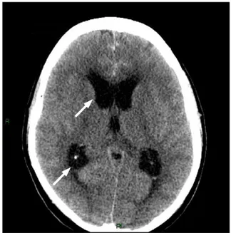
Fig. 4. CT scan performed 24 hours after the procedure showed ventricular shrinking to a normal size (white arrows).
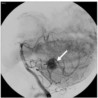
Fig. 2. Angiographic shape of the hemangioblastoma before devascularization (white arrow).
http://dx.doi.org/10.1016/j.ijgo.2014.09.023
0020-7292/© 2014 International Federation of Gynecology and Obstetrics. Published by Elsevier Ireland Ltd. All rights reserved.