Generation of
β cell-specific human cytotoxic T cells by lentiviral
transduction and their survival in immunodeficient human leucocyte
antigen-transgenic mice
J. Babad,* G. Mukherjee,* A. Follenzi,†
R. Ali,* B. O. Roep,‡L. D. Shultz,§
P. Santamaria,¶**††O. O. Yang,‡‡§§¶¶
H. Goldstein,*,*** D. L. Greiner†††and
T. P. DiLorenzo*‡‡‡
Departments of *Microbiology and Immunology, ***Pediatrics and‡‡‡Medicine (Division of Endocrinology), Albert Einstein College of Medicine, Bronx, NY, USA, Departments of ‡‡Medicine and§§Microbiology, Immunology, and Molecular Genetics, Geffen School of Medicine, University of California, Los Angeles, CA, USA, ¶¶AIDS Healthcare Foundation, Los Angeles, CA, USA,§The Jackson Laboratory, Bar Harbor, ME, USA,†††Program in Molecular Medicine, University of Massachusetts Medical School, Worcester, MA, USA,†Department of Health Sciences, University of Piemonte Orientale ‘A. Avogadro’, Novara, Italy,‡Department of Immunohematology and Blood Transfusion, Leiden University Medical Center, Leiden, the Netherlands,¶Julia McFarlane Diabetes Research Centre and **Department of Microbiology, Immunology, and Infectious Diseases, Faculty of Medicine, University of Calgary, Calgary, Canada, and††Institut d’Investigacions
Biomediques August Pi i Sunyer, Barcelona, Spain
Summary
Severalβ cell antigens recognized by T cells in the non-obese diabetic (NOD) mouse model of type 1 diabetes (T1D) are also T cell targets in the human disease. While numerous antigen-specific therapies prevent diabetes in NOD mice, successful translation of rodent findings to patients has been difficult. A human leucocyte antigen (HLA)-transgenic mouse model incorporating humanβ cell-specific T cells might provide a better platform for evaluating antigen-specific therapies. The ability to study such T cells is limited by their low frequency in peripheral blood and the difficulty in obtaining islet-infiltrating T cells from patients. We have worked to overcome this limitation by using lentiviral transduction to ‘reprogram’ primary human CD8 T cells to express three T cell receptors (TCRs) specific for a peptide derived from the β cell antigen islet-specific glucose-6-phosphatase catalytic subunit-related protein (IGRP265–273) and recognized in the context of the human class I major histocompatibility complex (MHC) molecule HLA-A2. The TCRs bound peptide/MHC multimers with a range of avidities, but all bound with at least 10-fold lower avidity than the anti-viral TCR used for comparison. One exhibited antigenic recognition promiscuity. Theβ cell-specific human CD8 T cells generated by lentiviral transduction with one of the TCRs released interferon (IFN)-γ in response to antigen and exhibited cytotoxic activity against peptide-pulsed target cells. The cells engrafted in HLA-A2-transgenic NOD-scid IL2rγnull mice and could be detected in the blood, spleen and pancreas up to 5 weeks post-transfer, suggesting the utility of this approach for the evaluation of T cell-modulatory therapies for T1D and other T cell-mediated autoimmune diseases.
Keywords: autoimmunity, CD8 T cells, type 1 diabetes
Accepted for publication 6 October 2014 Correspondence: T. P. DiLorenzo, Albert Einstein College of Medicine, Microbiology and Immunology, 1300 Morris Park Avenue, Bronx, NY 10461, USA.
E-mail: [email protected]
Introduction
Type 1 diabetes (T1D) is an autoimmune disease resulting in part from the CD8 T cell-mediated killing of insulin-producing pancreatic β cells. The non-obese diabetic (NOD) mouse has been a widely used model of this disease for many years [1]. Many autoantigens found to be targeted by T cells in NOD mice have also been found to be targets of T cells in the human disease [2]. Thus, the NOD mouse
has potential for testing therapies that eliminate antigen-specific T cells or induce tolerance. However, while many aspects of the pathogenesis of diabetes in NOD mice have been elucidated and numerous treatments based on these insights have prevented diabetes in mice [2,3], the disease process in humans is more complex, and much remains unknown [4]. Clinical trials based on rodent data have shown only temporary and partial efficacy in a subset of those treated [5,6]. An improved understanding of human
diabetogenic T cells would help to ascertain more clearly which antigen-specific therapies are likely to be successful.
The study of human diabetogenic T cells has been limited by their low frequency in peripheral blood [7], the difficulty in obtaining islets from T1D patients and the challenges inherent in propagating islet-autoreactive T cell clones. However, it is possible to ‘reprogram’ human T cells to express a defined, new T cell receptor (TCR) using retroviral or lentiviral transduction [8–12]. This strategy is being used clinically to generate autologous antigen-specific T cells against tumour or viral antigens in order to confer a protective T cell response to patients [13]. Conversely, we reasoned that human T cells redirected to recognizeβ cell antigens could be transferred to an appropriate murine host and used as targets for the development of antigen-specific therapies for T1D.
The NOD-severe combined immunodeficiency (SCID) IL2rγnull(NSG) mouse strain is a highly effective model for the engraftment of both human haematopoietic stem cells [14] and peripheral blood mononuclear cells (PBMC) [15]. The interleukin (IL)-2Rγ-chain deficiency eliminates the residual natural killer (NK) cell activity present in NOD-SCID mice that reduces engraftment efficiency [14]. As these mice lack a competent immune system of their own, particularly CD4 and CD8 T cells essential for disease devel-opment, they cannot develop autoimmune diabetes [16]. However, they provide a potential system for the in-vivo study of human autoreactive T cells. Transgenic NSG mice have been developed to express the human class I major his-tocompatibility complex (MHC) molecule HLA-A2 [17,18], which is a T1D susceptibility allele in humans [19–21]. These NSG-A2 mice develop islet inflammation (insulitis) when engrafted with PBMC from HLA-A2+T1D patients [22], demonstrating the potential use of this mouse model for studying humanβ cell-specific T cells.
Islet-specific glucose-6-phosphatase catalytic-subunit related protein (IGRP) is an antigen recognized by autoreactive T cells in both NOD mice [23–25] and humans [7,26–30]. The epitope IGRP265–273(VLFGLGFAI), identical
in mice and humans, was first found to be recognized by islet-infiltrating CD8 T cells in NOD mice transgenic for HLA-A2 [31], and also shown later to be a target of CD8 T cells in the peripheral blood [7,27,29] and islets [26] of HLA-A2+human T1D patients. We have generated lentiviral vectors encoding three distinct human TCRs specific for IGRP265–273/HLA-A2, two isolated from T1D patients and
one from a healthy donor. The TCRs were compared in vitro by transduction of a TCR-deficient Jurkat cell line and were found to vary in their avidity for peptide/MHC (pMHC) multimers and to support antigen-specific responses to varying degrees. Lentiviral transduction of primary human CD8 T cells redirected them to be specific for the β cell antigen IGRP, and to exhibit antigen-dependent cytokine secretion and cytotoxic activity. After transfer into NSG-A2 mice, the transduced human CD8 T cells could be detected
in the blood, spleen and pancreas of recipient mice up to 5 weeks post-transfer. We propose NSG-A2 mice engrafted with human β cell-specific T cells, generated by lentiviral TCR transduction, as a new system for the study of human autoreactive T cells and the development and testing of antigen-specific therapies for T1D.
Materials and methods Cells and cell culture
Human C1R [32] and T2 cells [33] were obtained from the American Type Culture Collection (ATCC; Manassas, VA, USA). C1R cells stably expressing HLA-A2 (C1R-A2) [34] were obtained from V. Engelhard. Human Jurkat cells expressing a chimeric class I MHC molecule consisting of the α1 and α2 domains of HLA-A2 and the α3, transmembrane and cytoplasmic portions of H-2Kb
(Jurkat-A2/Kb) [35] were provided by L. Sherman.
Jurkat/MA cells, a TCR-β chain-deficient Jurkat derivative modified to express human CD8α and to contain a luciferase reporter gene controlled by nuclear factor of acti-vated T cells (NFAT) [36], were obtained from E. Hooijberg and then modified further by lentiviral transduction to increase human CD8α expression. All cell lines were main-tained in Iscove’s modified Dulbecco’s medium (IMDM) (Invitrogen, Grand Island, NY, USA) containing 10% heat-inactivated fetal bovine serum (FBS) (Hyclone, Logan, UT, USA) and penicillin/streptomycin (Invitrogen). For lentiviral production, the 293T cell line [37] was used at no more than 15 passages of a stock obtained from the ATCC.
Cloning of the TCR-α and -β chains from human CD8 T cell clones specific for IGRP265–273/HLA-A2
CD8 T cells specific for IGRP265–273/HLA-A2 were cloned
from the peripheral blood of HLA-A*0201-positive T1D patients (clones 7 and 32) or an HLA-A*0201-positive healthy blood donor (clone FSB) and characterized as described [29]. Following nomenclature of the international ImMunoGeneTics information system (IMGT; http:// www.imgt.org), the TCR-α and -β chain gene usage of clones 7, FSB and 32 was, respectively, TRAV41/TRAJ48/ TRBV6-2 (or -3)/TRBJ2-7; TRAV29/TRAJ29/TRBV28/ TRBJ2-7; and TRAV12-1/TRAJ48/TRBV20-1/TRBJ2-1. The TCR-α and -β chains of each of the three T cell clones were linked with the self-cleaving 2A peptide derived from porcine teschovirus-1 by polymerase chain reaction (PCR), as described previously [9], to allow equimolar expression of both TCR chains [38]. The PCR product was then cloned into a lentiviral transfer construct regulated by the spleen focus-forming virus promoter [39] and followed by the 2A peptide derived from Thoseassigna virus and the coding sequence for green fluorescent protein (GFP). The transfer
constructs encoding the control HLA-A2-restricted TCRs 1803 and 1·9 A2B, specific for HIV-1 p17gag77–85
(SLYNTVATL; SL9), have been described [9,40]. With the exception of 1803, all TCR transfer constructs were codon-optimized for expression in human cells [41].
293T cell transfection and lentiviral vector production Lentiviral vectors were produced by calcium phosphate transfection of 293T cells, as described previously [39]. Briefly, the transfer construct encoding the TCR-α–2A– TCR-β sequence was co-transfected into 293T cells with three additional plasmids: a packaging construct expressing the gag and pol genes, a construct expressing rev and a construct expressing the VSV-G envelope. Culture superna-tant was replaced 16 h after transfection and lentiviral supernatant was collected 24 and 48 h later and passed through a 0·22μm filter. Lentivirus was concentrated by ultracentrifugation, resuspended in sterile PBS and frozen in aliquots at−80°C until use. Viral titres ranged from 3 to 11× 109transducing units/ml.
Jurkat/MA cell transduction and lentiviral titring Jurkat/MA cells [36] were transduced in complete IMDM containing 4μg/ml polybrene in 24-well plates (1 × 10⁵cells/
well in 500μl). After the addition of an infectious dose of lentivirus sufficient to obtain greater than 95% transduction, the plates were centrifuged at 1350 g for 30 min. Cells were incubated for 16 h at 37°C and 500μl fresh medium without polybrene was added. Transduced cells were cultured an additional 3–5 days before checking transduction efficiency by flow cytometry. Additionally, lentivirus was quantified by titring in Jurkat/MA cells. For this, cells were plated into six-well plates (1× 10⁵ cells/well) and transduced with 10-fold
serial dilutions of lentivirus. Transduction efficiency was determined by flow cytometric analysis of GFP expression. Titre was determined from the viral dilution that gave GFP expression in 1–10% of cells [39].
Primary human T cell transduction
PBMC were isolated from HLA-A2+ leucopacks from anonymous donors (New York Blood Center) by Ficoll density gradient centrifugation. HLA-A2 expression was determined by flow cytometry using a fluorescein isothiocyanate (FITC)-conjugated anti-HLA-A2-specific antibody (BB7·2; BD Biosciences, San Jose, CA, USA). For some experiments, cryopreserved HLA-A2+ PBMC were obtained from AllCells (Alameda, CA, USA). CD8 T cells were isolated from PBMCs by positive selection with Miltenyi magnetic beads and cultured in RPMI-1640 (Invitrogen) containing 10% heat-inactivated FBS (Hyclone), penicillin/streptomycin (Invitrogen), non-essential amino acids (Invitrogen) and sodium pyruvate
(Invitrogen). Prior to transduction, CD8 T cells were acti-vated with 30 ng/ml anti-CD3 (OKT3; eBioscience, San Diego, CA, USA) and 1μg/ml anti-CD28 (CD28·2; BD Biosciences) for 2 days and 50 U/ml of recombinant human (rh)IL-2 (Peprotech, Rocky Hill, NJ, USA) for 1 day. Cells were transduced with lentivirus at a multiplicity of infec-tion of≥ 50 in 24-well plates (5 × 10⁵cells/well in 500μl) in
the presence of 8μg/ml polybrene while being centrifuged at 1350 g for 1 h. The next day an equal volume of complete medium with 50 U/ml IL-2 was added, and cells were cul-tured for 2–5 days before monitoring transduction effi-ciency by flow cytometry.
Analysis of transduced cells by flow cytometry
Transduced Jurkat/MA and primary human CD8 T cells were stained for 30 min on ice with an antibody to human TCR-αβ (T10B9·1A-31; BD Biosciences) as well as with anti-bodies to human TCR-β chain variable regions: anti-Vβ2-phycoerythrin (PE) (MPB2D5; Beckman Coulter, Brea, CA, USA), anti-Vβ3-RPE (JOVI-3; Ancell, Bayport, MN, USA) and anti-Vβ13·2-PE (H132; eBioscience). TCR-Vα2 was detected using anti-Vα2 (F1; Pierce, Rockford, IL, USA) and rat anti-mouse IgG2a-allophycocyanin (344701; R&D Systems, Minneapolis, MN, USA). Transduced Jurkat/MA cells were stained with HLA-A2/IGRP265–273 or HLA-A2/SL9
tetramer-PE at 34 nM for 1 h at room temperature. To evalu-ate tetramer staining, gevalu-ates were set based on unstained con-trols. Transduced primary human CD8 T cells were stained with HLA-A2/IGRP265–273 or HLA-A2/SL9
dextramer-allophycocyanin (Immudex, Copenhagen, Denmark) for 10 min at room temperature according to the manufacturer’s recommendations. To evaluate dextramer staining, gates were set based on the irrelevant dextramer controls. In certain experiments, cells were pretreated with 50 nM dasatinib (Axon Medchem, Groningen, the Netherlands) for 1 h at 37°C, followed by tetramer or dextramer staining in the pres-ence of dasatinib. For detection of granzyme B, cells were treated with 1× fixation and permeabilization buffer (eBioscience) and anti-granzyme B-AlexaFluor 700 (GB11; BD Biosciences). 4’,6-Diamidino-2-phenylindole (DAPI; Sigma-Aldrich, St Louis, MO, USA) was added just prior to acquisition on a BD LSRII flow cytometer, allowing for the exclusion of dead cells from further analysis. Data were ana-lysed with FlowJo software (Tree Star Inc., Ashland, OR, USA).
Jurkat/MA cell luciferase assay
T2 [33], C1R [32], C1R-A2 [34], Jurkat-A2/Kb [35] or
splenic dendritic cells (DCs) from HLA-A2-transgenic NOD.β2mnull.HHD mice [31] were loaded with the HIV gag peptide SL9 or IGRP265–273for 1 h at 26°C. An equal number
of transduced Jurkat/MA cells (generally greater than 95% GFP+, as determined by flow cytometry) were added and
cells were co-cultured for 16 h at 37°C. In certain experi-ments, T2 cells were loaded with peptide for 30 min at 26°C, and then treated with 5, 10 or 20μg/ml HLA-A2 blocking antibody (BB7·2; BD Biosciences) for 30 min at 26°C before the addition of Jurkat/MA cells. The cells were washed with PBS, then lysed with Reporter Lysis Buffer (Promega, Madison, WI, USA) and frozen at −80°C and then thawed to complete lysis. Luciferase activity was meas-ured using the Promega Luciferase Assay System. Lumines-cence was measured using a Victor plate reader.
Determination of tetramer avidity
Jurkat/MA cells were transduced with TCRs 1803, 7, FSB and 32 and sorted by fluorescence-activated cell sorting (FACS) as needed to generate cells with comparable levels of TCR expression with a FACSAria. Equivalent numbers of GFP+ cells were stained with HLA-A2/IGRP265–273
tetramer-PE serial dilutions of 34, 17, 8·5, 4·25 and 2·125 nM in 25μl FACS buffer (1% FBS, 0·1% sodium azide in PBS) for 1 h at room temperature. Cells were washed once with 200μl FACS buffer and then resuspended in 200μl 1% paraformaldehyde. Data were collected using a BD LSRII flow cytometer and analysed with FlowJo soft-ware (Tree Star). GFP+cells were gated on for analysis of tetramer staining. To determine the avidity of tetramer binding to each TCR, non-linear regression analysis of tetramer concentration versus tetramer-PE mean fluores-cence intensity (MFI) was performed using GraphPad Prism software. The MFI of unstained cells was subtracted from all values when assessing tetramer avidity.
Human interferon (IFN)-γ enzyme-linked immunospot (ELISPOT) assay
T2 cells were plated at 5× 10⁴ cells/well in 50μl complete
RPMI in a 96-well multi-screen filter plate (Millipore, Billerica, MA, USA) precoated with anti-IFN-γ antibody (MAB285; R&D Systems) and blocked with 1% bovine serum albumin (BSA), and loaded with 10μM peptide SL9 or IGRP265–273 for 1 h at 26°C. Transduced human CD8 T
cells and untransduced control cells were added at 5× 10⁴
cells/well in 50μl complete RPMI and incubated for 40 h. IFN-γ was detected with a second, biotinylated anti-IFN-γ antibody (BAF285; R&D Systems), and spots were devel-oped using streptavidin–alkaline phosphatase (Zymed Laboratories, Carlsbad, CA, USA) and 5-bromo-4-chloro-3-indolyl-phosphate/nitro-blue tetrazolium (NBT) substrate (Sigma-Aldrich). Spots were counted by an automated ELISPOT reader system (Autoimmun Diagnostika, Strasberg, Germany).
Lactate dehydrogenase (LDH) cytotoxicity assay T2 cells were plated in 96-well round-bottomed plates at 2× 10⁴cells/well in 50μl RPMI with 5% FBS and loaded
with 10μM SL9 or IGRP265–273 peptide for 1 h at 26°C.
Transduced human CD8 T cells and untransduced controls were added at effector : target ratios of 10 : 1 and 2·5 : 1 in 50μl RPMI with 5% FBS. Cells were co-cultured for 4 h at 37°C, centrifuged, and lactate dehydrogenase (LDH) was detected in the supernatant using an LDH Cytotoxicity Assay Kit (Pierce). Percentage of cytotoxicity was calculated as 100× (measured reading – effector only – target only)/ (target maximum lysis – target only).
Engraftment of transduced human CD8 T cells in NSG-A2 mice
NOD.Cg-PrkdcscidIl2rgtm1WjlTg(HLA-A2·1)1Enge/SzJ (NSG-A2) mice [18] were obtained from The Jackson Laboratory (Bar Harbor, ME, USA). Human HLA-A2+CD8 T cells were transduced with lentiviral vectors as described above. Four days later, 4× 10⁶cells were combined with 8× 10⁶CD8 T
cell-depleted PBMC from the same donor and transferred via tail vein into NSG-A2 mice. The CD8 T cell-depleted PBMC had been activated with anti-CD3, anti-CD28 and IL-2 and maintained in culture until use, as described above for the transduced CD8 T cells. Blood was taken weekly from the tail of the mice starting at week 1 and analysed for engraftment by flow cytometry. At week 5 mice were euthanized, and blood, spleen and pancreas were analysed for engraftment by flow cytometry. Cells were stained with human CD45-V450 (HI30; BD Biosciences), anti-human CD8-allophycocyanin-cyanine 7 (Cy7) (RPA-T8; BD Biosciences), anti-human CD4-peridinin chlorophyll (PerCP)-Cy5·5 (L200; BD Biosciences), allphycocyanin-labelled HLA-A2/IGRP265–273 or HLA-A2/SL9 dextramers
(Immudex), anti-mouse CD45-PE-Cy7 (30-F11; BD Biosciences) and LIVE/DEAD Fixable Yellow (Invitrogen) prior to fixing with 1% paraformaldehyde. NSG-A2 mice that did not receive human cells were used as negative con-trols for identifying the human CD45+ population, and human PBMC were used as single-stained controls for the anti-human antibodies. All animal studies were approved by Albert Einstein College of Medicine’s Institutional Animal Care and Use Committee.
Results
Lentiviral transduction of TCR-deficient Jurkat/MA cells
The TCR-α and -β chains from three distinct human T cell receptors specific for HLA-A2/IGRP265–273 were linked
with a self-cleaving viral 2A sequence to achieve equimolar expression of both TCR chains [38] and inserted into a lentiviral vector regulated by the spleen focus-forming virus promoter. Despite their common specificity, the α and β chains of the TCRs utilized different Vα and Vβ gene segments (Table 1). However, sequence analysis of the
complementarity determining regions (CDRs) revealed several common residues, particularly in the CDR3α sequences of TCRs 7 and 32, and the CDR3β sequences of TCRs 7 and FSB.
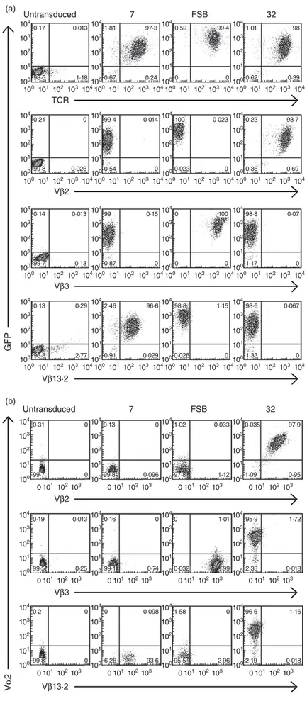
Expression of the lentivirus-encoded TCRs was evaluated by transducing the CD8+ TCR-β-deficient cell line Jurkat/MA [36], which does not express endogenous TCR on its surface. Transduction with lentiviral vectors 7, FSB and 32 resulted in the expression of vector-encoded GFP and TCR in greater than 95% of the cells (Fig. 1a). To further characterize the TCRs, transduced cells were stained with TCR Vβ-specific antibodies (Fig. 1a). Vβ staining was comparable to TCR staining, as nearly all GFP+ cells also stained with the Vβ antibody specific for that TCR (i.e. Vβ13·2 for 7, Vβ3 for FSB and Vβ2 for 32). While Vα anti-bodies specific for TCRs 7 and FSB were not available, we were able to verify co-expression of the lentivirus-encoded TCR-α and -β chains of TCR 32 by co-staining transduced Jurkat/MA cells with antibodies specific for Vα2 and Vβ2 (Fig. 1b). Note that the TCR nomenclature used to desig-nate commercial anti-Vα and -Vβ antibodies is according to Arden [42], and differs from the IMGT nomenclature provided in the Materials and methods.
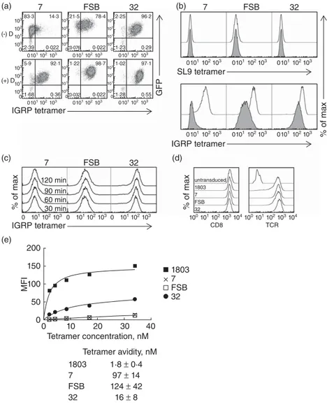
Tetramer staining of transduced Jurkat/MA cells reveals varying avidities for pMHC among the three TCRs To verify the specificity of the transduced TCRs for IGRP265– 273, transduced Jurkat/MA cells were stained with
PE-labelled HLA-A2/IGRP265–273 tetramers (Fig. 2a). All
three TCRs were stained successfully with the tetramers, although with varying intensities. TCR 32 stained very brightly, while FSB stained moderately well and 7 stained weakly, with little separation between positive and negative cells. Tetramer staining has been shown to be correlated with TCR/pMHC monomer affinity [43,44]. To verify that the weak tetramer staining of TCRs 7 and FSB was due to low-affinity binding and not the result of the transduced
TCR-β chain pairing with the endogenous Jurkat/MA TCR-α chain, transduced cells were pretreated with the protein kinase inhibitor dasatinib prior to tetramer stain-ing. Dasatinib has been shown to improve the tetramer staining of low-affinity TCRs by preventing TCR internali-zation that occurs during tetramer staining and by reducing tetramer-induced cell death [45]. Dasatinib pretreatment of transduced cells resulted in substantial improvements in tetramer staining for all three TCRs (Fig. 2a,b). The most significant effect was seen with TCRs 7 and FSB, for which distinct tetramer-positive populations were now observed. As TCR 32-transduced cells already stained well with tetramer, the effect was less dramatic, resulting in only a slightly higher intensity of staining. These results indicated that the weaker tetramer staining of TCRs 7 and FSB was due probably to a lower-affinity TCR/pMHC interaction. Dasatinib pretreatment did not cause an increase in non-specific binding to a negative control HLA-A2/SL9 tetramer (Fig. 2b).
To investigate further these differences in tetramer stain-ing intensities, transduced cells were stained with serial dilutions of the tetramers, and equilibrium binding results were used to determine the avidity of tetramer binding to each TCR. Binding was carried out for 1 h, as staining intensity was increased only minimally beyond this time-point (Fig. 2c). Because the observed avidity of tetramer binding could be influenced by the expression level of both CD8 and TCR, transduced Jurkat/MA cells were FACS-sorted as needed to generate cells with equivalent TCR expression. These sorted cells expressed comparable levels of both CD8 and TCR (Fig. 2d). As suggested by the initial tetramer staining, of the three IGRP TCRs, TCR 32 bound the most avidly to the HLA-A2/IGRP265–273tetramer,
exhibit-ing a tetramer avidity of 16± 8 nM, compared to 124± 42 nM for FSB and 97 ± 14 nM for TCR 7 (Fig. 2e). As expected, the HIV-specific TCR 1803 bound more avidly to its cognate tetramer (1·8± 0·4 nM) than did any of the IGRP-specific TCRs (Fig. 2e).
Table 1. Amino acid comparison of human islet-specific glucose-6-phosphatase catalytic subunit-related protein (IGRP)265–273-specific T cell receptor
(TCR)-α and -β chains. (a) TCR-α chain CDRs*
TCR Vα† CDR1α CDR2α CDR3α
7 Vα19 VGISA LSSGK AVTSNFGNEKLT†
FSB Vα21 NSMFDY ISSIKDK AASAGSGNTPLV
32 Vα2 NSASQS VYSSGN VVNILSNFGNEKLT
(b) TCR-β chain CDRs*
TCR Vβ† CDR1β CDR2β CDR3β
7 Vβ13·2 MNHEY SVGEGT ASSSRFVGEGLFRYGYEQY
FSB Vβ3 MDHEN SYDVKM ASSSISGYEQY
32 Vβ2 DFQATT SNEGSKA SASRQGWVNEQF
104 (a) Untransduced TCR 7 FSB 0·17 98·6 0·013 1·18 104 103 103 102 102 101 101 100 100 104 1·81 0·67 97·3 0·24 104 103 103 102 102 101 101 100 100 104 0·59 0 99·4 0 104 103 103 102 102 101 101 100 100 32 104 1·01 0·62 98 0·39 104 103 103 102 102 101 101 100 100 104 Untransduced 7 FSB 0·31 99·7 0 0 103 103 102 102 101 101 100 0 0101102103 0101102103 0101102103 104 0·13 99·8 0 0·096 103 102 101 100 104 1·02 97·8 0·033 1·12 103 102 101 100 32 104 0·035 1·09 97·9 0·95 103 102 101 100 104 Vβ2 Vβ2 104 0·19 99·5 0·013 0·25 103 103 102 102 101 101 100 0 0101102103 0101102103 0101102103 104 0·16 99·1 0 0·74 103 102 101 100 104 0 0·032 1·01 99 103 102 101 100 104 95·9 2·33 1·72 0·018 103 102 101 100 Vβ3 104 0·2 99·8 0 0 103 103 102 102 101 101 100 0 0101102103 0101102103 0101102103 104 0 6·26 0·098 93·6 103 102 101 100 104 1·58 95·5 0 2·96 103 102 101 100 104 96·6 2·19 1·16 0·018 103 102 101 100 Vβ13·2 0·21 99·8 0 0·026 104 103 103 102 102 101 101 100 100 104 99·4 0·54 0·014 0 104 103 103 102 102 101 101 100 100 104 100 0·023 0·023 0 104 103 103 102 102 101 101 100 100 104 0·23 0·36 98·7 0·69 104 103 103 102 102 101 101 100 100 104 Vβ3 0·14 99·7 0·013 0·13 104 103 103 102 102 101 101 100 100 104 99 0·87 0·15 0 104 103 103 102 102 101 101 100 100 104 0 0 100 0 104 103 103 102 102 101 101 100 100 104 98·8 1·17 0·07 0 104 103 103 102 102 101 101 100 100 104 Vβ13·2 GFP (b) V α 2 0·13 96·8 0·29 2·77 104 103 103 102 102 101 101 100 100 104 2·46 0·91 96·6 0·029 104 103 103 102 102 101 101 100 100 104 98·8 0·026 1·15 0 104 103 103 102 102 101 101 100 100 104 98·6 1·33 0·067 0 104 103 103 102 102 101 101 100 100
Fig. 1. Lentiviral transduction of T cell receptor
(TCR)-deficient Jurkat/MA cells. (a) Jurkat/MA cells were transduced with lentiviruses encoding the indicated islet-specific
glucose-6-phosphatase catalytic subunit-related protein (IGRP)265–273-specific TCRs and evaluated for TCR expression 4 days after transduction. Cells were stained with anti-human TCR and anti-Vβ antibodies specific for the three TCRs. (b) As in (a), except that an anti-Vα antibody specific for TCR 32 was also included.
Lentivirus-encoded TCRs support signalling in Jurkat/MA cells to varying degrees
In addition to its TCR deficiency, the Jurkat/MA cell line expresses luciferase under the control of the TCR-induced transcription factor NFAT [36]. In order to test the func-tional activity of the three lentivirus-encoded TCRs, we measured the luciferase activity of the transduced Jurkat/MA cells in response to 1μM IGRP265–273presented
by the human HLA-A2+ cell line T2 [33] (Fig. 3a). Jurkat/MA cells transduced to express the 1803 TCR [46], specific for the HIV gag epitope SL9, were also used in these experiments. The FSB TCR responded to its cognate peptide, although not as strongly as did the HIV TCR 1803, which has a much higher avidity for its pMHC. TCR 7 did not give a detectable response to IGRP265–273. Unexpectedly,
TCR 32 responded strongly to T2 cells, regardless of whether or not an exogenous peptide was provided.
To correlate further the TCR binding avidity results with functional activity, we performed a dose–response luciferase
experiment using the TCR-equalized transduced Jurkat/MA cells (Fig. 3b). Cells transduced with the high-avidity TCR 1803 maintained a strong luciferase response to the SL9 peptide, even at 0·01μM. The lower-avidity FSB TCR responded to IGRP265–273 at concentrations of 1μM and
above. TCR 7 did not have an observable response, even at 100μM peptide. TCR 32 continued to demonstrate a non-specific response to the T2 cells, which was unaffected even with high concentrations of IGRP265–273.To verify that these
were HLA-A2-restricted responses, T2 cells were loaded with peptide, then treated with an HLA-A2 blocking anti-body prior to the addition of Jurkat/MA cells (Fig. 3c). At 20μg/ml blocking antibody, responses from TCRs FSB and 32 were nearly abolished, while 1803 maintained a reduced but robust response. TCR 32 also responded in varying degrees to other HLA-A2+ human cell lines tested as antigen-presenting cells (APC), including C1R-A2 [34] and Jurkat-A2/Kb[35], although it did not respond to the
HLA-A2−cell line C1R [32] (Fig. 3d). These results suggest that, in addition to recognizing HLA-A2/IGRP265–273, TCR 32 also 10383·3 14·3 7 (a) (c) (e) 200 150 100 50 0 0 10 Tetramer concentration, nM Tetramer avidity, nM MFI 20 30 40 (b) (-) D 2·39 0·022 103 102 102 101 101 100 0 0 10321·5 78·4 FSB 0·076 0·022 103 102 102 101 101 100 0 0 1032·25 96·2 32 7 120 min 90 min 60 min 30 min FSB 32 (d) 7 FSB 32 1·23 0·29 103 102 102 101 101 100 0 0 103 102 101 0 0101102103 0101102103 1035·9 92·1 (+) D 1·68 0·36 103 102 102 101 101 100 0 0 103 102 101 0 0 1011021030 101102103 IGRP tetramer IGRP tetramer SL9 tetramer 103 102 101 0 103104 102 CD8 TCR 101 100 103 102 101 0 0101102103 IGRP tetramer untransduced 1803 7 FSB 32 1803 7 FSB 32 1803 7 FSB 32 1·8 ± 0·4 97 ± 14 124 ± 42 16 ± 8 103104 102 101 100 1031·22 98·7 0·032 0·022 103 102 102 101 101 100 0 0 1031·02 97·1 1·28 0·55 103 GFP % of max % of max % of max 102 102 101 101 100 0 0
Fig. 2. Human T cell receptors (TCRs) specific
for islet-specific glucose-6-phosphatase catalytic subunit-related protein (IGRP)265–273
demonstrate varying avidity for peptide/major histocompatibility complex (MHC) tetramers. (a) Jurkat/MA cells were transduced with lentiviruses encoding the indicated TCRs and stained with human leucocyte antigen (HLA)-A2/IGRP265–273tetramers alone (−D) or following treatment with 50 nM dasatinib (+D). (b) Transduced cells were stained with HLA-A2/SL9 (upper panel) or
HLA-A2/IGRP265–273tetramers (lower panel) with (not shaded) or without (shaded) 50 nM dasatinib pretreatment. Dasatinib pretreatment improves tetramer staining of low-affinity TCRs but does not increase non-specific tetramer staining. (c) To determine the time required for IGRP tetramer staining to reach equilibrium, transduced Jurkat/MA cells were stained with IGRP tetramer for 30, 60, 90 or 120 min at room temperature and analysed by flow cytometry. (d) Transduced Jurkat/MA cells were sorted by fluorescence-activated cell sorting (FACS) for equivalent TCR levels and evaluated for CD8 (left panel) and TCR expression (right panel). (e) The avidity of the TCR/tetramer interaction was determined by staining TCR-equalized Jurkat/MA cells with serial dilutions of HLA-A2/IGRP265–273tetramers and performing non-linear regression analysis. The mean fluorescence intensity (MFI) of unstained cells was subtracted from all values when assessing tetramer avidity. The graph shown is representative of three separate experiments. Values below the graph are mean± standard deviation of the three experiments.
cross-reacts with a second peptide presented in the context of HLA-A2 by the three human cell lines examined. This idea is supported by the finding that Jurkat-A2/Kb, which
expresses less than half as much HLA-A2 on its surface as T2 and C1R-A2 (data not shown), elicited a weaker response from TCR 32 than did the other two cell lines. Similarly, when DCs from mice transgenic for HLA-A2 (Fig. 3e) were used as the APC, the response of TCR 32 in the absence of exogenous peptide was reduced compared to that observed with the human HLA-A2+cell lines. These reductions in IGRP peptide-independent signalling were sufficient to allow a modest response of TCR 32 to IGRP265– 273to be observed (Fig. 3d,e). When DCs were used as the
APC, TCRs 7 and FSB also responded to IGRP265–273
although, like 32, TCR 7 responded only to the highest con-centration of peptide (100μM) (Fig. 3e). Taken together, these experiments demonstrate the differing functional activities of the lentivector-encoded TCRs 7, FSB and 32 in transduced Jurkat/MA cells and suggest TCR FSB as the best candidate for the in-vivo aspects of our work.
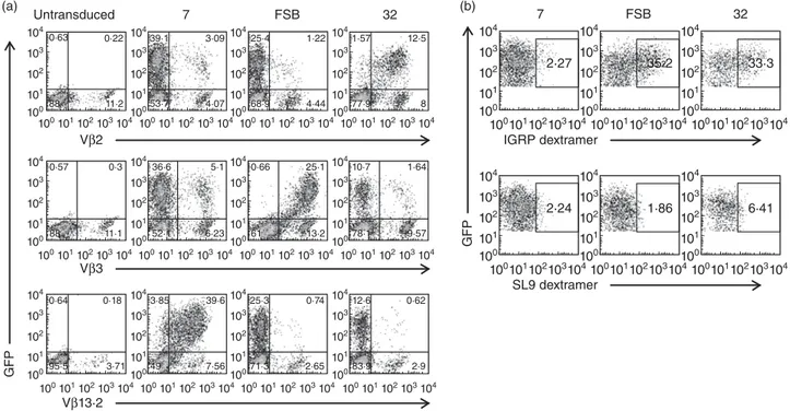
Reprogramming human primary CD8 T cells with IGRP-specific TCRs
We next sought to evaluate the ability of the lentiviral vectors to transduce primary cells and to confirm FSB as the
most useful of the three IGRP-specific TCRs. CD8 T cells were purified from peripheral blood of HLA-A2+human donors, activated to increase transduction efficiency, and transduced separately with the three lentiviral vectors. Transduction efficiency was determined by flow cytometric analysis of GFP expression and staining with the appropri-ate TCR Vβ-specific antibodies (Fig. 4a). Untransduced cells lacked GFP expression, but had variable levels of endogenous expression of Vβ2, Vβ3 and Vβ13·2 TCRs. Transduction efficiencies varied depending on the donor, but≥ 90% of GFP+cells were found consistently to express the lentiviral-encoded TCR-β chain. Additionally, the inten-sity of Vβ staining was comparable to endogenous levels, indicating that the transduced TCRs were being expressed at normal levels. To verify the proper pairing of the transduced TCR chains, cells were pretreated with dasatinib and stained with HLA-A2/IGRP265–273 dextramers [47],
pMHC multimers demonstrated to enable improved detec-tion of low-affinity TCRs compared to standard tetramers [48]. Human primary CD8 T cells transduced with TCRs FSB and 32 both stained with the dextramers (Fig. 4b), although no staining was detected in the case of TCR 7. As primary human CD8 T cells have endogenous TCR expres-sion, it is likely that there is some mixed pairing of transduced TCR-α and -β chains with endogenous chains, resulting in reduced expression of the lentivirus-encoded (a) 25 000 No T2No peptide SL9 IGRP 265−273 20 000 15 000 R elativ e light units 10 000 5000 2500 2000 1500 1000 500 0 U 1803 1803 7 7 FSB FSB 32 (b) R elativ e light units 5000 4000 3000 2000 1000 800 600 400 200 0 0 Cognate peptide, μM 0·01 0·1 1 10 100 (d) No peptide *** * ** ** ** SL9 IGRP 265-273 20 000 15 000 R elativ e light units 10 000 5000 1000 500 0 U C1R C1R-A2 Jurkat-A2/Kb 1803 32 U 1803 32 U 1803 32 (e) No peptide No DC *** *** **** * ** ** ** 10 μM SL9 10 μM IGRP 265−273 100 μM SL9 100 μM IGRP 265−273 R elativ e light units 2500 4000 3500 3000 1000 1500 2000 500 0 U 1803 7 FSB 32 32 1803 7 FSB 32 (c) 20 000 15 000 R elativ e light units 10 000 2000 4000 6000 0 0 HLA-A2 blocking antibody, μg/ml 5 10 20
Fig. 3. Lentivirus-encoded T cell receptors
(TCRs) are functional in Jurkat/MA cells. (a) T2 cells were preincubated with 1μM SLYNTVATL (SL9) or islet-specific glucose-6-phosphatase catalytic subunit-related protein (IGRP)265–273 for 1 h before addition of Jurkat/MA cells transduced with lentiviruses encoding the indicated TCRs. After overnight co-culture, luciferase activity was measured. Graph depicts mean± standard deviation (s.d.) of seven independent experiments. U, untransduced Jurkat/MA cells; **P< 0·005. (b) As in (a), except T2 cells were preincubated with 10-fold serial dilutions of SL9 or IGRP265–273from 0·01μM to 100 μM. (c) As in (a), except after 30 min incubation with 1μM SL9 or IGRP265–273, T2 cells were treated with 5, 10 or 20μg/ml human leucocyte antigen (HLA)-A2 blocking antibody before addition of
transduced Jurkat/MA cells. (d) As in (a), except that the indicated cell lines were used as antigen-presenting cells (APC) and 10μM peptide was used. Graph depicts mean± s.d. of technical replicates. *P< 0·05; **P < 0·005; ***P< 0·0001. (e) As in (a), except that splenic dendritic cells (DCs) from non-obese diabetic (NOD).β2mnull.HHD mice were used as APC.
Graph depicts mean± s.d. of technical replicates. *P< 0·05; **P < 0·01; ***P < 0·005; ****P< 0·0001.
TCR and lower pMHC multimer staining than observed in the case of transduced Jurkat/MA cells (Fig. 2a).
FSB-transduced human CD8 T cells release IFN-γ in response to their cognate antigen and lyse
peptide-pulsed target cells
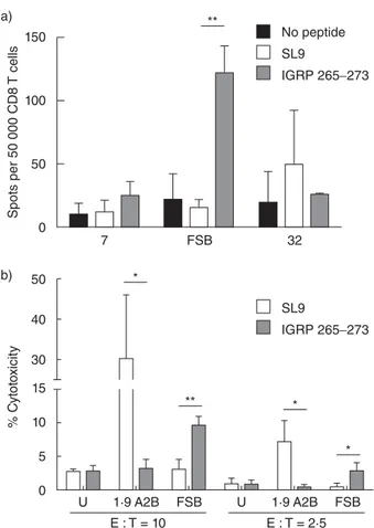
After demonstrating that human primary CD8 T cells could be engineered to express β cell-specific TCRs, we next examined the effector functions of the transduced cells. To do this, transduced cells were co-cultured with T2 cells pre-treated with IGRP265–273, and their response was measured by
IFN-γ ELISPOT (Fig. 5a). In agreement with the luciferase results obtained with transduced Jurkat/MA cells, IFN-γ production by FSB-transduced primary human CD8 T cells was the most pronounced. TCR 7 trended towards an increase in IFN-γ-producing cells in the presence of T2 cells loaded with IGRP265–273, but this was not significantly
differ-ent from the response to an irrelevant peptide or to T2 cells alone. TCR 32-transduced cells did not show a specific response to IGRP265–273-loaded T2 cells.
To evaluate further the ability of the transduced CD8 T cells to act as cytotoxic T lymphocytes (CTL), an LDH release assay was performed to measure their cytotoxic activity. T2 cells loaded with IGRP265–273 were specifically
lysed by FSB-transduced cells compared to T2 cells loaded with the irrelevant SL9 peptide (Fig. 5b). Cells transduced
with the SL9-specific TCR 1·9 A2B lysed SL9-loaded T2 cells, but not IGRP265–273-loaded T2 cells. In contrast to TCR
FSB, cells transduced with TCRs 7 or 32 did not exhibit spe-cific lysis of IGRP265–273-pulsed target cells (data not shown).
Taken together, these experiments demonstrate successful transduction of primary human CD8 T cells with IGRP-specific TCRs, with FSB yielding cells possessing peptide-specific CTL functions including IFN-γ production and cytotoxic activity. TCR FSB is clearly the preferred TCR for in-vivo studies, given that it responds in a dose-dependent manner to peptide concentrations as low as 1μM (Fig. 3a,b). Furthermore, primary human T cells transduced to express FSB show cytotoxic activity against peptide-pulsed target cells in vitro even at low effector : target ratios (Fig. 5b).
Long-term survival of engrafted transduced cells in NSG-A2 mice
In-vivo studies of the autoimmune activity of the TCR-transduced human CD8 T cells would require long-term engraftment in NSG-A2 hosts. It has been shown that intra-venous injection of at least 1× 10⁷ PBMC yields the best
engraftment outcome in NSG mice [15]. As CD8 T cells have been found to engraft poorly in NOD-SCID mice when transferred alone [49], we injected NSG-A2 mice intravenously with 4× 10⁶untransduced or FSB-transduced
104 0·63 0·22 88 11·2 Untransduced (a) (b) Vβ2 IGRP dextramer 7 FSB 32 104 103 103 102 102 101 101 100 100 104 0·57 0·3 88 11·1 104 103 103 102 102 101 101 100 100 104 0·64 0·18 95·5 3·71 104 103 103 102 102 101 101 100 100 104 3·85 39·6 49 7·56 104 103 103 102 102 101 101 100 100 104 25·3 0·74 71·3 2·65 104 103 103 102 102 101 101 100 100 104 12·6 0·62 83·9 2·9 104 103 103 102 102 101 101 100 100 104 36·6 5·1 52·1 6·23 104 103 103 102 102 101 101 100 100 104 0·66 25·1 61 13·2 104 103 103 102 102 101 101 100 100 104 10·7 1·64 78·1 9·57 104 103 103 102 102 101 101 100 100 104 39·1 3·09 53·7 4·07 104 103 103 102 102 101 101 100 100 104 25·4 1·22 68·9 4·44 104 103 103 102 102 101 101 100 100 104 1·57 12·5 77·9 8 104 103 103 102 102 101 101 100 100 7 FSB 32 104 2·27 104 103 103 102 102 101 101 100 100 104 35·2 104 103 103 102 102 101 101 100 100 104 33·3 104 103 103 102 102 101 101 100 100 SL9 dextramer 104 2·24 104 103 103 102 102 101 101 100 100 104 1·86 104 103 103 102 102 101 101 100 100 104 6·41 104 103 103 102 102 101 101 100 100 Vβ3 Vβ13·2 GFP GFP
Fig. 4. Lentiviral transduction can be used to generate humanβ cell-specific CD8 T cells. Primary human CD8 T cells were transduced with
lentiviruses encoding the indicated T cell receptors (TCRs) and (a) stained with anti-Vβ antibodies specific for each of the three TCRs or (b) pretreated with 50 nM dasatinib and stained with human leucocyte antigen (HLA)-A2/ islet-specific glucose-6-phosphatase catalytic subunit-related protein (IGRP)265–273or human leucocyte antigen (HLA)-A2/SLYNTVATL (SL9) dextramers. Plots shown in (b) are from the green fluorescent protein (GFP)+gate.
human CD8 T cells in combination with 8× 10⁶ CD8 T
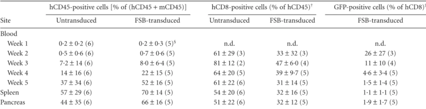
cell-depleted PBMC from the same donor. Blood engraftment levels were monitored weekly, and at 5 weeks after transfer mice were euthanized to examine spleen and pancreas engraftment as well. The experiments were termi-nated at 5 weeks, because xenogeneic graft-versus-host (GvH) disease occurs in NSG mice repopulated with PBMC between 30 and 45 days post-transfer [15,50]. The level of engraftment of hCD45+ cells in mice receiving FSB-transduced CD8 T cells was similar to engraftment in mice receiving untransduced cells (Table 2). The percentage of engrafted cells in the blood increased each week, and higher engraftment was seen in the spleen than in the blood.
Human CD45+cells were also observed in the pancreata of both untransduced and FSB-transduced recipient mice. Recipients that received untransduced cells tended to have a higher percentage of engrafted CD8 T cells, but this was only statistically significant at the 3-week time-point. In FSB-transduced recipient mice, GFP+ CD8 cells could be detected as early as 2 weeks after transfer, and remained detectable in blood, spleen and pancreas 5 weeks after transfer (Fig. 6a; Table 2). Histological analysis of pancreas sections revealed mild infiltration around some islets in both untransduced and FSB-transduced recipient mice (Fig. 6b) due probably, at least in part, to a GvH response to murine class I MHC molecules [15,50]. Because of this non-specific infiltration, we were unable to discern a differ-ence in histopathology between the two groups, and diabe-tes was not observed in any of the recipients during the 5-week experimental period. To evaluate whether the transduced cells maintained their CTL capabilities after engraftment, splenocytes from untransduced, FSB-transduced and 1803-FSB-transduced recipients were analysed 5 weeks after engraftment for granzyme B expression (Fig. 6c). In all mice tested, granzyme B expression was restricted to the CD8 T cell population. Importantly, granzyme B expression in GFP+FSB and 1803-transduced cells was comparable to granzyme B expression of GFP− cells in the same host, as well as those from untransduced recipients. This result confirms that the transduced human cells retain CTL function after 5 weeks in the mouse host.
Discussion
The NOD mouse has been the prevailing model for the study of T1D for many years and has greatly improved our understanding of this autoimmune disease [1]. However, while the disease can be prevented, and even reversed, in the mice [2,3], a robust immunological therapy for human T1D has not yet been achieved [5,6]. We reasoned that the ability to investigate the impact of therapeutic approaches target-ing humanβ cell-specific T cells might support the transla-tion of rodent data to the human disease. To that end, we have used lentiviral transduction to generate human CD8 T cells specific for IGRP265–273/HLA-A2. Cells transduced with
TCR FSB exhibited characteristics of CTL, including antigen-dependent IFN-γ secretion and lysis of peptide-pulsed targets, suggesting in-vitro uses for such cells, e.g. investigation of mechanisms of β cell killing by human T cells or antigen identification in the case of β cell-specific TCRs of unknown specificity. Importantly, the transduced cells survived for up to 5 weeks in NSG-A2 hosts and will thus permit the future evaluation of T cell-modulatory interventions in an in-vivo system that incorporates human T cells interacting with human MHC molecules. Although TCR-transduced human T cells have been used to achieve anti-tumour or anti-viral activity in patients [13], their 150 (a) ** * * * ** No peptide SL9 IGRP 265−273 SL9 IGRP 265−273 100 Spots per 50 000 CD8 T cells 50 0 50 40 15 (b) 30 % C y to to xicit y 10 5 0 U E : T = 10 1·9 A2B FSB U E : T = 2·5 1·9 A2B FSB 7 FSB 32
Fig. 5. T cell receptor (TCR)-transduced primary human CD8 T cells
release interferon (IFN)-γ and are cytotoxic. (a) Primary human CD8 T cells were transduced with lentiviruses encoding the indicated TCRs and incubated with T2 cells loaded with 10μM SLYNTVATL (SL9) or islet-specific glucose-6-phosphatase catalytic subunit-related protein (IGRP)265–273. IFN-γ production was detected by enzyme-linked immunospot (ELISPOT). Graph depicts mean± standard deviation (s.d.) of three independent experiments. **P< 0·005. (b) Transduced primary human CD8 T cells were incubated with peptide-loaded T2 cells at an effector : target (E : T) ratio of 10 : 1 or 2·5 : 1. Cytotoxicity was measured by lactate dehydrogenase (LDH) release into the supernatant. Graph depicts mean± s.d. of technical replicates. U, untransduced primary human CD8 T cells; *P< 0·05; **P < 0·005.
long-term survival and utility in immune-deficient murine models has not been reported previously.
In the course of developing this system, we first character-ized three human TCRs specific for IGRP265–273/HLA-A2 in
terms of their structural and functional avidities. As deter-mined by measurement of tetramer avidity values, the TCRs exhibited a range of structural avidities, and all were of lower avidity than the HIV-specific TCR 1803 studied for compari-son. While the functional avidities of the three IGRP-specific TCRs did not correlate strictly with the measured structural avidities, all were reduced compared to the functional avidity observed for the HIV-specific TCR. This is perhaps not sur-prising, as it is unlikely that T cells bearing high-affinity autoreactive TCRs would escape negative selection in the thymus [51]. It is becoming clear that autoreactive TCRs in both mice and humans often exhibit low avidity, either because of the TCR itself (as in our work) or because its cognate peptide binds poorly to MHC [52–59]. It is not unusual for autoreactive T cells to exhibit responses only to relatively high concentrations of peptide in vitro (compared to anti-viral T cells, for example) [52,56,57,59]. Some of these T cells are nonetheless pathogenic, presumably because the local peptide concentration in the target organ is high [60], or because the peptide is modified there in a way that improves recognition [61]. The relatively high tetramer avidity values for the IGRP-specific TCRs, the poor tetramer staining observed in the absence of dasatinib for TCR 7 (and, to a lesser extent, FSB) and the reduced functional avidities compared to the anti-viral TCR support the autoreactive nature of our TCRs and emphasize some of the challenges inherent in working with autoreactive TCRs. TCR 7 is of par-ticular interest, because its CDR3β loop is unusually long (19 residues). It is tempting to speculate that a loop of this length may impair the formation of intimate contacts between the
TCR and the pMHC and, in so doing, may account for the poor tetramer staining and low functional avidity observed for this autoreactive TCR.
Another characteristic that has been reported for autoreactive T cells is antigenic recognition promiscuity (cross-reactivity) [62–69]. Our results show that this prop-erty, exemplified by TCR 32, can also be studied using lentiviral transduction of T cells. TCR 32 had the highest structural avidity of the three TCRs examined here (as measured by tetramer avidity); however, it was difficult to document a functional peptide-specific response from transduced cells due to its robust cross-reaction to human cell lines expressing HLA-A2 and used as APC. Apparently, in addition to recognizing IGRP265–273/HLA-A2, TCR 32 also
responds to one or more endogenous peptides presented by HLA-A2 in these cell lines. When murine HLA-A2-positive DCs were used as APC, the stimulation in the absence of exogenous peptide was considerably reduced, but still present. Surprisingly, however, the peptide-specific response was quite low and required a high peptide concentration for detection. Taken together, these data suggest not only that TCR 32 is promiscuous, but also that one of its peptide ligands may be an antagonist [70] that can dampen its response to IGRP265–273. Alternatively, it is possible that the
binding characteristics of TCR 32 to IGRP265–273/HLA-A2 are
not suitable to elicit a strong functional response [71]. These results illustrate that careful in-vitro analysis of can-didate TCRs, as performed here, is necessary before under-taking in-vivo experiments, because tetramer binding does not guarantee a measurable functional response to peptide. Interestingly, we observed that FSB, isolated from a healthy donor, was the most functionally active of the three TCRs. This finding suggests that useful islet-specific human TCRs need not be derived from diabetic patients, while also
Table 2. Human cell engraftment in NSG-A2 mice*.
Site
hCD45-positive cells [% of (hCD45+ mCD45)] hCD8-positive cells (% of hCD45)† GFP-positive cells (% of hCD8)‡ Untransduced FSB-transduced Untransduced FSB-transduced FSB-transduced Blood Week 1 0·2± 0·2 (6) 0·2± 0·3 (5)§ n.d. n.d. n.d. Week 2 0·5± 0·6 (6) 0·7± 0·6 (5) 61± 29 (3) 33± 32 (3) 26± 27 (3) Week 3 7·2± 14 (6) 8·0± 6·4 (5) 81± 12 (2) 47± 6·0 (4) 11± 10 (4) Week 4 14± 16 (6) 22± 15 (5) 64± 20 (5) 39± 9·7 (5) 4·6± 3·4 (5) Week 5 37± 34 (6) 52± 16 (5) 61± 22 (6) 31± 14 (5) 1·5± 1·4 (5) Spleen 57± 29 (6) 70± 14 (5) 54± 20 (6) 32± 16 (5) 1·1± 1·1 (5) Pancreas 44± 35 (6) 66± 16 (5) 51± 22 (6) 32± 12 (5) 1·9± 1·7 (5)
*Untransduced or T cell receptor (TCR) FSB lentivirus-transduced human leucocyte antigen (HLA)-A2+human CD8 T cells (4× 10⁶) were com-bined with CD8 T cell-depleted peripheral blood monunclear cells (PBMC) from the same donor (8× 10⁶) and transferred via tail vein into HLA-A2-transgenic non-obese diabetic (NOD)-severe combined immunodeficient (SCID) interleukin (IL)-2rγ null (NSG-A2) mice. Blood was taken weekly from the tail starting at week 1 and analysed for engraftment by flow cytometry. At week 5, mice were killed and blood, spleen and pancreas were ana-lysed for engraftment. Numbers of mice are indicated in parentheses. Sample fluorescence activated cell sorter (FACS) plots are shown in Fig. 6a. n.d., not determined.†Expressed as a % of hCD45-positive cells. Mice having a hCD45-positive cell percentage less than 0·5% for a given time-point were excluded from subsequent analysis for that time-point.‡Expressed as a % of hCD8-positive cells.§A sixth mouse had a splenic hCD45-positive cell fre-quency of less than 1% at week 5 and was excluded from further analysis.
mCD45 GFP hCD8 GFP hCD45 hCD8 Untransduced Untransduced, hCD45+ Granzyme B FSB-transduced 1803, hCD8+ FSB, hCD8+ 103 Blood (a) (b) (c) Spleen Pancreas 1 week 2 weeks 3 weeks 4 weeks 5 weeks 5 weeks 5 weeks
102 100101102103 0 0 0 103 102 100101102103 0 0 0·56 103 102 101 100101102103 0 0 1·3 10 3 102 101 100101102103 0 0 2·43 103 102 100101102103 0 0 57·1 103 102 101 100 101 102 103 104 0 0 103 102 101 100 101 102 103 104 0 4·49 103 104 102 101 100 100 101 102 103 104 0·011 103 104 102 101 100 100 101 102 103 104 20·4 103 104 102 101 100 100 101 102 103 104 0·025 103 104 102 101 100 100 101 102 103 104 23·9 103 104 102 101 100 100 101 100 101 102 103 104 23·8 103 104 102 101 100 100 101102103 104 43·3 0·92 33·4 22·4 103 104 102 101 100 100 101102 103104 0·098 70·7 29·1 0·047 103 104 102 101 100 100 101102 103104 1·24 68·4 30·1 0·32 103 104 102 101 100 100 101 102 103 104 7·56 10 3 104 102 101 100 100 101 102 103 104 1·95 103 102 101 100 100101102103 0 0 0 103 102 101 100 100101102103 0 0 1·37 103 102 101 100101102103 0 0 0 103 102 101 100101102103 0 0 68 103 102 101 100101102103 0 0 0·044 103 102 101 100101102103 0 0 48
Fig. 6. Transduced human CD8 T cells remain detectable and functional 5 weeks after transfer to human leucocyte antigen (HLA)-A2-transgenic
non-obese diabetic (NOD)-severe combined immunodeficient (SCID) interleukin (IL)-2rγ null (NSG-A2) mice. NSG-A2 mice were injected intravenously with 4× 10⁶untransduced, FSB-transduced, or 1803-transduced human CD8 T cells in combination with 8× 10⁶CD8-depleted peripheral blood mononucleaer cells (PBMC). Blood samples were taken weekly from 1 to 5 weeks after transfer and analysed by flow cytometry. Spleen and pancreas were analysed similarly at 5 weeks post-transfer. (a) Engraftment of a representative FSB recipient mouse (middle panels), with an NSG-A2 mouse that did not receive human cells shown for comparison (top panels). Top and middle panels, total hCD45+mCD45−cell engraftment (% of total cells is shown). Bottom panels, engraftment of green fluorescent protein (GFP)+FSB-transduced CD8 T cells (% of hCD45+mCD45−hCD8+cells is shown). Due to the necessity of analysing the time-points on different days, variations in fluorescence intensity were observed. Summary data from all mice are shown in Table 2. (b) Pancreata from untransduced and FSB-transduced recipient mice were fixed 5 weeks after transfer, sectioned, and stained with aldehyde fuchsin. Representative images of islets from untransduced and FSB recipient mice are shown. (c) Splenocytes from untransduced, 1803-transduced and FSB-transduced recipient mice were analysed for intracellular granzyme B expression by flow cytometry 5 weeks after transfer. Left panel, granzyme B expression of untransduced hCD45+mCD45−cells. Middle and right panels, granzyme B expression of hCD45+mCD45−hCD8+cells from 1803 and FSB recipient mice.
once again highlighting the need for rigorous in-vitro analy-sis of TCRs.
Multiple antigen-specific therapies have demonstrated potential for preventing diabetes development in NOD mice by inducing deletion of β cell-specific CD8 T cells [72–74]. We have found that delivery ofβ cell antigens to DCs via the endocytic receptor DEC-205 can lead to the deletion of both transferred [75] and endogenous CD8 T cells [76] specific for the delivered antigen. Our ability to generate human CD8 T cells specific forβ cell antigens and their survival in NSG-A2 mice will now allow such deletional strategies to be explored in a system incorporat-ing human T cells. This line of investigation does not require that the recipient mice develop diabetes, as T cell deletion in response to treatment can be monitored even in the absence of overt disease by evaluating GFP expression. However, our goal is to optimize our system further so that diabetes is observed. The T cell clones from which the TCRs 7, FSB and 32 were obtained originally were able to lyse human β cells (data not shown), supporting their potential diabetogenic nature. It is possible that we did not observe diabetes upon transfer of transduced IGRP-specific CD8 T cells to NSG-A2 mice because this single specificity may be insufficient to cause disease. However, our experi-mental design was based in part on our finding that trans-fer to NOD-SCID recipients of cultured islet infiltrates from 8·3 TCR (specific for H-2Kd/IGRP
206–214)-transgenic
NOD mice induced diabetes in all recipients (data not shown). These cultured infiltrates contained 98% CD8 T cells and, of these, 80% were specific for IGRP206–214,
sug-gesting at least the possibility that a T cell population highly enriched for CD8 T cells having a single specificity can indeed transfer disease to an immunodeficient host. However, our lentiviral transduction approach will allow human CD8 T cells having multiple defined antigenic spe-cificities to be engineered and if necessary transferred in the future. Furthermore, we have found that human CD4 T cells can also be engineered by lentiviral transduction to be specific forβ cell antigens (data not shown), suggesting the possibility of simultaneously transferring β cell-specific CD8 and CD4 T cells to NSG mice transgenic for human class I and class II MHC molecules. The contribution of CD4 T cells to islet pathology in such a model is suggested by the recent report that insulitis was observed when immortalized human CD4 T cells specific for HLA-DR4-bindingβ cell peptides were transferred to NSG mice trans-genic for HLA-DR4 [77].
In addition to the possible requirements for multiple antigenic specificities or both CD8 and CD4 T cells, the duration of our experiments may have been insufficient for diabetes development to take place. We followed the recipient mice for only 5 weeks after transfer of the engi-neered IGRP-specific T cells, as a xenogeneic GvH disease develops in NSG mice engrafted with human PBMC within 4–5 weeks after transfer [15]. This GvH disease is
due largely to a T cell response to murine class I MHC molecules, as NSG mice that are murine class I MHC-deficient are relatively resistant to this disease [50]. Murine class I-deficient NSG-A2 mice will probably be an improved recipient for the engraftment of engineered β cell-specific human CD8 T cells, as they will allow the experimental duration to be extended. They should also allow increased numbers of transduced CD8 T cells to be transferred.
We demonstrate here for the first time, to our knowledge, the ability to generate human islet-specific cytotoxic T cells at will by TCR lentiviral transduction and their survival for up to 5 weeks in NSG-A2 mice. We believe that this strategy has the potential to allow for a better understanding of T1D pathogenesis and the evaluation of antigen-specific thera-pies. Our work highlights the difficulties in working with autoreactive TCRs, including their low affinity for pMHC and their propensity for promiscuity. However, our proof-of-concept study none the less suggests broad applicability of our approach to investigations concerning autoreactive T cells involved in other human T cell-mediated autoimmune diseases.
Acknowledgements
The authors thank Wendy Unger for T cell cloning and Arno van der Slik for TCR sequencing. This work was sup-ported by National Institutes of Health grants R01 DK094327 (T.P.D.), R01 DK064315 (T.P.D.), P60 DK020541 (Albert Einstein College of Medicine’s Diabetes Research Center), P30 AI051519 (Einstein-Montefiore Center for AIDS Research), P01 AI046629 (D.L.G., L.D.S), U01 DK089572 (D.L.G., L.D.S.), R01 DA033788 (H.G.) and R01 AI043203 (O.O.Y.), and by grants from the Canadian Insti-tutes of Health Research (P.S.) and The Leona M and Harry B. Helmsley Charitable Trust (2012PG-T1D018; University of Massachusetts Medical School Diabetes Center of Excel-lence). The flow cytometry facility at Albert Einstein College of Medicine is supported by National Institutes of Health Cancer Center grant P30 CA013330. The Julia McFarlane Diabetes Research Centre is supported by the Canadian Diabetes Association. T. P. D. is the Diane Belfer, Cypres and Endelson Families Faculty Scholar in Diabetes Research. H. G. holds the Charles Michael Chair in Autoim-mune Diseases. P. S. is a Scientist of the Alberta Inno-vates – Health Solutions and a scholar of the Instituto de Investigaciones Sanitarias Carlos III. The contents of this publication are solely the responsibility of the authors and do not necessarily represent the official views of the National Institutes of Health.
Disclosure
References
1 Driver JP, Serreze DV, Chen YG. Mouse models for the study of autoimmune type 1 diabetes: a NOD to similarities and differences to human disease. Semin Immunopathol 2011; 33:67– 87.
2 Chaparro RJ, DiLorenzo TP. An update on the use of NOD mice to study autoimmune (Type 1) diabetes. Expert Rev Clin Immunol 2010; 6:939–55.
3 Shoda LK, Young DL, Ramanujan S et al. A comprehensive review of interventions in the NOD mouse and implications for transla-tion. Immunity 2005; 23:115–26.
4 Herold KC, Vignali DA, Cooke A, Bluestone JA. Type 1 diabetes: translating mechanistic observations into effective clinical out-comes. Nat Rev Immunol 2013; 13:243–56.
5 Greenbaum CJ, Schatz DA, Haller MJ, Sanda S. Through the fog: recent clinical trials to preserve beta-cell function in type 1 diabe-tes. Diabetes 2012; 61:1323–30.
6 Staeva TP, Chatenoud L, Insel R, Atkinson MA. Recent lessons learned from prevention and recent-onset type 1 diabetes immu-notherapy trials. Diabetes 2013; 62:9–17.
7 Mallone R, Martinuzzi E, Blancou P et al. CD8+T-cell responses identifyβ-cell autoimmunity in human type 1 diabetes. Diabetes 2007; 56:613–21.
8 Hughes MS, Yu YY, Dudley ME et al. Transfer of a TCR gene derived from a patient with a marked antitumor response conveys highly active T-cell effector functions. Hum Gene Ther 2005;
16:457–72.
9 Joseph A, Zheng JH, Follenzi A et al. Lentiviral vectors encoding human immunodeficiency virus type 1 (HIV-1)-specific T-cell receptor genes efficiently convert peripheral blood CD8 T lym-phocytes into cytotoxic T lymlym-phocytes with potent in vitro and in vivo HIV-1-specific inhibitory activity. J Virol 2008; 82:3078–89. 10 Morgan RA, Dudley ME, Wunderlich JR et al. Cancer regression
in patients after transfer of genetically engineered lymphocytes. Science 2006; 314:126–9.
11 Morgan RA, Dudley ME, Yu YY et al. High efficiency TCR gene transfer into primary human lymphocytes affords avid recogni-tion of melanoma tumor antigen glycoprotein 100 and does not alter the recognition of autologous melanoma antigens. J Immunol 2003; 171:3287–95.
12 Varela-Rohena A, Molloy PE, Dunn SM et al. Control of HIV-1 immune escape by CD8 T cells expressing enhanced T-cell recep-tor. Nat Med 2008; 14:1390–5.
13 Zhang L, Morgan RA. Genetic engineering with T cell receptors. Adv Drug Deliv Rev 2012; 64:756–62.
14 Shultz LD, Lyons BL, Burzenski LM et al. Human lymphoid and myeloid cell development in NOD/LtSz-scid IL2Rγnull mice
engrafted with mobilized human hemopoietic stem cells. J Immunol 2005; 174:6477–89.
15 King M, Pearson T, Shultz LD et al. A new Hu-PBL model for the study of human islet alloreactivity based on NOD-scid mice bearing a targeted mutation in the IL-2 receptor gamma chain gene. Clin Immunol 2008; 126:303–14.
16 Christianson SW, Shultz LD, Leiter EH. Adoptive transfer of dia-betes into immunodeficient NOD-scid/scid mice. Relative contri-butions of CD4+and CD8+T-cells from diabetic versus prediabetic NOD.NON-Thy-1adonors. Diabetes 1993; 42:44–55.
17 Shultz LD, Saito Y, Najima Y et al. Generation of functional human T-cell subsets with HLA-restricted immune responses in
HLA class I expressing NOD/SCID/IL2rγnull humanized mice. Proc Natl Acad Sci USA 2010; 107:13022–7.
18 Strowig T, Gurer C, Ploss A et al. Priming of protective T cell responses against virus-induced tumors in mice with human immune system components. J Exp Med 2009; 206:1423–34. 19 Fennessy M, Metcalfe K, Hitman GA et al. A gene in the HLA class
I region contributes to susceptibility to IDDM in the Finnish population. Childhood Diabetes in Finland (DiMe) Study Group. Diabetologia 1994; 37:937–44.
20 Nejentsev S, Howson JM, Walker NM et al. Localization of type 1 diabetes susceptibility to the MHC class I genes HLA-B and HLA-A. Nature 2007; 450:887–92.
21 Robles DT, Eisenbarth GS, Wang T et al. Identification of children with early onset and high incidence of anti-islet autoantibodies. Clin Immunol 2002; 102:217–24.
22 Whitfield-Larry F, Young EF, Talmage G et al. HLA-A2-matched peripheral blood mononuclear cells from type 1 diabetic patients, but not nondiabetic donors, transfer insulitis to NOD-scid/γcnull/ HLA-A2 transgenic mice concurrent with the expansion of islet-specific CD8+T cells. Diabetes 2011; 60:1726–33.
23 Han B, Serra P, Amrani A et al. Prevention of diabetes by manipu-lation of anti-IGRP autoimmunity: high efficiency of a low-affinity peptide. Nat Med 2005; 11:645–52.
24 Lieberman SM, Evans AM, Han B et al. Identification of theβ cell antigen targeted by a prevalent population of pathogenic CD8+T cells in autoimmune diabetes. Proc Natl Acad Sci USA 2003;
100:8384–8.
25 Mukherjee R, Wagar D, Stephens TA, Lee-Chan E, Singh B. Identi-fication of CD4+T cell-specific epitopes of islet-specific glucose-6-phosphatase catalytic subunit-related protein: a novel β cell autoantigen in type 1 diabetes. J Immunol 2005; 174:5306–15. 26 Coppieters KT, Dotta F, Amirian N et al. Demonstration of
islet-autoreactive CD8 T cells in insulitic lesions from recent onset and long-term type 1 diabetes patients. J Exp Med 2012; 209:51– 60.
27 Jarchum I, Nichol L, Trucco M, Santamaria P, DiLorenzo TP. Identification of novel IGRP epitopes targeted in type 1 diabetes patients. Clin Immunol 2008; 127:359–65.
28 Standifer NE, Ouyang Q, Panagiotopoulos C et al. Identification of novel HLA-A*0201-restricted epitopes in recent-onset type 1 diabetic subjects and antibody-positive relatives. Diabetes 2006;
55:3061–7.
29 Unger WW, Pearson T, Abreu JR et al. Islet-specific CTL cloned from a type 1 diabetes patient cause beta-cell destruction after engraftment into HLA-A2 transgenic NOD/scid/IL2RG null mice. PLOS ONE 2012; 7:e49213.
30 Yang J, Danke NA, Berger D et al. Islet-specific glucose-6-phosphatase catalytic subunit-related protein-reactive CD4+ T cells in human subjects. J Immunol 2006; 176:2781–9.
31 Takaki T, Marron MP, Mathews CE et al. HLA-A*0201-restricted T cells from ‘humanized’ NOD mice recognize autoantigens of potential clinical relevance to type 1 diabetes. J Immunol 2006;
176:3257–65.
32 Storkus WJ, Alexander J, Payne JA, Dawson JR, Cresswell P. Rever-sal of natural killing susceptibility in target cells expressing transfected class I HLA genes. Proc Natl Acad Sci USA 1989;
86:2361–4.
33 Salter RD, Howell DN, Cresswell P. Genes regulating HLA class I antigen expression in T–B lymphoblast hybrids. Immunogenetics 1985; 21:235–46.