Abstract. – OBJECTIVE: The therapeutic ap-plication of ozone and its derivatives in the den-tal field has been used for many purposes. How-ever, there has yet to be a consistent evaluation of the outcomes, due to the lack of standardiza-tion of the treatment operating procedures.
MATERIALS AND METHODS: The keywords “ozone”, “ozonated”, “ozonation” “ozonized”, “ozonization”, “dentistry”, “periodontolo-gy”, “oral surgery”, “oxygen-ozone therapy” were used to perform a literature review using PubMed, Cochrane, Google Scholar, Zotero da-tabases with the temporal restriction for manu-scripts published between 2010 and 2020. Clin-ical trials and case reports of good, neutral, as well as negative results related to ozone treat-ment, specifications were evaluated.
DISCUSSION: A better understanding of the mechanisms of action of this bio-oxidative ther-apy could open new horizons related to the per-sonalization of treatments and the quality of dental care. The critical condition to achieve these goals is an improved knowledge of the qualitative/quantitative characteristics of ozone and its derivatives.
Key Words:
Ozone, Ozonated water, Ozonated oil, Bio-oxida-tive therapy, RegeneraBio-oxida-tive medicine, Quality of den-tal care.
Abbreviations
ATP: adenosine triphosphate. BRONJ: bisphospho-nate-related osteonecrosis of the jaws. CFU: colo-ny-forming units. DGG: deepithelialized gingival grafts. DUWL: dental unit water lines. EDTA: ethylenedi-aminetetraacetic acid. LOPs: lipid oxidation products. LPM: litre per minute. MDP: 10-methacryloyloxydecyl dihydrogen phosphate. MDPB:
12-methacryloyloxydo-decylpyridinium bromide. MTT: 3-[4,5-dimethylthi-azole-2-yl]-2,5-diphenyltetrazolium bromide. NTP: nor-mal temperature and pressure conditions. ppm: parts per million. ppmv: parts per million volume. qPCR: quan-titative polymerase chain reaction. ROS: reactive oxy-gen species. SEM: scanning electron microscopy. SRP: scaling and root planning. TMD: temporomandibular disease. TUNEL: Terminal deoxynucleotidyl transferase dUTP Nick End Labeling.
Introduction
The positive therapeutic effects of ozone and its derivatives have been studied in multiple fields of medicine. However, there is no limited agree-ment in the medical community on its use and benefits. This may be due to the fact that, unlike other drugs, ozone does not act directly through traditional drug-receptor interactions. When ad-ministered in the gaseous form, it is a gaseous mixture where ozone represents at most 5% of the total, while the remaining part is generally made up of oxygen, acting as a gas transmitter. On the other hand, ozone, due to its extreme reactivity, cannot be used for the transmission of chemical signals to induce physiological or biochemical changes. Moreover, ozone cannot be considered a pro-drug in the common sense of the term. A pro-drug is a biologically inactive molecule that, once introduced into the body, requires chemical transformations, generally of enzymatic nature, for its activation. Ultimately, ozone can be classified as an effector molecule generator. Depending on method of administration, admin-istration site, dosage and derivative formulations, different hydrophilic (mainly hydrogen peroxide)
European Review for Medical and Pharmacological Sciences 2020; 24: ???-???
G. TRICARICO
1, J. RODRIGUES ORLANDIN
2,3, V. ROCCHETTI
4,
C.E. AMBROSIO
2, V. TRAVAGLI
31Department of Dentistry, Sant’Andrew Hospital Vercelli, Italy
2Department of Veterinary Medicine, Faculty of Animal Science and Food Engineering (FZEA-USP), University of São Paulo, Pirassununga, São Paulo, Brazil
3Dipartimento di Biotecnologie, Chimica e Farmacia – Dipartimento di Eccellenza Nazionale 2018-2022, Università degli Studi di Siena, Italy
4Dipartimento di Scienze della Salute, Università del Piemonte Orientale, Novara, Italy
A critical evaluation of the use of ozone and
its derivatives in dentistry
and lipophilic (mainly alkenals) small molecules will be produced. These molecules selectively interact with protein moieties, regulating their biological activity epigenetically. Therefore, ef-fector molecules acting as ligands can increase or decrease enzyme activity, gene expression or cellular signals.
In general, oxygen-ozone therapy is classified as regenerative medicine, provided that the cor-rect conditions of the use of these substances are respected1,2. In this sense, it is possible to foresee
the use of ozone in personalized therapy based on the patients’ clinical history3.
Oxygen-ozone therapy has multiple methods of application in dental practice4-18. Given the
increased attention to this subject, further stud-ies and reviews are expected to be published19.
However, analytical evaluation of the published clinical results has not been performed. The pres-ent study addresses knowledge gaps related to research protocols and resulting outcomes related to the use of ozone and its derivatives in dentistry, using the following classification (Table I).
The present study will also address the op-erative protocols (in terms of ozone generators, ozone concentrations, ozone derivatives and so on) adopted by practicing dentists.
Four databases (PubMed, Cochrane, Goo-gle Scholar, Zotero) were consulted, using the keywords “ozone”, “ozonated”, “ozonation” “ozonized”, “ozonization”, “dentistry”, “periodon-tology”, “oral surgery”, “oxygen-ozone therapy”. For homogeneity’s sake, the terms “ozonation” and “ozonated” were used in the present manuscript.
The aim of this review is to analyze clinical trials and case reports, confronting good and negative results with respect to ozone treatment specifications. In the “Presentation of the papers” section, summaries of both usual Materials and Methods, as well as Results units, are reported. In order to make reading easier, the specific part relating to the characteristics of use of ozone, where present, is specifically indicated at the end of each summary. The clinical results obtained from the various works are grouped by similarity of treatment in the Discussion section.
Presentation of the Paper
A) General Dentistry for Sterilization of the EquipmentOkubo et al20 studied the bactericidal effects
of low-concentrated ozonated water on
microor-Table I. Classification of dental procedures and specialties in relation to the original articles from the last ten years using ozone and its derivatives.
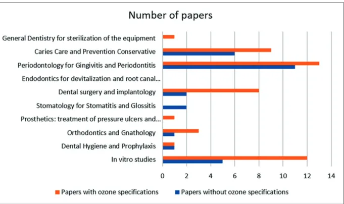
A) General Dentistry for Sterilization of the Equipment (n=1) B) Caries Care and Prevention Conservative (n=15)
i) Conservative for deciduous teething
ii) Conservative for permanent dentition, (treatment of reversible pulpits of mild degree)
C) Periodontology for Gingivitis and Periodontitis (n=24)
i) Marginal gingivitis ii) Periodontitis
iii) Mucositis (inflammation of peri-implant tissues) iv) Periimplantitis (destruction of peri-implant tissues)
D) Endodontics for Devitalization and Root Canal Treatments (n=0) E) Dental Surgery and Implantology (n=10)
i) Oral surgery and prevention of post-extraction alveolitis
ii) Surgical wound protection in implantology with flap-less technique iii) Protection of sites exposed during tissue biopsy
iv) Protection of the donor site during self-transplantation for periodontal surgical therapy (flap grafting) v) Protection of post-surgical sutured wounds of any nature present in the oral cavity
vi) Dental fistulas
vii) Prevention and treatment of Drug-Related Osteo Necrosis of Jaw – DRONJ
F) Stomatology for Stomatitis and Glossitis (n=2)
G) Prosthetics: Treatment of Pressure Ulcers and Mucosal Contact Lesions (n=1) H) Orthodontics and Gnathology (n=4)
i) Hygiene of orthodontic products ii) ATM pain and hypofunction
I) Dental Hygiene and Prophylaxis (n=2) J) In vitro studies (n=17)
ganisms and biofilms involved in bacterial con-tamination of dental unit waterlines (DUWL) and their harmfulness to the components of dental unit. Solutions compared to low concentrated ozonated water were 0.1% cetylpyridinium chlo-ride as positive control, as well as chlorinated tap water and phosphate-buffered saline as negative controls. Adenosine triphosphate (ATP) amounts of the microbes were measured and the biofilms of these microbes were observed using scanning electron microscopy (SEM). The ozonated water halved ATP levels in microbes compared to the others, while the former reduced it below detec-tion limits. No modificadetec-tions were observed on the surfaces of dental unit components.
The ozone concentration in ozonated water changed over time, with a maximum concen-tration of 0.4 mg/L immediately after sampling. Subsequently, it reduced to half within 2 h, and further decreased to the lowest concentration of 0.1 mg/L by 5 h.
B) Caries Care and Prevention Conservative
i) Conservative for deciduous dentition
Gökçen et al21 performed a study using 40
deciduous teeth in total. The teeth were divided into four study groups: a commercial calcium hy-droxide dental cement radiopaque; gaseous ozone application; a commercially available self-etching adhesive system containing monomer 12-meth-acryloyloxydodecylpyridinium bromide (MDPB) as antibacterial; physiological saline as negative control group. On the basis of the results ob-tained, the authors conclude that ozone treatment could be considered to exert an antibacterial effect in the treatment of deciduous teeth, even if further research on the long-term effects of ozone on microorganisms, and a more detailed compar-ison of ozone with dentine-bonding systems and Ca(OH)2 is necessary. Ozone therapy was applied to the experimental cavity in gaseous form, ac-cording to the manufacturer’s instructions. No further information about ozone concentration was given.
ii) Conservative for permanent dentition
Mosallam et al22 exposed the pulps of teeth
from 9 mixed breed dogs. In each dog, the right canines were capped with calcium hydroxide, and the left canine with a thin layer of ozonated olive oil (Oleozon). Then, the cavities were restored. 3 dogs were sacrificed at day 7, other 3 dogs at day
30 and the others after 90 days. No reparative dentin was detected. Histo-micro-morphological analysis concluded Oleozon induced less degrees of irritation to the dental pulp compared to that with calcium hydroxide, when used for pulp cap-ping.
Ozonated olive oil paste was prepared through incorporation of ozone gas (O3), with a concentra-tion of 70 µg/mL (5%).
Yazicioǧlu et al23 performed a study in vivo by
testing gaseous ozone for 40 seconds and other anti-cariogenic agents in patients with carious lesions. After 18 months, despite no differences in visual examination, it was observed significant improvement in radiographic and laser examina-tion, comparing to the initial examination. On the other hand, when compared with control group, no significant progression was seen.
No information about ozone specifications were given.
Cangul et al24 tested the effect of ozone and
boric acid on microleakage. Thus, 80 teeth were extracted, cleaned and divided into 8 groups. A cavity was made and disinfected with: gaseous ozone; 2% chlorhexidine solution; 2.5% sodium hypochlorite; 1%, 3%, 5% and 7% boric acid, for 10 seconds with a bonding brush. No treatment was applied in the control group. The teeth were submitted to thermal cycles and incubated in fuchsin for 24 hours. An analysis performed by a stereo-optic microscope revealed the microle-akage level was similar to all groups, including control. It was concluded ozone does not interfere in microleakage level.
The authors give no information about ozone specifications, except the type of ozone generator, which is not available anymore.
Prabhakar et al25 extracted non-carious teeth
to evaluate bond strength and microleakage after treatment with ozone. For the first evalu-ation, after embedded in acrylic resin and pol-ishing, the hemi-sectioned teeth were divided into 3 groups, and treated with: i) distilled wa-ter for 20 seconds; ii) 2% chlorhexidine gluco-nate for 20 seconds; and iii) ozogluco-nated water at a concentration of 1000 mg/mL for 80 seconds. For the microleakage evaluation, cavities were made, the same three disinfectants and condi-tions above were used in each group and the restorations were performed. No differences were observed between the groups regarding the bond strength. When compared to ozone, chlorhexidine group had a significantly greater microleakage.
Ozonated water was obtained sparging 5 mL of distilled water with ozone gas from an ozone-gen-erating device with a range of 300s, at a stated concentration of about 1000 mg/L.
Krunić et al26 evaluated the effect of ozone
in carious dentin. For this purpose, two studies were performed. In the first one, they selected 48 patients with primary carious lesions and treated them with 2% chlorhexidine for 60 seconds or gaseous ozone for 40 seconds. In the other study, 38 patients indicated for pulp removal due to prosthetic rehabilitation or extraction received gaseous ozone for 40 seconds, or a sterile cotton pellet, in control group. In both studies, a bio-logical sample was collected before and after the treatment. qPCR was performed to quantify the total load of Lactobacillus spp. in caries samples. It was observed in both studies that ozone is able to decrease the number of total bacterial. No sig-nificant differences were seen between ozone and 2% chlorhexidine.
The ozone disinfection was performed using a specific ozone generator and the ozone was ap-plied to the cavity for 40 s by the special dispos-able silicone cup provided by the manufacturer. However, the authors gave no information about ozone amount.
On the other hand, Durmus et al27 divided teeth
with carious lesions into three groups, treated without a disinfectant agent, 2% chlorhexidine for 60 seconds and gaseous ozone for 60 seconds. After 4-months follow-up, teeth treated with ch-lorhexidine or ozone had their dentin harder, drier and darker. Despite eliminated 93.33% of the bac-teria with ozone, chlorhexidine had significantly greater reduction: 98.39%.
An ozone concentration of 2100 ppm was used during the treatment period.
Zoi et al28 selected 40 patients with dentin
hypersensitivity to analyses the effectiveness of a probe that produces ozone by electrometric field. The treatment was applied for 1 minute, weekly, for 4 weeks. The other group used a commercial film-like desensitizing varnish in the same con-ditions that ozone. The paint group showed an immediate improvement after the first applica-tion. However, in the long-term, ozone was more effective.
The ozone group was treated with a specific medical ozone generator set to a specific pro-gram. However, the amount of applied ozone is lacking.
Karlsson and Kjaeldgaard29 and Azarpazhooh
et al30 found comparable results with respect to
dentin hypersensitivity. The first study recruited 26 patients with dentin hypersensitivity surfaces, with a total of 52 teeth in different quadrants. By using a split-mouth model, they treated the test teeth with a rubber delivering cup and gas-eous ozone, for 12 minutes at baseline and after 3 months. The second study also used delivery cups, but for 40 seconds, in a total of 17 patients at baseline and 4 weeks later. In both studies, no significant differences between ozone and control group were seen.
In the first study, the test tooth was treated with ozone using a specific tip according to the manu-facture’s instruction. The authors did not indicate ozone specifications.
In the second study, an ozone concentration of 2100 ppm at a flow rate of 615 mL/min has been indicated.
Libonati et al31 divided 75 patients with at least
two class I carious lesions into 2 groups. After the complete caries removal, only one group re-ceived a gaseous ozone application with a deliv-ery device. Dentin samples were collected before the treatment and after six months. There was a significant CFU count reduction in group treated with ozone, more evident in Lactobacillus than
Streptococcus mutans.
Gaseous ozone at a concentration of 32 g/m3
for 60 seconds has been applied.
In contrast, Polydorou et al32 observed no
sig-nificant differences in the number of
Lactobacil-lus casei in extracted teeth treated with ozone,
while there was a significant decreased in the number of Streptococcus mutans after treatment with gaseous ozone for 60 s, incubated for 4 and 8 weeks.
A concentration of 2.100 ppmv ± 5% at a flow rate of 615 cm³/min for 60 seconds are the applied conditions.
Analogously, Hauser-Gerspach et al33
random-ly divided 40 children with at least two carious lesions into 2 groups, which they received ozone or a gel with the unusual 1% chlorhexidine for 30 seconds. Before and immediately after the treat-ment, biological samples were collected. In both groups, there was no significant decrease in the number of bacteria.
The Authors refer to the use of a portable ozone delivery system with an ozone genera-tor which delivers ozone at a concentration of 2,100±200 ppm (615 mL/min of O2-O3 at a low concentration of 4 μg/mL).
After removing and cleaning the caries lesion, Kirilova et al34 filled the cavity with gaseous
ozone for 24 seconds. Before and after the appli-cation, microbiological samples were collected. Ozone administration was able to eliminate 27 different species of microorganisms isolated from caries lesions.
The authors did not inform the ozone concen-tration in the oxygen-ozone mixture apart from the maximum ozone production per patient from a commercial ozone generator (Prozone, TIP TOP TIPS Sarl, Switzerland) was an unspecified 5 × 24s.
Anumula et al35 freshly prepared ozonated
wa-ter for patients with high caries incidence daily oral rinse for 45-60 seconds for 14 days. Patients treated with ozone had a significant decrease in the S. mutans count when compared to those who oral rinsed with 0.2% chlorhexidine. Ozonated water was prepared by using a table top ozone generating device by bubbling ozone gas into the distilled water. The concentration of the gas displaced from table top ozone generator was analyzed to be 2.4 mg/L (>2 ppm O3).
C) Periodontology for Gingivitis and Periodontitis
i) Marginal gingivitis
Priya et al36 performed a split-mouth study in
28 patients with fixed orthodontic treatment by irrigating one quadrant of the mouth with 900 mL of ozonated water and the other with saline solution. By evaluating the gingival crevicular fluid patients for up to 4 months, they concluded ozone was able to reduce aspartate aminotrans-ferase significantly and improving gingival in-dex, thus reducing inflammation. The test area was irrigated with ozonated water through ozone water jet set in a mode so that it equalizes with the air water syringe pressure. A total of 900 ml of ozone water was used to irrigate on test side and same quantity of saline irrigation was used on the control side each time.
The authors gave no information about ozone concentration.
Sandra et al37 conducted a split-mouth study by
irrigation one quadrant with 0.2% chlorhexidine solution and the other with ozonated water for 15 seconds. Reassessed 14 and 28 days after the irrigation, patients treated with a single ozone irrigation had a significant clinical improvement compared to those treated with chlorhexidine.
The adopted experimental conditions are 0.01 mg/L ozonated water that was released from a
dental jet at an ozone output of 0.082 mg/h, at a noise output of <70 dB and a water outflow of ≥450 mL.
Parkar et al38 irrigated the mouth of patients
with chronic gingivitis with water, 0.2% chlor-hexidine and ozonated water. After a 15-days follow-up, despite chlorhexidine had shown a greater improvement in reducing plaque, ozonat-ed water provozonat-ed to be equally effective.
The authors do not give information on the concentration of ozone.
Al-Chalabi and Mohamed39 treated patients
with gingivitis induced by plaque with chlorhex-idine gel and ozonated gel, immediately after the scaling. After 7 days, it was observed that gin-gival crevicular fluid volume and IL-1β concen-tration was significantly lower in the ozone gel group. Ozone gel directly reacts with the bacterial plaque allowing it to exert its optimal bactericidal effect during exposure and subsequently reduce gingivitis.
Neither information about ozone concentration nor time of application of the treatment are pro-vided.
ii) Periodontitis
Dengizek et al40 performed scaling and root
planing (hereafter referred to as SRP) in 40 pa-tients with chronic periodontitis and randomly treated them with: gaseous ozone in the gingival sulcus for one minute; or placebo. Two appli-cations were performed in a period of 4 days. Ozone showed no statistical differences when compared with placebo in plaque index, gingival index and probing depth. Ozone was applied to the periodontal pockets in accordance with the manufacturer’s protocol.
No specifications about ozone was given, ex-cept for the likely indication of the program adopted (3W).
On the other hand, Abreu et al41 combined
diverse ozone applications to treat periodontitis in 50 patients, which were divided into 5 groups, that received: gaseous ozone (3 seconds in each pocket); ozonated oil (2 drops in each pocket, twice a day by the patient itself); ozonated water (20 mL in each pocket – weekly during a month); ozonated water + gas + oil; and the conventional treatment, with saline solution. Clinically, all the groups treated with ozone improved in the first month, especially the one with combined ther-apies. Besides less gingivorrhagia and decrease of depth of probe, after 6 months, the number of pathogens dropped to less than detectable. It was
concluded that combined modalities of ozone therapy were more efficient in treating periodon-titis.
The authors gave no specifications about ozone treatment, except for commercial ozonated sun-flower oil.
Saglam et al42 evaluated histopathological and
immunohistochemical changes in 3 groups of rats with periodontitis, treated with: systemic gaseous ozone injected intraperitoneally at a concentration 0.7 mg/kg; topical gaseous ozone for 30 seconds; no treatment. Both treatments were performed ev-ery two days, for 14 days. Two days after the last application, the rats were sacrificed. Both ozone applications were equally effective on reducing periodontitis in rats. The topical ozone applica-tion was performed describing the modalities of a commercial ozone generator (Ozone DTA, Apoza Enterprise Co., New Taipei, Taiwan).
Hayakumo et al43 performed a double-blind
study with 22 patients with chronic periodonti-tis to a treatment with mechanical debridement with ultrasound, using ozone nanobubble water. The placebo group was treated only with water. Despite a significant reduction on number of bac-teria after 8 weeks in ozone group, there was no significant improvement in clinical analysis and it was concluded that, despite the benefits were mi-nor and of unknown clinical significance, ozone could be adjunct to periodontal treatment. Ozone nanobubble is stabilized over a long period in aqueous solution and the method to prepare them is protected by patent.
The ozone concentration of 1.5 mg/L is provided. Cosola et al44 divided 28 orthodontic patients
with brackets and arch wires both in the 2 groups: control, which received traditional oral hygiene session + 0,05% chlorhexidine mouthwash twice a day; and ozone group, with besides traditional hygiene session, received also ozonated water. After one month, ozone had more improvement in plaque index and bleeding on probe score, when compared with chlorhexidine.
Patients were instructed to use ozonated water mouthwash twice a day, through a device that de-livered ozone at 50 mg/h (20°C) and a mass flow rate of 0.2 L/min.
In contrast, Al Habashneh et al45 irrigated the
pockets of 41 patients with periodontitis with ozonated water or distilled water for 30-60 sec-onds. After 3 months, despite a significant im-provement in clinical parameters before and after treatment, no significant differences were ob-served between ozone and control group.
A detailed aqueous ozone preparation method is reported by treating bi-distilled water with gaseous ozone (75-85 µg/mL) for 10-15 min using a commercial ozone generator (Hypernedezon Comfort, Iffezheim, Germany), resulting in a final ozone concentration in water of about 20 µg/mL.
Corroborating, Kshitish and Laxman46
per-formed a split-mouth study by treating 16 pa-tients with chronic and aggressive periodontitis with oral irrigation of ozonated water or 0,2% chlorhexidine for 4 days and after 18 days. Ozone showed higher potential in plaque and bleeding index reduction when compared to chlorhexidine.
A detailed description of the irrigation and ozone output of 0.082 mg/h at a water outflow of ≥450 mL for a total time of 5-10 min is reported. After SRP, Niveda and Malaiappan47 irrigated
with ozonated water the mouth of patients with chronic generalized periodontitis. The plaque samples collected from the patients who received ozonated water was significantly lower anaerobic bacterial load comparing to those who received distilled water.
The final concentration of ozone in the water is missing.
On the other hand, Vasthavi et al48 performed
ozonated water subgingival irrigation for 30-45 seconds after SRP in patients with chronic periodontitis. The control group was irrigated with distilled water. By evaluation and samples collected at baseline and after 14 and 21 days and 2 months, they observed both groups improved clinical and microbiological analysis, comparing to baseline, but no significant differences be-tween them.
Similar results were found by Dodwad et al49,
that treated patients with chronic periodontist with ozonated water at baseline, 1 and 4 weeks after. In comparison with 0.2% chlorhexidine and povidone iodine, patients treated with ozone had a higher reduction in gingival and plaque index and pocket probing depth. Besides, all three ther-apies had similar results in bacteria reduction.
After SRP on patients with chronic periodon-titis, Issac et al50 performed an ozonated water
subgingival irrigation for 60 seconds each pock-et. The treatment was made at the first, second and third week. The last evaluation happened on the fourth week. Comparing to baseline and to the control sites, ozone irrigation improved clinical and microbiological parameters. All the authors gave no information about ozone con-centration.
Instead, Tasdemir51 treated patients with
gen-eralized periodontitis with gaseous ozone appli-cation into periodontal pockets for 30 seconds twice a week for 2 weeks. For this, a split-mouth study was performed and the patients were reas-sessed after 3 months. All the biochemical pa-rameters were lower after the follow-up, but only pentraxin-3 decrease was statistically significant. All periodontal parameters had improved, but no significant difference was observed between two sides.
Ozone applications at a concentration of 75 µg/ mL were performed by an experienced investi-gator.
After performed SRP in 20 patients with two sites of periodontitis located in separated quad-rants, Carinci et al52 performed a split-mouth
treatment with a single application of ozonated water delivered for 30 seconds. Patients were re-assessed after 1 week. Despite a reduction in total bacterial loading in sites treated with ozone, only one sample reached a statistical significance. The authors believe the results would be better if other applications were performed.
The indicated concentration of dissolved ozone is between 0.01 and 0.03 ppm (average 0.02 ppm).
Similarly, Uraz et al53 performed a split-mouth
study in patients diagnosed with chronic peri-odontitis, treated with SRP alone or SRP fol-lowed by ozone therapy. After 1 and 3 months, significant improvement in clinical, microbiolog-ical and biochemmicrobiolog-ical parameters was observed in patients treated with ozone comparing to base-line. On the other hand, no significant differences were observed between both groups. Despite no adverse effects or postoperative complications were reported, ozone was worthless in the treat-ment of periodontitis.
Gaseous ozone was applied at a fixed concen-tration of 2100 ppm with 80% oxygen 3 times for 30 s (every 3rd day) for 1 week, using a
com-mercial device equipped with a periodontal tip (Ozone DTA Ozone Generator with PA Probe, Denta Tec Dental AS, Norway), as per the manu-facturer’s instructions.
Çalışır et al54 conducted a half-mouth study
in patients with aggressive periodontitis by per-forming SRP followed by irrigation with physi-ological saline solution or ozonated water. After a 6-weeks follow-up, despite both treatments result in a significant decrease in the levels of proinflammatory cytokines, the levels in ozone group were statistically significantly lower when compared to the control group. Ozone was
ad-ministered for 60 s by periodontal probe every two days for one week according to the manufac-turer’s instructions.
The final ozone concentration has not been reported.
Vadhana et al55 used freshly prepared ozonated
sesame oil against S. mutans. For this, 75 teenag-ers were recruited. After an oral prophylaxis, the participants rinsed with 10 mL ozonated sesame oil (OSO), sesame oil (SO) itself or 0.12% chlor-hexidine mouthwash every weekday for 15 days. Before the treatment, and after 15 and 30 days, salivary samples were collected. Despite show-ing significant reduction in the S. mutans count in all the groups after 15 days when compared to baseline, after 30 days, only SO and OSO had a statistically significant reduction. Parallel, an in vitro trial tested the same agents above in agar well-diffusion seeded with S. mutans. While none inhibition was observed in the sesame oil group, chlorhexidine group has the greatest zone of inhibition.
The authors state that “ozonated sesame oil was prepared by passing ozone gas through com-mercially available sesame oil using ozone gener-ator, whose output was titrated to 2 g/h for about 2 min to adjust the concentration of ozone to 0.01 ppm”.
Patients with aggressive periodontitis were treated with ozonated olive oil gel by Shoukhe-ba & Ali56. Subgingival administrations were
performed immediately after SRP and 7, 14 and 21 days after SRP. After one month, there was an improvement in all clinical parameters in pa-tients treated with ozonated oil gel. After 3 and 6 months, the improvement was minor, thus still significant comparing to control group.
The brand name of the commercial ozonated olive oil gel (Oxactiv gel, Pharmoxid Arznei GmbH&Co., Iffezheim, Germany) has been men-tioned.
Gandhi et al57 selected patients with
periodon-titis to perform a split-mouth study, where two quadrants were treated with SRP and 0.2% ch-lorhexidine and the other two were treated with SRP and ozonated oil, applied subgingivally im-mediately after the SRP and after 2 weeks. Up to 3 months, both groups demonstrated significant clinical and microbiological improvements when compared to baseline, but no difference was ob-served between the groups, concluding ozone is equally effective as chlorhexidine and had no side effects. No information about ozonated olive oil has been given.
Patel et al58 conducted a randomized
split-mouth study in patients with minimum 3 teeth in each quadrant diagnosed with chronic periodon-titis, divided in 4 groups, which received: con-ventional SRP; SRP + topical ozonated olive oil; topical ozonated olive oil as monotherapy; topical 1% chlorhexidine gluconate gel as monotherapy. Treatments were performed at baseline and after 2, 4 and 6 weeks. Comparing to control group, ozone combined with SRP significantly improved all clinical parameters. Ozone therapy as mono-therapy also showed a significant improvement; however, it results in iatrogenic dentinal hyper-sensitivity. Despite this, the authors concluded ozone is efficient in improving periodontal con-ditions, as adjunctive therapy, as monotherapy.
The ozone amount in the olive oil has been es-timated at 140 mg/mL, in the absence of further indications.
iv) Periimplantitis (destruction of peri-im-plant tissues)
Similar outcomes were found by Hauser-Ger-spach et al59. They colonized dental implant with
bacteria and treated them with gaseous ozone. As control, samples were treated with 2% ch-lorhexidine for 30 seconds. Gaseous ozone at longer exposure time was also able to reduce the number of P. gingivalis below the detect-able, besides it did not change the adhesion and proliferation of the material. On the other hand, chlorhexidine eliminated S. sanguinis, while ozone reduced > 90%.
In vitro application of gaseous ozone at 140 ppm and 2 L/min for 6 and 24 seconds are the experimental conditions adopted.
E) Dental Surgery and Implantology
i) Odontostomatological surgery and prevention of post-extraction alveolitis
Buyuk et al60 performed premaxillary sutural
expansion in 48 rats, during 10 days of the reten-tion period. The animals were randomly divided into 3 groups, treated for 5 days with 1 mL gas-eous ozone at increasing concentration and 1 mL of saline solution in control group. The density of a new bone was measured using cone beam computed tomography. After the experimental part, animals were sacrificed and histomorpho-metric evaluations were performed. When com-pared with control, ozone enhances new bone formation, fibrotic area, number of osteoblast
and osteoclast and vascularity, especially at 25 µg/mL, where it was observed the faster bone regeneration.
Ozone gas concentrations equal to 10, 25 and 40 µg/mL were used.
Sivalingam et al61 removed bilateral impacted
mandibular third molars of 33 patients. Only one tooth was removed at a time: the second one with an interval of 3 weeks. A split-mouth study was performed, where one side received ozone gel, while the other was assigned for systemic anti-biotics for 5 days. Patients received analgesics for 2 days. Besides using less analgesics, patients treated with ozone gel showed significative less postoperative pain, swelling and trismus. No fur-ther information about ozone gel was given.
ii) Surgical wound protection in implantology
According to a study where it was observed intimate contact between the surface of rabbit tibial implant and new bone formation around titanium implants in ozonated oil-treated group62,
the Authors suggest its use for influencing bone density and quality of dental implant integration. About the modality of application, a volume of 0.550 mL of ozonated sunflower oil was applied directly into each implant osteotomy site to fill the site and excess ozonated oil was allowed to flood over surrounding bone and soft tissues.
No information about the concentration of ozone derivatives has been given.
iii) Protection of the donor site during self-transplantation for periodontal surgical therapy
Patel et al63 divided 18 patients that needed a
gingival autograft: 8 in the test group and 10 as control. A standard donor site wound of 10x9 mm was made. The patients were instructed to apply on the wound 2 mL, either ozonated or pure olive oil daily for 1 week. The wound was evaluated by digital photographs for up to 28 days. The exfoliative cytological technique was used in the study to evaluate epithelial keratini-zation, regeneration, and degeneration for up to 21 days. Comparing to the control group, palatal wounds treated with ozonated oil significantly enhanced re-epithelization, either cytological or size measuring. The indication of cold-pressed olive oil treated with ozone at a concentration of 14 µg/mL is reported.
Debated results were observed by Taşdemir et al64. 33 patients with inadequate or no attached
gingiva in the lower incisor region were selected. All the patients were submitted to deepithelial-ized gingival grafts (DGG) and divided into 2 groups: DGG + ozone; and DGG alone. Plaque index, gingival index, bleeding on probing, prob-ing depth, quality of life and pain were evaluated before and up to 13 days after surgery. Besides an increase in quality of life and a decrease in postoperative pain, ozone also increased blood perfusion units in the first postoperative week. Such a fact could improve wound healing. There was a significant increase in keratinized tissue when compared with presurgical, but there was no significant difference between the graft alone and with ozone. Gaseous ozone was applied on donor and recipient sites immediately after sur-gery and at days 1 and 3 post-sursur-gery in the test group. The first and second ozone applications were at 75 µg/mL for 30 s, while the third was at 30 µg/mL for 30 seconds.
Isler et al65 compared laser and ozone
thera-py on the reepithelization of palatal donor. For that, free gingival grafts were performed in pa-tients, which were randomly divided into three groups, treated with: gaseous ozone, diode laser and control group. Treatments were immedi-ately performed after surgery and at day 1, 3 and 7, post-operatively. Although both therapies demonstrated less discomfort post-operatory, af-ter 30-days reassessment, ozone demonstrated a significant improvement on palatal wound, while diode laser did not.
Ozone was applied at five different points at a fixed concentration of 2100 ppm for a total of 30 s (6 s for each application point).
Oldoini et al66 reported a case of a 69-years-old
male patient who suffered from lymphoblastic Ph+ leukemia, diagnosed with major aphthous ulcers, an oral lesion lasting more than 25 days. Besides chemotherapy, the patient also received antibiotic, antifungal, analgesic and opioid med-ications without improvements. During the first session of ozone therapy, the patient received 5 application of 2 minutes each, and concentration gradually increasing from 10 to 100 μg/mL. An-other 10 min application of ozone was performed every 2 days. The treatment ended after 22 days, with gaseous and ozonated water administra-tion. After 31 days of the first administration, a complete resolution was observed. The ulcer was treated using an ozone generator device, which develops ozone from environmental oxygen.
As previously stated, the ozone concentrations declared by the Authors were between 10 and
100 µg/mL. As for ozonated water is concerned, 2 cycles of 1.5 min using a patented professional device is indicated.
v) Protection of post-surgical sutured wounds of any nature present in the oral cavity
Patel and Gujjari67 reported a case of a
42-years-old female patient with a mild to moderately painful 10x14 mm exophytic fibrous and ulcer-ated lesion on gingiva. Before starting the ozone treatment, a small tissue mass was obtained. 2 mL of ozonated olive oil was applied. The patient received instructions to apply the same quantity 3 times a day, for 7 days, after meals. No antibiotics or analgesics were prescribed. After that, 0.5 mL of ozonated oil was applied on the lesion, fol-lowed by complete excision of the gingival lesion under local anesthesia. The authors reported less bleeding than usual and a visual and histopatho-logical section showed a reduction in chronic inflammation post-ozone treatment.
A concentration of 80 µg/mL estimated for ozonated olive oil applied on the lesion has been reported.
vii) Prevention and treatment of Drug-Related Osteo Necrosis of Jaw – BRONJ
Ripamonti et al68 selected ten patients with
osteonecrosis of the jaw after bisphosphonates, treated before, but without outcomes. In this study, they have received azithromycin for 10 days before ozone therapy. It was performed with 10-minutes ozone oil suspension applied in situ, once every 3 days for 10 applications maximum. An eight-month follow-up concluded that besides this protocol was able to reduce the risk of infec-tion; all the patients were free from any invasive dental procedure or surgery, 70% of them with less than ten applications.
The ozone concentration in the oil was not reported.
Similarly, Ripamonti et al69 selected 24 adult
patients with osteonecrosis of the jaw due to solid tumors and multiple myeloma or patients with osteoporosis due to hormonal therapy, who previously received nitrogen-containing bisphos-phonates treatment and had no benefit after the treatment. Patients with lesions +2.5 cm were considered for ozone therapy. All the patients were pre-treated with azithromycin 500 mg/day for 10 days. After, medical ozone gas was applied with an “insufflation chamber” for 10 minutes.
Every patient was treated for a minimum of 10 applications every 3 days. Evaluations were per-formed after each ozone application and during the follow-up monthly, up to four months after the last application, and then every six months. Pain intensity was evaluated by a numerical rating scale. Six patients did not conclude the therapy. Other six had the sequestrum and complete or partial expulsion of the necrotic bone sponta-neously, follow by re-epithelization after 4 to 27 insufflations. In 12 patients, it was observed sequestrum of the necrotic bone, but it was nec-essary to perform a surgery to remove it. Those patients had larger or deeper lesions. There was no control group.
The ozone concentration used for the study is reported equal to 20 ppm ± 1.
F) Stomatology for Stomatitis and Glossitis
Kumar et al70 selected 50 patients with oral
le-sions: candidiasis (n=20), angular cheilitis (n=10), oral lichen planus (n=5) and herpes labialis (n=5). After mouthwash, ozonated oil was applied and massaged for 1 minute, twice a day. Although no control group was included, all patients showed cured at maximum 4-6 days after the first appli-cation.
The authors provide information on the ozonat-ed oil company, but not on other characteristics.
Based on previous experiments revealing the hemostatic activity of ozonated water and gel in animals71, Fukui et al72 verified oral mucosa
irri-tation produced by ozone gel in five guinea pigs. 0.25 g of ozone gel was swabbing for 30 seconds into the left cheek, daily for 4 days. The right cheek received no treatment. No histopathologic study was performed. However, it was concluded that ozone gel is nontoxic to the oral mucosa.
Information about ozone concentration, when present, are 1000 ppm of ozone for commercial ozonated gel and 4ppm of ozone for authors’ lab-oratory-made ozonated water72.
G) Prosthetics: Treatment of Pressure Ulcers and Mucosal Contact Lesions
AlZarea73 investigates the efficacy of gaseous
ozone in the treatment of denture-related trau-matic ulcers in a blinded, controlled cohort ob-servational investigation (n=75). It was applied
to the traumatic ulcers in the study group for 60 seconds. A control group was also recruited and treated with air (n=75). Ozone decreased levels of pain, ulcer size and treatment time, enhancing ulcer healing. Future research is recommended to reveal the effects of various ozone concentrations and application protocols to ulcers among various populations.
Ozone gas has been produced by an ozone-gen-erating machine at 2350 ppm concentration and a flow rate of 615 mL/min.
H) Orthodontics and Gnathology
ii) ATM pain and hypofunction
Celakil et al74 selected 40 patients
complain-ing of pain due to temporomandibular disease (TMD). The ozone group was treated with a probe in masseter and/or temporalis for 10 minutes, 3 times weekly, for 2 weeks. Patients were exposed to ozone application from a 2-mm distance while seated in a dental chair. The other group used occlusal splint every night for 4 weeks. In the pressure pain threshold test, occlusal splint was statistically better than ozone, but the pain inten-sity by visual analogue scale showed no differ-ences between both groups. Both treatments had statistically relieved pain in patients with TMD. No control placebo group was used.
The concentration of ozone in the operation field was 10-100 μg/mL.
Tortelli et al75 compared low-intensity laser,
acupuncture and ozone therapy in patients with TMD. The 808 nm laser treatment was applied at target points, with an intensity of 100 mW ± 20%, every 72 hours, for 6 sessions. The 30 min-utes acupuncture protocol was performed once a week. The ozone was injected intramuscularly with a 0.8 x 40 mm needle (0.1 to 0.3 to 1 mL each side) into the trigger points twice a week for 6 sessions. Independent of the treatment, all patients related decreasing of pain and improve-ment of mouth opening capacity, besides no statistical differences has been observed between the groups.
A commercial ozone generator was used (Philozon Medplus® ozone generator, Philozon,
Santa Catarina, Brazil), with ozone-oxygen con-centration of 10-20 μg/mL, placed in a 5 mL syringe.
Özalp et al76 evaluated the efficacy of
transder-mal ozone application in 40 patients with TMD. Ozone was applied 3 times in a week. Despite
a non-statistically significant increase value of maximal interincisal open values, significant de-crease in pain score was observed. This allowed patients to get physiotherapy improvements with-out side effects.
The only information regarding ozone treat-ment is that of using 80% of intensity for 10 min-utes bilaterally on each temporomandibular joint. Conversely, using the same protocol, concen-tration and intensity above reported, Celakil et al77 randomly divided 40 women with
mastica-tory muscle pain in ozone and placebo groups. When compared with placebo group, ozone de-creased dental pain significantly, but placebo one also showed significant improvements in some tested parameters.
The Authors state the “ozone intensity of 60% was used for the greatest points of pain in the related muscle (masseter and/or TMD with mus-cular origin temporalis) and the concentration of ozone in the operation field was 10-100 µg/mL”.
I) Dental Hygiene and Prophylaxis
Sürmelġoğlu & Torun78 divided 100 patients
into 5 groups, that received 2 sessions of: ozone; chemical bleaching with 40% Hydrogen Peroxide (HP) gel; 35% HP gel; 40% HP gel + diode laser activation; 35% HP gel diode laser activation. The hosepipe of the ozone-releasing machine was attached to the custom tray and ozone was applied to the external surfaces of teeth by using a special setting bleaching. Color was measured by spectrophotometry device before and 2 weeks after the treatment. Despite being less effective than HP in tooth bleaching, ozone did not irritate soft tissues, while the other groups showed hy-persensitivity.
A total amount of 600,000 ppm is indicated. Ozone was applied twice to each arch for 15 minutes.
Jijo Mon79, in his research for the degree of
Master of Dental Surgery, verified oral hygiene in kids before and after mouth rinsed with: wa-ter (control); herbal wawa-ter; ozonated wawa-ter; and chlorhexidine mouthwash. The rinse should be performed daily for one minute for 15 days. Sali-va samples were collected at before and 15 and 30 days after the beginning of the treatment to eval-uate S. mutans count: all the treatments showed a significant decrease, despite chlorhexidine was more effective. Regarding to debris reduction, ozonated water was more efficient, followed by herbal water and chlorhexidine. In vitro, the au-thor performed and agar gel diffusion test against
S. mutans in which herbal water had the greatest
zone of inhibition, followed by chlorhexidine. The final concentration of all the antimicrobials agents has not been informed by the author.
As regards ozonated water, it was freshly pre-pared every day by ozonation of water by using an ozone generator, and it was immediately given in a container to the children.
J) In Vitro Studies
To evaluate the cytotoxic and apoptotic effect of diverse substances on human deciduous teeth stem cells, Tunç et al80 cultivated and divided
these cells into 13 groups. Double-distilled ozo-nated water, EDTA, NaOCl and diode laser ener-gy represent the methods of treatment under com-parison. The control group received no treatment. The cells were incubated at 37ºC for 5, 10 and 15 minutes. Cell viability was verified using MTT method, measuring the absorbance. Cytotoxicity was measured by spectrophotometer. To verify apoptotic cells, TUNEL method was performed. In all the intervals, the groups treated with ozone had the highest survival rate and all concentra-tions of ozonated water and laser showed prolif-erative effects on cells, but no significant differ-ences between them and control. There were no apoptotic differences between control, ozone and laser groups, proofing those therapies could be used, without harming healthy tissues.
As for ozone treatment is concerned, bidis-tilled water was ozonated with bubbling for 5 min at concentrations of 5, 10 and 20 μg/mL, respec-tively. The so-obtained ozonated water stocks were diluted with culture medium at a ratio of 1:1 and 200 μL of fresh test solutions were applied to relevant wells in the experimental groups.
Al-Omiri et al81 extracted 70 teeth and
random-ly divided them into 2 groups. In the first one, teeth were treated in peroxide professional whitening gel exposed to gaseous ozone for 60 seconds and left there for 20 minutes. The second group was left in the whitening gel alone for 20 minutes. After evaluated the shade of the teeth using a colorimeter method, the second group was treated with ozone for 60 seconds, and then the shade was recorded again. Teeth treated with ozonated peroxide had significantly shade improvements when compared to those treated only with peroxide. Also, the teeth treated with ozone after the peroxide treatment alone obtained significantly lighter shades after the ozone application.
The delivery of ozone was at a concentration of 2350 ppm at a flow rate of 615 cc/min.
An in vitro study performed for a PhD degree by Del Pilar82 observed antibacterial activity of
ozonated sunflower oil against Porphyromonas
gingivales. Comparing to 0.12% chlorhexidine
+ 0.05% cetylpyridinium chloride and pure sun-flower oil, after 72 hours of anaerobic incubation, the ozonated oil had a greater inhibition halo.
A commercial ozonated sunflower oil with 8-12.8 g of unsaturated triglyceride hydroxyl-hy-droperoxides as active oxygen has been used.
In vitro, Montevecchi et al83 confronted three
antimicrobial agents against S. aureus and P.
gingivalis: 0,2% chlorhexidine, 10%
povidone-io-dine and a commercial ozonated oil, Disks con-taining different concentrations of agents were cultivated with the microorganisms in a culture plate. Povidone-iodine was more efficient than ozone at dilutions 1:4 and 1:8. At other dilutions, ozone had better antibacterial efficacy. In all sam-ples, ozone showed a significant greater zone of inhibition than 0.2% chlorhexidine.
Ozonated extra virgin olive oil with a peroxide value of 560/590 mmol-equiv/kg has been tested.
Pietrocola et al84 compared ozonated oil with
other two gel agents based on chlorhexidine against A. actinomycetemcomitans, P.
inter-media and S. mutans, by performing a direct
contact agar diffusion test. The inhibition zone around the chlorhexidine wells was greater than ozone, which proved to be a moderate antiseptic.
The ozone derivative concentration in a new ozonated olive oil identified only by the trade name (O-zone gel, Alnitec, Cremosano, Italy) was not specified by the authors.
Leewananthawet et al85 treated human primary
periodontal ligament fibroblasts and human gin-gival epithelial cells with ozone ultrafine bubble water. After 3 hours of incubation, intracellu-lar ROS generation, western blot, immunoflu-orescence and RNA sequencing analysis were performed. The generation of reactive oxygen species (ROS) and the activation of signaling pathways. They observed that ozone can pro-duce ROS and stimulate cellular signaling and responses to oxidative stress.
The concentration of ozone in the ultrafine bubble water was 2.5 ppm and the particle con-centration of was 1.68 × 10⁹ particles/mL.
In vitro, Eick et al86 tested the effect of ozone
against 23 different microbial strains involved in the pathogenesis of periodontitis. Most of the
strains were completely eliminated after 18 sec-onds of exposure to ozone, excepted A.
actino-mycetemcomitans. They concluded ozone could
be useful as an adjunctive in periodontitis treat-ment, especially against anaerobic periodontal pathogens.
The microorganisms were exposed to ozone at 140 ppm/min, for 6, 12, 18 and 24 seconds. In two case, they were exposed to ozone for a second time (2 × 18 and 2 × 24 seconds).
Complementary, Huth et al87 cultivated A.
ac-tinomycetemcomitans, P. gingivalis, T. forsythia
and P. micra, and treated them with either gas-eous ozone or aqugas-eous ozone; 0.2%, 1% and 2% Chlorhexidine; phosphate buffer solution as control. The microorganisms were exposed to the agents for 60 seconds. While concentra-tions of gaseous ozone ≥4 g m-3 were effective
as chlorhexidine, the highest concentration of aqueous ozone (20 µg/mL) proofed to be even more effective than chlorhexidine in killing oral pathogens. Ozone gas at a concentration of 1 g m-3 (the minimum concentration detectable by the
measuring device used) to 53 g m-3 (the highest
concentration achievable with the experimental set-up and the ozone generator), was applied to the test microorganisms.
Aqueous ozone was prepared by treating bidis-tilled water with gaseous ozone 75 µg/mL for 15 min using the ozone generator, resulting in a final ozone concentration in water of 20 µg/mL. The solution was then diluted to 1.25 µg/mL.
Hayran et al88 tested many samples of oral
pathogens in vitro, present on denture base resins front of gaseous ozone. They observed that con-centration was most important than the duration.
The most efficient disinfection was at 100 µg/ mL for 10 minutes. Ozone application was per-formed in two different ways. Firstly, a fixed con-centration of ozone (100 µg/mL), was applied for 10, 20 and 30 minutes. Secondly, different doses as 25, 50 and 100 µg/mL were used for a standard time of 5 minutes.
Celebi et al89 observed the level of
Escherich-ia coli, Staphylococcus aureus, Pseudomonas aeruginosa and Enterococcus faecalis in
poly-vinyl siloxane exposed to gaseous ozone. After 5 minutes, ozone was not able to reduce the counts of S. aureus and E. faecalis significantly. The counts in 5 and 10 minutes were significant-ly lower in the samples treated with NaOCl. On the other hand, the number of microorganisms went to an undetectable level in both groups after 30 minutes.
Ozone was directly generated from atmospher-ic oxygen by a lab-scale generator. The airflow rate in the tube connected to the inlet port was adjusted to 2 L/min using a flowmeter. The ozone concentration in the airflow was determined as 12.8 mg/L by the iodometric method. The time necessary for the ozone concentration inside the treatment chamber to reach a maximum level was also calculated.
In addition, Hayakumo et al90 performed a
study in vitro, which ozone nanobubbles were mixed with agar medium, seeded with P.
gingi-valis and A. actinomycetemcomitans. The number
of CFUs/mL dropped below the lower limit of detection when they were exposed to ozone, same result obtained after treating with chlorhexidine. Furthermore, no cytotoxicity was observed when it was used human buccal and gingival tissue models. The concentration of ozone gas dispersed as microbubbles in seawater was about 50 g/ Nm3. During the ozone microbubble dispersion
for more than 3h, the total organic content of the water decreased to less than 3 mg/L and iron levels were less than 0.03 mg/L because of the strong oxidation ability of ozonation. It was then stored in a cool dark place for at least one month before the tests.
The aqueous ozone concentration evaluated by the indigo method was about 1.5 mg/L at the tests.
Pires et al91 tested the effect of the gaseous
ozone in enamel bonds. For this, 60 bovine teeth were extracted and cleaned. A flat surface was cre-ated to stimulate a smear layer and a polyester film with a hole to confine the adhesion area. The teeth were divided into 4 groups, where they tested two different adhesives with and without ozone. After storage and disinfection, the teeth were evaluated. It was concluded that both adhesives had on influ-ence by pretreatment with ozone gas.
Conditioning for 20 s with a continuous stream of ozone gas at 615 cc/min with a concentration of 2,100 ppm ± 5% are the stated experimental conditions.
Since 2006, Polydorou et al92 observed a
de-crease in the number of Streptococcus mutans in extracted teeth treated with gaseous ozone at a concentration of 2100 ppm ozone ± 5% at a flow rate of 615 cc min-1. While the treatment
during 40 seconds was able to reduce significant-ly the number of bacteria, but not as good as den-tin-bonding agent, the treatment for 80 seconds was able to reduce the number of bacteria below detection limits.
In 2007, Müller et al93 seeded six different
mi-croorganisms in extracted bovine teeth to form biofilms. The teeth were exposed to dry or wet ozone for 60 seconds, besides others antimicro-bial agents. Except for 5% hypochlorite, ozone and all the other therapies failed on reduction microbiota.
Apart from the indication of the type of gener-ator used for both ozone dry and wet treatment, the authors gave no other specifications about ozone concentration.
For orthodontic reasons, Karawia & Mo-hamed94 extracted 60 health premolars, which
were randomly divided into 3 groups: I – Gas-eous ozone for 60 seconds + fluoridated tooth-paste daily; II – Gaseous ozone for 60 seconds + remineralizing solution for 60 seconds + Ozone tooth paste and Ozone spray daily; III – Gaseous ozone for 60 seconds + thin layer of 5% sodium fluoride varnish + fluoridated toothpaste daily. Each group was subdivided into two subgroups: the crowns were cut from the roots, sectioned into two, so one half was treated and the other wasn’t. After 4 weeks, teeth were evaluated by scanning electron microscopy with x-ray analysis which could inform which elements (Ca, P, Zn) are dis-tributed on the surface. All the treatments showed significant remineralization when compared with their controls. Statistically, both group II and III have the same effect on remineralizing, but a bet-ter effect on surface remineralization was seen in group II due to the presence of zinc.
Apart from the indication of the type of gener-ator used the authors gave no other specifications about ozone concentration.
Savitri et al95 exposed five microorganisms (E.
faecalis, S. aureus, S. mutans, C. albicans and K. rhizophila) common in endodontic diseases to
ozonated water, and evaluated the zone of inhibi-tion after 24 and 48 hours of incubainhibi-tion. Despite significantly reduced the number of microorgan-isms, when comparing to 2% chlorhexidine and 5.25% sodium hypochlorite, ozone showed less antimicrobial activity in vitro.
Ozonated water at 4 mg/L was tested.
Gökçen et al96 performed cavities in
extract-ed teeth and seextract-edextract-ed them with S. mutans. The teeth were divided into five experimental study groups: commercial calcium hydroxide dental cement radiopaque; gaseous ozone applications at different contact times (30 sec and 60 sec); commercially available bonding agent containing monomer 12-methacryloyloxydodecylpyridini-um bromide (MDPB) as antibacterial bonding
agent and 10-methacryloyloxydecyl dihydrogen phosphate (MDP) as acidic adhesion-promoting monomer; commercially available bonding agent containing MDP alone. A negative control group was also studied. All teeth had 2 cavities, which one was for the treatment and the other as control. The teeth were incubated at 36ºC for 72 hours. The ozone treatment, especially for 60 seconds, showed a significant decrease in levels of S.
mutans when compared to antibacterial bonding
systems and calcium hydroxide.
The system used in this study was an ozone generator with a set of probes which offers a wide array of therapeutic uses for treatment and prevention. No further information about ozone concentration was given.
Discussion
The first important aspect concerns the limited specification of the methods used for ozone and its derivatives. As specified below, of the 76 orig-inal works published in the last 10 years, even 38 works (50%) do not indicate the ozone specifica-tions (Figure 1).
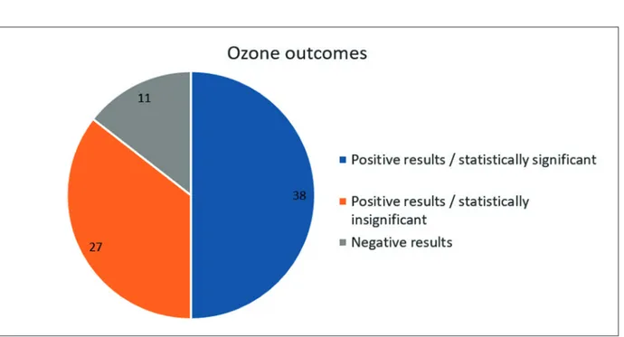
According to Figure 2, we also evaluated the outcomes as: i) positive results and statistically
significant; ii) positive results, but statistically insignificant; iii) poor or negative results.
It should be noted that physicians habitually handle drug dosages in mass units, mainly mg or g. On the other hand, the possibility of dealing with ozone in the gaseous state mixed with an-other gas (air, oxygen) or it is still in the gaseous state but solubilized in a liquid (water, buffer solutions) leads to having to clarify some aspects of the mode of expression of the concentrations units97.
As it can be observed, concentrations of gas-eous ozone in oxygen for medical use, more prop-erly than in air, are typically measured in units of the mass of ozone (i.e., µg or mg or g) per volume of air (i.e., cm3 or dm3 or m3 equivalent to mL or
L or 1,000 L, respectively) and they are generally validated by wet chemistry98.
Moreover, the same concentrations may also be expressed as %. When these conditions occur, it is necessary to introduce conversion factors mainly based on the molecular weight of ozone. Also, both atmospheric temperature and pres-sure affect the calculation. Typically, conversion factors for ozone in oxygen are made assuming a pressure of 1 bar (in some cases 1 atm) and a temperature of 20°C. They approximately equal to 20 in case of % by volume and to 13.5 in case
of % by weight (e. g., 2%w/v and 3%w/w equals to about 40 g/m3, respectively).
On the contrary, if the ppm values are ex-pressed, it should first be specified if they are ppmv (parts per million volume) or ppmw (parts per million weight). At this point and in accor-dance with the above, 1 ppmv O3 in O2 equals approximately 2 mg/m3.
On the other hand, when ozone is solubi-lized in a liquid, parts per million can be also expressed as milligrams of ozone per liter of a solvent (mg/L). This measurement is the mass of a chemical or contaminate per unit volume of water (note that ppm or mg/L on a lab report are equivalent).
Other than these aspects, together with the operating modes of the ozone generators, it is also important to underline the dependence of the ozone quality based on the feed gases. In particular, concentrated oxygen does not cause the formation of nitrogen dioxides in the ozone cell that prevents the formation of nitric acid as a factor damaging metallic details of the ozone generator with air as the feed gas. A further aspect to consider, sometimes present in the orig-inal researches, is the ozone output value of the different generators. Such a productivity level, P [g/h], is shown in a mass over time value, i.e., the mass of ozone produced by ozone generator in a
given period of time. This value can be calculated through the flow rate of the feed gas, R [m3/h] and
ozone concentration, C [g/m3] by:
P = R × C (1)
It is commonly known that the flow rate is measured in liters per minute (LPM); if this is the case, the output is calculated as:
P = [R × (LPM × 60) × 0.001] × C (2) Thus, knowing the ozone output and the flow rate of the feed gas, the ozone concentration can be calculated.
Moreover, the flow rate of feed gas controls the mass of oxygen molecules that interacts with plasma state in the cell to produce ozone. Low flow rate causes an increase of the contact time of oxygen molecules with the radicals (high en-ergy electrons) in corona and on the other hand, yielded ozone slowly is released from ozone cell. In total, it results in keeping high ozone concen-tration in ozone cell. The increase of flow rate leads to a decrease of ozone concentration. From an application point of view, high ozone concen-tration enhances the solubility of ozone in liquids. Eventually, the flow rate would first cause an increase of output, and then at a certain high flow
rate, the output reaches saturation or even slightly decreases. So, there is a need to choose the opti-mal flow rate for every sort of ozone generator99.
A completely different reasoning occurs in the case of ozonated derivatives deriving from vege-table matrices such as oils. In fact, ozone does not solubilize or dissolve in oil, but it reacts chemi-cally at the level of double bonds, mainly turning into the 1,2,4-trioxolane moiety100. To improve
the general knowledge of this topic, the Authors should provide the specific characteristics of the products. These features are very important be-cause the therapeutic activity of these functional dermatological matrices depends upon both the overall ozonation process and the variety of oil in use (such as olive, sunflower, sesame, and so on)101.
Furthermore, the lack of a standard method to ex-press the quantity of peroxidic derivatives present has to be considered102. To improve the general
knowledge of this topic, the modalities of ozo-nation (amount of oil, reaction time, temperature just to name a few) are aspects of primary interest other than the ozone gas concentration (generally up to 70 µg/ml, corresponding to about 5% of the gaseous mixture with the remaining 95% of oxy-gen) used during the ozonation process.
Once these aspects have been defined, the studies will be critically analyzed below accord-ing to the type of ozone usage: i) gaseous ozone; ii) ozonated water; iii) ozonated derivatives.
Gaseous ozone
In the case of applications of gaseous ozone as a therapeutic agent, some studies showing positive results, in qualitative and/or statistically significant terms, cannot be further commented as they are completely without indications of the quantity of ozone used21,23,24,28,41,42,94,96. The mere indication
of the model of the ozone generator used is not sufficient. For example, from consulting the tech-nical specifications of the equipment used, ozone concentrations from 1,000 to 100,000 ppm on the contact surface of the ozone electrodes are indicat-ed21. Moreover, the methods of use may be
man-ifold and it may no longer be in production24. A
study also reports the concomitant intraperitoneal systemic route of ozone administration43.
Going into the details of the studies where the concentrations are indicated and in light of all that has been previously said, the gaseous ozone concentration of 2100 ppm is equivalent to about 4 mg/L. This data justifies the low antibacterial activity27,32,33 as well as the inefficacy on dentin
hypersensitivity29,30, periodontitis53. However, the
same 2100 ppm can result in the improvement of palatal wound closure65, enamel bonds91 and
bac-teria number reduction92.
Analogously, the results of microbial reduction were not strong by continuous application of 140 ppm59, or the treatment of osteonecrosis of the
jaw with application of 20 ppm, a very low ozone concentration68.
Microorganisms exposed to ozone at 140 ppm/ min, for 6, 12, 18 and 24 s were completely elim-inated, excepted A. actinomycetemcomitans86.
On the other hand, few studies evaluate a comparison of the effects deriving from different dosages of use. Among them, noteworthy is the research about the effect of different concentra-tions of topical ozone administration on bone formation in orthopedically expanded suture in rats61. Gaseous ozone concentration of 25 µg/
mL in comparison to 10 µg/mL and 40 µg/mL provides faster bone regeneration, confirming the typical hormetic effect of ozone103.
Positive results were also obtained in either blood perfusion increase and quality of life64 or
aphthous ulcer resolution66, with well-described
dosage schedule twice ozone applications at 75 µg/mL for 30 sec, followed by a third at 30 µg/ mL and gradual concentrations between 10 and 100 µg/mL, respectively. For completeness’ sake, the maximum concentration of 100 µg/ml it is hard to explain using environmental oxygen from air as feeding gas66.
Ozone applications at a fixed concentration of 75 µg/mL, even if performed by an experienced investigator appears to be too high51, while
sig-nificant CFU count reduction in group treated with ozone, more evident in Lactobacillus than
Streptococcus mutans was obtained using 32 µg/
mL for 1 min31.
Ozone gas at a concentration greater than 4 g m-3 (the minimum concentration detectable by the
measuring device used) to 53 g m-3 (the highest
concentration achievable with the experimental set-up and the ozone generator), proved to be even more effective than chlorhexidine in killing oral pathogens87.
As expected, oral pathogens present on den-ture base resins were efficiently disinfected by gaseous ozone at 100 µg/mL for 10 minutes88,
while after 5 minutes, gaseous ozone at the con-centration of 12.8 mg/L was not able to reduce the counts of S. aureus and E. faecalis significantly89.
In conclusion, it can be deduced that the cor-rect concentration range of gaseous ozone is be-tween 15 and 40 µg/mL.