1
UNIVERSITÀ DEGLI STUDI DI CATANIA
Dottorato di Ricerca in Biotecnologie
XXVI Ciclo
Andrea Magrì
VDAC and SOD1: two major players
in mitochondrial metabolism and in ALS
TESI DI DOTTORATO
Relatore: Chiar.mo Prof. Vito De Pinto
Coordinatore: Chiar.mo Prof. Vito De Pinto
2
ABSTRACT
The Thesis work presented here has been devoted to two proteins, the
Voltage-Dependent Anion Channel (VDAC) and the Superoxide Dismutase I (SOD1) and has
been especially focused on the relationships between them in physiological or pathological conditions of the cell.
VDAC is a pore-forming protein located in the outer mitochondrial membrane, where it is suspected to play a key role in metabolism regulation, as the interface between mitochondria and cytosol, and in apoptosis regulation. In the small family of VDAC proteins, composed of three isoforms in chordates, VDAC3 is the least known. Conversely from isoforms 1 and 2, its ability to form pores has been questioned. In this thesis, we present the first complete electrophysiological characterization of VDAC3, showing that this protein is able to forms smaller pores compared to VDAC1, under physiological condition of pH. Another point examined here has been the gating of VDAC1. This protein, in vitro, shows the important feature of gating the pore in dependence of high voltage applied. It is believed that the N-terminal domain has a crucial role in voltage-dependent gating and in the stabilization of the protein through its interaction with the pore-wall.
By producing several mutants of VDAC1, in this work we have shown that the Voltage-dependence may be modulated in an asymmetrical way by modifying sequences or deleting β-strands required for the interaction with N-terminal domain.
The Superoxide Dismutase I (SOD1) is the most important antioxidant enzyme of all eukaryotic cells, since it inactivates the superoxide anion. Many recent evidences suggest that SOD1 is important for mitochondrial function, both in physiological and pathological conditions. SOD1 protects, among others, VDAC from oxidative stress, and may affect mitochondrial proteins expression levels. In addition, in the neurodegenerative disease Amyotrophic Lateral Sclerosis (ALS), SOD1 mutants were reported to directly bind the cytosolic surface of mitochondria, using VDAC1 as docking site. To understand the relationships between VDAC and SOD1, in this work, we studied the influence of hSOD1 on mitochondria when it was overexpressed in a yeast strain devoid of yeast
3
endogenous VDAC. Our results sturprisingly indicate that SOD1 may have a metabolic role and can support the mitochondrial recovery in the strain por1, heavily slowed down by the lack of porin1. Our results support the recent claim that SOD1 may act on the expression of other mitochondrial proteins. In addition, the characterization of the interaction between VDAC1 and two of the most diffused ALS-linked SOD1 mutants was also obtained and might be considered the molecular basis in understanding the mitochondrial involvement in ALS.
4
SUMMARY
1. INTRODUCTION ... 8
1.1 Yeast, a perfect model organism for metabolic studies ... 8
1.1.1 A story of success ... 8
1.1.2 Biology of S. cerevisiae ... 9
1.1.3 Yeast fermentative metabolism ... 10
1.1.4 Mitochondria and the respiratory metabolism ... 13
1.2 The VDAC family protein ... 15
1.2.1 Organization of the Outer Mitochondrial Membrane ... 15
1.2.2 The Voltage-Dependent Anion Channel. General features. ... 17
1.2.3 The structure of human VDAC1 ... 19
1.2.4 Electrophysiological features of VDAC1 ... 21
1.2.5 The role of VDAC1 in the cell ... 22
1.2.6 VDAC proteins in S. cerevisiae... 24
1.2.7 VDAC N-terminal domain ... 26
1.3 The Superoxide Dismutase I ... 27
1.3.1 Appearance of antioxidant enzymes ... 27
1.3.2 The Superoxide Dismutase family ... 29
1.3.3 Genomic organization of SOD1 ... 32
1.3.4 The structure of human SOD1 ... 32
1.3.5 SOD enzymes in S. cerevisiae ... 35
1.3.6 Distribution and localization of SOD1 ... 36
1.3.7 Antioxidant-independent roles of SOD1 ... 36
1.3.8 Relationship of SOD1 with VDAC1 ... 39
1.4 The Amyotrophic Lateral Sclerosis ... 40
1.4.1 Clinical features of ALS ... 40
1.4.2 SOD1 mutations in ALS1 ... 41
1.4.3 Impact of ALS-linked SOD1 mutants on mitochondria ... 42
2. AIM OF THE WORK ... 45
3. MATERIALS AND METHODS ... 46
3.1 Cloning ... 46
5
3.1.2 Plasmids ... 46
3.1.3 Cloning of human VDAC genes in pET-21a ... 47
3.1.4 Construction of VDAC1 deletion mutants ... 47
3.1.5 Cloning of human VDAC genes in pYX212 ... 50
3.1.6 Cloning of human SOD1 in pET-52b ... 50
3.1.7 Construction of ALS-linked SOD1 mutants ... 50
3.1.8 Cloning of human hSOD1 in pYX142 ... 51
3.1.9 Constructs pEGFP-N1 ... 52
3.2 Expression and purification of recombinant proteins ... 52
3.2.1 Expression and purification of VDAC proteins in E. coli ... 52
3.2.2 Refolding VDAC1 and VDAC1 mutants ... 52
3.2.3 Refolding of VDAC3 ... 53
3.2.4 VDACs purification from yeast mitochondria ... 54
3.2.5 Expression and purification of SOD1 proteins ... 54
3.3 In vitro analysis and characterization of recombinant proteins ... 55
3.3.1 Gel electrophoresis and Immunoblotting ... 55
3.3.2 Planar Lipid Bilayer ... 55
3.3.3 SOD1 activity and oligomerization assay ... 57
3.3.4 Microscale Thermophoresis ... 58
3.4 Yeast cellular biology ... 58
3.4.1 Yeast strain and growth conditions ... 58
3.4.2 Complementation assay... 59
3.4.3 Flow Cytometry analysis ... 60
3.4.4 Determination of mitochondrial membrane potential ... 61
3.4.5 Determination of total ROS ... 61
3.4.6 Treatment of yeast cells with hydrogen peroxide ... 61
3.4.7 Yeast spheroplast preparation by glass beads ... 61
3.5 Analysis of gene expression levels ... 62
3.5.1 Reverse Trascription PCR ... 62
3.5.2 Quantitative Real-Time PCR ... 62
3.5.3 Relative quantification of expression levels ... 64
3.6 Neuronal cell line ... 64
3.6.1 Maintenance of cell line and transfection ... 64
3.6.2 Determination of mitochondrial membrane potential ... 64
6
4.1 Electrophysiological characterization of human VDAC3 ... 66
4.1.1 Impact of human VDAC3 expression on Δpor1 yeast cells ... 67
4.1.2 Purification of VDAC proteins from yeast ... 67
4.1.3 Purification of VDAC proteins from E. coli ... 68
4.1.4 Electrophysiological characterization of human VDAC3 from yeast ... 69
4.1.5 Electrophysiological characterization of recombinant human VDAC3 ... 69
4.1.6 Conclusions ... 71
4.2 Electrophysiological characterization of VDAC1 mutated in domains possibly involved in voltage gating ... 73
4.2.1 Impact of N1-VDAC3 expression on yeast Δpor1 cells ... 74
4.2.2 Electrophysiological characterization of N1-VDAC3 ... 74
4.2.3 Voltage dependence analysis of N1-VDAC3 ... 75
4.2.4 Purification of VDAC1 deletion mutants from E. coli ... 76
4.2.5 Electrophysiological characterization of VDAC1 deletion mutants ... 77
4.2.6 Voltage-dependence of VDAC1 deletion mutants ... 79
4.2.7 Ion selectivity analysis ... 80
4.2.8 Conclusions ... 80
4.3 The hSOD1 expression in S. cerevisiae Δpor1 strain: impact on the mitochondrial metabolism ... 83
4.3.1 Expression of hSOD1 in Δpor1 yeast cells ... 84
4.3.2 Complementation assay of transformed Δpor1 yeast cells on glucose and glycerol ... 84
4.3.3 Complementation assay of transformed Δpor1 yeast cells on mitochondrial substrates ... 85
4.2.4 Growth curves of transformed Δpor1 yeast cells in glucose and glycerol ... 86
4.2.5 Measurement of mitochondrial membrane potential of transformed Δpor1 yeast cells ... 88
4.3.6 Determination of total ROS content in transformed Δpor1 yeast cells ... 90
4.3.7 Resistance to hydrogen peroxide of transformed Δpor1 yeast cells ... 91
4.3.8 Real-Time PCR analysis of the expression of genes for mitochondrial proteins in cDNA from transformed Δpor1 yeast cells... 92
4.3.9 Conclusion ... 96
4.4 Interaction between VDAC and ALS-linked SOD1 mutants: molecular characterization ... 100
4.4.1 Expression of hSOD1 proteins ... 101
4.3.2 Purification of hSOD1 proteins ... 102
7
4.4.4 VDAC1-SOD1 interactions by Microscale Thermophoresis ... 104 4.4.5 The influence of SOD1 proteins on the mitochondrial swelling ... 105 4.4.6 The influence of SOD1 proteins on the mitochondrial membrane potential 106 4.4.7 Conclusion ... 107 5. DISCUSSION ... 110 6. BIBLIOGRAPHY ... 114
8
1. INTRODUCTION
1.1 Yeast, a perfect model organism for metabolic studies 1.1.1 A story of success
The unicellular yeast Saccharomyces cerevisiae is one of the most known, studied and used model organisms. First traces of its use date back to ancient Egypt, even though, only in the XIX century Louis Pasteur fully realized the enormous potential of this microorganism for the development of biotechnology. Today, yeast is widely used for research and plays a key role for food and pharmaceutical industry, where it used for beer brewing, bakery, and production of recombinant molecules. The extraordinary success of this organism as research model organism depends on several aspects.
First, yeast is extremely easy to manipulate, requires simple and inexpensive culture media, has a short generation time and is not pathogenic. These properties allow for the swift production and maintenance of multiple specimen lines at low cost.
Second, S. cerevisiae genome was completely sequenced in 1996. The genome of 12068 Kb is organized in 16 chromosomes, and includes approximately 6200 ORFs (open reading frame) of which 5800 ORFs correspond to protein coding genes [1]. The complete genome knowledge has allowed to genetically modifying the yeast DNA, producing many deletion mutants, which played a key role in understanding the function of many proteins.
Third, S. cerevisiae is evolutionarily close to higher eukaryotes. It has been estimated that around 30% of yeast genes have homologous genes in humans; moreover, yeast shows absolutely conserved molecular mechanisms, such as DNA replication, RNA synthesis and processing, protein synthesis and post-traslational modifications, the main metabolic pathways, cellular respiration, signal transduction, cell cycle regulation, apoptosis [2]. Thus, higher eukaryotes genes are often successfully expressed in S. cerevisiae and, in many case, the heterologous protein is able to complement the absence of the endogenous one. The heterologous expression in yeast allows to study protein properties and
9
behavior, the effects of mutations and protein-protein interactions in a relatively simple system.
1.1.2 Biology of S. cerevisiae
S. cerevisiae is a unicellular yeast belonging to the class of Ascomycetes. Cells show
an ellipsoidal shape, with a diameter between 5 and 10 micrometers, and are characterized by a resistant outer wall essential for yeast survival (Fig. 1A). Cell wall is composed prevalently by polysaccharides, β(1→3)-d-glucan and β(1→6)-d-glucan, that appear to have a structural function, and mannoproteins that act as “filler” and are important for the permeability of the wall [3]. As an eukaryote, S.
cerevisiae shares the complex internal cell structure of plants and animals cells,
with a well-defined nucleus, containing a genome that is devoid of the high percentage of non-coding DNA (typical of higher eukaryotes), and all organelles (Fig. 1B).
S. cerevisiae generally reproduces by budding, doubling their population around
every 100 minutes, although growth rates vary depending from several conditions. Yeast growth is synchronized with bud growth, which reaches the size of the mature cell by the time it separates from the parent cell; both mother and daughter cells can initiate bud formation before cell separation has occurred.
A
B
C
Fig 1 Yeast morphology and life cycle. A) Yeast S. cerevisiae morphology showed by SEM microscopy. B) A thin-section of budding yeast cell by electron microscopy; N indicates nucleus, M mitochondria.
Picture taken from [4]. C) Schematic representation of life cell cycle of yeast; it is showed the alternation between diploid and haploid population.
10
S. cerevisiae is a haplodiplont organism, which may alternates haploid or diploid
generations (Fig. 1C). The haploid stage represents the asexual form of yeast and undergoes a simple life cycle of mitosis and growth; however, under stress conditions, haploid cells generally die. Conversely, the diploid stage represents the sexual and the preferential form of yeast and, similarly to the haploid, undergoes a simple life cycle of mitosis, growing with a faster rate.
Under stress conditions, diploid cells are more resistant and are subjected to
sporulation by meiosis, a process that promotes the production of four haploid
spores characterized by two different mating type, called a and α (alpha) [5]. Thus, the haploid cells of opposite mating type can mate to form diploid cells that can sporulate, to form another generation of haploid cells, or continue to exist as diploid cells. As in many other eukaryotes, aim of mating is promoting the genetic recombination, through the production of novel combinations of chromosomes and combine plasmids and proteins.
Yeast metabolism was well characterized. Yeast is able to grow in presence of many carbon sources, but independently from them, yeast normally grows in three main phases. When yeast cells are inoculated in a fresh growth medium, cells enter an adaptation phase, called lag phase, in which they are biochemically active but not dividing and number of cells remains relatively constant. Length of lag phase strongly depends from carbon source, but it also influenced by several factors, such as temperature, oxygen and pH. During this phase, cells activate the most appropriate metabolic pathways, according to nutrients availability in growth medium. Once metabolism is completely active, cells start DNA replication and cell division: this is the second phase of growth, called exponential phase, in which the cells grow most rapidly. The third phase of growth is the stationary phase: metabolism slows and the cells stop rapid cell division. The factors that cause cells to enter stationary phase are related to change in the environment, such as lower availability of nutrients, normally caused by high cell density.
1.1.3 Yeast fermentative metabolism
S. cerevisiae is characterized by fermentative or respiratory metabolism,
11
media and oxygen availability have a strong impact on metabolism physiology. Yeast can use a broad set of carbon sources such as alcohols, amino acids and organic acids, which can support their growth; however, yeasts preferentially metabolizes sugars, and glucose certainly represents the favorite carbon source also for S. cerevisiae [6].
Glucose utilization starts with the uptake of the sugar into the yeast cells, thanks to the presence of specific carriers located on the plasma membrane. S. cerevisiae has a large gene family codifying for hexose transporters, that consists of more than 20 members, including 18 genes encoding for transporters (HXT family, GAL2) and 2 genes encoding for sugar sensors (SNF3, RGT2) [7]. Once in the cytosol, glucose metabolism occurs prevalently through glycolysis and pentose phosphate pathway, although this last pathway is predominantly used for NADPH production [8-9]. The concentration of sugars in the growth medium strongly influences the enzyme levels of several metabolic pathways. Glucose acts on an extensive transcriptional regulation of a large number of genes, leading to the adaptation to fermentative metabolism such as genes required for the glycolytic pathway.
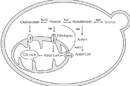
Fig 2 Yeast fermentative metabolism. Schematic representation of glucose metabolism
that occurs prevalently by glycolysis and alcoholic fermentation. Glucose undergoes through glycolysis, producing 2 ATP and 2 NADH molecules; the final product of glycolysis, pyruvate, undergoes decarboxylation up to ethanol, that is normal secreted outside the cell. Picture taken from [6].
12
At the same time, glucose acts as repressor of genes encoding for proteins involved in the gluconeogenic and respiratory pathways, a process known as catabolite
repression [10].
Through glycolysis, glucose is metabolized to pyruvate, whereby production of energy, in form of two molecules of ATP per each molecule of glucose, is coupled to the generation of reducing intermediates as NADH [11]. Pyruvate can follow different metabolic fates, which strongly depends on the environmental conditions.
In anaerobic conditions, pyruvate metabolism occurs through the alcoholic
fermentation, in which it is decarboxylated to give acetaldehyde and CO2; then,
acetaldehyde is converted in ethanol that is normally secreted outside the cell together with CO2, and this process promotes the re-oxidation of NADH to NAD+,
making it available again for glycolysis. The alcoholic fermentation is the basis of some industrial processes, such as bakery: CO2 is responsible for the formation of
bubbles in the bread while ethanol evaporates during cooking. Absence of oxygen does not represent a strict rule for alcoholic fermentation: indeed, even in the presence of high levels of oxygen, yeasts normally choose to use fermentation instead of respiration, when glucose is fully accessible. This phenomenon, known as Crabtree effect, consists in the inhibition of gene expression involved in aerobic metabolism when glucose is available, which may occur both in the presence or absence of oxygen [12]. In fact, although fermentation is not by itself a highly energetically efficient process, the high concentration of glucose strongly promotes the acceleration of glycolysis, producing appreciable amounts of ATP and allows the yeast to grow in a rapid and exponential way.
As the level of glucose drops, the diauxic shift occurs, that promote the yeast adaptation from fermentative to respiratory metabolism. The repression of respiration mediated by glucose is left and synthesis of proteins involved in the mitochondrial respiration machinery occurs. In this stage, ethanol becomes the primary mode of ATP generation by respiratory metabolism and the overall growth rate becomes much slower: this is the main feature of the stationary phase [13].
13
1.1.4 Mitochondria and the respiratory metabolism
Respiratory metabolism starts with the presence of the acetyl–cofactor A (acetyl-CoA), considered the first intermediate of Tricarboxylic Acid (TCA) Cycle. Acetyl-CoA can be obtained from metabolism of both fermentable and non-fermentable carbon sources, through pyruvate formation, which undergoes, through oxidative decarboxylation, catalysis by the pyruvate dehydrogenase enzymatic complex. Other source of acetyl-CoA is the oxidation of fatty acids, as well as the final product of fermentation, ethanol: under certain conditions, it is re-converted in acetaldehyde up to the formation of acetyl-CoA.
In TCA cycle, acetil-CoA molecules are totally oxidized to CO2, furnishing a set of
intermediates commonly used by other metabolic reactions [14]. Electrons, obtained from decarboxylation, are able to reduce NAD+ and FAD molecules to
NADH and FADH2 respectively, that are high-energy intermediates; then, NADH
and FADH2 produced will be re-oxidated during oxidative phosphorylation, a
process that convert potential energy in ATP molecules [15].
TCA cycle, as well as oxidative phosphorylation, occurs in a separate cellular compartment, the mitochondria. They are membrane bound organelles that represent the "power plants" of most eukaryotic cells. Their primary role, indeed, consist in the conversion of most part of the energy to ATP, even if they are involved in a set of interconnected metabolic functions, such as storage of calcium ions and modulation of cell cycle and apoptosis [16-17].
Mitochondria show a diameter between 0.2 and 1 μm with a peculiar structure that strongly reflects the function: they are characterized by two separate and functionally distinct membranes, the outer and the inner mitochondrial
membranes, that encapsulate respectively the intermembrane space and the matrix. In addition, mitochondria are the only cellular organelles in animals that
have their own DNA, which codifies for few mitochondrial proteins, while most of the proteins are encoded by nuclear genes and then imported into mitochondria. Matrix is the site of mitochondrial DNA, of enzymes involved in DNA replication and in the TCA cycle: it is in the matrix that the oxidative degradation of metabolites occurs.
14
The inner mitochondrial membrane (IMM) contains the matrix and represents the principal site of ATP generation. Its composition is different from the others cellular membranes, since the lipid content resembles that of the prokaryotic membranes. In addition, IMM shows high level of impermeability to ions and small molecules, a critical property for maintaining the proton gradient that drives oxidative phosphorylation. IMM surface area is substantially increased by its folding into
cristae that contain an unusually high percentage of proteins, including ATP
synthase and a variety of cytochromes used in oxidative phosphorylation, as well as proteins involved in metabolites transport. Indeed, the high-energy electrons from NADH and FADH2 are transferred to molecular oxygen through a series of
carriers, located in the inner membrane of mitochondria and ranked according to a decreasing redox potential. The electron transfer reactions generates energy that is converted to potential energy stored in a proton gradient across the membrane, which is then used to drive ATP synthesis through ATP synthase.
Despite the electron transfer along the transport chain takes place in a controlled and selective way, it can generate as intermediates oxygen species partially reduced, called Reactive Oxygen Species (ROS), which can be harmful to the mitochondria and to the entire cell because of their reactivity. However, cells have developed enzymatic defense mechanisms against ROS aimed to destroy oxygen radicals.
Fig 3 Yeast respirative metabolism. Schematic representation of pyruvate metabolism by
TCA cycle. Pyruvate is transformed in acetil-CoA, that represent also the final product of fatty acids oxidation. Acetil-CoA is totally decarboxilated to CO2 producing high-energy
equivalents, which represent a source of potential energy for ATP production. Picture taken from [6].
15
In contrast to the IMM, the outer mitochondrial membrane (OMM) is highly permeable to small molecules, making the ion composition of the intermembrane space (IMS) similar to the cytosol. Thus IMM is considered the functional barrier against the free passage of small molecules between the cytosol and the matrix. It is able to maintain the proton gradient between the two sides of the membrane. Consequently, OMM plays a key role in the metabolic exchanges between cytosol and mitochondria; it owes its high permeability to its high content of pore-forming proteins, called porins, which form channels that allow the diffusion of ions and small molecules up to around 6000 Da, and specific transport proteins for larger molecules, which can cross OMM by active transport.
1.2 The VDAC family protein
1.2.1 Organization of the Outer Mitochondrial Membrane
The Outer Mitochondrial Membrane (OMM) represents the interface between cytosol and mitochondria. It shows a peculiar protein composition. OMM contains proteins involved in mitochondrial mobility (through its interaction with microtubules), apoptotic factors, enzymes, and others, involved in active and passive transport of proteins, ions and metabolites across OMM [18-19].
Most of these mitochondrial proteins are synthetized in cytosol, and are targeted to mitochondria by a pre-sequence of 20-35 amino acids, located at the N-terminal domain, which will be removed by proteolytic cleavage once the protein is imported into the organelle. Thus, OMM contains several proteins involved in traslocation of proteins: the Translocator of Outer Membrane, TOM complex, binds the pre-sequence of the polypeptide and promotes its insertion into OMM or the passage into the IMS, and the Translocator of Inner Membrane, TIM complex, able to allow the sorting of the protein to the mitochondrial matrix and to the other subcompartments [20].
One of the most abundant protein families of OMM is represented by the mitochondrial porins. They are integral membrane proteins that form a pore through which small molecules can selectively cross the OMM; porins are also suspected to modulate the metabolite flux by a not yet elucidated mechanism of
16
gating of the channel. Porins were initially discovered in 1974 in the outer membrane of the Gram-negative bacteria Escherichia coli; two years later, the existence of porins was proved also in eukaryotes: they were found in mitochondria of Paramecium tetraaurelia [21]. Afterward, porins with very similar features were identified in mitochondria from fungi, plants, metazoans and invertebrates.
The bacterial porin Omp F represents a typical example of porin: it has inspired also the structural investigations of the eukaryotic porins. Omp F is a non-specific pore with a weak cation selectivity that allows the passive diffusion of small and polar molecules of around 600-700 Da, such as water, ions, glucose as well as waste products [22].
Omp F forms a trimeric complex in the bacterial outer membrane (Fig. 4). Each Omp F subunit spans the membrane with 16 β-strands that form a β-barrel structure. It is characterized by hydrophilic amino acid residues, protruding towards the aqueous inside of the pore, alternating with hydrophobic residues, in contact with the lipid bilayer of the membrane. Furthermore, the connections between the β-strands are very short ("turns") from the periplasmic side of the membrane and long loops on the external side of the membrane. In particular a major loop about 40 amino acids long (L3) is folded inside the pore’s lumen and modulates its diffusion properties [23].
B A
Fig 4 Tridimensional structure of Omp F. The bacterial porin Omp F forms trimeric structure on the
surface of outer membrane of Gram-negative. A) The side view of the trimer complex of Omp F. B) The view from above of the complex. In yellow are indicated the loops inserted in the lumen of each sibunit, that are involved in gating process.
17
Omp F was characterized using electrophysiological methods, such as reconstitution in planar lipid bilayer, and incorporation in proteoliposomes loaded with sugars of permeable size. These experiments have demonstrated the voltage-dependence of Omp F, which consists in the modulation of opening/closing activity of the channel, a phenomenon happening only when the applied voltage is high [24].
Comparison of the sequences of prokaryotic porins with eukaryotic counterparts (VDAC; see below) shows only about 13% of sequence homology; despite this, the two proteins have similar secondary structure.
1.2.2 The Voltage-Dependent Anion Channel. General features.
Voltage-Dependent Anion Channel or VDAC indicates a family of pore-forming
proteins of similar size of 28–32 kDa and around 280 amino acids, characterized by pores slightly selective for anions, with a conductance of around 4 nS. Their name indicates that the conductance may vary as well as the selectivity in dependence of the voltage applied when proteins are reconstituted on planar lipid bilayer [25]. Mammals, as well as most chordates, express three clades corresponding to three VDAC isoforms, called VDAC1, VDAC2 and VDAC3. From such evolutionary analysis, it is generally accepted that VDAC3 is the oldest protein, while VDAC1 is considered the most recent [26]. The organization of the three VDAC genes is very similar: genes are made of the same number of coding exons, sharing exactly the same size, with the VDAC2 gene containing an additional first exon encoding for the short pre-sequence of 11 amino acids, a peculiar feature of this isoform [27]. VDAC isoforms 1 and 2 are expressed more or less ubiquitously in all eukaryotes, while VDAC3 is more abundant in celebral cortex, liver, hearth, testis and spermatozoa [28]. In addition, analysis of the expression levels of human VDAC isoforms in HeLa cells, determined by Real-Time PCR, have shown that VDAC1 is the most abundant isoform, 10 times more abundant compared to VDAC2 and hundred times more abundant compared to VDAC3. Moreover, the over-expression of each single VDAC isoform affects the mRNA levels of the other two isoforms, suggesting that the ratios between VDAC isoforms are subject to a reciprocal control that avoids an imbalance among these proteins [29].
18
The multi-alignment of the three human VDAC protein sequence shows a high level of sequence conservation. Moreover, comparison between human and yeast VDAC1 homologous, POR1, suggests a sequence conservation of porins through evolution. Since the sequences are highly conserved, the structures of the three human VDAC isoforms results very similar and characterized by the typical β-barrel conformation. Despite this, the three human isoforms display different functional properties.
Several experiments have demonstrated that recombinant VDAC1 and 2 are able to form pores in planar lipid bilayers (PLB), while VDAC3 shows a reduced ability to insert into the membrane [30]. However, in a very recent work it was demonstrated that VDAC3, in certain conditions, forms very small pores compared to the others isoforms, with a lower conductance [31]. These different features showed by VDAC3 may be partially explained by the presence of a higher content in cysteine residues, which can modulate its insertion on PLB under certain conditions. Moreover, these features have suggested an unknown and peculiar role for this isoform.
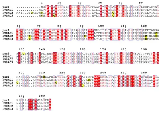
Fig 5 Comparison between yeast and human VDAC proteins sequences. The yeast porin POR1 was
aligned with the three human VDAC isoforms. Sequences show high levels of identity, which can be calculated to be around 70%. In yellow are underlined the cystein residues, considered particularly important for VDAC3 peculiar features. Picture taken from [27].
19
1.2.3 The structure of human VDAC1
VDAC1 is the most studied isoform of the three mammal VDAC proteins. In the past years, several studies based on structure prediction have suggested the presence of a transmembrane β-barrel domain associated to α-helix domain with amphipathic feature, corresponding to the N-terminal sequence of the protein [32]. This structural organization, common to others transmembrane proteins with different function, such as the subunit of TOM complex, TOM40, was confirmed in 2008 by the analysis of data obtained combining NMR and X-ray crystallography and independently by three groups [33-35].
The pore resembles an elliptical shape of approximately 3.1 x 3.5 nm in the horizontal dimensions and approximately 4 nm in the vertical directions; the diameter of the pore’s lumen is reduced to approximately 1.5 x 1 nm (Fig. 6). Surprisingly, at variance with bacterial porins formed by an even number of strands (usually 16 strands), VDAC1 barrel is organized with 19 antiparallel β-strands, approximately 10 amino acids long. The odd number of strands requires one parallel interaction between the adjacent β-strands 1 and 19.
The N-terminal region has been much studied. CD and NMR studies have shown that a synthetic peptide corresponding to VDAC1 residues 2–20 exists as an unstructured peptide in aqueous solvent, while it forms α-helix structure from residues 5–16 in SDS [36]. The amphipathic α-helix is not part of the transmembrane domain: in particular, the portion including residues between Tyr7 and Val17 is located halfway into the pore’s lumen, approximately in a central position corresponding to the hydrophobic layer of the membrane [33]. The N-terminus interacts with several strands, less stable than other regions of the β-barrel, probably due to the possibility of VDAC1 N-terminus to assume different conformations. As a cause or an effect of these conformational changes, the hydrogen bonds between the α-helix and the β-barrel are temporarily destroyed and rebuilt. Moreover, the fact that the α-helix is not a hydrophobic domain suggests that it could not be inserted permanently in the lumen, but can resides transiently on one side of the phospholipid bilayer [37]. The features of N-terminal domain will be discussed later.
20
A
B C
Fig 6 Architecture of human VDAC1. A) Schematic representation of the secondary structure of
VDAC1, in which the amino acids in squares denote β-strands structure while all others amino acids are represented in circle. Red lines indicate the interaction between amino acids of the two interacting β-strands. Picture taken from [34] B) Tridimensional structure of VDAC1 from side view; in the enlargement it is showed the C-terminal domain of the protein. C) Structure of VDAC1 view from above; it shows the α-helix N-terminal domain located into the pore’s lumen. The enlargement shows the interaction between the helix and specific residues of the β-barrel. Pictures taken from [33].
21
Due to its conformation, VDAC1 needs a specific transport system to the OMM. Mitochondria have developed several different system for the import of nuclear-encoded proteins. The fate of VDAC is inextricably related to the presence of the
Sorting and Assembly Machinery complex, SAM complex, which promotes the
incorporation of β-barrel proteins into the OMM. SAM complex consists of a membrane-embedded component, the subunit SAM50 that has been shown to have a conserved role in VDAC biogenesis from yeast to mammalian. SAM complex is also important for the incorporation of TOM40, the main component of TOM complex, that, in turn, facilitates the recognition of most pre-proteins with a mitochondrial targeting sequence, their transfer through the membrane and the insertion of resident outer membrane proteins. Interestingly, as VDACs, also TOM40 and SAM50 are predicted to have a β-barrel topology [38].
1.2.4 Electrophysiological features of VDAC1
Functional properties of VDAC proteins have been examined using typical methods used for transmembrane proteins with a transfer function, which consist in their reconstitution in artificial phospholipid bilayers, such as vesicles or PLB. Despite the phylogenetic distance among eukaryotes as well as the divergence between protein sequences, analysis of reconstituted VDAC has shown a highly conserved pore formation activity and electrophysiological properties [39].
VDAC channels show a conductance or 4-4.5 nS in 1M KCl, which corresponds to the maximum theoretical value of a channel with VDAC dimension [40-41]. In addition, VDAC shows a weak anion selectivity and this feature can be perfectly explained by the presence of positively charged residues on the pore’s lumen [35]. The main VDAC feature is its dependency from voltage.
At low voltages, from 0 up to around ± 10-20 mV, VDAC is stable in the open state, while at high voltages, either positive or negative, and starting from ± 30-40 mV, VDAC switch in a set of multiple partially closed sub-states with different ionic selectivities and permeabilities [42], as showed in Fig. 7.
A direct involvement of N-terminal domain in voltage-gating properties of VDAC1 was proposed.
22
Indeed, the removal of N-terminal domain from VDAC1 results in the loss of voltage dependency: reconstituted VDAC1 in PLB maintains the open state at applied potentials higher than ± 40 mV [43]. The N-terminal domain is considered the most flexibly moiety of the entire protein; it was hypothesized that this domain can interact with different portion of the β-barrel lumen, modulating the opening and closing of the channel [44]. This hypothesis is supported by the presence of several hydrophobic residues on the β-barrel and on N-terminal domain, that can transient interact through hydrophobic interaction (see VDAC1 crystal structure in [34]).
1.2.5 The role of VDAC1 in the cell
VDAC proteins play a primary role in cell metabolism since they are considered responsible of the metabolic flux across OMM, which is crucial for the mitochondrial function and cell viability (Fig. 8). Metabolites, such as pyruvate and succinate, small molecules such as ATP/ADP, ions such as phosphate, magnesium and calcium, they all use VDAC pores to cross OMM [45-46]. In addition, VDAC interacts with several enzymes involved in glycolysis, such as hexokinase,
glucokinase and glycerol kinase: this location guarantees to the enzymes a
preferential access to mitochondrial ATP.
In general, VDAC behaves as an interface between cytosol and mitochondria, interacting with many cytosolic and mitochondrial proteins; these interactions, at times, can modulate VDAC activity and affect several physiological and pathological cellular mechanisms.
Fig 7 VDAC1 voltage-dependence. The average
VDAC1 conductance analyzed at PLB is showed as function of the applied voltage; this voltage-dependence diagram for VDAC1 is a common and well known feature.
23
Hexokinase (HK) isoforms I and II catalyze the first step of glycolysis, using VDAC as mitochondrial docking site, and this interaction was proposed as a central point in promoting growth and survival of cancer cells [47]. Indeed, cancer cells show high propensity to utilize glucose via glycolysis at a much higher rate than normal cells, a phenomenon known as Warburg effect. It was found that HKI and II were overexpressed in many types of cancer, including colon, prostate, lymphoma, glioma, gastric adenomas, carcinomas and breast cancers; thus, the interaction between HK and VDAC proteins increases ATP availability and promotes the high rate of tumor growth [48-49].
In addition, the HK-VDAC1 binding protects cancer cells from mitochondria-mediated cell death. It has been demonstrated that VDAC regulates apoptosis through interaction with Bcl-2 proteins, a family of regulators proteins of apoptosis in mammals, which includes both pro- and anti-apoptotic members. Under certain stimuli, the pro-apoptotic proteins Bak and Bax translocate to mitochondria and interact specifically with VDAC1, where they promote a death signal leading to OMM permeabilization, Cytochrome C (Cyt C) release and caspase activation [50-51]. In cancer cells, HK interaction with VDAC1 protects cells from apoptosis, blocking the interaction of Bax with VDAC1 [48].
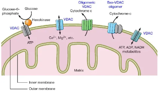
Fig 8 VDAC functions in OMM. VDAC represents one of the most important metabolic protein,
since its involvement in ATP, metabolites, ions transport across OMM, and its interaction with several enzymes, such as Hexokinases, involved in glycolysis. However, VDAC plays a key role also in apoptosis regulation. VDAC can interact with the pro-apoptotic protein Bax, and it is supposed to be directly involved in Cytochrome c releases, the most known hallmark of mitochondria-mediated apoptosis. Picture taken from [45].
24
Independently from HK interaction, VDAC1 regulates apoptosis in response to several stimuli, such as increased levels of cytoplasmic calcium or ROS: in these conditions VDAC promotes OMM permeabilization that culminates with release of many apoptotic factors, such as Cyt C and AIF. These factors are responsible of caspase activation, destroying the cells from within. It has been proposed a direct VDAC involvement in OMM permeabilization and Cyt C releases: VDAC may form oligomers or hetero-oligomers with pro-apototic proteins Bax, that may result in larger pores through which Cyt C is released [52].
1.2.6 VDAC proteins in S. cerevisiae
The most known mitochondrial porin of S. cerevisiae is encoded by the single copy genes por1 and is called POR1 or YVDAC1, which represents one of the most expressed cellular protein, with more of 67000 unit per cell. As in high eukaryotes, POR1 provides the metabolic exchanges and it is involved maintenance of mitochondrial osmotic stability and mitochondrial membrane permeability.
Por1 gene is the fifty-fifth ORF of chromosome XIV and is constituted by a single
exon of 852 bp that encodes for a protein of 283 amino acids. The protein shows a sequence homology of about 40% with the three isoforms of human VDAC and ability to form channel in PLB [53].
POR1 represents a fundamental protein for yeast viability under certain conditions. Yeast cells devoid of endogenous POR1 (Δpor1) show a partial growth defect on a non-fermentable carbon source, such as glycerol at 30°C, that is aggravated at elevated temperature, such as 37°C [54]. However, the ability of the
Δpor1 mutant of M22-2 yeast strain to grow at lower temperature means that
some other protein must be able to partially substitute for that function. The analysis of yeast genome resulted in the discovery of a por1 gene paralog, called
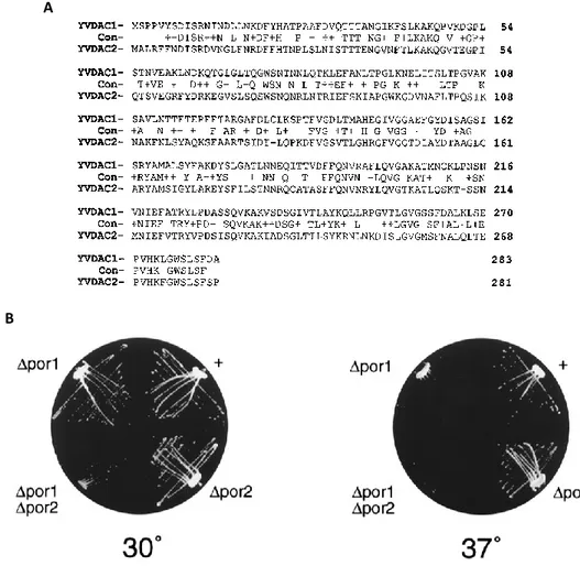
por2. The encoded protein POR2 or YVDAC2 is definitely less characterized. It
shares with POR1 around 49% of sequence homology (Fig. 9A) and, if overexpressed, is able to complement Δpor1 growth defect on glycerol. While deletion of por2 gene had no detectable phenotype, double mutant lacking both porins genes (Δpor1Δpor2) is viable but shows a more severe growth defect (Fig. 9B).
25
However, POR2 was not able to form channels in reconstitution experiments [55]. Indeed, mitochondrial membranes from wild-type cells and from single and double mutant overexpressing POR2, do not confer any permeability when incorporated to liposomes. These results strongly suggest that POR2 is normally not able to form channels.
The Δpor1 yeast strain has become a very convenient model system to study heterologous VDAC proteins from other sources. For example, it has been showed that mouse or human VDAC isoforms can partially or totally complement the absence of endogenous VDAC in yeast. While VDAC1 and 2 can fully recover the yeast growth defect, VDAC3 can partially recover the growth defect, underlining once again a difference in VDAC3 function [56-57].
Fig 9 Properties of yeast VDAC proteins sequences. A) Alignment between the two yeast VDAC
isoforms; sequence identities are indicated by the single-letter amino acid code in the line labeled Con. Dashes in the POR2 sequence indicate gaps introduced to optimize alignment. B) Growth of Δpor1, Δpor2 and double mutant yeasts compared to wild-type yeast (+). Strains were streaked on glycerol based medium and incubated at the indicated temperatures. Picture taken from [55].
A
26
1.2.7 VDAC N-terminal domain
The VDAC1 N-terminal domain has been the subject of considerable interest. This domain is not only involved in voltage gating, as previously described, but it plays a key role in interaction with other molecules such as HK [58] and in apoptosis regulation: N-terminal deletion, indeed, decreases the pro-apoptotic effect of VDAC1 over-expression [43]. In addition, N-terminus is considered responsible of the gating of the pore, due to conformational changes or movements mediated by this moiety [60]. Particularly, have been identified several hydrophobic residues on both β-barrel and N-terminus, that can transiently interact. NMR analysis have revealed that the contact between two part of the protein is based on the interaction between the methyl groups of Leucine 10, located on the N-terminal α-helix, and the Valine 143 and Leucine 150, located on the opposite side of the β-barrel. This data is supported by the fact that Val 143 and Leu 150 are the only residues with a hydrophobic side chains in the barrel wall pointing to the lumen. [34, 60]. A model of interaction is reported in Fig. 10.
Most part of the differences between human VDAC isoforms are present in the N-terminal domain. Compared to VDAC1, isoform 2 shows an N-N-terminal domain longer of 12 amino acids, even if this difference does not modify the voltage-dependency and the ability to complement the absence of endogenous porin in
Fig 10 N-terminus involvement in voltage gating. A gating models for VDAC1. Shown in yellow and
red is the 2.3 Å crystal structure of VDAC-1 (PDB: 3EMN). The side chains of residues Leu 10, Val 143 and Leu 150 are shown in stick representation and their hydrophobic contact point is marked with an asterisk. In each model, a certain amino acid segment, which is marked red, undergoes a conformational exchange to a fictive, magenta conformation, indicated by black arrows. Picture taken from [60].
27
Δpor1 strain. The N-terminal region of VDAC3 differs form VDAC1 for presence of
two cysteine residues in position 2 and 8. Cysteine content, that represents a peculiarity of isoform 3, seems to be the basis of the different behavior of this isoform. To fully understand the peculiar role of VDAC3 N-terminal domain, a set of chimerical protein were created by the swapping of the first 20 amino acids of VDAC3 with the corresponding regions of VDAC1 (N1-VDAC3 chimera) and VDAC2 (N2-VDAC3), and then expressed in yeast S. cerevisiae Δpor1 strain. It has been found that expression of these chimerical proteins in yeast cells confers an increased life span compared to VDAC3 and confers resistance to the yeast against incubation with reactive oxygen species [61].
1.3 The Superoxide Dismutase I
1.3.1 Appearance of antioxidant enzymes
The appearance of molecular oxygen on Earth can be considered the factor most responsible of the environmental changes started around 3.5 billions of years ago. Produced by the first photosynthetic cyanobacteria, oxygen has started to accumulate in the atmosphere and, for the first time, to interact with oxidizable elements such as iron, modifying the atmosphere composition through the conversion of methane in carbon dioxide [62]. This process provided new opportunities for the living organisms, energetically limited at that time, promoting the biological diversification: the oxygen availability has increased significantly the energy sources and imprinted an important thrust for metabolism evolution.
Life, as we know it today, requires oxygen for its existence and has developed cellular systems able to use oxygen efficiently, such as mitochondria; nevertheless, oxygen is considered a highly reactive molecule. The molecular oxygen (O2) is a
bi-radical containing two unpaired electrons in the outer shell; this molecule can only react with one electron at a time, producing a set of relatively stable intermediates known as ROS, Reactive Oxygen Species [63]. The product of one-electron reduction of O2 is the superoxide anion (O2-•). Quite toxic for the cell, superoxide
28
dismutation produces hydrogen peroxide (H2O2), which may be fully reduced to
water or partially reduced to hydroxyl radical (OH•) [64].
The primary source of superoxide anion in vivo is represented by mitochondrion: most steps in the mitochondrial electron transport chain involve single-electron reactions, further favoring the superoxide anion production [65].
Although small fluctuations in the steady-state concentration of ROS plays a key role in intracellular signaling, accumulation of ROS in the cell actually promotes damage in mitochondria and cellular organelles. In general, harmful effects of ROS are represented by damages of mitochondrial and nuclear DNA [66], lipid peroxidation [67], oxidations of amino acids in proteins, oxidation of co-factors in specific enzyme [68].
Many molecules, introduced by the diet, show an antioxidant activity: e.g., ascorbic acid (vitamin C), with its reducing activity, may be oxidized by ROS with the loss of two electrons (one by one, forming a radical intermediate) to form a stable molecule of dehydroascorbic acid. Other molecules, such as vitamin E, glutathione, polyphenol, uric acid, use a similar mechanism. However, cells have evolved well-defined strategies to defend themselves against ROS damage. Specific enzymes, such as Superoxide Dismutases (SOD), Catalases, Peroxiredoxins and Glutathione peroxidases, destroy ROS using a more complex mechanisms; indeed, all these enzymes contain one or more metal co-factors directly involved in the catalysis. Differently from antioxidant molecules, enzymes act in specific manner: e.g., SOD catalyzes dismutation of superoxide anion producing hydrogen peroxide that, in turn, represents the substrate for catalases and peroxidases. Despite the presence of this massive antioxidant arsenal in the living organisms, ROS can be responsible of several inflammatory states and pathologies. It has been demonstrated by many studies that excessive amounts of ROS can lead to cell injury and death, contributing to development of diseases as myocardial infarction, diabetes and cancer; particularly, ROS can react with genomic DNA, resulting in mutations that potentially affect cell cycle [69]. ROS accumulation is also associated to several neurodegenerative diseases, such as Parkinson’s and Alzheimer's disease and several forms of schizophrenia [70]. In addition, several
29
evidences correlated ROS accumulation with aging, proposing that free radicals underlie the aging process itself.
1.3.2 The Superoxide Dismutase family
The Superoxide Dismutase (SOD) enzymes are a big family belonging to the
oxidoreductase class. Initially classified as a metalloprotein with unknown
function, SOD activity was discovered in 1969 by Fridovich and McCord. SOD enzymes catalyze the dismutation of superoxide anion into molecular oxygen and hydrogen peroxide, due to the presence of a metal co-factor in the active site [71]. It has been estimated that under physiological conditions, the rate of the enzymatic reaction is around 104 times faster than that of the spontaneous
disproportionation of superoxide anion in solution. Reaction of dismutation mediated by SOD is composed by two sequential steps, and is described below:
M(n+1)+-SOD + O
2- → Mn+-SOD + O2
Mn+-SOD + O
2- + 2H+ → M(n+1)+-SOD + H2O2
in which M represents the metal co-factor and n is the oxidation state of metal cation, that oscillates between n and n+1.
Based on the metal co-factor, SOD proteins are divided into three major groups, as showed in Fig. 11. They are:
Cu/Zn-SOD, containing both copper and zinc ions, shows dimeric or
tetrameric structure and it is localized mainly in cytosol of all eukaryotes;
Fe-SOD and Mn-SOD, containing alternatively iron or manganese ions,
show tetrameric structure and were found prevalently in prokaryotes and protists, but also in plant chloroplast (Fe-SOD) and mitochondria (Mn-SOD);
Ni-SOD, containing nickel, shows hexameric structure and it was found
30
Humans, such as most mammals and chordates, contain three different SOD isoforms encoded by three different genes, whose structures have been extensively characterized [73]. The sod1 gene is located on chromosome 22 (21q22.1) and encodes for a dimeric Cu/Zn-SOD, located mainly in the cytosol. The protein SOD1 represents the most abundant isoform and performs around 95% of dismutase activity. SOD1 will be described subsequently.
Sod2 gene is located on chromosome 6 (6q25.3) and encodes for a Mn-SOD called
SOD2. Both genomic organization and encoding sequence of sod2 show high level of interspecies conservation but no homology with sod1, suggesting an independent origin for these two proteins. SOD2 protein is a tetramer formed by four identical subunit; each subunit shows a molecular weight of 23 kDa and contains one Mn2+ ion in the active site. Although the expression profiles of SOD1
A B
C D
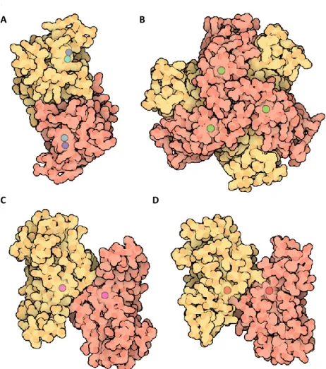
Fig 11 Structures of SOD proteins. Tridimensional structures of SOD proteins. A) The dimeric
form of Cu,Zn-SOD. B) The Ni-SOD structure. C) The Fe-SOD structure. D) The Ni-SOD. Picture taken from Protein Data Bank.
31
and 2 are really similar in humans, SOD2 is located exclusively in the mitochondrial matrix, where it is supposed to have a primary role in detoxification from ROS produced in mitochondria. The importance of SOD2 function in mammals was confirmed by disruption of the gene, which turns out to be lethal for mice due to neurodegeneration and damage to the heart [74]; moreover, mutations in sod2 gene have been associated with several pathologies, as idiopathic cardiomyopathy, cancer and motor neuron disease.
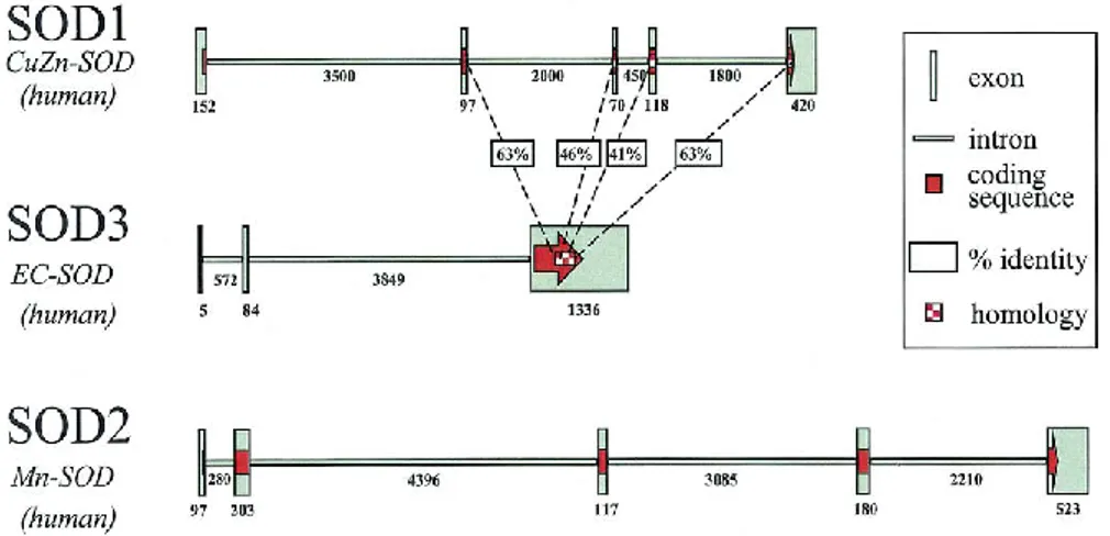
The sod3 gene is on chromosome 4 (4p15.3-p15.1) and encodes for a Cu/Zn-SOD called SOD3. Sod3 shares around 50% of similarity with sod1 at the exon level, but no similarity with sod2. SOD3 is the most recently discovered and least characterized member of SOD family. SOD3 is a tetramer formed by four identical subunit of around 33 kDa, with one Cu2+ and one Zn2+ ions per subunit; the enzyme
is also known as Extracellular-SOD (Ec-SOD) because it is exclusively present in extracellular fluids, such as human plasma, lymph, ascites and cerebrospinal fluids [75]. Thus, in contrast to intracellular SOD proteins, expression of SOD3 appears restricted to only a few cell types in specific tissues, such as alveolar type II cells, vascular smooth muscular cells, lung macrophages and few cultured fibroblast cell lines [76-77].
Fig 12 Genomic organization of human SOD genes. SOD3 was placed in the middle in order to
demonstrate areas of amino acid sequence homology between SOD1 and SOD3. SOD2 has no significant amino acid sequence homology with either SOD1 or SOD3. The size of each exon and intron, in base pairs, is shown in association with that fragment. Picture taken from [69].
32
1.3.3 Genomic organization of SOD1
The genomic organization of sod1 gene shows a striking similarity among species.
Sod1 gene is characterized by five exons and four introns, a promoter containing
TATA box, CCAAT box and several highly conserved GC-rich regions. Analysis of 5’-flanking sequence shows a high level of homology during evolution, assuming for this region a preserved key regulatory role for SOD1 expression. In addition, several putative binding sites for transcription factors have been found in Sod1 promoter, such as sites for NF1, Sp1, AP1, AP2, GRE, HSF, and NF-κB [78].
The 3'-end of sod1 gene is characterized by the presence of several poly(A) signal sequences that terminate the mRNA species with different lengths. To these, several consensus sequences, as YGTGTTYY and a G/T cluster, are associated, required for an efficient formation of 3'-termini.
As previously reported, sod1 gene is located on chromosome 21, which has been studied intensely due to the association of the trisomy of 21 with Down’s syndrome. Due to the presence of more copies of chromosome 21, the patients with Down’s syndrome show around 50% of SOD1 activity increase; however, the role of SOD1 in pathologies, associated with this disease remains questionable, since no obvious implication in the development of the major symptoms was demonstrated [79]. Conversely, a well-known correlation was found between mutations in sod1 gene and the onset of several familial form of Amyotrophic
Lateral Sclerosis (ALS), a neurodegenerative disease characterized by the loss of
motor neuron in brain stem and spinal cord. In 1993, 11 mutations in sod1 were described for the first time and associated to ALS, providing the first molecular insight into the pathogenesis of this disease [80]. Today, over 150 mutations ALS-associated have been characterized. However, SOD1-mediated disease mechanism is not still clear. Involvement of SOD1 in ALS will be further discussed subsequently.
1.3.4 The structure of human SOD1
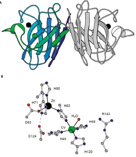
SOD1 protein is a homodimeric enzyme of 32 kDa formed by two subunit of 153 amino acids. The overall fold of the subunit consist in eight antiparallel β-strands connected by seven loops that form a flattened β-barrel with Greek key topology
33
[81]. A very important part of the single subunit is represented by the three external loops, which form a major structural element (Fig. 13).
Particularly, the loop IV includes a Zn-binding region involved in zinc coordination; the loop VII includes the electrostatic loop, which participates in electrostatic channel formation, through which superoxide is guided towards the active site. The electrostatic loop contains several charged residues, such as Arg143, that are directly involved in the electronic guidance of the substrate to the active site and contribute to the high specificity of SOD1 for the superoxide anion substrate. The smallest loop forms a Greek key connection across one end of the β barrel. The disulfide bond, which forms a left-handed spiral, covalently joins the largest loop to the beginning of the β-strand 8, representing another important structural element [82].
The SOD1 active site contains one Cu (II) and one Zn (II) ions per subunit, which interact with polypeptide chain through non-covalent interactions. It was demonstrated that Zn ion has mainly a structural role, being necessary to stabilize the active site; anyway, it was observed that its removal decreases the redox potential of Cu and the catalytic abilities of the entire enzyme. Zn is coordinated to the protein due to the presence of three histidine residues, in position 61, 69 and 79, and an aspartic acid residue, in position 81. The Cu2+ ion, which represents
the catalytic site, is coordinated by four imidazole of histidine residues, in position Fig 13 Structure of human SOD1 subunit. A) SOD1 subunit shows a secondary structure composed
by eight β-strands antiparallel and seven loops. B) The tridimensional structure of the subunit. Picture taken from Peter Doucette PhD Thesis, 2004, UCLA.
34
44, 46, 61, 118 and a fifth axial coordination position exposed to solvent and represented by a molecule of water; the side-chain of His61 forms a bridge between the Cu and Zn [82-83].
For the correct positioning of the metal cofactors, as well as for the proper achievement of protein folding, the newly synthesized polypeptide chain undergoes several post-translational modifications, which make the protein functional. The post-translational modifications are supported by the presence of specific chaperones, called CCS, Copper Chaperone of SOD1 [84].
B A
Fig 14 Structure of human SOD1 and its active site. A) The dimeric structure of human SOD1 formed by
two identical subuinit of 16 kDa each. Each subunit shows eight antiparallel β-strands connected by seven loops that form a flattened β-barrel with Greek key topology. B) Schematic representation of the active site of the single subunit, which contain both zinc and copper ions. Zn ion is coordinated to the protein due to the presence of His61, 69 and 79, and an aspartic acid residue, in position 81. Cu ion is coordinated by four imidazole of histidine residues, in position 44, 46, 61, 118 and a fifth axial coordination position exposed to solvent and represented by a molecule of water; the side-chain of His61 forms a bridge between the Cu and Zn. Picture taken from [81].
35
The process starts with the hetero-dimerization between SOD1 monomer and CCS followed by coordination of Zn ion, transfer of Cu ion and the formation of a disulfide bond between cysteine residues 57 and 146 that stabilizes the structure. The process ends with the dimerization of two perfectly folded subunits [85]. The reaching of correct protein folding protein leads to the formation of a channel, due to the presence of positively charged amino acid residues; the amino acids disposal allows the formation of an electrostatic field that facilitates the approach and the subsequent association of the superoxide radical with the metal.
1.3.5 SOD enzymes in S. cerevisiae
As mammals, the budding yeast S. cerevisiae expresses two intracellular SOD isoforms: a cytosolic Cu,Zn-SOD, called SOD1 and the mitochondrial Mn-SOD, SOD2. SOD1 accounts for 90–95% of the total superoxide dismutase activity in S.
cerevisiae: strains lacking SOD1 (Δsod1) show more severe defects than strains
lacking SOD2 (Δsod2) [86]. In addition, SOD1 is considered essential for yeast survival during the stationary phase, which is characterized by high rate of respiration, thus more ROS production. Indeed, viability of ∆sod1 strain significantly ameliorates by abolishing the mitochondrial respiration [87]: this underlines once again that SOD1 is involved in protection from oxidative stress promoted by respiration.
Therefore, function and activity of yeast enzymes is comparable to that performed by the homologous proteins of higher eukaryotes. In particular, yeast and human SOD1 share 70% homology and over 90% identity in their active-site residues, as showed by the followed sequence alignment:
hSOD1 MATKAVCVLKGDGPVQGIINFEQKESNGPVKVWGSIKG-LTEGLHGFHVHEFGDNTAGCT 59 ySOD1 -MVQAVAVLKGDAGVSGVVKFEQASESEPTTVSYEIAGNSPNAERGFHIHEFGDATNGCV 59 .:**.*****. *.*:::*** ... *..* .* * .:. :***:***** * **. hSOD1 SAGPHFNPLSRKHGGPKDEERHVGDLGNVTADKDGVADVSIEDSVISLSGDHCIIGRTLV 119 ySOD1 SAGPHFNPFKKTHGAPTDEVRHVGDMGNVKTDENGVAKGSFKDSLIKLIGPTSVVGRSVV 119 ********:.:.**.*.** *****:***.:*::***. *::**:*.* * .::**::* hSOD1 VHEKADDLGKGGNEESTKTGNAGSRLACGVIGIAQ 154 ySOD1 IHAGQDDLGKGDTEESLKTGNAGPRPACGVIGLTN 154 :* ******..*** ******.* ******:::
36
The high level of sequence conservation allows the expression of an active human SOD1 in yeast, which significantly increases resistance to both oxidative and heat stress of yeast cells. Overexpression of hSOD1 increase up to 50% of cell survival after treatment with oxidizing agents, such as menadione or paraquat, or heat shock compared to the controls [88].
1.3.6 Distribution and localization of SOD1
SOD1 is constitutively expressed and shows widespread distribution in a large variety of cells, where it represents one of the most abundant protein of the entire cell. Here, SOD1 plays a key role in controlling steady-state levels of ROS and oxidative stress. Moreover, the regulation of sod1 gene transcription may be subjected to control by both intra- and extra-cellular stimuli, such as heat shock, UV and X ray irradiation, increased levels of heavy metals or hydrogen peroxide [89], all cases in which SOD1 may help in oxidation control.
SOD1 is prevalently located into the cytosol [90], although under certain conditions SOD1 shows specific intra-organelles localization. For example, SOD1 was found in lysosomes and peroxisomes, even if the role of the enzyme in this compartment remains to be elucidated [91-92]. In yeast, a fraction of SOD1 localizes in IMS in association with its CCS, where it promotes prolonged survival in the stationary phase, diminishing the oxidative stress [93]; the redistribution of SOD1 in IMS was confirmed also in mammals [94].
1.3.7 Antioxidant-independent roles of SOD1
SOD1 is one of the most conserved enzymes throughout evolution due to its important antioxidant action. However, recent studies have suggested that the SOD1 role is much more extensive than its involvement in oxidative stress protection. For example, a series of metabolic alterations have been found in yeast cells devoid of endogenous SOD1 (Δsod1).
In presence of glucose, yeast cells grow in an exponential way using fermentation and repressing the expression of the respiratory machinery (catabolite repression), as long as the sugar is abundant. It was observed that Δsod1 yeast cells exhibit an impaired aerobic growth under fermentative conditions, while they grow better
37
on non-fermentable carbon sources, such as lactate and pyruvate. In these conditions, Δsod1 yeast shows also an increase in oxygen consumption and shorter survival times in stationary phase [86]. In addition, the mitochondrial mass in
Δsod1 yeast cells is approximately doubled compared to wild-type cells: this, in
association with observed higher oxygen consumption when glucose is abundant, strongly indicates the presence of a defect in glucose repression [95].
Initially, the growth defect shown by Δsod1 was attributed to an increased oxidative stress shown by the strain lacking endogenous SOD1; however, it has been shown that SOD1 plays a key role in respiration inhibition, independent of its antioxidant function, participating in the integration of signals from glucose and oxygen concentration [96].
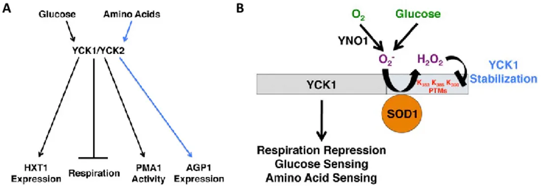
Glucose activates a signal transduction pathway via the glucose-sensor on the plasma membrane, SNF3 and RTG2, which modulates the glucose sensing and the repression of respiration; activation of this pathway requires also the yeast homologous casein kinase I γ (CK1γ) YCK1 and its paralog YCK2 (Fig. 15A) [97]. YCK1/2 directly act on the detachment of the transcriptional repressor RGT1, coupled to MHR1, promoting its phosphorylation and subsequent degradation that results in RGT1 detachment and leads to the constitutive expression of glucose transporter HXT genes [98-99].
SOD1 protein is directly involved in maintenance the stability of YCK1 and YCK2 kinases, for respiratory repression. SOD1 binds to a specific YCK region, identified in residues 367–412, and prevents its degradation through a mechanism involving lysines at the YCK C-terminus (Fig. 15B). Particularly, the superoxide anions
A B
Fig 15 Role of YCK1/2 proteins in glucose metabolism. A) YCK1/2 proteins are directly involved in
glucose sensing, repression of respiration, and in the extracellular amino acids signaling. B) SOD1 stabilizes YCK1/2 protein structure allowing to these proteins the activation of glucose metabolism. Pictures taken from [96].
38
generated during growth in the presence of glucose and oxygen feeds into cytosolic SOD1, and the concomitant production of hydrogen peroxide helps stabilizing the YCK kinases for nutrient signaling. In addition, SOD1 can maintain this function also for human CK1γ proteins, indicating that this mechanism is conserved through the evolution [96].
In a very recent work, it was shown in S. cerevisiae that the increase of oxidative stress induced by hydrogen peroxide or menadione promotes SOD1 translocation to the nucleus, where it is assumed that it plays a crucial role in DNA protection from oxidative damage. This change in SOD1 localization is specifically related to ROS increase, as demonstrated by the involvement of ATM/Mec1 kinase, a well known ROS sensor [100]. The activation of ATM pathway promotes the association of SOD1 with DUN1, that is responsible of specific phosphorylation of serine residues 60 and 99 of SOD1, conserved also in mammalian SOD1. Furthermore, once in nucleus, SOD1 acts as transcription factor: it directly binds promoters and regulates the expression of genes involved in ROS resistance and DNA damaging repair [101]. Mechanism is extensive explained in Fig. 16.
Fig 16 Proposed model of SOD1 traslocation into the nucleus. Hydrogen peroxide promotes SOD1
phosphorylation by ATM/Mec1 pathway activation, through SOD1 interaction with Dun1 kinase. Phosphorylated SOD1 traslocates to the nucleus where it acts a transcription factor involved in regulation of a set of gene that promote response to oxidative stress. Picture taken from [101].