is is an open-access article distributed under the terms of the Creative Commons Attribution-Non Commercial-Share Alike 4.0 License, which allows others to remix, tweak, and build upon the work non-commercially, as long as the author is credited and the new creations are licensed under the identical terms.
Original Article
Minimally invasive posterior fossa decompression with
duraplasty in Chiari malformation type I with and without
syringomyelia
Maria Caffo1,Salvatore M. Cardali1, Gerardo Caruso1, Elena Fazzari1, Rosaria V. Abbritti1, Valeria Barresi2, Antonino Germanò1
1Department of Biomedical and Dental Sciences and Morphofunctional Imaging, Unit of Neurosurgery, University of Messina, Messina, 2Department of Diagnostics and
Public Health, University of Verona, Verona, Italy.
E-mail: Maria Caffo - [email protected]; Salvatore M. Cardali - [email protected]; *Gerardo Caruso - [email protected]; Elena Fazzari - [email protected]; Rosaria V. Abbritti - [email protected]; Valeria Barresi - [email protected]; Antonino Germanò - [email protected]
*Correspondence author: Gerardo Caruso, Department of Biomedical and Dental Sciences and Morphofunctional Imaging, Unit of Neurosurgery, University of Messina, Messina, Italy. [email protected] Received : 03 November 18 Accepted : 02 April 19 Published : 10 May 19 DOI 10.25259/SNI-70-2019 Quick Response Code:
INTRODUCTION
e pathophysiology of Chiari malformation Type I (CM-I) involves frequently the small posterior
cranial fossa[8,12] with the consequent overcrowding of neural structures associated with a hyperdynamic
cerebrospinal fluid (CSF) flow in craniovertebral junction (CVJ). ese conditions result in an alteration
of pressure gradients at CVJ with the consequent compression of brainstem.[22] However, it is likely that,
in patients with CM-I, who do not present a crowded posterior fossa, other mechanisms, still unclear, can
cause the impaction of the tonsils in the foramen magnum.[26] In addition, it has been largely reported that
in 70% of cases CM-I is associated to syringomyelia.[2,16]
ABSTRACT
Background: Posterior fossa decompression (PFD), with and without duraplasty, represents a valid treatment in Chiari malformation Type I (CM-I) with and without syringomyelia. Despite a large amount of series reported in literature, several controversies exist regarding the optimal surgical approach yet. In this study, we report our experience in the treatment of CM-I, with and without syringomyelia, highlighting how the application of some technical refinements could lead to a good outcome and a lesser rate of complications.
Methods: Twenty-six patients with CM-I, with and without syringomyelia, underwent PFD through a 3 cm × 3 cm craniectomy with the removal of the most median third of the posterior arch of C1 and duraplasty. Signs and symptoms included sensory deficits, motor deficits, neck pain, paresthesias, headache, dizziness, lower cranial nerve deficits, and urinary incontinence. Postoperative magnetic resonance (MR) was performed in all patients.
Results: Signs and symptoms improved in 76.9% of cases. Postoperative MR revealed a repositioning of cerebellar tonsils and the restoration of cerebrospinal fluid circulation. In our experience, the rate of complication was 23% (fistula, worsening of symptoms, and respiratory impairment).
Conclusion: PFD through a 3 cm × 3 cm craniectomy and the removal of the most median third of posterior arch of C1 with duraplasty represents a feasible and valid surgical alternative to treat patients with CM-I, with and without syringomyelia, achieving a good outcome and a low rate of complications.
Keywords: Cerebellar tonsils, Chiari malformation type I, Duraplasty, Posterior fossa decompression, Syringomyelia
Surgical Neurology International
Editor-in-Chief: Nancy E. Epstein, MD, NYU Winthrop Hospital, Mineola, NY, USA.
SNI: General Neurosurgery Editor
Eric Nussbaum, MD
Surgical management of CM-I with and without syringomyelia is a still controversial topic in neurosurgery. Among the numerous methods proposed, the posterior fossa decompression (PFD) would seem to be the most suitable in the treatment of CM-I. PFD allows the relief of the cerebellar tonsils from the dorsal surface of the brainstem and upper cervical spinal cord, allowing physiologic and better CSF dynamics. A large amount of data demonstrated that PFD with or without removal of posterior arch of C1, and with or without duraplasty, allows an increase of volume of the posterior fossa, the repositioning of the herniated cerebellar tonsils, and the restoration of CSF circulation. However, few data exist regarding the dimension of craniectomy correlating this aspect to clinical outcome, neuroradiological findings, and rate of complications.
We performed, in 26 patients, a 3 cm × 3 cm craniectomy, with the removal of the most median third of the posterior arch of C1 and duraplasty with heterologous patches. In the light of our results and after a careful evaluation of the relevant literature, we think that this surgical strategy could represent a reliable and feasible approach to achieve a good outcome and a low rate of complications.
MATERIALS AND METHODS
Patients selection and data collection
A retrospective review of medical records of 26 patients, affected by CM-I with and without syringomyelia, undergone surgery from 2004 to 2014, was conducted. e epidemiological information, clinical presentation, duration of symptoms, radiological findings, and the clinical outcome were determined. All patients underwent brain and spine magnetic resonance (MR) study preoperatively.
Surgical technique
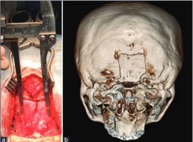
The patients were positioned in prone position, with the head in slight flexion and fixed with the Kees-Mayfield three-pin head holder. A midline skin incision is performed starting 1 cm above the inion to the spinous process of C2. The fascia and muscles were incised and dissected in a subperiosteal fashion until the occipital bone and the posterior arch of C1 were exposed. A 3 cm × 3 cm suboccipital craniectomy was realized. The decompression was completed through the removal of the most median third of the posterior arch of C1 beneath and contiguous to the craniectomy [Figure 1a and b]. The posterior atlanto-occipital membrane and the fibrous band of tissue covering the dura were gently dissected preserving the dural plane. Aiming to preserve the arachnoid layer, a careful Y-shaped dural incision was performed. In some cases, the dura appeared thick and required a more careful incision and dissection from the arachnoid plane. The cerebellar tonsils were dislocated downward through the foramen magnum, resulting in an obliteration of cisterna
magna. In 11 of our cases, we visualized the pulsation from downward to upward of cerebellar tonsils. Duraplasty with a bovine pericardium graft was performed in 10 cases, whereas a patch of Goretex was used in 16 cases, and finally, a watertight closure was realized. The closure was reinforced by Gelfoam and a fibrin glue layer.
Follow-up and outcomes
Patients’ clinical examinations were compared with their
preoperative examinations in the 1st, 6th, and 12th months after
surgery. A postoperative MR imaging study was performed at each follow-up control [Figures 2a and b, 3a and b]. In each, control was determined the outcome (improved, no change, or worse).
Figure 1: (a) e image shows the 3 cm × 3 cm craniectomy and the removal of the most median third of the posterior arch of C1, (b) ree-dimensional computed tomography scan reconstruction demonstrates the suboccipital craniectomy and removal of the most median third of posterior arch of C1.
a b
Figure 2: (a) T2 magnetic resonance (MR) weighted images showing the descent of cerebellar tonsils through the foramen magnum and the compression of the medulla oblongata, (b) T2-weighted postoperative MR in sagittal plane demonstrates the repositioning of cerebellar tonsils and the enlargement of subarachnoid spaces of posterior cranial fossa.
RESULTS
e study included 26 patients, 12 males and 14 females. e mean age at surgery was 35.8 years. e patients were divided into two groups. e former is made up of CM-I patients without syringomyelia, while the second is from patients with CM-I associated with syringomyelia. e characteristics of both series are summarized in Tables 1 and 2.
e first series included patients with CM-I without associated syringomyelia. is series included 15 patients, aged between 15 and 60 years. Symptoms at presentation are listed in Table 1. e most frequent presenting symptoms were cervical pain (9 patients, 60%), headache (8 patients, 53.3%), paresthesias (8 patients, 53.3%), sensory deficits (7 patients, 46%), motor deficits (6 patients, 40%), and dizziness (1 patient, 6.6%). e mean tonsillar descent measured from the rim of the foramen magnum was 12.1 mm (range 5–17 mm). In 4 patients (26.6%), CM-I was associated with hydrocephalus. ere were no long-term neurological or surgical complications. ere were no deaths. Mean postoperative follow-up was 27.5 months (range 5–72 months). At follow-up, 12 patients (80%) demonstrated a complete resolution of symptoms and 3 patients (20%) demonstrated a partial resolution of clinical signs and symptoms. Postoperative studies also showed a reduction of hydrocephalus in all cases.
e second series consisted of patients affected by CM-I with associated syringomyelia. is series included 11 patients, aged
between 16 and 67 years. Symptoms at presentation are listed in Table 2. Preoperative symptoms were sensory deficits in all patients, motor deficits (9 patients, 81.8%), paresthesias (7 patients, 63.6%), cervical pain (7 patients, 63.6%), headache (4 patients, 36.3%), lower cranial nerve deficits (3 patients, 27.2%), and bladder incontinence (2 patients, 18.1%). e mean tonsillar descent, measured from the rim of the foramen magnum, was 13.2 mm (range 5–18.3 mm). In 2 patients (18.1%), CM-I was also associated with hydrocephalus and a patient (9%) additionally presented scoliosis and tethered cord. One patient (9%) developed a CSF leakage. ere were no long-term neurological complications. ere were no deaths. Mean postoperative follow-up was 27.5 months (range 5–72). At follow-up considered, 8 patients (72.7%) demonstrated a complete resolution of symptoms. Of the remaining 3 patients (27.2%), one patient showed a worsening of the preoperative symptoms, while another patient revealed the appearance of paresthesias and respiratory impairment. e third patient presented no change in the clinical picture. Postoperative studies showed a moderate reduction of hydrocephalus in all cases.
Progressive reduction in syrinx volume was observed in 8 patients (72.7%) [Figure 3a and b], while it remained unchanged in the remaining 3 cases (27.2%).
DISCUSSION
CM-I includes a group of entities of congenital or acquired etiology that has in common the caudal displacement of the cerebellar
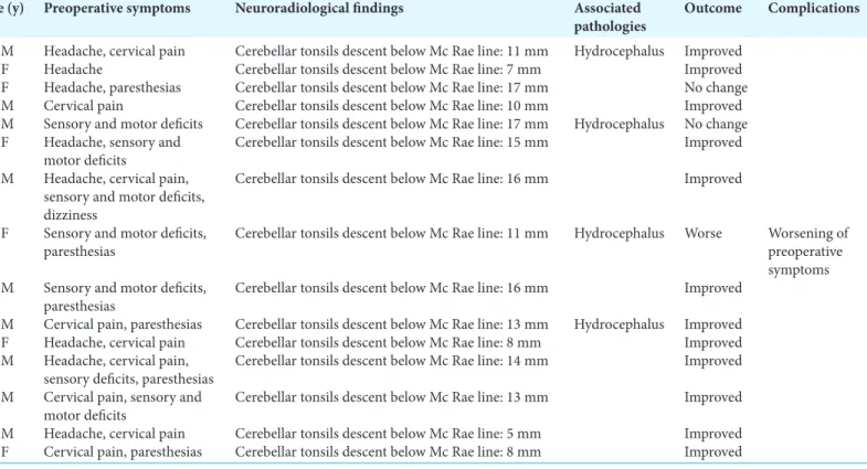
Table 1: Summary of nosological, clinical, and neuroradiological data of patients with Chiari malformation type I without associated syringomyelia. Age (y)
sex Preoperative symptoms Neuroradiological findings Associated pathologies Outcome Complications 40, M Headache, cervical pain Cerebellar tonsils descent below Mc Rae line: 11 mm Hydrocephalus Improved
22, F Headache Cerebellar tonsils descent below Mc Rae line: 7 mm Improved 38, F Headache, paresthesias Cerebellar tonsils descent below Mc Rae line: 17 mm No change 60, M Cervical pain Cerebellar tonsils descent below Mc Rae line: 10 mm Improved 27, M Sensory and motor deficits Cerebellar tonsils descent below Mc Rae line: 17 mm Hydrocephalus No change 30, F Headache, sensory and
motor deficits Cerebellar tonsils descent below Mc Rae line: 15 mm Improved 35, M Headache, cervical pain,
sensory and motor deficits, dizziness
Cerebellar tonsils descent below Mc Rae line: 16 mm Improved 26, F Sensory and motor deficits,
paresthesias Cerebellar tonsils descent below Mc Rae line: 11 mm Hydrocephalus Worse Worsening of preoperative symptoms 57, M Sensory and motor deficits,
paresthesias Cerebellar tonsils descent below Mc Rae line: 16 mm Improved 15, M Cervical pain, paresthesias Cerebellar tonsils descent below Mc Rae line: 13 mm Hydrocephalus Improved 21, F Headache, cervical pain Cerebellar tonsils descent below Mc Rae line: 8 mm Improved 36, M Headache, cervical pain,
sensory deficits, paresthesias Cerebellar tonsils descent below Mc Rae line: 14 mm Improved 28, M Cervical pain, sensory and
motor deficits Cerebellar tonsils descent below Mc Rae line: 13 mm Improved 17, M Headache, cervical pain Cerebellar tonsils descent below Mc Rae line: 5 mm Improved 28, F Cervical pain, paresthesias Cerebellar tonsils descent below Mc Rae line: 8 mm Improved
tonsils through the foramen magnum into the cervical canal. is may be due to the underdevelopment of occipital somites from paraxial mesoderm. As a result, a small posterior fossa is developed predisposing to a downward herniation of its contents, i.e., cerebellar tonsils migrating to cervical spinal canal. In 50%– 76% of patients, the malformation is associated with hydromyelic
cavitation of the spinal cord and medulla oblongata.[2,16] In these
cases, it has been postulated that obstruction of the outlets of the fourth ventricle, with diversion of the CSF pulse wave into the central canal or the foramen magnum, may cause a pressure gradient between the intracranial and intraspinal compartments,
pulling in fluid from the fourth ventricle into the central canal.[29]
CM-I usually presents after the second or third decade of life. Patients with CM-I present a wide range of signs and symptoms as headache and cervical pain. Weakness and lower extremity spasticity are also frequent, associated with disorders related to myelopathy (motor and sensory losses, hypo- and hyper-reflexia, and Babinski response), cerebellar (ataxia), and brainstem dysfunctions (respiratory irregularities, nystagmus, and lower cranial nerve dysfunction). Upper limbs deficit associated with
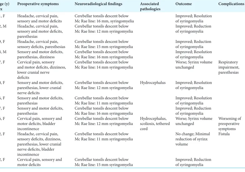
Table 2: Summary of nosological, clinical, and neuroradiological data of patients with Chiari malformation type I with associated syringomyelia. Age (y)
sex Preoperative symptoms Neuroradiological findings Associated pathologies Outcome Complications 51, F Headache, cervical pain,
sensory and motor deficits Cerebellar tonsils descent below Mc Rae line: 16 mm, syringomyelia Improved; Resolution of syringomyelia 62, M Headache, cervical pain,
sensory and motor deficits, paresthesias
Cerebellar tonsils descent below
Mc Rae line: 12 mm syringomyelia Improved; Reduction of syringomyelia 59, F Headache, cervical pain,
sensory deficits, paresthesias Cerebellar tonsils descent below Mc Rae line: 15 mm syringomyelia Improved; Reduction of syringomyelia 18, M Sensory and motor deficits,
paresthesias, dizziness Cerebellar tonsils descent below Mc Rae line: 16 mm syringomyelia Improved; Resolution of syringomyelia 67, F Cervical pain, sensory
and motor deficits, dizziness, lower cranial nerve
deficits
Cerebellar tonsils descent below
Mc Rae line: 14 mm syringomyelia Worse; Syrinx volume unchanged Respiratory impairment, paresthesias 40, F Sensory and motor deficits,
paresthesias, lower cranial nerve deficits
Cerebellar tonsils descent below
Mc Rae line: 12 mm syringomyelia Hydrocephalus Improved; Resolution of syringomyelia 36, F Sensory and motor deficits,
paresthesias Cerebellar tonsils descent below Mc Rae line: 11 mm syringomyelia Improved; Resolution of syringomyelia 47, F Sensory and motor deficits,
paresthesias Cerebellar tonsils descent below Mc Rae line: 16 mm syringomyelia Improved; Reduction of syringomyelia 16, F Cervical pain, sensory and
motor deficits, bladder incontinence
Cerebellar tonsils descent below
Mc Rae line: 12 mm syringomyelia Hydrocephalus, scoliosis, tethered cord
Worse; Syrinx volume
unchanged Worsening of preoperative symptoms 32, F Headache, cervical pain,
sensory deficits, dizziness, paresthesias, lower cranial nerve deficits, bladder incontinence
Cerebellar tonsils descent below
Mc Rae line: 11 mm syringomyelia No change; Minimal reduction of syrinx volume
Fistula
22, F Cervical pain, sensory and
motor deficits Cerebellar tonsils descent below Mc Rae line: 15 mm syringomyelia Improved; Reduction of syringomyelia
Figure 3: (a) T2 magnetic resonance (MR) weighted images showing the descent of cerebellar tonsils through the foramen magnum and T2–T9 syringomyelia, (b) T2 MR weighted 6-month postoperative picture reveals the decrease in size of syringomyelia.
signs of syringomyelia (thermic and pain dissociation, and hand muscle atrophy) is described in patients with syringomyelia. Few studies have been performed on the histopathological alterations
present within the cerebellar tonsils of patients with CM-I.[27]
Purkinje cells loss and reactive gliosis were demonstrated to be the most frequent findings and their genesis was connected to local
trauma depending on cerebellar tonsils herniation.[27]
Although the pathogenesis of CM-I is still mattered of debate, the posterior fossa volume mismatch is the leading cause of CM-I. Marin-Padilla showed that CM-I may be caused by a mesodermal
insufficiency occurring after closure of the neural folds.[24]
According to this hypothesis, a small posterior cranial fossa may
be an essential reason in the hindbrain hernia formation.[13] is
event leads to herniation of the tonsils below the foramen magnum. However, it is widely accepted that CM-I depends on a premature stenosis of spheno-occipital synchondrosis with transformation of posterior cranial fossa in a narrow funnel shape without statistically significant differences of anterior-posterior diameter
of the foramen magnum between CM-I and control group.[16]
Morphometric studies demonstrated that the disproportion of posterior cranial fossa is due attributable to basioccipital
hypoplasia.[25] In addition, it should be considered that CM-I is
characterized by occurrence of frequent and concomitant severe adhesions between the dura, arachnoid, and neural tissue. is arachnoid scarring may cause itself clinical symptoms, which are independent of brain stem compression and syringomyelia, acting
as an additional factor for impaired CSF flow.[13]
MR represents the optimal tool in the diagnosis since it is noninvasive and has a good correlation with clinical findings. Cine flow MR studies have recently been introduced into clinical use and have gained importance in decision-making of the ideal
surgical procedure.[15] e use of intraoperative imaging for Chiari
I malformation is usually limited by the CSF flow dynamics across the foramen magnum which significantly change when the patient
is positioned for surgery.[26] Computed tomography gives data in
axial, sagittal, and coronal sections and makes achievable three-dimensional reconstructions, for a more correct knowledge of the anatomy of the CVJ.
Indications for surgery include the presence of neurological symptoms, their progression, and/or headache caused by herniation of the cerebellar tonsils and significantly deteriorating the patients’ quality of life. However, nowadays, there is still considerable controversy about the optimal surgical procedure for the treatment of CM-I with or without syringomyelia. In cases characterized by symptoms clearly attributable to syringomyelia, shunting of the syringomyelic cavity is indicated, based on fluid diversion in the subarachnoid space or into extracavitary locations (peritoneal and pleural cavities). However, in these cases, treated with shunting procedures, the cause is not removed and the pathogenetic mechanism persists. PFD with and without cervical laminectomy is the preferred treatment option that allowing a
satisfactory CSF flow restoration and tonsillar repositioning.[1,7]
e goal of decompression is the improvement of symptoms and the reduction of CSF pressure at the CVJ. e surgical approach may be limited to a merely extradural decompression with lysis of extradural adhesions. It may consist of the opening of the outer
layer of dura mater (dura splitting),[6,9] performing transverse
microincisions of the outer layer of the dura mater[13] or achieving
duraplasty with or without lysis of adhesions around the cerebellar tonsils, and reducing in size of the latter through coagulation or partial tonsillectomy.[4,19,23]
Despite several series have been published about the choice of approach in terms of PFD, the dimension of craniectomy still represents matter of debate, due to few data exist in literature. e optimal extent of bony removal varies from surgeon to surgeon. A reduced craniectomy could cause an inadequate decompression, while a large craniectomy might theoretically allow an abnormal dural distention and the descent of the cerebellar tonsils through the bony defect. Sindou et al. analyzed the clinical outcome of a series of 44 patients affected by CM-I
with and without syringomyelia.[28] eir technique consisted of
a large craniectomy extended to the occipital condyles on either side to achieve an optimal decompression of cerebellar tonsils, with dural opening and preservation of arachnoid membrane. ey reported an improvement of the Karnofsky score in 83% and 80% of patients, respectively, with CM-I alone and with
associated syringomyelia.[28] As well, Chotai et al. demonstrated
a good rate of symptoms improvement performing a large craniectomy, stressing the importance to create a new artificial
cisterna magna.[9] On the other hand, Klekamp et al. referred that
large craniectomy is correlated to a worse outcome than smaller
craniectomy.[17] However, smaller decompressions are linked with
few rate of complications.[6,9] As well, the removal of the posterior
arch of C1 during PFD has to consider. Few authors focused on
this aspect.[28] In particular, two studies showed encouraging both
clinical and radiological results, (90% improvements), performing
a PFD with C1 arch resection.[9]
e objective of the craniectomy should be to restore the volume of the cisterna magna and decompress the brainstem. An excessively wide craniectomy could cause cerebellar herniation into spinal canal and compression on brainstem, obstruction of the liquoral circulation, and formation of arachnoidal scarring. In the light of these observations, in our series, we performed a suboccipital
3 cm × 3 cm2 craniectomy combined with the removal of the most
median third of the posterior arch of C1, below and contiguous to the craniectomy. is phase allows us a sufficient decompression of the posterior cranial fossa and the restoration of CSF flow at CVJ.
Published series reports clinical improvement rates after surgical
treatment from 71% to 100%[10,20] and syringomyelia resolution
rates from 52% to 91%.[18,20] Our results seem to be aligned. e
incidence of clinical improvement of our patients was 76.9% (72.7% and 80% in patients with and without syringomyelia, respectively). In literature, the reported rate of complications is between 3% and
40%.[1,7,15] In our experience, the rate of complications was 23%
(20% in the first series and 27.2% in the second one). Compared
to other series,[1,7,15] our data demonstrate a good postoperative
outcome and, overall, a low incidence of complications.
Fenestration of the arachnoid plane is still mattered of debate; some authors claim that preservation of arachnoidal layer, likewise dura mater, is associated to lower risk of CSF-related complication and is a safe and effective surgical option, mainly when no evidence of arachnoiditis or obstruction of the outflow
at the level of foramen of Magendie occurs.[20,26] It is our opinion
that opening of the arachnoid exposes the subarachnoid space to cellular debris that increases the risk of adhesive arachnoiditis, aseptic meningitis, and CSF leak. Conversely, others argue the need to explore the arachnoid flat and identify arachnoid strands and veils, potential obstacles to the ascent of cerebellar tonsils and to the reestablishment of normal CSF flow.[4] Several authors[14,19,23]
emphasized the usefulness and the utility of cerebellar tonsils reduction to restore the CSF pathway. Guyotat et al. supported the clinical outcome improvement with tonsils resection,
especially in patients with syringomyelia.[14] For other authors, it
is enough to remove herniated cerebellar tonsils from the cervical canal through atlanto-occipital interspace without perform any craniectomy or C1 laminectomy; this route could be considered a safe procedure, as a matter of fact that in CM-I cerebellar tonsils
have often a sclerotic or an atrophic histopathologic pattern.[19] We
believe that a tailored surgical technique is necessary, with tonsils reduction/tonsillectomy, only, if an optimal intraoperative restore of CSF flow is not achieved. e need to create a wide cisterna magna to allow the cerebellar tonsils ascent is widely reported in literature.[8,12]
Another critical issue is the duraplasty. Some authors published
that PFD with duraplasty is more effective than PFD alone.[1,4,15]
A systematic review of literature[11] showed a higher reoperation
rate in patients who underwent PFD alone compared with PFDD group, conversely, CSF-related complications were more frequent in PFDD, but the difference was not statistically significant. is study highlighted, however, that clinical outcome is similar in both
groups.[11] A meta-analysis was recently conducted to compare the
validity of PFD with duraplasty and PFD in treating patients with CM-I. is study confirmed that the decrease in syringomyelia was better for patients treated with PFDD than for patients treated
with PFD alone.[5] In line with these data, our number two series,
although numerically limited, has shown, in all cases, a decrease in size of the syringomyelic cavity.
e choice of dural substitute is a critical topic. Attenello et al. compared the use of pericranium autograft and synthetic expanded polytetrafluoroethylene (ePTFE) in pediatric patients. Both dural grafts were associated to a great rate of clinical improvement and a minimal incidence of complications; ePTFE provided an earlier improvement in syringomyelia than autologous graft, without
differences in absolute incidence.[3] Other authors compared
unsutured and suturable dural substitutes, obtaining similar
clinical outcome and rates of complication.[30] In another study by
Lee et al., the use of porcine and bovine dural grafts was matched;
the rate of pseudomeningocele was higher in porcine ones.[21]
In our institution, we always realized a duraplasty using bovine pericardium or Gore-Tex (a synthetic polytetrafluoroethylene graft), performing a watertight closure to obtain a wide space around cerebellar tonsils.
CONCLUSION
Although CM-I physiopathology is still unclear, signs and symptoms could be attributed to a CSF flow impairment due to an overcrowding of posterior fossa structures. e main goal of surgical treatment is to restore a normal outflow at CVJ. Our treatment option is a suboccipital 3 × 3 craniectomy, the removal of the median part of C1 arch, and duraplasty with bovine dural graft or Gore-Tex patch. We can consider satisfactory our clinical results and our rate of complications, even more if compared with the data reported in literature. is technique can be considered a valid and safe choice for the treatment of CM-I.
Financial support and sponsorship Nil.
Conflicts of interest
ere are no conflicts of interest.
REFERENCES
1. Alfieri A, Pinna G. Long-term results after posterior fossa decompression in syringomyelia with adult chiari Type I malformation. J Neurosurg Spine 2012;17:381-7.
2. Arnautovic A, Splavski B, Boop FA, Arnautovic KI. Pediatric and adult chiari malformation Type I surgical series 1965-2013: A review of demographics, operative treatment, and outcomes. J Neurosurg Pediatr 2015;15:161-77.
3. Attenello FJ, McGirt MJ, Garcés-Ambrossi GL, Chaichana KL, Carson B, Jallo GI, et al. Suboccipital decompression for chiari I malformation: Outcome comparison of duraplasty with expanded polytetrafluoroethylene dural substitute versus pericranial autograft. Childs Nerv Syst 2009;25:183-90.
4. Batzdorf U, McArthur DL, Bentson JR. Surgical treatment of chiari malformation with and without syringomyelia: Experience with 177 adult patients. J Neurosurg 2013;118:232-42.
5. Chai Z, Xue X, Fan H, Sun L, Cai H, Ma Y, et al. Efficacy of posterior fossa decompression with duraplasty for patients with chiari malformation Type I: A systematic review and meta-analysis. World Neurosurg 2018;113:357-650.
6. Chauvet D, Carpentier A, Allain JM, Polivka M, Crépin J, George B, et al. Histological and biomechanical study of dura mater applied to the technique of dura splitting decompression in chiari Type I malformation. Neurosurg Rev 2010;33:287-94.
7. Chen J, Li Y, Wang T, Gao J, Xu J, Lai R, et al. Comparison of posterior fossa decompression with and without duraplasty for the
surgical treatment of chiari malformation Type I in adult patients: A retrospective analysis of 103 patients. Medicine (Baltimore) 2017;96:e5945.
8. Chotai S, Kshettry VR, Lamki T, Ammirati M. Surgical outcomes using wide suboccipital decompression for adult chiari I malformation with and without syringomyelia. Clin Neurol Neurosurg 2014;120:129-35.
9. Chotai S, Medhkour A. Surgical outcomes after posterior fossa decompression with and without duraplasty in chiari malformation-I. Clin Neurol Neurosurg 2014;125:182-8.
10. El-Ghandour NM. Long-term outcome of surgical management of adult chiari I malformation. Neurosurg Rev 2012;35:537-47. 11. Forander P, Sjavik K, Solheim O, Riphagen I, Gulati S, Salvesen Ø,
et al. e case for duraplasty in adults undergoing posterior fossa decompression for chiari I malformation: A systematic review and meta-analysis of observational studies. Clin Neurol Neurosurg 2014;125:58-64.
12. Furtado SV, akar S, Hegde AS. Correlation of functional outcome and natural history with clinicoradiological factors in surgically managed pediatric chiari I malformation. Neurosurgery 2011;68:319-28. 13. Gambardella G, Caruso G, Caffo M, Germanò A, La Rosa G,
Tomasello F. Transverse microincisions of the outer layer of the dura mater combined with foramen magnum decompression as treatment for syringomyelia with chiari I malformation. Acta Neurochir 1998;140:134-9.
14. Guyotat J, Bret P, Jouanneau E, Ricci AC, Lapras C. Syringomyelia associated with Type I chiari malformation. A 21-year retrospective study on 75 cases treated by foramen magnum decompression with a special emphasis on the value of tonsils resection. Acta Neurochir 1998;140:745-54.
15. Hu Y, Liu J, Chen H, Jiang S, Li Q, Fang Y, et al. A minimally invasive technique for decompression of chiari malformation Type I (DECMI study): Study protocol for a randomized controlled trial. BMJ Open 2015;5:e007869.
16. Hwang HS, Moon JG, Kim CH, Oh SH, Song JH, Jeong JH. e comparative morphometric study of the posterior cranial fossa: What is effective approaches to the treatment of chiari malformation Type 1? J Korean Neurosurg Soc 2013;54:405-10.
17. Klekamp J, Batzdorf U, Samii M, Bothe HW. e surgical treatment of chiari I malformation. Acta Neurochir 1996;138:788-801. 18. Klekamp J. Surgical treatment of chiari I malformation analysis
of intraoperative findings, complications, and outcome for 371 foramen magnum decompressions. Neurosurgery 2012;71:365-80. 19. Lazareff JA, Galarza M, Gravori T, Spinks TJ. Tonsillectomy without
craniectomy for the management of infantile chiari I malformation. J Neurosurg 2002;97:1018-22.
20. Lee HS, Lee SH, Kim ES, Kim JS, Lee JI, Shin HJ, et al. Surgical results of arachnoid-preserving posterior fossa decompression for chiari I malformation with associated syringomyelia. J Clin Neurosci 2012;19:557-60.
21. Lee CK, Mokhtari T, Connolly ID, Li G, Shuer LM, Chang SD, et al. Comparison of porcine and bovine collagen dural substitutes in posterior fossa decompression for chiari I malformation in adults. World Neurosurg 2017;108:33-40.
22. Linge SO, Haughton V, Løvgren AE, Mardal KA, Helgeland A, Langtangen HP. Effect of tonsillar herniation on cyclic CSF flow studied with computational flow analysis. Am J Neuroradiol 2011;32:1474-81.
23. Ma J, You C, Chen H, Huang S, Ieong C. Cerebellar tonsillectomy with suboccipital decompression and duraplasty by small incision for chiari I malformation (with syringomyelia): Long term follow-up of 76 surgically treated cases. Turk Neurosurg 2012;22:274-9.
24. Marin-Padilla M, Marin-Padilla TM. Morphogenesis of experimentally induced Arnold-chiari malformation. J Neurol Sci 1981;50:29-55.
25. Noudel R, Jovenin N, Eap C, Scherpereel B, Pierot L, Rousseaux P. Incidence of basioccipital hypoplasia in chiari malformation Type I: Comparative morphometric study of the posterior cranial fossa. J Neurosurg 2009;111:1046-52.
26. Oldfield EH. Pathogenesis of chiari I-pathophysiology of syringomyelia: Implications for therapy: A summary of 3 decades of clinical research. Neurosurgery 2017;64 Suppl 1:66-77.
27. Pueyrredon F, Spaho N, Arroyave I, Vinters H, Lazareff J. Histological findings in cerebellar tonsils of patients with chiari Type I malformation. Childs Nerv Syst 2007;23:427-9.
28. Sindou M, Chàvez-Machuca J, Hashish H. Cranio-cervical decompression for chiari Type I-malformation, adding extreme lateral foramen magnum opening and expansile duroplasty with arachnoid preservation. Technique and long-term functional results in 44 consecutive adult cases comparison with literature data. Acta Neurochir 2002;144:1005-19.
29. Williams B. e distending force in the production of communicating syringomyelia. Lancet 1970;2:41-2.
30. Williams LE, Vannemreddy PS, Watson KS, Slavin KV. e need in dural graft suturing in chiari I malformation decompression: A prospective, single-blind, randomized trial comparing sutured and sutureless duraplasty materials. Surg Neurol Int 2013;4:26. How to cite this article: Caffo M, Cardali SM, Caruso G, Fazzari E, Abbritti RV, Barresi V, et al. Minimally invasive posterior fossa decompression with duraplasty in Chiari malformation type I with and without syringomyelia. Surg Neurol Int 2019;10:88.