TOR VERGATA UNIVERSITY
POTENTIAL ROLE OF IL-10-TREATED DENDRITIC
CELLS IN THE CONTROL OF THE IMMUNE
RESPONSE TO ALLERGENS
Valentina Pacciani
DOCTORAL DISSERTATION
Faculty opponent Supervisor
Prof. Maria Grazia Roncarolo Dr. Federica Angelini Vita-Salute San Raffaele University Tor Vergata University
HSR-TIGET, Milano Rome
Department of Public Health and Cellular Biology,
Tor Vergata University, Rome, Italy
TABLE OF CONTENTS
ABSTRACT
pg 6
LIST OF PAPERS
pg 8
ABBREVIATIONS
pg 9
INTRODUCTION
pg 10
Allergic Disease
pg 10
Epidemiology of Asthma
pg 10
Immunopathogenesis of Allergic Disease
pg 12
Allergic Inflammation
pg 12
Immunological Mechanisms in Asthma
pg 16
Role of Dendritic Cells in Immune Regulation and Allergic
Immune Response
pg 21
Airway DC
pg 21
Antigen Uptake and Limphnode Migration of Airway DC
pg 22
Sensitization to Inhaled Allergens and Th2 Polarization
by Airway DC
pg 24
Immunological Tolerance
pg 30
Peripheral Tolerance
pg 33
T regulatory Cells
pg 36
Natural Occuring T regulatory Cells
pg 36
Adaptive T reg pg 40
CD4+CD25+ T Regulatory Cells
pg 40
Type-1 Regulatory Cells (Tr1) pg 41
Mechanisms of Tolerance to Allergens
pg 46
Role of CD4+CD25+ in allergy
pg 48
Role of Tr1 in allergy
pg 50
The Interplay between IL-10 Dendritic Cells, and
T regulatory Cells in Allergy
pg 52
IL-10
pg 52
Immunoregulation of Dendritic Cells by IL-10 pg 56 Induction of T Regulatory Cells by Dendritic Cells
pg 57
Control of Dendritic Cell Function by T regulatory Cellspg 59
AIM OF THE STUDY
pg 61
MATERIALS AND METHODS
pg 62
RESULTS
pg 68
CONCLUDING REMARKS
pg 103
REFERENCES
pg 104
Ringraziamenti
pg 135
ABSTRACT
Several lines of evidence indicate that a defect in immunoregulatory mechanisms is involved in the pathogenesis of allergic asthma.
The aim of this study was to determine whether IL-10-treated dendritic cells (DC) are able to modulate allergen-specific T cell responses in children affected by allergic asthma.
Fourthy-one children, aged between 4-14 years, allergic to House Dust Mite (HDM), and 10 healthy age-matched children were recruited. DC were differentiated from peripheral blood CD14+ precursors and cultured with GM-CSF and IL-4 for 5 days. Der p2 (a major HDM allergen) was added alone or in combination with IL-10 for 48 hours to obtain Dp2-DC and IL10 Dp2-DC, respectively. Alternatively, DC were differentiated in the presence of IL-10 and pulsed with Der p2 during the 2 last days of culture (Dp2-DC10).
The ability of the resulting DC to stimulate allergen-specific autologous T cells and to promote allergen-specific T cell anergy was analyzed.
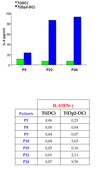
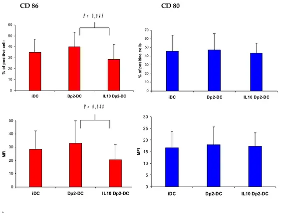
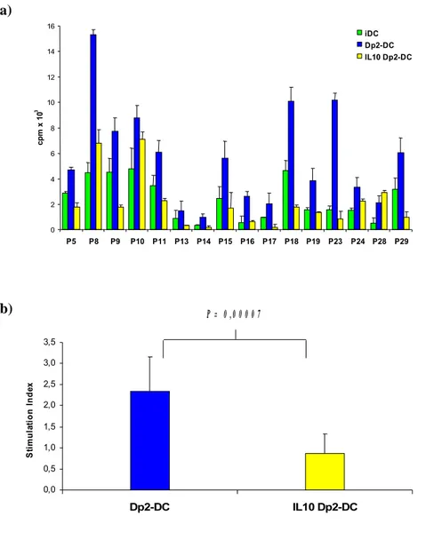
Dp2-DC induced allergen-specific T cell proliferation in 32 out of 41 patients but not in healthy controls. In 25 out of 26 allergic patients both IL10 Dp2-DC and Dp2 DC10 induced a significantly lower allergen-specific T cell proliferation. The analysis of DC phenotype showed that IL-10 treatment during the last 2 days of culture down-regulated the expression of CD86 on Dp2-DC. However, no correlation between the reduction of CD86 expression and of T cell proliferation was observed. Dp2-DC promoted a selective activation of autologous CD4+ T cells (CD25 and intracellular CTLA-4 up-regulation), which remained unchanged after IL-10 treatment. Moreover, Dp2-DC stimulation induced a Th2 cytokine profile characterized by an increase of IL-5, IL-13 and IL-4 production and IL-5/IFN-γ ratio. In the same patients, the co-culture with both IL10 Dp2-DC and Dp2 DC10 caused a marked reduction of IL-5 and IL-13 production by T cells, with a parallel decrease of IL-5/IFN-γ ratio. Moreover, in 8 children we observed an increase of IL-10 production with IL10 Dp2-DC stimulation.
T cell lines generated with Dp2-DC10, compared to those generate with Dp2-DC, were hyporesponsive to reactivation with Der p2 in 4 out of 5 patients tested, both in terms of proliferation and cytokine production: IL-5, IL-13 and IL-5/IFN-γ ratio.
Our data show that IL-10 reduced the stimulatory capacity of DC through a mechanism independent from the downregulation of costimulatory signals. IL-10 treatment of DC promoted a suppression of allergen-specific Th2 cytokine production without a skewing to a Th1 profile, but with a parallel increase of IL-10 production by T cells. Moreover, Dp2-DC10 were able to promote T cell anergy associated with a reduction in Th2 cytokine production. These results represent an important step forward to the prospective clinical application of Dp2-DC10 to modulate allergen-specific T cell responses in vivo.
LIST OF PAPERS
I. Pacciani V, Gregori S, Chini L, Chianca M, Corrente S, Rossi P, Roncarolo MG and Angelini F. Potential role of IL-10 treated dendritic cells in controlling the immune response to allergens. Manuscript in preparation. II. Angelini F, Piccinini S, Pacciani V, Di Cesare S, Rapini N, Del Duca E.,
Paone FM, Rossi P, Manca Bitti ML. Increase of α4β7 integrin on T lymphocytes in children affected by Type 1 diabetes. 2007. Submitted.
III.
Gregori S, Pacciani V, Tomasoni D, Scirpoli M, Battaglia M, Magnani C, Hauben E, Roncarolo MG. Differentiation of type 1 T regulatory cells through the IL-10-dependent ILT4/HLA-G pathway. 2007. Submitted.IV.
Angelini F, Pacciani V, Di Pede A, Polito A, Riccardi C, Rossi P and Chini L. Role of dendritic cells in the mechanism of sublingual immunotherapy in children affected by allergic asthma. 2007. Submitted.V.
Angelini F, Del Duca E, Piccinini S, Pacciani V, Rossi P and Manca Bitti ML. Altered phenotype and function of dendritic cells in children with type 1 diabetes. Clin Exp Immunol 2005; 142: 341-346.ABBREVIATIONS
ACD Apoptotic cell death AHR Airway hyper reactivity AICD Activation-induced Cell Death APC: Antigen presenting cells BAL bronchoalveolar lavage
DC: Dendritic cell
Der p2 Major group 2 allergen from Dermatophagoides Pteronyssinus GVHD Graft versus host disease
HDM House dust mites
IBD Inflammatory bowel disease IDO 2,3 Indoleamine deoxygenase
LPS Lypopolisaccarides
mDC Myeloid dendritic cells
MHC: Major histocompatibility complex mTEC: medullar thyimic epithelial cell NKT Natural killer T cells
nTreg Natural occurring T regulatory cells PBMC Peripheral blood mononuclear cells pDC Plasmacytoid dendritic cells
SIT Specific immunotherapy SLIT Sublingual immunotherapy
TCR: T cell receptor
TD1 Type 1 diabetes
Th1 T helper 1
Th2 T helper 2
Tr1 Type 1 regulatory cells Treg T regulatory cells
INTRODUCTION
ALLERGIC DISEASE
Allergy can be defined as the clinically evident reaction to ubiquitous allergens. Immunological sensitization to common environmental allergens, such as house dust mites, grass and tree pollens and cat dander, can result in diseases such as allergic rhinitis and conjunctivitis, asthma and atopic dermatitis, and in the most extreme cases, in anaphylaxis and death (Kay AB et al., 2001; Holgate S et al., 2003).
Clinical symptoms vary, partly depending on how the allergen is introduced into the body. Aeroallergens are frequently associated with asthma, causing characteristic tightness in the chest, wheezing and shortness of breath. Most individuals with allergic asthma also suffer from allergic rhinitis, and the two conditions have a common immunopathology.
Epidemiology of Asthma
Allergic rhinitis, asthma and atopic eczema are among the commonest causes of chronic ill health care costs. In Sweden, for example, the number of children with allergic rhinitis, asthma or eczema roughly doubled over a 12-year period (Aberg N et
al., 1995) and in the United States the annual cost of treating asthma is about $6 billion (SmithDH et al., 1997).
The prevalence of allergy varies between countries, as reported in the ISAAC study,
(ISAAC 1998), with one study of young adults showing that the prevalence of
sensitization to at least one allergen varies from 16% in Albacete, Spain, to 46% in Christchurch, New Zealand (Burney P et al., 1997). The prevalence is also generally much higher in the developed, rather than the developing, world. The recent increase in the prevalence of allergy and diseases associated with allergy in the developed world has major health and economic consequences.
Asthma prevalence in childhood, reaches in Germany 20% and in Australia 29%
(Sears MR et al., 1997). In the USA asthma affects approximately 8-10% of the
population and is the leading cause of hospitalization among children less than 15 years of age (Elias JA et al., 2003). Eighty percent of childhood asthma is reported to be IgE mediated.
There is a considerably lower prevalence of pediatric allergy in developing countries and there are also substantial differences between rural and urban areas. According to Von Ehrenstein, in a study performed in rural Bavaria, the prevalence of a doctor's diagnosis of asthma (3.4 vs. 6.4%) and current wheeze (5.6 vs. 8.1%) was significantly lower in farmers' children, whereas no such difference was observed for eczema. The effects were stronger in children of farmers with full-time activity as compared with their peers with only part-time farming parents, suggesting a dose-response relationship (Von Ehrenstein OS et al., 2000).
This and other observations support the so-called hygiene hypothesis. According to this theory bacterial and viral infections in early life might direct the immune system towards a prevalent type 1 helper T (Th1) cell response which produces interleukin (IL)-2 and interferon-γ (IFN-γ), thus preventing the development of allergic diseases which are characterized by a Th2 cell response with prevalent by IL-4, IL-5 and IL-13 production. Nevertheless, this theory is not easily reconciled with the increased prevalence of allergic asthma among poor blacks in the USA associated with sensitization to cockroaches and house-dust mite, and with some evidence about the increased prevalence of Th1 mediated diseases complicate the picture (Yazdanbakhsh
Immunopathogenesis of Allergic Diseases
Allergic Inflammation
All of us come across many allergens is our life, but a person without atopy mount a low grade immunological response mainly characterized by production of allergen specific IgG1 and IgG4 antibodies, and by a modest cell proliferation and production of IFN-γ by Th1 cells. By contrast, when susceptible or atopic individuals are initially exposed or sensitized to allergens, this induces an exaggerated allergen-specific response which is characterized by CD4+ T cells producing a T helper 2 (Th2) profile
of cytokines (IL-4, IL-5, IL-9 and IL-13 rather than IFN-γ and IL-2) and the presence of allergen-specific IgE (Tab. 1).
Table 1. Components of Immune Response to Allergens in Healthy and Allergic Individuals.
Specific antibody response in serum
Healthy: -No response
Detectable IgG1, IgG4 and IgA
High amounts of IgG4, relatively low amounts of IgG1 and detectable IgA and IgE
Allergic: -Relatively high amounts of IgE, together with low or high amounts of IgG1, IgG4 and IgA
T-cell response in allergen-specific cells and in PBMC
Healthy: -No response
Th0 response in PBMC and specific T-cell clones with low frequency Tr1, particularly IL-10-dominating response with relatively high frequency
Allergic: Th2 response with varying quantities of IL-4, IL-5 and IL-13, in the presence of detectable IL-10 and IFN-γ
Clinical outcome
Healthy: -Healthy
Skin-prick test and IgE positivity; clinical disease cannot be induced
Skin-prick test and IgE positivity and healthy in normal circumstances; clinical disease can be induced by provocation tests and it is dose-dependent
In utero, T cells of the fetus are primed by common environmental allergens that cross the placenta. As a result, the immune response of virtually all newborn infants is dominated by Th2 cells (Prescott S et al., 1998). It has been proposed that during subsequent development the normal (i.e., nonatopic) infant’s immune system shifts in favor of a Th1-mediated response to inhaled allergens (a process termed “immune deviation”) (Holt PG et al., 1999), whereas in the potentially atopic infant there is a further increase in Th2 cells that were primed in utero. Microbes are probably the chief stimuli of protective Th1-mediated immunity. Macrophages that engulf microbes secrete IL-12, which induces Th1 cells and natural killer cells to produce IFN-γ, thereby shifting the immune system into an “allergy-protective” Th1-mediated response. Other factors may also influence whether Th1 or Th2 cells dominate the response, including the amount of allergen, the duration of exposure to the allergen, and the avidity of allergen-specific interactions between T cells and APC (Constant
SL et al., 1997; Rogers PR et al., 1999).
Although the picture is more complex, the immunopathological hallmark of allergic disease is the infiltration of affected tissues by Th2 cells.
In particular, the allergen is taken up by phagocytic cells located in peripheral lung tissue and subsequently transported by DC to draining limph nodes (step 1) where is presented to T cells (step 2). Naïve T cells recognizing the allergen for the first time become activated and undergo a differentiation into Th2 effector T cells (step 3). The fully activated Th2 effector T cells initiate and sustain the local inflammatory process through secretion of soluble mediators (step 4). Concomitantly, the Th2 effector T cells instruct allergen specific B cells to differentiate to antibody-secreting plasma cells (step 5) and memory B cells (step 6). Differentiated allergen-specific T cells develop into long-lived memory T cells (step 7). Memory T cells are quiescent in the absence of antigen, but on allergen re-exposure, they quickly become activated Th2 effector T cells again (step 3) (Ngoc LP et al., 2005) (Fig. 1).
from Kroczek R et al. Allergy Clin Immunol 2005
Fig. 1. Schematic representation of the development and function of Th2 cells in airway disease
The cascade of events leading to the allergic inflammation is initiated by the process of tethering, activation, and adhesion to the endothelium followed by extravasation of inflammatory cells. This requires specific glycoprotein adhesion molecules, such as integrins and selectins, on both leukocytes and on endothelial cells, which are upregulated and show increased binding affinity in response to various inflammatory stimuli. Once the inflammatory cells have infiltrated into the tissue, they respond to chemotactic gradients established by chemoattractant cytokines and chemokines, which emanate from sites of injury or infection. More than 50 different chemokines are now recognized to be involved in the recruitment of inflammatory cells via the activation of more than 20 different surface receptors (Rossi D et al.,
2000). These molecules play a central role in defining the nature of the inflammatory
infiltrate in allergy (Kay AB 2001).
Exposure to an allergen following allergic sensitization leads to crosslinking of allergen specificIgE bound to the surface of mast cells andbasophils, degranulation of these cells and release of histamine, preformed granule-associated mediators,
membrane-derived lipids, cytokines, and chemokines that cause the symptoms associated with early or acute allergic reactions, including wheezing and conjunctivitis
(Kay AB 2001). The release of mediators and leukotrienes causes increases in vascular
permeability, smooth-muscle contraction and mucus secretion.
Late-phase allergic responses are characterized by the additional recruitment and activation of eosinophils and Th2 cells at the site of allergen challenge (Fig. 2).
from Kay AB et al N Engl J Med 2001
Immunological Mechanisms in Asthma
Over the decades, the prominent involvement of eosinophils, macrophages, mast cells, and lymphocytes in the inflammatory response in the airways of patients with asthma and the efficacy of steroids in the majority of patients with asthma became more clear and then led to the present-day concept that asthma is a chronic inflammatory disorder of the airways and that T cells are pivotal initiators and regulators of this response. Structural alterations including airway wall thickening, fibrosis in the lamina reticularis and adventitia of the airway, mucus metaplasia, myocyte hypertrophy and hyperplasia, and neovascularization are all readily appreciated in the asthmatic airway.
This led to the hypothesis that the inflammatory response in the asthmatic airway causes these remodeling events, and to the belief that these events contribute to disease pathogenesis (Elias JA et al., 2003).
Immediate hypersensitivity is the basis of acute allergic reactions. It is caused by molecules released by mast cells when an allergen interacts with membrane bound IgE. The complex of allergen, IgE on the surface of the mast cell triggers a release of preformed histamine and other mediators prostaglandins. These mast-cell mediators have a critical role in anaphylaxis, rhinoconjunctivitis, and urticaria. The role of histamine in chronic asthma and eczema is probably minimal, however, as shown by the relative ineffectiveness of histamine antagonists in controlling these conditions.
Mast cells produce the three cysteinyl leukotrienes C4, D4, and E4, which cause
the contraction of smooth muscles, vasodilatation, increased vascular permeability, and the hypersecretion of mucus when they bind to specific receptors (Drazen JM et al.,
1999).
Eosinophils, macrophages, and monocytes are also major sources of cysteinyl leukotrienes. Mast cells also contain tryptase, a four-chain neutral protease that activates the protease-activated receptors on endothelial and epithelial cells. The activation of these receptors initiates a cascade of events, including the up-regulation of adhesion molecules that selectively attract eosinophils and basophils (Holgate ST
In the cutaneous late-phase reaction, eosinophils and neutrophils accumulate, and then CD4+ T cells and basophils infiltrate the site (Ying S et al., 1999). Late-phase
asthmatic (Robinson DS et al., 1993) and nasal (Durham SR et al., 1992) reactions have a similar pattern of cellular infiltration, although basophils are not prominent in the lower airways (Macfarlane AJ et al., 2000).
Th2-type cytokines such as interleukin-4, 5, 9, and 13 influence a wide range of events associated with chronic allergic inflammation.
IL-4 has a role in the initial derivation of the allergen-specific Th2-lineage cells, and IL-4 and IL-13 induce IgE class switching.
IL-5 is a lineage-specific eosinophil differentiation and activator factor. It can be detected in the serum of mice with eosinophilia, and antibody to IL-5 blocks the development of parasite-induced eosinophilia. Although eosinophilia is often associated with high levels of IgE antibody, IL-5 appears not be involved in this response, where IL-4 appears to be main controlling factor (Sanderson CJ 1990). IL-4 and IL-9 promote the development of mast cells; IL-9 and IL-13 help promote airway hyperresponsiveness; and IL-4, IL-9, and IL-13 promote the overproduction of mucus (Kay AB 2001; Robinson DS 2000; Romagnani S 1994; Lloyd CM et al.,
2001).
In the last years the important role of IL-13 in asthma has been elucidated. Originally discovered as an IL-4 like molecules, it is nowadays clear that these cytokines differ in their effector properties, IL-4 playing a more prominent role in the initiation and IL-13 in the effector phase of Th2 allergic inflammation (Zhou Y at al 2001). Several studies using overespression-transgenic animal models have provided impressive insights in the mechanism of IL-13 induced inflammation, trough the chemokine receptor CCR2, and tissue fibrosis. They demonstrated, for instance, that the fibrotic response in asthma results from the ability of IL-13 to stimulate the production and activation of the fibrogenic cytokine TGF-β (Lee CG et al., 2001).
Moreover, the observation that other cytokines such us IL-9, mediate their effect in the lung through the induction of IL-13, suggests that IL-13 might be a final common pathway for Th2-mediated inflammatory response (Tab.2).
Tab.2. The role of Cytokines produced by Th2 Cells in Chronic Allergic Inflammation
Other players involved in pathogenesis of asthma have been recently studied. Among these proinflammatory factors, the main are thymic stromal lymphopoietin (TSLP), IL-25, IL-21, tumor necrosis factor a (TNF-α) and natural killer T cells (NKT cells).
TSLP, which is highly expressed in Hassall’s corpuscles in the thymic medulla, appears to be important in the periphery, where it enhances the capacity of DC to induce the development of Th2 cells (Ito T et al., 2005). In particular, overexpression of TSLP in the lungs of mice results in the development of severe allergic airway inflammation (Zhou B et al., 2005; Al-Shami A et al., 2005). TSLP is, in fact, expressed at high levels in the lungs of patients who have asthma (Ying S et
al., 2005), which suggests that expression of TSLP by lung epithelial cells can indeed
cause airway DC to enhance the development of Th2-driven inflammation.
IL-25 (also known as IL-17E) is produced by Th2 cells and mast cells and was found to induce the production of large quantities of Th2 cytokines in models of both infectious and pulmonary allergic disease (Fort MM et al., 2001). IL-25 might also enhance Th2 responses by actively inhibiting IFN-γ and IL-17 production, suggesting
that IL-25-producing cells might have a regulatory function, limiting pathologic (Th1-biased) inflammation at mucosal sites. Because IL-25 enhances Th2 cytokine production, however, it might also enhance the development of allergic inflammatory responses at mucosal sites by inducing eosinophilia, airway hyperreactivity and increased mucus production (Hurst SD et al., 2002).
IL-21 is a newly described T cell-produced cytokine related to IL-2, IL-4, and IL-15 that is capable of regulating T, NK, and, especially, Bcells (Parrish-Novak J et
al., 2000; Kasaian MT et al., 2002). The role of IL-21 in Th differentiation is still
somewhat controversial. In the mouse IL-21 is preferentially expressed by Th2 lymphocytes and inhibits IFN-γ production from developing Th1 cells (Wurstel AR et
al., 2002). Paradoxically, though, exposure of human primary T and NK cells to IL-21
has also been reported to up-regulate IFN-γ, T-bet, and IL-12Rβ2 (Strengell M et al.,
2002).
TNF-α is a cytokine produced by mast cells and T cells, but its role in asthma has been controversial. However, a recent clinical study, in which patients who had refractory asthma were treated with the soluble TNF-α receptor ‘etanercept’, suggests that the TNF-α axis is upregulated and is proinflammatory in asthma (Berry MA et
al., 2006). Whether the role of TNF-α is as important in mild-to-moderate asthma as it is in refractory asthma remains to be seen.
Another pro-allergic pro-asthmatic factor that is part of the innate immune system is the invariant T-cell receptor (TCR) natural killer T (iNKT) cell compartment. Some years ago, iNKT cells were shown to be required for the development of allergen-induced airway hyperreactivity (AHR) in mouse models of asthma (Akbari O
et al., 2003). More recently, the activation of iNKT cells has been shown to be
sufficient to induce AHR (Meyer EH et al., 2006) and it has been shown that iNKT cell-driven AHR can occur in the complete absence of adaptive immunity. These results suggest that glycolipids from respiratory pathogens might activate iNKT cells and directly cause wheezing and AHR.
Although the studies of allergen-induced AHR in mice strongly suggested that iNKT cells might be important in human asthma, direct assessment of the role of iNKT cells
in human asthma was necessary to establish this possibility. Frequency and distribution of iNKT cells in the lungs and in the circulating blood of patients with moderate-to-severe persistent asthma was assessed (Akbari O et al., 2006). Surprisingly, 60% of the pulmonary CD4+CD3+ cells in the lungs of these patients who had asthma were iNKT cells. These studies strongly suggest that iNKT cells play a prominent pathogenic role in human asthma. The presence of a large numbers of iNKT cells in the lungs of patients with asthma is surprising, and suggests that these cells might have been mistakenly identified in the past as conventional CD4+ Th2 cells. Most of the
iNKT cells in the lungs of patients with asthma expressed CD4 and produced IL-4 and IL-13, but not IFN-γ, suggesting that a Th2-like subset of iNKT cells was recruited or expanded in the lungs of patients with asthma (Akbari O et al., 2006). In this and in another study by Ikegami et al, iNKT cells were not increased in the peripheral blood of patients with asthma, nor did circulating iNKT cells show any change in functionality (Ikegami Y et al., 2004); this indicates that the immunology of asthma must be studied not by the examination of peripheral blood alone but rather by the evaluation of cells from within the lung. The specific mechanisms by which the Th2-like subset of iNKT cells enters or expands in the lungs, and whether the number of iNKT cells in the lungs correlates with disease severity, are not yet clear.
Nevertheless, a very recent study showed low number of iNKT in airway biopsy, bronchoalveolar lavage (BAL), and sputum of allergic patients with no significant differences in the percentage of iNKT in atopic subjects compared to healthy controls
(Pandurangan V et al., 2007).
Therefore, it seems clear that the precise role of this potent group of immunoregulatory cells in airway inflammation remain to be understood.
Role of Dendritic Cells in Immune Regulation and in Allergic
Immune Response
Antigen-presenting cells are critical in initiating and controlling allergic inflammation and DC are particularly important in asthma.
DC are professional APC specialized for the initiation of T cell immunity
(Banchereau J et al., 1998; Cella M et al., 1997). Depending on their maturational
state and their location, DC perform different functions within the immune system. DC normally reside in nonlymphoid tissues, such as the skin or the bronchial mucosa, in an immature form, where they are specialized for antigen capture. Activation of DC and subsequent migration from nonlymphoid tissues to regional lymph nodes have been shown to be early steps during inflammatory processes and critical events in the generation of cell-mediated immune responses against various pathogens. After antigen uptake, inflammatory stimuli are necessary to switch DC to a T cell stimulatory mode. This process has been called “maturation” and is associated with changes in the phenotype and function of DC, including upregulation of costimulatory molecules and adhesion molecules, expression of chemokine receptors, with concomitant down-regulation of tissue homing receptors for RANTES (CCL5), eotaxin (CCL11), and MIP3a (CCL20) (Power CA et al., 1997; Stumbles PA et al., 1998;
Beaulieu S et al., 2002) and upregulation of lymph node homing chemokine receptors,
which direct the migration of DC to the T-cell zones of draining lymph nodes where DC interact with recirculating T cells and initiate T cell immunity (Cella M et al.,
1997; De Smedt TB et al., 1996; Romani N et al., 1989).
Airway DC
Over the last 10 years, it has become increasingly clear that airway DC are crucial to the process of allergic Th2 sensitization, particularly through the use of mouse models of asthma. Transgenic and inducible knock out models have been developed to specifically study the role of DC in the pulmonary allergic response. From these
studies, a model has emerged in which airway DC are not only crucial for regulating the process of sensitization to inhaled antigens leading to allergy, but also for controlling established allergic inflammation. In addition to chemokines, other mechanisms are likely to regulate DC migration, including integrin and cadherin expression and extracellular matrix degrading enzymes.
Antigen Uptake and Limphnode Migration of Airway DC
DC form a network in the upper layers of the epithelium and lamina propria of the airways. Here DC are said to be in an immature state, specialized for internalizing foreign antigens but not yet able to activate naïve T cells. With antigen uptake in the presence of a danger signal, DC undergo a maturation, whereby they lose their capacity to take up antigen and acquire a phenotype of professional APC expressing all the costimulatory molecules and chemokines to attract and stimulate naïve T cells (Fig. 3).
A molecular basis for DC activation has been provided with the discovery of pattern-recognition receptors (PRR). PRR recognize conserved microbial structures, termed pathogen-associated molecular patterns (PAMP), and signalling via PRR leads to DC activation, defined by upregulation of MHC class II and co-stimulatory molecules. The significance of this finding for induction of pulmonary immunity is underscored by the fact that lipopolysaccharide (LPS) is necessary for Th2 sensitization in mouse models of asthma (Eisenbarth SC et al., 2002; Dabbagh K et
al., 2002).
DC transport the antigen from the mucosa to the draining lymph nodes of the lung, after degradation of the antigen in short immunogenic peptides and loading the peptide on major histocompatibility complex II (MHCII) molecules. DC migrate to the T cell-rich area of draining lymph nodes where naïve T lymphocytes continuously pass by (Vermaelen KY et al., 2001; Lambrecht BN et al., 2000).
In the lymph node mature DC form an immunologic synapse with T cells in which the MHC peptide interacts with the T-cell receptor, costimulatory molecules interact with T cell–expressed coreceptors, and cytokines are released to polarize the T-cell response. The process of migration to the lymph node is driven by chemokine signals
acting on the CCR7 receptor (Marsland BJ et al., 2005). The recognition of danger induces the surface expression of CCR7 on peripheral DC, but the responsiveness of CCR7 to CCL19 and CCL21 and the consequent lymph node migration of DC is controlled by lipid mediators, such as the leukotrienes and prostaglandins.
Like skin DC, lung DC used the CCR8 receptor for the chemokine CCL-1 (also known as I-309 in human subjects and TCA-3 in mice) in concert with CCR7 for emigration of DC from the skin and lung, although the pathways governing DC migration from different tissues partially differ in molecular regulation. (Jakubzick C et al., 2006).
from Kuipers H et al. Curr Opin Immunol 2004
Sensitization to Inhaled Allergen and Th2 Polarization by Airway DC
It was therefore long enigmatic how sensitization to natural allergens occurres. One requisite for priming to occur is that DC need to be activated by a sense of danger. This danger signal could be found in the allergen itself or in some accompanying microbial contaminant. Most clinically important allergens, such as the major Der p1 allergen from house dust mite (HDM), are proteolytic enzymes that can directly activate DC or epithelial cells (Hammad H et al., 2001). The biological function of the HDM allergen Der p2 is still unknown, although the possibility of a protease activity has been recently ruled out, since it had no effect on human alveolar cell lines (Kauffman
HF et al. 2006). Other allergens, such as the experimental allergen OVA, do not have
any intrinsic activating properties. For these antigens, contaminating molecules or environmental exposures (respiratory viruses and air pollution) might pull the trigger on DC activation (Dahl ME et al., 2004).
Based on studies on the functional interaction between mucosal T cells and DC, it is clear that effector Th2 responses in vivo in the lung continuously depend on antigen-presenting DC. One possible explanation would be that effector T cells in vivo remain dependent on costimulation.
Pulmonary DC upregulate the expression of CD40, CD80, CD86, ICOS-L, programmed death ligand (PD-L) 1, and PD-L2 during eosinophilic airway inflammation, particularly on contact with Th2 cells. (De Heer HJ et al., 2004; Van
Rijt LS et al., 2005; Van Rijt LS et al., 2004). Costimulatory molecules might be
involved in activation of effector T cells in the tissues.
Numerous models of asthma have demonstrated that blocking the interaction of costimulatory molecules of the B7 superfamily (CD80, CD86, ICOS-L) or tumour necrosis factor (TNF)-R family (OX40L) can reduce features of asthma (Coyle AJ et
al., 2000; Deurloo DT et al., 2003; Harris N et al., 1997). However, challenge with
DC derived from the BM of CD80/86 double knockout mice in sensitized mice induced similarly strong airway inflammation similar to the one observed after challenge with wild-type DC. It is likely that other costimulatory molecules besides CD80/CD86 are involved in activating T cells in secondary immune responses. Several
molecules on the surface of the DC, like ICOSL, OX40L, 4-1BBL, CD40 and ICAM-1, have been reported to have costimulatory capacity, and may be responsible for the induced reaction in the absence of CD80/CD86 (Watts TH et al., 1999). Strikingly, it was also observed an increase in the B7 family members PDL-1 and PDL-2, ligands of the inhibitory PD-1 receptor, on DC within eosinophilic inflammation (Van Rijt LS et
al., 2005). PD-1 is generally seen as an inhibitory signal but recent data suggest that
PDL-1 might also provide a costimulatory signal to T cells (Shin T et al., 2003). Another costimulatory pathway able to compensate for the lack of CD80/86/B7RP-1 costimulation on DC would be OX40L. OX40 (CD134), a member of the TNFR family, is a major regulator of anti-apoptotic proteins such as Bcl-xL and Bcl-2, and strongly promotes the survival of antigen-activated primary CD4 T cells. In addition, OX40 is preferentially expressed by memory Th2 cells. Blocking of OX40–OX40L interaction impaired all features of asthma induced by adoptive transfer of OVA-specific Th2 cells (Salek-Ardakani S et al., 2003). The requirement of CD40 and CD40L interaction has been investigated in several murine models for asthma with different protocols. It appeared that sensitized CD40-/- and CD40L-/- mice develop, respectively, less eosinophilia and airway hyper-reactivity in response to an allergen challenge when compared with wild-types (Lei XF et al., 1998; Mehlhop PD et al.,
2000). However, an inevitable disadvantage of using transgenic mice to study
secondary immune responses is also that during sensitization, CD40-CD40L interaction is absent. Therefore, it is possible that the final consequence of asthma development in mice is determined by the sensitization phase because mice need to be sensitized before they can develop the effector phase. In another model where they could circumvent sensitization by using the OVA transgene in CD40-deficient mice to obtain sufficient OVA-specific T cells without sensitization, the results were completely opposite. Mice without CD40 developed more eosinophilia and a higher AHR compared with CD40+/+ OVA animals (Takahashi H et al., 2003). These results suggest that during sensitization, CD40/ CD40L can act as a pro-inflammatory signal while having a protective role during the effector phase. The role of CD40/CD40L interaction between DC and T cells during a secondary immune
response in allergic airway inflammation still has to be further elucidated.
Besides their capacity to provide costimulation, an alternative characteristic that makes DC so important during secondary immune responses could be that lung DC are essential for the recruitment of Th2 cells by producing Th2-selective chemokines and cytokines.
In allergen-challenged mice, mDC might also be a prominent source of the chemokines CCL17 and CCL22, which are involved in attracting CCR4+ Th2 cells to the airways
(Kohl J et al., 2006). The proallergic cytokine TSLP induces the production of large
amounts of CCL17 by mDC, thus contributing to the recruitment of a large number of Th2 cells to the airways (Zhou B et al., 2005). In human subjects the HDM allergen Der p1 induces the production of CCL17 and CCL22 in monocyte-derived DC derived from asthmatic subjects with HDM allergy (Hammad H et al., 2003).
Mature DC produce two important polarizing cytokines: IL-12 recognized as the most powerful Th1 inducing cytokine and IL-10, a regulatory cytokine which has been shown to influence indirectly T cell polarization through its capacity to downregulate IL-12 production by DC (Smedt T et al., 1997; Trinchieri G 2003; Smits HH et al,.
2004). It has been shown that retroviral overexpression of IL-12 in myeloid DC is
sufficient to turn these cells into strong Th1 inducers, even in the Th2-prone milieu of the lung (Kuipers H et al., 2004). However, IL-12 is not necessary for Th1 development by DC, as LPS-stimulated IL-12p40-/- DC still induce Th1 development in the lung (Kuipers H et al., 2003).
In contrast to the signals governing Th1 development, the mechanisms for DC-driven Th2 development have remained somewhat enigmatic. According to one theory, Th2 development occurs as a default in the absence of polarizing IL-12. Alternatively, some regard Th2 development as an instructive event requiring specific cytokines (such as IL-4 and IL-13) or cell surface molecules on DC. Although the prototypic Th2 cytokine IL-4 is important for Th2 development in vivo, it is not produced by DC directly, but can be induced in other cells by DC contact. Early sources of IL-4 (and IL-13) might be naïve T cells, eosinophils or CD1d-restricted NKT cells, reacting to antigens presented by CD1d on airway DC (Voehringer D et al., 2004). Recent work
from Amsen identified that the cell surface Notch ligand families delta and jagged influence Th1 and Th2 differentiation, respectively. Th2 differentiation induced by APC is abrogated in T cells lacking the Notch effector RBPJkappa. Notch directs Th2 differentiation by inducing GATA3, a transcription factor important for Th2 development and by directly regulating IL4 gene transcription through RBPJkappa sites in a 3' enhancer (Amsen D et al., 2004). The expression patterns of these ligands on DC correlate with the ability of known Th1- or Th2-inducing stimuli, such as cholera toxin, prostaglandin E2 or LPS to induce T cell differentiation (Kapsenberg
ML 2003). Ectopic expression of jagged1 and delta1 skewed naïve CD4+ T cells
towards Th2 and Th1, respectively.
Other cytokines are important during Th1 or Th2 development. IL-6 is a cytokine produced by DC and other cell types and has recently discovered roles in the abrogation of tolerance and in Th2 induction in several non-allergic models of disease. In addition, the production of IL-6 by pulmonary DC may favor Th2 differentiation by inhibiting Th1 responses (Dodge IL et al., 2003). IL-27 and IL-23 are produced by macrophages and DC. IL-27 and IL-23 can function as a proinflammatory cytokine because they synergize with IL-12 to induce IFN-γ production from NK cells and to promote Th1 responses. IL-27 also has anti-inflammatory properties. Addition of recombinant IL-27 to naïve T cells in culture under Th2-polarizing conditions results in decreased expression of GATA-3. Concurrent with the decrease in GATA-3 was a decrease in IL-4 production. The decrease in Th2 cytokines caused by IL-27 is a result of inhibition of Th2 cell development. These results suggest that IL-27 might serve a dual role in T-cell development and the immune response by stimulating production of Th1 responses while inhibiting production of Th2 inflammatory responses (Villarino
AV et al., 2004).
Others important players are involved in Th polarization. Among the transcription factors, T-bet and Gata-3 are the most important involved in Th1 and Th2 development, respectively. The transcription factor T-bet is necessary to induce helper T cells to differentiate into Th1 cells and to produce IFN-γ. For these reasons, T-bet is thought to be central to the feedback loops that regulate Th1 and Th2 cells, and in this
way it could be important in asthma. Without any allergic sensitization of the animals, the bronchi in the T-bet-/- mice were infiltrated with eosinophils and lymphocytes and showed signs of the airway remodelling typical of allergic asthma. Moreover, these animals had AHR, and their BAL contained increased amounts of cytokines produced by the Th2 cells. These spontaneous changes in the T-bet-/- knockout mice were similar to those found in bronchi of wild-type mice that had been sensitized with a foreign protein and then challenged with an aerosol containing the allergen. These findings constitute strong evidence of the modulating role of IFN-γ in asthma and provide support for the hypothesis that an imbalance between Th1 and Th2 cells contributes to asthma (Finotto S et al., 2002). Furthermore, the transcription factor T-bet was recently found to be expressed in DC in addition to T cells. T-T-bet is required for optimal production of IFN-γ by DC under certain conditions. T-bet-/- DC were less potent in inducing Th1 responses and produced less proinflammatory cytokines. These observations suggest that T-bet could regulate type 1 and 2 immunity by influencing genetic programs in both adaptive and innate immunity (Wang J et al., 2006).
Gata-3 is present at low levels in naïve CD4+ T cells. Its expression strongly increases during Th2 differentiation and decreases during Th1 differentiation. Expression of Gata-3 using retroviral vectors or in transgenic mice, up-regulates expression of all Th2 cytokine genes, even when CD4+ cells are stimulated under Th1-polarizing
conditions (Ouyang W et al., 1998; Zheng W et al., 1997; Ferber IA et al., 1999;
Lee HJ et al., 2000). In addition, Gata-3 strongly inhibits the production of IFN-γ and down-regulates IL-12Rβ2 expression in an IL-4-independent manner T (Ouyang W et
al., 1998, Ferber IA et al., 1999). Gata-3 mRNA is increased in the airways of atopic
asthmatic patients compared with those of normal control subjects (Nakamura Y et
al., 1999). Moreover, transgenic T cell expression of a dominant-negative Gata-3 allele
attenuates allergic inflammation in a murine model of asthma, with decreased eosinophilia, mucus production, and IgE levels associated with decreased IL-4, IL-5, and IL-13 production (Zhang DH et al., 1999).
Finally, it has been shown that exposure of DC to agents such as epithelial cell-derived TSLP (Soumelis V et al., 2002) and PGE2 (Kalinski P et al., 1997), mast
cell-derived histamine (Caron G et al., 2001), and b2 agonists (Panina-Bordignon P
et al., 1997), polarize the maturation of myeloid DC into Th2-promoting DC. Mice that
conditionally overexpress thymic stromal lymphopoietin (TSLP) in the lungs mount vigorous Th2 responses in the airways in a process driven by DC (Zhou B et al., 2005;
Al-Shami A et al., 2005). TSLP is increased in the airways of asthmatic patients (Ying S et al., 2005) and it can activate myeloid DC to prime naïve CD4+ T cells to
differentiate into proinflammatory Th2 cells (Watanabe N et al 2005). The Th2 skewing effect induced by TSLP-activated DC was found to be dependent on OX40 ligand, a costimulatory molecule shown to play a critical role in the development of allergic lung inflammation (Ito T et al 2005).
IMMUNOLOGICAL TOLERANCE
The ability of the immune system to discriminate between self and non-self results in immunological tolerance, which can be defined as lack of responsiveness towards certain molecules, and can be acquired by central and peripheral mechanisms. CENTRAL TOLERANCE occurs during the ontogeny of T cells and leads to the elimination of self-reactive T cells by clonal deletion in the thymus. PERIPHERAL TOLERANCE takes place throughout life, and is usually designed to control responses towards foreign antigens, which are not harmful or antigens recognized with low affinity. Several not exclusive mechanisms are operational in peripheral lymphoid organs.
Central Tolerance
The thymus is the unique lymphoid organ responsible for the generation of the peripheral T lymphocytes repertoire with the ability to eliminate foreign pathogens while being tolerant to self-antigens. The process of differentiation of thymocytes into mature T cells is a dynamic process with a series of selection steps that allows only fully competent T cells to be exported to the periphery. During the development in the thymus only a very small percentage of the thymocytes generated are able to survive and eventually reach the periphery (Shortman K 1992; Tough DF et al., 1994). The thymic cellular environment allows the generation of the peripheral T-cell repertoire through three main mechanisms: a) intrathymic random recombination of gene segments coding for the variable parts of the T-cell antigen receptor (TCR); b) clonal deletion of T cells bearing a TCR that binds with high avidity/affinity MHC-self peptides; c) generation of self-antigen specific regulatory T cells (Treg).
Hemopoietic progenitor T cells enter the thymus by the cortico-medullary boundary and migrate to subcapsular cortex where they undergo intense proliferation. Their differentiation depends on positive or negative selection, and is mediated by
transient synapses of immature thymocytes with thymic stromal cells. Thymic selection tunes the T cell repertoire through the testing of the signaling thresholds of newly rearranged molecules for peptides presented within the thymus. This process depends on several parameters: concentration and density of thymic MHC/self antigens complexes, as well as the affinity of randomly recombined for those complexes
(Sprent J, Webb SR 1995; Sebzda E et al., 1994). Positive selection of immature
thymocytes is the result of low avidity/MHC-self peptide interactions. Thymocytes with TCR that binds to MHC-self peptide complex with high affinity are deleted during negative selection whereas those with TCR that does not bind to MHC fail positive selection and will die by default (neglect) (Ashton-Rickardt PG et al., 1994;
Jameson SC et al., 1995; Jameson SC et al., 1994; Surh CD et al., 1994).
Recent studies on autoimmune diseases that result from single gene mutations like autoimmune polyendocrinopathy syndrome type 1 (APS1) have provided new insights into how self-tolerance is maintained. APS1 is a syndrome characterized by mucocutaneous candidiasis, hypoparathyroidism and Addison Disease and results from homozygous mutations of the autoimmune regulator gene Aire, encoding a protein with structural and functional features suggestive of a transcription factor. Aire has recently been identified as an important mediator of central tolerance. The highest levels of Aire expression are detected in the thymus (Anderson MS et al., 2002;
Gotter J et al., 2004) with its highest levels within the thymic medullary epithelial
cells (mTEC), followed by thymic dendritic cells (Heino M et al., 2000). Importantly, Aire is undetectable in the organs targeted by the autoimmune disease in APS1 patients. Studies on Aire-deficient mice have demonstrated that Aire drives the expression of many self-proteins in medullary mTEC (Anderson MS et al., 2002). These organ-specific proteins are presented on the surface of mTEC by MHC molecules to developing T cells and thymocytes that recognize these proteins undergo negative selection. In the absence of Aire, there is a defect in the negative selection of organ specific T cells (Liston A et al., 2003). Recently, it has been shown that also lymph node stromal cells can present endogenously expressed peripheral-tissue antigens and are therefore functionally akin to mTEC (Lee JW et a., 2007). (Fig. 4).
from Makai IR et al N Engl J Med 2001
Peripheral Tolerance
Thymic deletion is not sufficient for preventing the escape of self-reactive T cells in the periphery. Indeed, self-reactive T cells exist in normal healthy individuals (Pette M
et al., 1990; Lohmann T et al., 1996; Semana G et al., 1999), suggesting that to
control self-reactive T cells in order to maintain self-tolerance additional mechanisms occur in the periphery. Peripheral tolerance is operational during the entire lifespan and controls immune responses to self-antigens that are not expressed in the thymus, and to foreign antigens that are encountered in peripheral tissues. Mechanisms of peripheral T-cell tolerance include immunological ignorance, clonal deletion (Munn DH et al.,
1996; Varadhachary AS, Salgame P. 1998), anergy (Sharpe AH. 1995), and active
suppression mediated by regulatory cells (Chen L. 1998) (Fig. 5).
from Makai IR et al N Engl J Med 2001
Several mechanisms can cause immunological ignorance: the antigen concentration that may be below the threshold required to induce the activation or deletion of T cells (Akkaraju S et al., 1997; Ferber I et al., 1999), antigens that may be physically separated from T cells (e.g., by the blood-brain barrier) (Barker CF et
al., 1977), or antigens presented by MHC molecules in the absence of costimulation
and therefore cannot induce T cell responses (Janeway CA Jr. 1992).
Anergy is a process that occurs when a T cell encounters its proper MHC-peptide complex under particular conditions and results in the induction of a hyporesponsive state, which affect IL-2 production and proliferation upon re-stimulation (Lamb JR et al., 1983). Several mechanisms are responsible for the induction of anergy including presentation of peptide antigens by a non-professional APC, which lack co-stimulatory molecules, such as CD80 and CD86. Moreover, during the course of inflammatory processes cell types other than professional APC, such as endothelial or epithelial cells, may express MHC class II alleles and are able to present peptides to T cells in the absence of co-stimulatory signals (Powell JD. 2006;
Asnagli H et al., 2001). Alternatively, T cell anergy can occur when peptide
recognition is not followed by CD86/CD28 interaction, but with the CD80-CD86/cytotoxic T lymphocyte associated antigen (CTLA)-4 (CD152), which delivers inhibitory signals (Powell JD. 2006; Asnagli H et al., 2001).
Deletion is an alternative mechanism of peripheral tolerance by which mature T cells undergo apoptosis when they encounter the specific antigen at high concentrations and/or are heavily activated. This process is known as activation-induced cell death (AICD), and it is mediated by the surface expression of Fas (CD95). The interaction between Fas e FasL on the proliferating T cells activates the cascade of caspase enzymes that results in the induction of apoptosis (Fas SC et al., 2006).
Regulatory T (Treg) cells are crucial players in the induction of peripheral tolerance to both self and not harmful foreign antigens. Cells with regulatory function exist within all major T and NK cell subsets, although most attention has been focused on Treg cells comprise in the CD4+ lymphocyte subset (Tab 3). The two most relevant
2002; Wood KJ et al., 2003) and regulatory T type 1 (Tr1) cells (Roncarolo MG et al., 2000; Battaglia M et al., 2006). These two Treg subsets differ in a number of
important biological features, including their specific cytokine secretion profile, cellular markers, ability to differentiate in response to antigen specific stimuli, and dependency on cytokines vs. cell–cell contact mechanisms for mediating suppressive activity (Wood KJ et al., 2003; Levings MK et al., 2005).
Regulatory T Cells
Natural Occurring T Regulatory Cells
Naturally occurring CD4+CD25+ Treg (nTreg) cells represents a minor population of
CD4+ T cells (~10%), which emerge from the thymus. Thymic development of natural
Treg appears to require a high avidity interaction between their and self-peptide/MHC ligands, and they expand in the periphery by recognizing the selecting self-peptide/MHC ligands (Picca CC et al., 2006). nTreg are characterized by the constitutively expression of CD25 (IL-2 receptor) molecule, as well as CTLA-4, glucocorticoid-induced tumor necrosis factor receptor (GITR), OX40 (CD134), L-selectin (CD62L) and the transcription factor forkhead box protein 3 (Foxp3)
(Sakaguchi S 2005). Although the expression of CD25 has been used to specifically
identify nTreg in naïve mice, the value of CD25 as a marker of human nTreg is limited because CD25 is highly expressed (as are CTLA-4 and GITR) on activated CD4+ T
cells. Based on the level of expression of CD25, freshly isolated human T cells can be split into suppressive (CD25high) and non-suppressive (CD25low) cells . However,
analysis at the clonal level revealed that even the small fraction of CD25high cells is not
an homogeneous population of suppressor cells . In addition, within the freshly isolated CD25high subset, the HLA-DR positive cells can mediate early contact-dependent
suppression that is associated with high Foxp3 expression, whereas HLA-DR negative CD25high T cells promote early IL-10 and IL-4 production and induce late
Foxp3-associated contact-dependent suppression . In contrast to CD25, the expression of Foxp3 is highly restricted to a subset of αβ TCR T cells. Studies on mice suggest that Foxp3, which binds to DNA, localizes to the nucleus, acts as transcriptional repressor
(Shubert L et al., 2001) and functions as a nTreg lineage specification factor (Fontenot JD et al., 2005; Fontenot JD et al., 2003; Hori S et al., 2003). It has been
shown that Foxp3 expression correlates with suppressor activity (Fontenot JD et al.,
2005), primarily in mice and less clearly in humans. It has been demonstrated that
vitro and in vivo (Ramsdell F. 2003). However, in contrast to results described in murine cells, in humans it is still controversial whether Foxp3 overexpression in naïve CD4+ T cells is sufficient to confer a regulatory function (Yagi H et al., 2004; Allan
SE et al., 2005). Of note is that in human cells, Foxp3 is also expressed by activated
non-suppressive CD4+CD25- T cells (Allan SE et al., 2007; Wang J et al., 2007)
suggesting that in addition to this transcription factor other components may be required for optimal suppressor activity (Schofield L et al., 1999).
Genetic mutation of Foxp3 gene, and the resulting deficiency or dysfunction of nTreg, is the primary cause of IPEX (immunodysregulation, polyendocrinopathy, enteropathy, X–linked syndrome), a monogenic human disease characterized by an allergic phenotype with dermatitis and hyper IgE and an autoimmune phenotype with enteropathy, type I diabetes, thyroiditis, haemolytic anemia and thrombocytopenia, and results from mutations of Foxp3 (Chatila TA et al, 2000).
nTreg cells do not produce IL-2 and are anergic in vitro . This hypo-responsiveness can be reversed by potent stimulation via TCR and high concentrations of IL-2. Importantly, IL-2 is not required for thymic development of nTreg cells, but it plays a central role in their peripheral homeostasis and function . Indeed, the presence of IL-2 is required for nTreg cells to exert their suppressive function.
Several mechanisms of nTreg-mediated suppression have been proposed including a direct T cell–T cell interaction, perforin and/or granzyme A-dependent killing, inhibitory cytokine-mediated suppression, modification of the DC function, and IL-2 consumption (Von Boehmer H 2005; Kim JM et al., 2006). In vitro studies suggest a role for cell–cell contact rather than secretion of inhibitory cytokines
(Thornton AM et al., 1998). Whereas some groups have linked high levels of
membrane-bound TGF-β to nTreg function in vitro (Nakamura K et al., 2001;
Nakamura K, et al., 2004), others have shown that nTreg can suppress in the absence
of TGF-β (Piccirillo CA et al., 2002). A recent report, which suggests that TGF-β is important for maintaining expression of Foxp3 and thus the suppressive function of nTreg, but it is not directly involved in their mechanism of action, may reconcile these discrepancies (Marie JC et al., 2005). However, in vivo, at least part of the
suppressive function of nTreg can be ascribed to inhibitory cytokines such as IL-10 and TGF-β, but the source of TGF-β that is needed for suppression may vary in different situations (Fahlen L et al., 2005; Chen ML et al., 2005; Huang X et al.,
2005). Inhibitory receptors such as CTLA-4 have also been implicated as a direct or
indirect effector mechanism of Treg that is associated with TGF-β, but this association remains controversial (Sullivan TJ et al., 2001; Chen W et al., 1998; Shevach EM et
al., 2002). Finally, cytolytic activity has been invoked as possible mechanism of
suppression. Human CD4+CD25+Foxp3-expressing T cells can be activated by a
combination of antibodies to CD3 and CD28 to express Granzyme A (GZ-A) and kill activated CD4+ and CD8+ T cells by a perforin-dependent mechanism in a mechanism
that does not involve Fas-Fas L interaction (Grossmann WJ et al., 2004). However, a recent study in mouse have been demonstrated that Granzyme B (GZ-B), but not perforin, is one of the key mechanism through which nTreg induce cell contact-mediated suppression. Indeed, mice GZ-B-/- showed reduced suppression ability of nTreg (Gondek DC et al., 2005). nTreg may exert their inhibitory function by indirectly affecting APC or other cell types such as NK cells. Recently, it has been shown by two-photon microscopy that the presence of nTreg alters the migration behavior of autoreactive diabetogenic T cells in vivo (Tang Q, et al., 2006). This study suggests that nTreg affect the formation of immunological synapses of effector cells with their APC in vivo, either directly or indirectly. nTreg may compete for self-antigen and thus efficiently outcompete pathogenic T cells. Indeed, despite the fact that nTreg possess a diverse repertoire, their specificity seems to be predominantly directed to self-antigen (Hsieh CS et al., 2004; Cozzo C et al., 2003). Therefore, more than one mechanism of suppression mediated by nTreg cells is operational in vivo.
The regulatory function of nTreg cells has been demonstrated in vivo in several pre-clinical models of autoimmune pathology including autoimmune diabetes in non-obese diabetic (NOD) mice , experimental autoimmune encephalomyelitis (EAE) , colitis , and arthritis . In addition, nTreg cells have been demonstrated to exert regulatory function in pre-clinical mouse models of allogenic BM transplantation
(BMT). Depletion of either recipient or donor nTreg cells induced markedly accelerated graft-versus host disease (GvHD) lethality , whereas addition of freshly isolated nTreg cells to the donor graft not only delayed GvHD lethality , but also favoured the allogeneic hematopoietic engraftment . Importantly, it has been shown that following BMT, inhibition of GvHD by nTreg cells does not abolish graft-versus-tumor and graft-versus-leukaemia activity . Furthermore, depletion of nTreg provokes effective tumor immunity to autologous tumor cells in otherwise non responding animals, enhances immune responses to invading or cohabiting microbes (Shimizu J
et al., 1999; Onizuka S et al., 1999; Jones E et al., 2003). Elevated numbers of
CD4+CD25+ T cells have been found in various human cancers, and these cells have
been demonstrated to mediate suppressive function and to be present at tumor sites, envisaging nTreg depletion as an interesting strategy for promoting tumor rejection. The relevance of immune suppressive function of Foxp3+CD4+CD25+ Treg cells has
been studied also in human allergies, and impaired skin infiltration of Foxp3+CD4+CD25+ T cells was observed in acute atopic dermatitis lesions (Verhagen
J et al., 2006). Therefore, nTreg not only play a central role in maintaining tolerance
towards self-antigens but also suppress a variety of physiological and pathological immune responses to non-self antigens (Sakaguchi S et al., 2006). A growing number of infections have been shown to be also associated with nTreg development (Belkaid
Y, Rouse BT. 2005). nTreg in this scenario may tamper immune responses and thus
diminish tissue damage in the host; however, this activity may come at the cost of prevention of pathogen clearance, thus leading to increased persistence or transmission of the microbe.
Adaptive T Regulatory Cells
CD4+CD25+ Treg
It has been generally accepted that a population of Treg cells phenotypically and functionally similar to nTreg can be generated in the periphery from naïve precursors, perhaps under particular conditions (Walker MR et al., 2003; Walker MR et al.,
2005). De novo generation of human CD4+CD25+ T cells can be obtained using high
doses of anti-CD3 or anti-CD3 together with anti-CD28 . Alternatively, Treg specific for particular peptide-MHC complexes can be generated in vitro by culturing CD4+CD25- T cells with allogeneic DC pulsed with peptide. The resulting
peptide-specific Treg cells can be isolated by using HLA class II tetramers (Walker MR et al.,
2005). Peripheral development of CD4+CD25+ Treg appears to be a stochastic process,
with a certain fraction of activated cells remaining CD25bright, beginning to express
Foxp3, and acquiring suppressive capacity. Alternatively, CD25+ Treg cells expressing
Foxp3 and presenting suppressive functions can be generated by priming both human and mouse T cell in the presence of TGF-β . Indeed, polyclonal stimulation CD4+CD25- T cells, but not CD4+CD25+ T cells, in the presence of exogenous TGF-β
promotes the expression of Foxp3 (Fantini MC et al., 2004; Fantini MC et al., 2006). The resulting Foxp3 expressing T cells are anergic and suppress T cells responses in vitro , and in vivo . Moreover, it has been shown that activation of CD4+CD25- T cells
with allogeneic APC in the presence of exogenous TGF-β results in the generation of Treg cells phenotypically and functionally similar to nTreg cells . In this system the ability of TGF-β to promote Treg cells is strickly dependent on IL-2 . Importantly, TGF-β-induced Treg cells have the ability to educate other CD4+CD25– cells to
become Treg cells , suggesting that IL-2 and TGF-β stimulation triggers a cytokine-dependent self-perpetuating loop to sustain specific Treg cell activity and expansion .
Similarly, it has been reported that Treg cells can be induced in vivo. Thorstenson et al., (Thorstenson KM et al., 2001) used the DO11.10 transgenic adoptive transfer system to show that CD4+CD25+ T cells may also arise in the course
of peripheral tolerance induction. CD25+ DO11.10 cells could be induced in hosts that
had been adoptively transferred with DO11.10 cells and immunized with OVA peptide intravenously or orally. In the studies, the absolute number of CD25+ cells that was
generated following adoptive transfer and immunization, and the relative percentage of CD25+ DO11.10 cells, inversely correlated with the amount of antigen present and the
strength of immunogenic stimulus. Although Foxp3 staining was not feasible at the time of these studies, the CD25+ cells were shown to be defective in IL-2 production
and to mediate suppressive function in vitro. These studies suggested that CD25+
suppressor cells could be generated from mature peripheral CD4+ cells. Additionally,
two studies have demonstrated that delivery of antigen coupled to the DC-specific anti-DEC205 monoclonal antibody led to the appearance of CD25+ Treg, in the periphery
(Kretschmer K et al., 2005; Mahnke K et al., 2003). Similar to previously described
results, the highest percentage of Treg was seen with low doses of antigen in the absence of inflammatory stimuli. These Treg had undergone fewer cell divisions than CD25- antigen-specific cells. However, immunization with a high dose of
anti-DEC205HA and anti-CD40 (the latter to activate DC) led to the presence of approximately fivefold more Foxp3+ cells at earlier time points and twofold fewer at
later points than with a low dose of anti-DEC205HA alone, and this population seemed to undergo as many divisions as did CD25- cells.
Type-1 Regulatory T cells
In 1997, Groux et al. described antigen-specific Treg cells characterized by the ability to secrete high levels of 10 and suppress T cell responses in vitro and in vivo via IL-10 and TGF-β . This subset was defined as Tr1 cells based on the fact that it was the first Treg cell population characterized at the clonal level. Tr1 cells can be generated in vitro and in vivo upon priming of naïve T cells with antigen in the presence of IL-10 and are characterized by their unique pattern of cytokine production, which is distinct from that of classical Th1 and Th2 cells . Tr1 cells, upon TCR-mediated activation, produce high levels of IL-10 and TGF-β, and IL-5, but low IL-2 and IFN-γ and no IL-4. Tr1 cells display low proliferative capacity, but can expand in the presence of IL-2 and IL-15, independently from their activation via the TCR (Bacchetta R et al., 2002). Activated Tr1 cells express the IL-2 receptor (IL-2R) α chain, and high levels of the IL-15Rα chain, together with both the IL-2/15Rβ and IL-2Rγ chains. Upon activation Tr1 cells also express activation markers such as CD40L, CD69, CD28, CTLA-4 and HLA-DR (Bacchetta R et al., 2002). In contrast to CD25+ Treg cells, Tr1 cells do not