ALMA MATER STUDIORUM - UNIVERSITÀ DI BOLOGNA
DOTTORATO DI RICERCA IN SCIENZE FARMACOLOGICHE E
TOSSICOLOGICHE, DELLO SVILUPPO E DEL MOVIMENTO UMANO
XXVI CICLO
Settore concorsuale di afferenza: 05/A1 Settore scientifico disciplinare: BIO 15
PHARMACOLOGICAL SCREENING AND BIOTECHNOLOGICAL
PRODUCTION OF ALKALOIDS FROM TISSUES AND CELLS CULTURED
BY PLANTS OF THE AMARYLLIDACEAE FAMILY
Tesi di Dottorato presentata da:
Dott.ssa Carmelina Iannello
Coordinatore del Dottorato: Relatore:
Chiar.mo Prof. Giorgio Cantelli Forti Chiar.mo Prof. Ferruccio Poli
INDEX
INTRODUCTION 4
CHAPTER 1 :
PLANTS OF AMARYLLIDACEAE FAMILY 8
1.1 Introduction 8
1.2 The Amaryllidaceae Alkaloids and their biological activities 9
1.3 Crinum angustum Steud. 12
1.4 Pancratium illyricum L. 13
1.5 Leucojum nicaeense Ard. 14
CHAPTER 2 : OXIDATIVE STRESS AND ANTIOXIDANTS AGENTS 15
2.1 Introduction 15
2.2 Radical Formation 15
2.3 Antioxidant Systems 16
2.4 Oxidative Stress and related diseases 17 CHAPTER 3 : ALZHEIMER DISEASE 18
3.1 Introduction 18
3.2 Therapy in Alzheimer’s Disease 19
3.3 Cholinesterase Inhibitors 19
CHAPTER 4 : MATRIX METALLOPROTEINASES AND REGULATION OF CELL BEHAVIOR 21
4.1 Introduction 21
4.2 MMPs and Disease 24
5.2 Role of Tyrosinase for Man and Nature 27 5.3 Tyrosinase Inhibitors and their importance 28
CHAPTER 6 :
THE PROBLEM OF MULTIDRUG RESISTANCE (MDR)
AND NOSOCOMIAL INFECTIONS 30
6.1 Introduction 30
6.2 Medicinal Plants and treatment of microbial infections 31
CHAPTER 7 :
PLANT CELL CULTURE SYSTEMS: A POTENTIAL
RENEWABLE SOURCE OF VALUABLE MEDICINAL COMPOUNDS 32
CHAPTER 8 :
AIM OF THE STUDY 33
CHAPTER 9 :
MATERIALS AND METHODS 35
9.1 Extraction and Isolation of Alkaloids 35
9.2 GC-MS analysis 35 9.3 HPLC-DAD analysis 36 9.4 General 36 9.5 NMR analysis 36 9.6 Cytotoxicity assay 37 9.7 Antioxidants Assays 37
9.7.1 DPPH and ABTS Assay 37
9.7.2 -Carotene Bleaching Assay 38
9.8 Study of the enzymatic inhibition activity 39
9.8.1 Inhibition of the Acetylcholinesterase (AChE) enzyme 39 9.8.2 Inhibition of the Collagenase enzyme with synthetic substrate 39 9.8.3 Inhibition of the Collagenase enzyme with collagen substrate 40
9.9 Antimicrobials Assays 42 9.9.1 Antibacterial and antifungal assay on ATCC strains 42 9.9.2 Antibacterial and antifungal assay on Clinical Isolates 43
9.10 Developments of in vitro plant cell cultures 44
9.10.1 Selection of Media for in vitro development 44
9.10.2 Explants from immature fruits 44
CHAPTER 10 :
RESULTS AND DISCUSSION 45
10.1 Qualitative analysis of the extracts by GC-MS 45
10.2 Quantitative analysis of the extracts by HPLC-DAD 45 10.3 One and two-dimensional NMR characterization
of the new molecule 11α-hydroxy-O-methylleucotamine 49
10.4 Cytotoxicity 52
10.5 Antioxidant activity 54
10.6 Enzymatic inhibition activity 56
10.6.1 Acetylcholinesterase Inhibition 56
10.6.2 Collagenase Inhibition 58
10.6.3 Tyrosinase Inhibition 61
10.7 Antimicrobial Inhibition 62
10.7.1 Bacterial and Fungal growth inhibition on ATCC strains 62 10.7.2 Bacterial and Fungal growth inhibition on Clinical Isolates 65 10.8 In vitro development of plant cell cultures from
C. angustum, P. illyricum, L. nicaeense 69
CONCLUSIONS 71
INTRODUCTION
Throughout the ages humans have entrusted on Nature to cater for their basic needs, not the least of which are medicines for the treatment of a wide spectrum of diseases. Plants, in particular, have formed the basis of refined traditional medicine systems, with the earliest records, dating from around 2600 BCE, documenting the uses of approximately 1000 plant-derived substances in Mesopotamia [1]. The World Health Organization (WHO) had already estimated in 1985 that approximately 65% of the population of the world predominately relied on plant-derived traditional medicines for their basic health care, while plant products also play an important, though more indirect role in the health care systems of the remaining population who mainly reside in developed countries. A research of plant-derived pure compounds used as drugs in countries hosting WHO-Traditional Medicine Centers indicated that, of 122 compounds recognized, 80% were used for the same or correlated ethnomedical purposes and were derived from only 94 plant species [2].
Some relevant examples show how molecules that have been discovered from natural plant source can treat various aspects relating to human health care, in particular : galegine, from Galega officinalis L., which was the model for the synthesis of metformin and other bisguanidine-type antidiabetic drugs; ephedrine, from Ephedra sinica, a plant long used in traditional Chinese medicine, and the basis for the synthesis of the anti-asthma agents (beta agonists) like salbutamol and salmetrol; quinine, from the bark of Cinchona species (e. g., C. officinalis), that formed the basis for the synthesis of the commonly used antimalarial drugs, chloroquine and mefloquine; vinblastine and vincristine, isolated from the Madagascar periwinkle, Catharanthus roseus, important plant-derived anticancer drugs in clinical use; galantamine, a natural product discovered through an ethnobotanical lead and first isolated from Galanthus woronowii Losinsk., to date used to treat symptoms due to Alzheimer's disease.
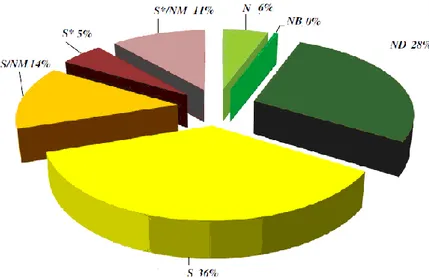
The importance of natural products lies not only in their direct utilization, but also, in order to constitute the pharmacophore or synthons for new molecules, reason is that they often possess highly selective and specific biological activities based on mechanisms of action. From this perspective, the products arising from natural compounds or their direct modifications represent 50% of the new chemical entities (NCEs), as shown in Figure 1 [3].
Fig. 1 Cragg et al. (2013), sources of current drugs represented by pie chart
an unmodified natural product a natural product botanical
amodified natural product
a synthetic compound with no natural product conception
a synthetic compound showing competitive inhibition of the natural product substrate a synthetic compound with a natural product pharmacophore
a synthetic compound with a natural product pharmacophore that showing competitive inhibition of the natural product substrate
The activity of natural products is mainly related to secondary metabolites from plants, compounds that can be generally classified into [4]:
Phenols
Compounds Sulphur-Containing
Terpenes
Alkaloids
Each group has different pharmacological activities, and can be divided into different classes according to own chemical structure.
Phenols, through phenolic ring structure, can play directly activity in free radical scavenger, reducing agents, prooxidants metal chelating and quencher in formation of
protective activities against many pathological conditions, particularly where is involved a component of oxidative stress [5].
Compounds Sulphur-Containing, derived from two main sources in the diet: those derived from the glucosinolate–myrosinase (substrate–enzyme) system found in cruciferous crops, such as cabbages, broccoli (Brassica oleracea) and watercress (Nasturtium officinale), and those derived from the alliin-alliinase system found within Allium crops, such as garlic (A. sativum), onions (A. cepa) and leeks (A. porrum). At these molecules are due the flavor and aroma, as well as many of the alleged therapeutic effects associated with these plants, including the potential chemopreventive, anti-thrombotic, hypoglycemic and hypolipidemic [6].
Terpenes represent one of the largest families of natural products, including more than 40.000 compounds. Their most important structural property is the long series of conjugated double bonds in the central part of the molecule. This kind of structure makes the molecule vulnerable to oxidation and cis-trans isomerisation [7].
Alkaloids are heterogeneous group of low molecular weight, nitrogen-containing compounds mostly derived from amino-acids and found in about 20% of plant species. As secondary metabolites, alkaloids are considered to play a defensive role in the plant against herbivores and pathogens. Due to their potent biological activity, many of the approximately 12.000 known alkaloids have been exploited as pharmaceuticals, stimulants, narcotics and poisons [8].
Since has been said previously, plants play a key role in human health care. In the same way at the development of the research of novel natural compounds, has increased the demand for more and most easily available plant sources. At the UN (United Nations) Conference held in Rio de Janeiro in 1992, have been established parameters for the preservation of biological diversity by promoting sustainable use of sources, this involves the use of natural resources in ways and times that do not lead the depletion of biodiversity. For this reason in the last decade have been increasingly investigated methods for the development of in vitro plant cell cultures. Advances in biotechnology for culturing plant cell cultures, today allow to obtain secondary metabolites in large quantity, bypassing problem to impoverish endangered natural sources, and by using suitable elicitors, get a possibility to increase or select the secondary metabolites production.
According to what said above, in this work particular attention was given to the study of secondary metabolites produced by some plants belonging to the Amaryllidaceae family, in the specific case isoquinoline alkaloids.
At the first instance were characterized both qualitatively and quantitatively three different plants belonging to Amaryllidaceae family, such as: Crinum angustum Steud., Pancratium illyricum L., and Leucojum nicaeense Ard. The alkaloids extracts obtained were separately tested against enzymes involved in specific diseases or liable in multifactorial pathologies. Considering the protection role against external bodies carried out by these metabolites in plant, extracts were also assayed against ATCC microorganisms and clinical isolates. Plants with promising pharmacological activities have been the basis for development of in vitro plant models.
CHAPTER 1
PLANTS OF AMARYLLIDACEAE FAMILY 1.1 INTRODUCTION
The Amaryllidaceae are a family of monocotyledonous plants, represented by 59 genera and over 850 species all over the world [9], assigned by APG (Angiosperm Phylogeny Group III) classification to the order of Asparagales. These plants have mostly tropical and subtropical distribution and their presence in temperate areas is less significant. South America (28 genera) and South Africa (19 genera) are the regions with major diversity. The Mediterranean region, being the source of numerous horticultural introductions, has only eight genera, whereas Australia has only three genera. Amaryllidaceae plants occupy many different habitats: seasonally dry places, ephemeral pools, rainforests understory, and rivers. Currently, molecular evidence places the most ancient lineages and the origin of the family in Africa [10]. The family is essentially composed by bulbous plants that in temperate areas, in the winter season losing their entire epigeal portion stems and leaves, so as to disappear completely at the sight. Though several plants belonging to this family have a considerable size, indeed in some cases can exceed 2 meters high, in other instances are less than 10 centimeters tall. Having regard to the use made of these plants in the research of novel natural compounds, the urban areas development at the expense of natural one, and in some cases their limited size, to date most of the Amaryllidaceae plants family which grow in the Mediterranean area are endangered.
1.2 THE AMARYLLIDACEAE ALKALOIDS AND THEIR BIOLOGICAL ACTIVITIES
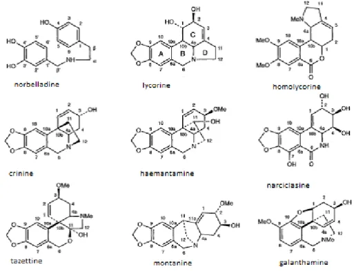
A peculiar property of the Amaryllidaceae plants is a consistent presence of an exclusive group of alkaloids, which have been isolated from plants of all the genera of this family. In fact, the Amaryllidaceae alkaloids represent a large, and still expanding, group of isoquinoline alkaloids, the majority of which are not known to occur in any other family plants. Since the isolation of lycorine from Narcissus pseudonarcissus in 1877, over 300 alkaloids have been isolated from plants of this family [11], and though their structures vary significantly, these alkaloids are considered to biogenetically related. The large number of structurally different Amaryllidaceae alkaloids are classified mainly into nine skeleton types as reported by the Ghosal’s model, for which the representative alkaloids are: norbelladine, lycorine, homolycorine, crinine, hemanthamine, narciclasine, tazetine, montanine and galanthamine (Fig 1.1).
Fig. 1.1 Amaryllidaceae Alkaloid types
investigation for nearly 200 years. Over the past two decades many have been isolated, and screened for different biological activities by a number of research groups. A relevant issue about these alkaloids is their availability, in fact are obtained only in small quantities from natural source. For this reason, is highly developed the practice of synthesize or semi synthesize these alkaloids and their derivatives [12].
In spite of the great variety of pharmacological and biological properties exhibited by Amaryllidaceae alkaloids, only some of the activities of a reduced number of these molecules have been reported. The relationship of chemical structure and biological activity is largely unknown, and further studies are needed to explore the full therapeutic potential of these alkaloids.
Generally, activity displayed by the Amaryllidaceae alkaloids can be traced to principal groups to which they belonging:
Lycorine types, with lycorine, their most representative and characteristic Amaryllidaceae alkaloids, have been reported to be strong inhibitors on parasite development and antifungal activity [13]. Additionally, the alkaloids of lycorine types are potent inducers of apoptosis in human leukemia cells, are selective inhibitors of human ovarian cancer cell, and may exert antiviral effects on several RNA- and DNA-containing viruses [14].
Homolycorine types, some alkaloids of this series, such as homolycorine, being moderately active in inhibiting the in vivo and in vitro growth of a variety of tumor cells, such as Molt 4 lymphoma, HepG2 human hepatoma, and LNCaP human prostate cancer [15]. Other alkaloids homolycorine types show DNA binding activity comparable to that vinblastine [16].
Hemanthamine types and Crinine types, display pronounced cell growth inhibitory activities against a variety of tumor cells, such as Rauscher viral leukemia, Molt 4 lymphoma, BL6 mouse melanoma, HepG2 human hepatoma, HeLa, LNCaP human prostate cancer [17]. Vittatine, as reported in Evidente et al. (2004) an alkaloids of this group, has antibacterial activity against the Gram-positive Staphylococcus aureus and Gram-negative E. coli.
Tazettine types, displays weak hypotensive and antimalarial activities and interacts with DNA. Pretazettine, alkaloids of this group and labile precursor of
tazettine, shows cytotoxicity against fibroblastic LMTK cell lines and inhibits HeLa cell growth, being therapeutically effective against advanced Rauscher leukemia, Ehrlich ascites carcinoma, spontaneous AKR lymphocytic leukemia, and Lewis lung carcinoma. It is one of the most active of the Amaryllidaceae alkaloids against Molt 4 lymphoid cells [18] [19].
Narciclasine types, are an antimitotic and antitumoral alkaloids, affects cell division at the metaphase stage and inhibits protein synthesis in eukaryotic ribosomes by directly interacting with the 60S subunit [20]. The peculiar effects of narciclasine seem to arise from the functional groups and conformational freedom of its C-ring, with the 7-hydroxyl group believed to be important in its biological activity [21]. Narciclasine, is one of the most important antineoplastic Amaryllidaceae alkaloids. It inhibits HeLa cell growth, and is active against a variety of tumor cells. No effect has been observed toward solid tumors. Some alkaloids of this type, such as trisphaeridine, possess high antiretroviral activities [22].
Montanine types, are a group with little information. Evidente el al. (2004), reported some data about pancracine, which shows antibacterial activity against S. aureus and Pseudomonas aeruginosa.
Galanthamine types, have like founder galanthamine. This alkaloid, originally isolated from Galanthus nivalis L. in the 1940, is a long-acting, selective, reversible and competitive inhibitor of acetylcholinesterase, in fact this product is marketed as a hydrobromide salt under the name Razadyne, formely Reminyl, for the treatment of Alzheimer’s disease. Galanthamine has other noteworthy pharmacological actions, including an ability to amplify the nerve-muscle transfer. It is also known to cause bradycardia or atrioventricular conduction disturbance, has long been used as a reversal agent in anesthetic practice, inhibits traumatic shock, and has been patented for use in the treatment of nicotine dependence [23].
1.3 CRINUM ANGUSTUM STEUD.
The genus Crinum, belonging to Amaryllidaceae family, includes approximately 160 species present in warm temperate regions of the world [24]. Cross-hybridization of Crinum plants is rather widespread, mainly due to their particular interest as ornamental plants. Indeed, several species of this genus are cultivated for both ornamental and therapeutic purposes [15]. The ethnobotanical use of Crinum plants have been developed in different parts of the world. For instance, the bulbs of C. asiaticum L. were used in India as tonic, laxatives and expectorants. The C. latifolium L. from India was applied to treat rheumatism, abscesses, earaches and as a tonic. The roots of some Crinum species were used in Africa to treat urinary infections, coughs and colds, renal and hepatic conditions, sores, sexually transmitted disease and backache [25].
In this work was examined Crinum angustum Steud., a hybrid between C. asiaticum var. asiaticum and C. zeylanicum [26]. Plant at the pre-flowering stage were collected in the greenhouse of the Botanical Garden of Bologna University, and was identified by Prof. Lucia Conte. A voucher specimen (No. BOLO0507744) was deposited in the Herbarium of the University Museum System (SMA), Bologna University, Italy. In figure 1.2 is reported a picture of Crinum angustum Steud.
1.4 PANCRATIUM ILLYRICUM L.
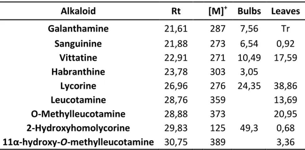
Amongst the Amaryllidaceae, the genus Pancratium comprises over 20 species widely spread throughout the Old World (Asia, Africa and Europe). Some of them have been studied for their potential therapeutical use [27]. Amongst these, Pancratium maritimum is one of the most exhaustively investigated species, and over half of all the alkaloids isolated from Pancratium plants have been identified in this species. Conversely, very few reports are available on Pancratium illyricum L., a species endemic to Sardinia, Corsica and the Tuscan archipelago [28]. In a recent work aimed at identifying new cytotoxic compounds from different Sardinian plants, a considerable activity against both human and bacterial topoisomerase has been demonstrated for three compounds isolated from bulbs of this species [29].
In the present work both bulbs and leaves of Pancratium illyricum L. was analysed. Plant was collected during the flowering period in the South of Sardinia (Punta San Michele, CA, Italy), and identified by Professor Mauro Ballero (University of Cagliari, Italy). A voucher specimen (CAG 1365) has been deposited in the Institute of Botany, University of Cagliari. In figure 1.3 is shown a picture of Pancratium illyricum L.
1.5 LEUCOJUM NICAEENSE ARD.
Leucojum L. is an interesting genus from a taxonomic and cytological point of view. It consists of 12 taxa, many of which are cultivated for their ornamental value. Its distribution, almost all over Europe, includes a wide variety of habitats. The main concentration of species however, is found in the Mediterranean region, which can be considered as the center of diversity for the genus [30]. The genus Leucojum with L. aestivum L., was one of the first sources of galanthamine, and the first plant of the Amaryllidaceae family that has been studied for in vitro development [31].
Leucojum nicaeense Ard., plant belonging to this genus, endemic from the Maritime Alps of southern France and NW-Italy, is included in the World Red List as an endangered species and it is classified as vulnerable. For the EU it is part of the Annex II of Habitat Directive (issued by WCMC) for threatened plants [32]. For this reason, even if potential source of alkaloids to pharmacological interest, is not yet investigated in phytochemical field.
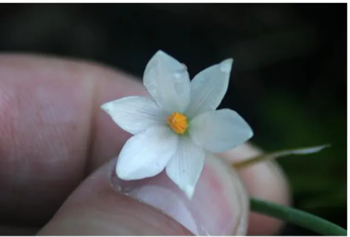
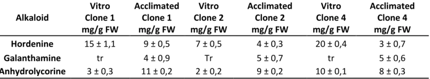
In this work have been examined, in collaboration with the Research Council for Agricultural Experimentation (CRA-FSO) of Sanremo, micropropagated plants (both in vitro and in greenhouse acclimatized) of L. nicaeense Ard.
A voucher specimen (B100341634) of original plant has been deposited in the Botanical Museum of Berlin (Germany). In figure 1.4 is shown a flower of Leucojum nicaeense Ard.
CHAPTER 2
OXIDATIVE STRESS AND ANTIOXIDANTS AGENTS 2.1 INTRODUCTION
Oxidative stress is a pathological condition caused by the breakdown of the physiological balance in organism, between production and elimination of oxidants chemical species by the antioxidant defence systems. All organisms keep a reducing environment within their cells. The cellular redox environment is preserved by enzymes which maintain the reduced state through a constant input of metabolic energy. Eventual disturbance in this normal redox state could cause toxic effects, producing peroxides and free radicals that damage all components of the cell, including proteins, lipids and DNA. The oxidant species and free radicals play important physiological roles, such as the defence against bacteria, the transmission of biochemical signals between cells, the blood pressure control. Is only their excess, generally refers to one or more classes of oxidants, to be implicated in oxidative stress.
Free radicals of importance in living organisms include hydroxyl (OH), superoxide (O2), nitric oxide (NO), and peroxyl (RO2). Peroxynitrite (ONOO), hypochlorous acid (HOCl), hydrogen peroxide (H2O2), singlet oxygen, and ozone (O3) are not free radicals but can easily lead to free-radical reactions in living organisms. The term “reactive oxygen species” (ROS) is often used to include not only the radicals OH, RO2, NO and O2 but also the non radicals HOCl, 1O2, ONOO−, O3, and H2O2 [33].
2.2 RADICAL FORMATION
The radicals are formed as result of acceptance or loss of an electron, and as result of the homolytic cleavage of a covalent bond: the two electrons are separated symmetrically forming two reactive intermediates each of which has an unpaired electron. The presence of electron in an aerobic environment leads to the formation, initially of the superoxide radical, then of hydrogen peroxide and finally the hydroxyl radical. The first step in the ROS generation, is represented by the oxygen acquisition of sufficient energy to reach the singlet state and the subsequent acquisition of an electron, thus giving rise superoxide anion (O2).
resulting in the formation of an oxygen molecule, a hydroxyl anion, and a radical hydroxyl (OH •) that between oxygen radicals, is the most toxic molecule and
more dangerous [34]. In addition to oxygen radicals are reactive species centered on the nitrogen such as nitric oxide (NO •), nitric dioxide (NO 2 •), which as ROS, play roles in physiological conditions of extreme importance, but that if produced in excess, can cause cellular stress.
Free radicals and other reactive species not radical type, are produced in biological systems, both through processes endogenous, that following exposure to exogenous sources. For example among the physiological processes include:
- Aerobic respiration in mitochondria - ROS formation in the cell membrane - Peroxisome activity
- ROS production in endoplasmic reticulum
ROS production can occur also by external agents, as chemical, physical, and natural.
2.3 ANTIOXIDANT SYSTEMS
Organisms have a complex system of defence against oxidative stress, constituted by antioxidant agents both endogenous and exogenous, enzymatic or not, with different mechanism of action and chemical characteristics. Antioxidants exert their action by counteracting or by preventing the oxidation of other molecules, preventing the radical formation, or giving to these an electron. The Human organism possess defense systems extremely effective, as the catalytic activity able to sequester ROS directly or indirectly, substances that act as chemical traps against free radicals, and which restore antioxidant defences. Among these are antioxidants enzymes that catalyze the transformation or destruction of ROS, chelating compounds, and molecules that can act as a scavenger, like many vitamins, GSH (reduced glutathione), co-enzymes and polyphenols.
The main antioxidant enzymes, present in the cytosolic, are:
- Catalase (CAT), located mainly in peroxisomes, and cytosolic fraction, is an enzyme able to protect cells from the toxic effects of hydrogen peroxide (H2O2) by catalysing its decomposition into molecular oxygen and water, without the production of free radicals.
- Glutathione peroxidase (GSH-Px), a metal- enzyme containing selenium, which performs the antioxidant function at the intracellular level. Oxidative stress produces peroxides that can be reduced by using the GSH through glutathione peroxidase into water and alcohol.
- Superoxide dismutase (SOD) is a metal-enzyme that catalyzes the dismutation of the superoxide ion to molecular oxygen and peroxide hydrogen.
- The NADP(H): quinone oxidoreductase 1 (NQO1), sometimes considered an enzyme of phase II, is an enzyme belonging to the class of oxidoreductases, that catalyzes the formation of semiquinone.
Quinones are a group of very common substrates, which may have deleterious effects, such as the ability to attack nucleophiles and generate reactive oxygen species, which are reduced with a mechanism of transfer of ions hydride to generate the corresponding derivative hydroquinonic. The NQO1 has also an important role in the metabolism of endogenous quinones such as vitamin E and ubiquinone. For example the reduction of ubiquinone to work of NQO1 regenerates ubiquinol, which possesses strong antioxidant properties [35].
2.4 OXIDATIVE STRESS AND RELATED DISEASES
Oxidative stress refers to all changes which produced in tissues, cells and biological macromolecules, when following an alteration of the balance between intracellular antioxidant defenses and pro-oxidant elements, with accumulation of radical species. The wide variety of mechanisms that can be activated as a consequence of redox imbalance can significantly contribute to the development of numerous pathological conditions, in particular, cancers, inflammatory pathologies, diabetes, ischemia, cardiovascular disease and neurodegenerative diseases. However, it is still unclear whether oxidative stress is among the main causes, or between the events that occur during the pathological process and that contribute to the progression of the disease. For example, in Parkinson's disease, is unlikely that oxidative stress is the primary event, of the degenerative process that leads to the depletion of dopaminergic neurons, but it is certain, however, that during the disease to occur oxidative phenomena involved substantially into progression of neuronal damage. ROS appear to be involved in many
CHAPTER 3 ALZHEIMER DISEASE 3.1 INTRODUCTION
Alzheimer’s disease (AD) is a debilitating neurodegenerative disorder associated with a rapid cognitive decline with an average of survival of 5-10 years after diagnosis. Furthermore, AD clearly differs from the normal aging in that it causes dramatic loss of synapsis, neurons and brain activity in specific anatomical regions, and results in massive atrophy and gliosis.
The factors that cause some individuals to depart from the relatively benign of normal aging brain and instead undergo the pathological cascade that leads to AD are unknown [36].
Although memory loss is usually the initial and most prominent problem, deficits in cognitive domains other than memory can occur in the early stages of disease in some patients. Final deterioration leads to a bedridden, mute, incontinent, and unresponsive state, which mimics the persistent vegetative state.
Pathologically AD is characterized by the presence of two insoluble protein aggregates, senile plaques formed from the peptide β-amyloid (Aβ) and neurofibrillary tangles composed of hyperphosphorylated tau protein [37]. In rare familial AD, the cause of disease is autosomal dominant mutations in Aβ precursor protein (APP) or the Aβ-producing enzymes presenilins (PSEN1 or PSEN2), which are all thought to lead to increased levels of aggregated Aβ [38]. Likewise, mutations in tau (MAPT) that predispose it to aggregation can cause specific diseases that involve profound neurodegeneration and dementia. Thus, like in other neurodegenerative diseases such as Huntington's disease (HD) and Parkinson's disease, the formation of toxic insoluble aggregates seems to be a key pathogenic step. However, it is not known why these Aβ and tau aggregates accumulate in AD patients nor how they contribute to neuronal dysfunction, particularly for Aβ deposits, which can often be found in the brains of elderly non-demented subjects [39].
An important goal of AD research is to identify interventions that maintain brain function, potentially by inhibiting the formation or improving the clearance of neurotoxic aggregates, or by promoting resistance to, or recovery from, damage.
The clinical presentation of AD is heterogeneous and insidious, and the psychological and financial effects of AD on caregivers and family members are significant.
3.2 THERAPY IN ALZHEIMER’S DISEASE
More than a decade after the first approval of the use of Acetylcholinesterase inhibitor on patients with Alzheimer’s disease, still not have a single treatment or combination therapy that can effectively stop or reverse the progression of such neurodegenerative disease.
Probably there is not one single cause, but several factors are important to describe the etiology of the disease. Therefore, combination of compounds, which act at more than one target site, could be useful for AD treatment.
To date, there are only 5 medications approved by Food Drug Administration (FDA) to treat AD.
They include 4 Acethylcholinesterase inhibitor (AChEIs), and one N-methyl-D-aspartate (NMDA) antagonist, molecule that regulates the activity of glutamic acid receptor.
3.3 CHOLINESTERASE INHIBITORS
The cholinergic hypothesis Of AD concludes that cholinergic system in the basal forebrain are affected early in the disease process including loss of the acetylcholine neurons, loss of enzymatic function for acetylcholine synthesis and degradation, resulting in memory loss as well as deterioration of other cognitive and noncognitive functions such as neuropsychiatric symptoms. A strategy to enhance the cholinergic transmission by using AChEIs to delay the degradation of acetylcholine between the synaptic cleft was then proposed. In 1993 the first FDA approved AChEIs, tacrine boomed out but it was no longer use because of its high prevalence of hepatotoxicity. FDA approved another three AChEIs: donepezil (1996), rivastigmine (2000), and galanthamine (2001) in the following years.
These drugs have been regarded as the standard and first-line treatment for of AD. Systemic reviews, showed benefit on cognitive functions, activities of daily living (ADL), and global functions for patients mild to moderate AD.
Certainly these drugs do not lead to the cure of AD, but to continue to identify AChEIs with fewer adverse effects and capable of alleviate symptoms of AD also in advanced stage, could represent a valid area to study, and Amaryllidaceae plants are good sources of molecules with this activity.
CHAPTER 4
MATRIX METALLOPROTEINASES AND REGULATION OF CELL BEHAVIOR
4.1 INTRODUCTION
Extracellular proteinases are required for numerous developmental and disease-related processes. The ability to degrade extracellular proteins is essential for any individual cell to interact properly with its immediate surroundings and for multicellular organisms to develop and function normally. On this basis, a family of related enzymes has been identified in species from hydra to humans and collectively called matrix metalloproteinases (MMPs), because of their dependence on metal ions for catalytic activity, their potent ability to degrade structural proteins of the extracellular matrix (ECM), and specific evolutionary sequence considerations that distinguish them from other closely related metalloproteinases.
Essentially Matrix metalloproteinases (MMPs) constitute a multigene family of over 25 secreted and cell surface enzymes that process or degrade numerous pericellular substrates. Their targets include other proteinases, proteinase inhibitors, clotting factors, chemotactic molecules, latent growth factors, growth factor–binding proteins, cell surface receptors, cell-cell adhesion molecules, and virtually all structural extracellular matrix proteins. Thus MMPs are able to regulate many biologic processes and are closely regulated themselves [40].
In addition to their ECM substrates, MMPs also cleave cell surface molecules and other pericellular non-matrix proteins, thereby regulating cell behavior in several ways [41]. Thus like the many proteins they modify, the MMPs influence diverse physiologic and pathologic processes, including aspects of embryonic development, tissue morphogenesis, wound repair, inflammatory diseases, and cancer [42].
At present, 25 vertebrate MMPs and 22 human homologues have been identified and have been classified according to substrate specificity and are often referred to as: collagenases, gelatinases, stromelysins, martilysins, and membrane-type MMPs. All contain Zn 2+ at the catalytic site as well as an additional zinc ion and calcium ions for stability. Individual MMPs are referred to by their common names or according to a
sequential numeric nomenclature reserved for the vertebrate MMPs. In addition, they are often grouped according to their modular domain structure. In this regard, all MMPs have an N-terminal signal sequence (or “pre” domain) that is removed after it directs
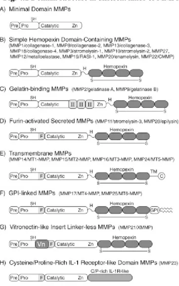
synthesis to the endoplasmic reticulum. Thus most MMPs are secreted; however, six display transmembrane domains and are expressed as cell surface enzymes. The pre domain is followed by a propeptide “pro” domain that maintains enzyme latency until it is removed or disrupted, and a catalytic domain that contains the conserved zinc-binding region. The catalytic domain dictates cleavage-site specificity through its active site cleft, through specificity sub-site pockets that bind amino acid residues immediately adjacent to the scissile peptide bond, and through secondary substrate-binding exosites located outside the active site itself [43]. In figure 4.1 are shown domain structure in different kinds of MMPs.
Fig. 4.1 Domain structure in different kinds of MMPs
Most of the MMPs are synthesized as inactive latent enzymes. Conversion to the active enzyme is generally mediated by activator systems that include plasminogen activator or the pro-hormone convertase, furin. MMP activity is regulated by a group of endogenous proteins, called, tissue inhibitor of metalloproteinases (TIMPs) that bind to active and alternative sites of the activated MMP.
The TIMPs represent a family of at least four 20–29-kDa secreted proteins (TIMPs 1–4) that reversibly inhibit the MMPs in a 1:1 stoichiometric fashion. Individual TIMPs differ in their ability to inhibit various MMPs. In addition, the TIMPs differ in terms of their gene regulation and tissue-specific patterns of gene expression [44].
To date, no TIMP receptors have yet been identified, suggesting that TIMPs may act as decoys for various signaling molecules. TIMPs are not the only endogenous MMP inhibitors. Indeed, α2-macroglobulin is a major endogenous inhibitor of the MMPs, and
its importance may have been overlooked due to the recent emphasis placed on the TIMPs. Because α2-macroglobulin is an abundant plasma protein, it represents the major inhibitor of MMPs in tissue fluids, whereas TIMPs may act locally. Moreover, α2-macroglobulin plays an important role in the irreversible clearance of MMPs, whereas TIMPs inhibit MMPs in a reversible manner.
4.2 MMPs AND DISEASE
Studies using relevant disease models in MMP-deficient animals have demonstrated the contribution of MMPs to disease processes. In the cardiovascular area, MMPs have been strongly associated with aneurysms [45], with atherosclerotic plaque rupture [46], with myocardial infarction, left ventricular remodeling and ultimate cardiac rupture [47], as well as cerebral ischemia events [48]. MMP expression is raised in multiple tumor types and mostly, these increases correlate with decreased survival. In both rheumatoid- and osteo-arthritis, MMPs are considered to be significantly responsible for the matrix degradation that characterizes these diseases [49]. Respiratory disorders including idiopathic pulmonary fibrosis, asthma, emphysema, and acute respiratory distress syndrome [50],are also strongly associated with MMP activity.
Is important to emphasize that in some pathologies the correct MMP activity leads to a positive resolution of the disease, while in others, this result is achieved by inhibiting the activity of MMPs. Rather than categorizing specific MMPs as “good” or “bad”, it is more helpful to consider activities in particular contexts. For instance expression studies in humans have shown protective roles of certain MMPs. In particular, high levels of MMP12 correlate with better prognosis in several tumor types including hepatocellular and colorectal carcinoma. This is in direct contrast to the destructive role of MMP12 in emphysema where the elastase-degrading activity of MMP12 clearly contributes to pathophysiology. By generalizing it can be stated that in chronic diseases, inflammatory, and in some tumors, leads to disease an hyperactivity of MMPs.
Is precisely on this type of disorders that will be tested extracts obtained in this work, using the collagenase enzyme from Clostridium histolyticum, as starting model for MMPs inhibition screening; in fact, scientific literature sees in the natural extracts, a great source of compounds with inhibitory activity on matrix metalloproteinase [51] [52].
CHAPTER 5 TYROSINASE ENZYME 5.1 INTRODUCTION
Tyrosinases also known as polyphenol oxidase (PPO), is a copper-containing enzyme widely distributed in microorganism, animals, and plants, which catalyze the oxidations of both monophenols (cresolase or monophenolase activity) and o-diphenols (catecholase or diphenolase activity) into reactive o-quinones. The term tyrosinase refers to its typical substrate, tyrosine. Both tyrosinase activities appear to have broad substrate specificities, although the enzyme has a higher affinity for the L-isomers of the substrates than for the corresponding D-isomers. The first biochemical investigations were carried out in 1895 on the mushroom Russula nigricans, whose cut flesh turns red and then black on exposure to air. Since this study, the enzyme has been found widely distributed throughout the phylogenetic scale from bacteria to mammals. The best-characterized tyrosinases are derived from Streptomyces glausescens, the fungi Neurospora crassa and Agaricus bisporus. In fungi and vertebrates, tyrosinase catalyzes the initial step in the formation of the pigment melanin from tyrosine.
The notable feature observed in tyrosinases from different sources is that the central copper-binding domain is conserved, which contains strictly conserved amino acid residues, including three histidines. One tyrosinase molecule can contain two copper atoms, and each atom of the binuclear copper cluster is ligated to three histidines. In the formation of melanin pigments, three types of tyrosinase (oxy-, met-, and deoxytyrosinase) with different binuclear copper structures of the active site are involved [53].
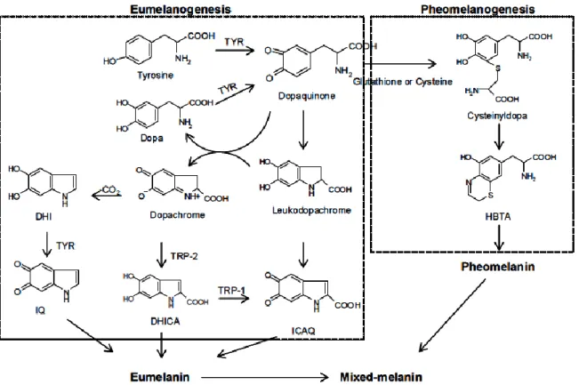
Melanogenesis is initiated with the first step of tyrosine oxidation to dopaquinone catalyzed by tyrosinase. This first step is the rate-limiting step in melanin synthesis because the remainder of the reaction sequence can proceed spontaneously at a physiological pH value. The subsequent dopaquinone is converted to dopa and dopachrome through auto-oxidation.
Dopa is also the substrate of tyrosinase and oxidized to dopaquinone again by the enzyme. Finally, eumelanin are formed through a series of oxidation reactions from
dihydroxyindole (DHI) and dihydroxyindole-2-carboxylic acid (DHICA), which are the reaction products from dopachrome. In the presence of cysteine or glutathione, dopaquinone is converted to cysteinyldopa or glutathionyldopa. Subsequently, pheomelanin is formed. In addition to eumelanin and pheomelanin, other “melanin” relying on phenolic monomers different from tyrosine is termed allomelanin. The browning phenomenon in fruit and fungi is also usually related to oxidative polymerization, conceptually similar to melanogenesis.
The main difference resides in the fact that allomelanin substantially does not contain dopaquinone-derived motifs as the main monomers in its structure and, on the contrary, is based on other quinoid building blocks [54].
Melanin plays an important role in protecting human skin from the harmful effects of sun UV radiation. Melanin also determines our phenotypic appearance.
Fig. 5.1 Biosynthetic pathway of melanin. TYR, tyrosinase; TRP; tyrosinase related
protein; dopa, 3,4-dihydroxyphenylalanine; DHICA, dihydroxyindole-2-carboxylic acid; DHI, 5,6-dihydroxyindole; ICAQ, indole-2-carboxylic acid-5,6-quinone; IQ, indole5,6-quinone; HBTA,
5-hydroxy-1,4-benzothiazinylalanine.
5.2 ROLE OF TYROSINASE IN MAN AND NATURE
Melanogenesis has been defined as the entire process leading to the formation of dark macromolecular pigments, i.e., melanin. Melanin is formed by a combination of enzymatically catalyzed and chemical reactions. It is a heterogeneous polyphenol-like biopolymer with a complex structure and color varying from yellow to black. The color of mammalian skin and hair is determined by a number of factors, the most important of which is in fact, the degree and distribution of melanin pigmentation. Melanin is secreted by melanocyte cells distributed in the basal layer of the dermis. The role of melanin is to protect the skin from ultraviolet (UV) damage by absorbing UV sunlight and removing reactive oxygen species (ROS). Various dermatological disorders result in the accumulation of an excessive level of epidermal pigmentation. These
Great interest has been shown in the involvement of melanins in malignant melanoma, the most life-threatening skin tumors. Is evident, like tyrosinase play a key role both correct pigmentation of skin and in development of melanoma. This enzyme in fact, shows antiproliferative and apoptotic activity against malignant melanocytes.
In the food industry, tyrosinase is a very important enzyme in controlling the quality and economics of fruits and vegetable. Tyrosinase catalyzes the oxidation of phenolic compounds to the corrisponding quinones and is responsible for the enzymatic browning of fruits and vegetable. In addition to the undesiderable color and flavor, the quinone compounds produced in the browning reaction may irreversibly react with the amino and sulfhydryl groups of proteins. The quinone-protein reaction decreases the digestibility of the protein and the bioavailability of essential amino acids, including lysine and cysteine [56].
At last, tyrosinase, plays an important role in the developmental and defensive functions of insects; is interested in fact in the insect molting, through the formation of the cuticle leads larva to insect [57].
5.3 TYROSINASE INHIBITORS AND THEIR IMPORTANCE
A number of tyrosinase inhibitors from both natural and synthetic sources have been identified. However, the definition of “tyrosinase inhibitor” is sometimes misleading: many authors use that terminology in reference to melanogenesis inhibitors, whose action mainly resides in some interference in melanin formation, regardless of any direct inhibitor/enzyme interaction. Many putative inhibitors are examined in the presence of tyrosine or dopa as the enzyme substrate, and activity is assessed in terms of dopachrome formation. Thus, experimental observation of the inhibition of tyrosinase activity can be accomplished by one of following:
1. Reducing agents, which causing chemical reduction of dopaquinone such as ascorbic acid, avoid dopachrome and melanin formations. For this reason are used as a melanogenesis inhibitor for their capacity to reduce back o-dopaquinone to dopa.
2. o-Dopaquinone scavenger such as most thio-containing compounds, which are well-known melanogenesis inhibitors and react with dopaquinone to form colorless products. The melanogenetic process is therefore slowed until all the scavenger is consumed, and then it goes at its original rate.
3. Alternative enzyme substrates such as some phenolic compounds, whose quinoid reaction products absorb in a spectral range different from that of dopachrome. When these phenolics show a good affinity for the enzyme, dopachrome formation is prevented, and they could be mistakenly classified as inhibitors.
4. Nonspecific enzyme inactivators such as acids or bases, which non-specifically denature the enzyme, thus inhibiting its activity.
5. Specific tyrosinase inactivators such as mechanism-based inhibitors, which are also called suicide substrates. These inhibitors can be catalyzed by tyrosinase and form covalent bond with the enzyme, thus irreversibly inactivating the enzyme during catalytic reaction. They inhibit tyrosinase activity by inducing the enzyme catalyzing “suicide reaction.”
6. Specific tyrosinase inhibitors. The compounds bind reversibly to tyrosinase and reduce its catalytic capacity.
Among the six types of compounds described above, only specific tyrosinase inactivators (5) and inhibitors (6) are regarded as “true inhibitors,” which actually bind to the enzyme and inhibit its activity [54].
The discovery and characterization of new tyrosinase inhibitors are useful for their potential applications, and also because there are few “true inhibitiors” available. The fields of interest for tyrosinase inhibitors is clear that are various: improving food quality and nutritional value, controlling insect pests, preventing pigmentation disorders and other melanin-related health problems in human beings.
CHAPTER 6 :
THE PROBLEM OF MULTIDRUG RESISTANCE (MDR) AND NOSOCOMIAL INFECTIONS
6.1 INTRODUCTION
According to the World Health Organization, infectious diseases are a significant cause of morbidity and mortality worldwide, accounting for approximately 50% of all deaths in tropical countries. Infectious and parasitic diseases remain primary causes of pediatric mortality in developing countries, partly as a result of the HIV/AIDS epidemic [58]. Despite the progress made in the understanding of microbiology and the control of microorganisms, sporadic incidents of epidemics due to drug resistant microorganisms and hitherto unknown disease causing microbes pose an enormous threat to public health. Such negative health trends call for a new global initiative for the development of new strategies for the prevention and treatment of infectious disease [59].
Proposed solutions outlined by the Center for Disease Control as a multipronged approach include: prevention, improved monitoring, and the development of new treatments that exceed the resistance bacterial.
It was just a few years after the introduction of penicillin that scientists began to notice the emergence of a penicillin resistant strain of Staphylococcus aureus, a common bacterium that makes up part of normal human bacterial flora.
Since then, resistant strains of gonorrhoea, dysentery causing Shigella (a major cause of premature death in developing countries) and Salmonella followed in the wake of S. aureus. From the initial case of resistant Staphylococcus aureus, the problem of antimicrobial resistance has grown into a serious threat to public with economic, social, and medical implications that are global in scope and cross all environmental and ethnic boundaries [60].
National Nosocomial Infection Surveillance (NNIS) System data demonstrate a steady increase in the incidence of nosocomial infections caused by methicillin-resistant S. aureus (MRSA) among ICU (intesive Care Units) patients over time. MRSA now accounts for more than 60% of S. aureus isolates in USA hospital ICUs. In addition, the medical issues of these infected patients have become more complex as a result of sophisticated medical system, and the parallel development of infections at already present bacterial resistance can lead to death of the patients [61].
In addition to the problem of multi-drug resistance (MRD), should consider the increasing development of nosocomial infections. One of the primarily responsible of hospital infections, is the Candida with its spp.
Candida spp. are an important cause of nosocomial bloodstream infections (BSIs). The incidence of BSIs caused by Candida spp. has risen in the past 20 years [62], and Candida spp. are currently the fourth leading cause of nosocomial BSI in the USA. Mortality rates range between 29% and 76%, with an attributable mortality high as 49% [63]. Several Candida spp. exhibit reduced susceptibility to fluconazole and others antifungal drugs, so, also in this case is important the development of new treatments.
6.2 MEDICINAL PLANTS AND TREATMENT OF MICROBIAL INFECTIONS Naturally derived compounds have made considerable contributions to human health and well-being, and have been a source of inspiration for novel drug development. There are thousands of published scientific papers from around the globe describing the antimicrobial activities of plants extract. The majority of the plants had activity against a range of bacteria, fungi and Mycobacterium. Many of the plant species with antimicrobial activity were native to tropical countries, due to the extraordinary biodiversity of these places.
Is important to note that, while many of the medicinal plant extracts purported to have antibacterial activity have only been subjected to in vitro screening, and the vast majority of these extracts have never been tested rigorously in animal models or controlled clinical trials [64].
In this study, the extracts and pure compounds tested, were analyzed both as regards the ATCC strains and on clinical isolated, with the aim to be able to approach the data obtained as much to a real result.
CHAPTER 7 :
PLANT CELL CULTURE SYSTEMS: A POTENTIAL RENEWABLE SOURCE OF VALUABLE MEDICINAL COMPOUNDS
Many higher plants are major sources of natural products used as pharmaceuticals, agrochemicals, and food additives. The search for new plant derived chemicals should thus be a priority in current and future efforts toward sustainable conservation and rational utilization of biodiversity. In the search for alternatives to production of desirable medicinal compounds from plants, biotechnological approaches, specifically, plant tissue cultures, are found to have potential as a supplement to traditional agriculture in the industrial production of bioactive plant metabolites. Cell suspension culture systems could be used for large scale culturing of plant cells from which secondary metabolites could be extracted. The advantage of this method is that it can ultimately provide a continuous, reliable source of natural products [65].
Discoveries of cell cultures capable of producing specific medicinal compounds at a rate similar or superior to that of intact plants have accelerated in the last few years. New physiologically active substances of medicinal interest have been found by bioassay. It has been demonstrated that the biosynthetic activity of cultured cells can be enhanced by regulating environmental factors, well as by artificial selection or the induction of variant clones. Some of the medicinal compounds localized in morphologically specialized tissues or organs of native plants have been produced in culture systems not only by inducing specific organized cultures, but also by undifferentiated cell cultures. The major advantages of a cell culture system over the conventional cultivation of whole plants are: 1) Useful compounds can be produced under controlled conditions independent of climatic changes or soil conditions; 2) Cultured cells would be free of microbes and insects; 3) The cells of any plants, tropical or alpine, could easily be multiplied to yield their specific metabolites; 4) Automated control of cell growth and rational regulation of metabolite processes would reduce of labor costs and improve productivity; 5) Organic substances are extractable from callus cultures.
Advances in tissue culture, combined with improvement in genetic engineering, specifically transformation technology, has opened new avenues for high volume production of pharmaceuticals, nutraceuticals, and other beneficial substances [66].
CHAPTER 8 : AIM OF THE STUDY
Historically, man has used plants to supply almost all of his basic needs, and plants have always played a central role in traditional systems of medicine for the prevention and treatment of disease around the world.
In the development of new drugs, the role of natural products has been two fold: 1) they may become the base for the development of a medicine, or, 2) a phytomedicine to be used for the treatment of disease.
On these basis, in this study, are analysed plants extract of Amaryllidaceae family, plants that are known to contain secondary metabolites of high pharmacological profile, since they have been proven to possess antibacterial, antifungal, antiprotozoal, antitumoral, and acetylcholinesterase inhibitory activities [67].
Since these plants have never been studied, the purpose is:
- to characterize with phytochemical studies the alkaloid extracts.
- In the event that encounter compounds not yet classified, carry out a complete characterization, by establishing the properties of the compounds.
- To test extracts on pharmacological targets, that representing both degenerative and chronic diseases.
- Achieve a development of in vitro plant cell cultures, with the goal of obtaining in vitro production of alkaloids, equivalent to that of plant.
The in vitro development will be made, not only for protection of the species biodiversity because the plants analyzed in this study are endangered, but also because in the case that plant will grow in vitro and will produce in the same manner of the original one, will be controlled the production of secondary metabolites with addition of elicitors.
The pharmacological targets chosen for screening of the extracts activity, are associated with a wide range of disorders or diseases, not only, their activity can also be extended to fields such as cosmetics, food and agrochemical; just think in fact of the applicability that could have the inhibition of enzymes such as: Collagenase, Tyrosinase and Acetylcholinesterase.
Not least, the extracts will be tested on Multi-Drug Resistant (MDR) microorganism, considering the original protective role against attacks from external agents that have the alkaloids in plants.
With the development of MDR and the consequent hospital infections, which daily involve discomfort and increased risk on the patient's health with a consequent increase in health spending, the development of new molecules with antimicrobial activity and not subject to bacterial resistance could open new ways for the development of new drugs.
CHAPTER 9 :
MATERIALS AND METHODS 9.1 EXTRACTION AND ISOLATION OF ALKALOIDS
The extracts of three Amaryllidaceae plants of C. angustum, P.illyricum and L. nicaeense (all 6 micropropagated plants) were separately obtained from fresh plants, as follows: were crushed separately and exhaustively extracted with MeOH at room temperature for 72 h. The extracts were evaporated under reduced pressure. These crude extracts were acidified by dissolving in 100 mL H2SO4 1% (v/v) and neutral material was removed with n-hexane (6 x 100 mL) and CHCl3 (4 x 100 mL). The acidified solutions were then basified with 25% NH4OH up to pH 9-10 and extracted with CHCl3 (4 x 100 mL) to give the chloroformic extract containing alkaloids. The extracts were dried with anhydrous Na2SO4, filtered and completely dried under reduced pressure. As regard leaf chloroformic extract of P. illyricum was subjected to vacuum liquid chromatography (VLC) [68] using a silica gel 60 A (6-35 μ) column with a diameter of 1 cm and height of 4 cm. Alkaloids were eluted using n-hexane gradually enriched with EtOAc, then gradually enriched with CHCl3 and finally with a mixture of EtOAc and CHCl3 gradually enriched with MeOH. Fractions of 10 mL were collected (200 in total), monitored by TLC (Dragendorff’s reagent, UV 254 nm) and combined according to their profiles. Four main fractions were obtained and subjected to preparative TLC (20 cm x 20 cm x 0.25 mm, silica gel 60F254). 11α-hydroxy-O-methylleucotamine (7 mg) was obtained in major quantities from fractions 40-53 (eluted from VLC with n-Hexane-EtOAc, 64:36 to 56:44) through preparative TLC (EtOAc-CHCl3-CH3OH 4:1.5:0.5 + 25% ammonia).
9.2 GC-MS ANALYIS
The extracts of C. angustum, P. illyricum and L. nicaeense (all 6 micropropagated plants) obtained separately were analyzed qualitatively by GC-MS analysis starting from a mother solution of 300 µg/mL.
EIMS were obtained on a GC-MS Hewlett-Packard 6890 + MSD 5975 operating in EI mode at 70 eV. A HP-5 MS column (30 m × 0.25 mm × 0.25 μm) was used. The temperature program was: 100-180 ºC at 15 ºC min-1, 1 min hold at 180 ºC, 180-300 ºC
of the carrier gas (helium) was 0.8 mL min-1. In most cases the split ratio was 1:20, but with more diluted samples a split ratio of 1:5.
9.3 HPLC-DAD ANALYSIS
The quantitative analysis of C. angustum, P. illyricum and L. nicaeense (all 6 micropropagated plants) obtained separately were carried out using an HPLC system (Jasco Corp., Tokyo, Japan) consisting of a PU-1580 pump, an LG-1580-02 ternary gradient unit, a DG-1580-53 three-line degasser, and a diode array (DAD) detector MD-2018 Plus operating at 280, 240 and 220 nm, linked to an autosampler (2055 Plus). A Spherisorb C18 column (5 μm, ODS2, 4.6 × 250 mm, Waters Corp., Milford, MA, USA) was used with a flow rate of 0.7 ml/min and elution was performed under 27 °C temperature. The mobile phase was made up of acetonitrile:ammonium acetate 80 mM pH 6.0 (30:70). Data were acquired and processed by the Chromnav Chromatography Data system software (Jasco).
9.4 GENERAL
All chemical products used were provided by Sigma Aldrich. UV spectra were obtained on a DINKO UV2310 instrument and IR spectra were recorded on a Nicolet Avatar 320 FT-IR spectrophotometer. Biological assays were performed using a microplate reader, Victor X3 Perkin-Elmer (Perkin-Elmer Inc., Boston, MA, USA) and dataanalyzed with the software Work Out 2.5 or in a Jasco V-530 spectrophotometer (Jasco Europe, Cremella, Italy). Statistical analyzes were obtained from software GraphPad Prism 5.
9.5 NMR ANALYSIS
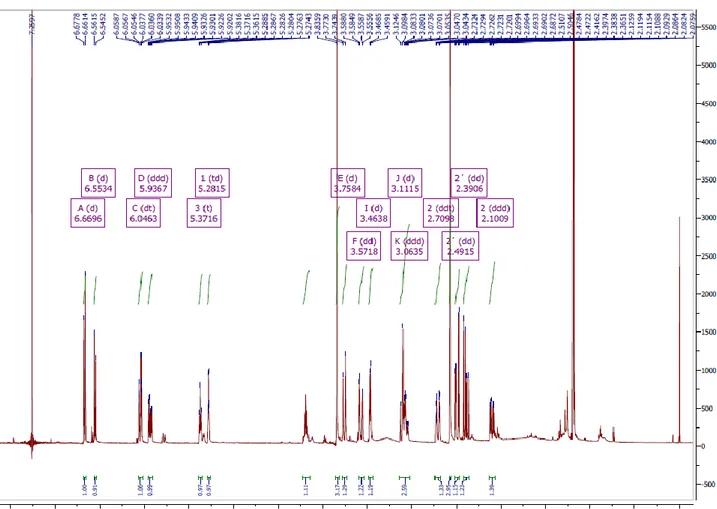
Compound isolated from the leaves extract of P. illyricum was subjected to NMR analysis as follows: NMR spectra (one and two dimensional) were recorded in a Varian VXR 500 MHz, instrument using CDCl3 as the solvent and TMS as the internal standard. Chemical shifts were reported in δ units (ppm) and coupling constants (J) in Hz.
9.6 CYTOTOXICITY ASSAY
The human embryo lung fibroblast (HEL) were grown in DMEM (Gibco®, Life technologies) supplemented with 10% foetal calf serum (Lonza, BioWhittaker), 10U penicillin and 50 μg/ml streptomycin at 37°C. For each set of experiments, cells were seeded into 96-well plates at 104 cells/well and incubated with 2-fold serial dilutions starting from 2.50 mg/mL of the extract sample; there were positive controls containing cells in regular medium and solvent controls containing cells in 2-fold serial dilutions of DMSO (from 6.25 %). Cells were grown for 24 h at 37°C, then 50 µL of XTT labeling mixture (Cell Proliferation kit II, XTT, Roche) were added to cells, followed by a 6-h incubation allowing formazan production. Finally, OD was measured at 450/630 nm by using the microplate reader. The cytotoxic activity was expressed as the IC20 (20% inhibitory concentration) after a 24 h-incubation period, relative to the control.
9.7 ANTIOXIDANTS ASSAYS 9.7.1 DPPH AND ABTS ASSAY
The DPPH assay was performed according to the method of Venditti et al. (2013) [69], with some modifications. Stock solutions of plants extract were prepared in water/DMSO (90-10) to obtain different final concentrations (from 5 to 200 μg/mL in the assay) to calculate the IC50 value. One and a half milliliters of a 0.05 mM DPPH methanol solution was added to different concentrations of plants extract and allowed to react at room temperature. The assay was performed in a final volume of 2 mL. After 20 min, the absorbance (Abs) values were measured at 517 nm and converted into percentage antioxidant activity using the following formula:
Scavenging capacity% = [1 (Abs sample /Abs control) 100]
The DPPH solution plus methanol was used as a negative control, whereas Trolox (TR) at different concentrations (from 5 to 40 μM) was used as reference antioxidant compound. The IC50 values were calculated by logarithmic regression of plots, where the x-axis represents the individual plants extract or reference compound concentrations and the y-axis the average percentage of scavenging capacity.
The ABTS assay was performed according to the method of Venditti et al. (2013) with some modifications. ABTS•+ radical was generated by mixing a 2 mM ABTS solution with 7 mM K2S2O8 and incubating in the dark for 24h at room temperature. Before usage, the ABTS •+ solution was diluted (1−25 mL methanol) to obtain an Abs value of 0.7 at 734 nm. Upon addition of 1 mL of the diluted ABTS•+ solution to 10 μL of reference compound or plants extract stock solutions (from 5 to 200 μg/mL), the Abs at 734 nm was recorded after 1 min. The final TEAC value of plants extract was calculated by comparing ABTS•+ decolorization with that of Trolox. The IC50 value was calculated as described above.
9.7.2 β-CAROTENE BLEACHING ASSAY
Prevention of the autoxidation of emulsified linoleic acid was determined by modifying method of Venditti et al. (2013). Briefly, 20 μL of different plants extract or reference compound stock solutions (50, 100, 150, and 200 μg/mL) was added to the microplate wells (Costar 3599) in duplicate. Then, 10 μL of linoleic acid, 47 μL of Tween 40, and 2.5 mL of β-carotene (2 mg/mL in chloroform) were placed in a flask. After removal of chloroform with nitrogen gas, 22,5 mL of distilled hot water (50 °C) and 2.5 mL of 0.1 M sodium phosphate buffer (pH 6.8) were added to the same flask and shaken well. An amount of 0.2 mL of the linoleic acid−β- carotene emulsion were transferred rapidly to each well, kept under constant temperature (50 °C), and the Abs at 490 nm monitored for 60 min. H2O (20 μL) and reference compound (20 μL) at different concentrations (0.25, 0.5, 1, 1.5, and 2.5 mM) were used as negative and positive controls, respectively. The difference in Abs at 50 and 5 min (Δ = Abs 50 min − Abs 5 min) was calculated. Results are expressed in terms of percentage bleaching inhibition of the initial linoleic acid−β-carotene emulsion by the test samples according to the following equation:
%bleaching inhibition = [1-(Δabs sample /Δ Abs negative control) 100]
IC50 values were calculated by logarithmic regression in terms of micromolar or µg/mL. The antioxidant activity was also expressed as TEAC, by comparing IC50 values obtained for plants extract with that of Trolox (TR).
9.8 STUDY OF THE ENZYMATIC INHIBITION ACTIVITY
9.8.1 INHIBITION OF THE ACETYLCHOLINESTERASE (ACHE) ENZYME The assay for measuring AChE activity was performed as described by López et al. (2002). In brief, 50 μL of AChE in buffer phosphate (8 mM K2HPO4, 2.3 mM NaH2PO4, 0.15 mM NaCl, 0.05% Tween 20, pH 7.6) and 50 μL of the sample dissolved in the same buffer were added to the wells. The plates were incubated for 30 min at room temperature before the addition of 100 μL of the substrate solution (0.1 M Na2HPO4, 0.5 M 5,5’-dithiobis-2-nitrobenzoic acid (DTNB), 0.6 mM acetylthiocholine iodide, pH 7.5). The absorbances were read in a microplate reader Victor X3 Perkin Elmer (Perkin Elmer Inc., Massachusetts USA) at 405 nm after five minutes. Galanthamine hydrobromide was used as a positive control. The IC50 of bulbs and leaves extract of P. illyricum, 11α-hydroxy-O-methylleucotamine and galanthamine hydrobromide was determined in triplicate and the results are presented as mean ± standard deviation.
9.8.2 INHIBITION OF THE COLLAGENASE ENZYME WITH SYNTHETIC SUBSTRATE
The alkaloid extracts of C. angustum, P. illyricum (bulbs and leaves) and L. nicaeense were tested on Collagenase assays that uses FALGPA as substrate.
The Clostridium hystolyticum collagenase (EC.3.4.23.3) assay was based on the assay described by van Wart et al. [70], slightly modified for use with a 96-well microtiter plate: enzyme was dissolved in 50 mM tricine buffer (with 10 mM CaCl2 and 400 mM NaCl), pH 7.5, to furnish 0.8 units/mL (according to the supplier’s activity data); substrate FALGPA was prepared 2 mM in that same buffer. An amount of 25 µL buffer, 25 µL H2O or inhibitor, and 25 µL enzyme was loaded each well of the 96-well microtiter plate, and after 15 min of preincubation, 50 µL of substrate was added. Absorbance was measured at 340 nm immediately and at 2-min intervals for 20 min. Enzyme activity was estimated by linear regression of the absorbance values recorded during that time. For comparative purposes we also assayed EGCG (epigallocatechin gallate) as a known inhibitor.