BIOS - Research Doctorate School in BIOmolecular Sciences
Ph.D. in BIOMATERIALS - XXII Cycle
Biocompatible Polymeric Materials for Regenerative
Medicine Applications
Mamoni Dash
Supervisor: Prof./Dr. Emo Chiellini
Tutor:
Dr. Federica Chiellini
Laboratory of Bioactive Polymeric Materials for Biomedical and
Environmental Applications (BIOlab)
Objectives ... xv
1. Chitosan- A Versatile Material For Regenerative Medicine Applications ... 1
1.1. Abstract ... 1
1.2. Introduction ... 1
1.3. General Aspects of Chitosan ... 2
1.3.1 Structure of Chitosan ... 2
1.3.2 Source & Availability of Chitosan ... 3
1.3.3 Physicochemical Properties of Chitosan ... 3
1.3.4 Biological Properties of Chitosan ... 5
1.3.5 Biodegradability of Chitosan ... 5
1.3.5.1 Biodegradation- In-vitro ... 6
1.3.5.2 Biodegradation- In-vivo ... 7
1.3.6 Biodistribution of Chitosan ... 7
1.3.6.1 Distribution after Intravenous Administration ... 8
1.3.6.2 Distribution after intraperitoneal administration ... 8
1.3.6.3 Tissue distribution after oral administration... 9
2.1.6.4 Intracellular chitosan distribution ... 9
1.3.7 Toxicity of Chitosan ... 10
1.3.7.1 In-vitro toxicity ... 10
1.3.7.2 In-vivo toxicity ... 11
1.4. Chitosan Based Systems for Regenerative Medicine Applications ... 11
1.4.1 Chitosan Micro/nano Particles ... 11
1.4.1.1 Methods for the preparation of Chitosan micro/nanoparticles ... 12
1.4.1.2 Drug loading into Chitosan micro/nanoparticles ... 20
1.4.1.3 Drug release & release kinetics ... 21
1.4.2.2 Crosslinked networks ... 28
1.4.2.3 Drug loading in chitosan hydrogels ... 30
1.4.2.4 Drug release from chitosan hydrogels... 31
1.5. Applications ... 32
1.5.1 Chitosan for Tissue Engineering Applications ... 32
1.5.1.1 Chitosan in bone tissue engineering ... 32
1.5.1.2 Chitosan in cartilage tissue engineering ... 34
1.5.1.3 Chitosan in liver tissue engineering ... 38
4.1.1.3 Chitosan in nerve tissue engineering ... 39
1.5.2 Drug Delivery Applications ... 41
1.5.3 Chitosan in Gene Therapy ... 47
1.5.4 Chitosan in Bioimaging Applications ... 50
1.5.5 Chitosan in wound healing applications ... 52
1.6. Conclusions ... 52
1.7. References ... 54
2. Statistical Approach of Chitin Deacetylation ... 79
2.1. Abstract ... 79
2.2. Introduction ... 79
2.3. Materials and Methods ... 81
2.3.1 Materials ... 81
2.3.2 Methods ... 81
2.3.2.1 Chitin Deacetylation ... 81
2.3.2.2 Fourier Transform Infrared Spectroscopy (FTIR) ... 82
2.3.2.3 Thermogravimetric Analysis (TGA) ... 82
2.3.2.3 Ultraviolet Spectrophotometry (UV) ... 83
2.4.2 TGA ... 88
2.4.3 UV Spectrophotometry ... 91
2.5. Conclusion ... 96
2.6. References ... 97
3. Chitosan Based Beads for Controlled Release of Proteins ... 101
3.1. Abstract ... 101
3.2. Introduction ... 101
3.3. Materials and Methods ... 104
3.3.1 Materials ... 104
3.3.2 Methods... 104
3.3.2.1 Preparation of crosslinked Chitosan beads ... 104
3.3.2.2 Preparation of protein loaded Chitosan beads ... 105
3.3.2.3 Morphological characterization... 105
3.3.2.4 Swelling of Chitosan-TPP beads ... 105
3.3.2.5 Degradation of Chitosan-TPP beads ... 106
3.3.2.6 Evaluation of protein encapsulation efficiency ... 106
3.3.2.7 Protein release studies ... 106
3.4. Results and Discussion ... 107
3.4.1 Preparation of Crosslinked Chitosan Beads ... 107
3.4.2 Morphological Observation ... 110
3.4.3 Swelling Ratio ... 111
3.4.4 Degradation of Chitosan-TPP Beads... 111
3.4.5 Protein Encapsulation and Release Studies ... 112
3.5. Conclusion ... 114
4.1. Abstract ... 119
4.2. Introduction ... 119
4.3. Materials and Methods ... 122
4.3.1 Materials ... 122
4.3.2 Methods ... 122
4.3.2.1 Synthesis of MAGlyGly ... 122
4.3.2.2 Preparation of Chitosan-MAGlyGly nanoparticles [CSx-(MAGlyGly)y] .... 123
4.3.2.3 Preparation of Chitosan-polymerized(MAGlyGly) nanoparticles [CSx -poly(MAGlyGly)y]... 123
4.3.2.4 Granulometry in suspension ... 124
4.3.2.5 Morphological analysis ... 124
4.3.2.6 Spectroscopic analysis ... 125
4.3.2.7 Surface chemical characterization ... 125
4.3.2.8 Zeta potential analysis... 126
4.3.2.9 Thermal analysis ... 126
4.4. Results And Discussion ... 126
4.4.1 Preparation & Characterization of MAGlyGly ... 126
4.4.2 Synthesis &Characterization of [CSx-(MAGlyGly)y] and [CSx-poly(MAGlyGly)y] nanoparticles ... 127
4.4.3 SEM Analysis ... 129
4.4.4 FT-IR Analysis ... 130
4.4.5 X-Ray Photoelectron spectroscopy ... 132
4.4.6 Zeta-potential studies ... 137
4.4.7 Thermal Analysis ... 138
4.5. Conclusion ... 139
Hydrogel Based on Chitosan and Poly(methacryloylglycylglycine) ... 145
5.1. Abstract ... 145
5.2. Introduction ... 145
5.3. Materials and Methods ... 148
5.3.1 Materials ... 148
5.3.2 Methods... 148
5.3.2.1 Preparation of the monomer ... 148
5.3.2.2 Silanization of glass ... 148
5.3.2.3 Synthesis of semi-IPN’s ... 149
5.3.2.4 Determination of swelling degree ... 149
5.3.2.5 Morphological Analysis ... 150
5.3.2.6 Fourier transform infrared (FTIR) spectroscopy measurements ... 150
5.3.2.7 Differential scanning calorimetry (DSC) measurements... 150
5.3.2.8 Thermogravimetric (TGA) studies ... 150
5.3.2.9 In-vitro degradation ... 151
5.4. Results and Discussion ... 151
5.4.1 Preparation of Chitosan-poly(MAGlyGly) semi-IPN’s ... 151
5.4.2 Degree of Swelling and Swelling Kinetics of Semi-IPN Hydrogel in Different Solvents ... 154
5.4.3 Morphological Analysis ... 155
5.4.4 FT-IR Analysis ... 155
5.4.5 Thermogravimetric (TGA) studies ... 157
5.4.6 Differential Scanning Calorimetric (DSC) studies ... 158
5.4.7 In-vitro degradation studies... 161
5.5. Conclusion ... 162
Acknowledgement ... 173
ANOVA: Analysis of Variance
DEX: Statistical Design Experiment DSC: Differential Scanning Calorimetry DTGA: Derivative Thermogravimetric Analysis FTIR: Fourier Transform Infrared Spectroscopy Td: Decomposition Temperature
Tg: Glass Transition Temperature
TGA: Thermogravimetric Analysis Tm: Melting Temperature
Tp: Peak Degradation Temperature
UV: Ultraviolet
Xc: Crystallinity Degree
LCST Lower Critical Solution Temperature PEC Polyelectrolyte Complex
ECM Extra-cellular Matrix BAL Bioartificial Liver
IBL Implanatable Bioartificial Liver ASGPR Asialoglycoproteins
RII Retrograde Intrabiliary Infusion
IL Inter Leukin
BMP Bone Morphogenetic Protein
FRET Fluorescence Resonance Energy Transfer
TE Tissue Engineering
HSA Human Serum Albumin
TPP Tripolyphosphate MW Molecular weight DD Degree of Deacetylation DA Degree of Acetylation BAPNA N-α-benzoyl-DL-arginine-4-nitroanilide MAGlyGly Methacryloylglycylglycine
SEM Scanning Electron Microscopy FEM Field Emission Microscopy
NMR Nuclear Magnetic Resonance Spectroscopy XPS X-ray Photoelectron Spectroscopy
DMSO Dimethyl sulphoxide
EGDMA Ethylene glycol dimethacrylate Semi-IPN Semi Interpenetrating Networks
Chitosan
Methacryloylglycylglycine
Ammonium persulfate
Sodium metabisulphite
The present doctorate thesis aims at studying in detail the behaviour and properties of a naturally derived semi-synthetic origin polymer, chitosan and its combination with a synthetic polymer belonging to the class of poly(acrylamides). To accomplish the above objective, micro/ nano particles as well as semi-interpenetrating hydrogel networks were prepared for biomedical applications. Various physico-chemical characterizations of the prepared materials have been performed and evaluated in detail.
Chitosan is a biodegradable polymer with great potential for various applications due to its biocompatibility, high charge density, non-toxicity and mucoadhesivity. It is a semi-crystalline polymer and most of its properties are known to be a function of the degree of acetylated monomeric units. Much of the potential of chitosan as a biomaterial stems from its cationic nature and high charge density in solution. The charge density allows chitosan to form insoluble ionic complexes or complex coacervates with a wide variety of water-soluble anionic polymers. Different strategies are adopted in this thesis to develop systems based on chitosan which would offer better application for Regenerative Medicine applications.
As mentioned above, the degree of deacetylation (DD) that represents indeed the number of acetylated amino glucosidic units and is one of the most important properties of chitosan. A simple, rapid and reliable method for the determination of DD of chitosan is essential. An economical and accurate determination of DD for highly acetylated amino polysaccharides has always been a challenge for researchers dealing with chitin and chitosan. Our aim was to prepare chitosan from its parent polymer chitin and to determine the DD values using spectroscopic and thermal techniques. Different reaction parameters were varied and using these data a statistical model was designed to define the best preparative condition for such reactions.
The use of microsphere or bead-based therapies allow drug release to be carefully tailored to the specific treatment sites through the choice and formulation of various drug– polymer combinations. Chitosan beads are used to provide controlled release of many drugs and to improve the bioavailability of degradable substances such as protein or enhance the uptake of hydrophilic substances across the epithelial compartments. Chitosan possess a unique capability of forming beads in the presence of non-toxic polyanion. We tried to exploit this ability of chitosan for the loading of two model proteins Human Serum Albumin (HSA) and Porcine Trypsin (PT). Both the proteins were successfully loaded into the beads and their release behaviour was studied.
A number of studies have been conducted with the aim of using chitosan-based nanoparticles as the carriers of drugs, vaccines and even DNA. Chitosan-based nanoparticles
solubilize various hydrophobic drugs, increase bioavailabilty and possess a long residence time in blood circulation system. With this objective in mind, chitosan nanoparticles were prepared by interaction with poly(methacryloylglycylglycine) (MAGlyGly). Poly(MAGlyGly) is an poly(acrylamide) based polymer with wide application in the delivery of anti-cancer drugs. Our main focus in this work has been in understanding the physico-chemical characteristics of the prepared nanoparticles as suited to be used in drug delivery practice.
The same underlying concept has been explored again to prepare hydrogels with semi- interpenetrating hydrogel networks (semi-IPN’s) composed of chitosan and Poly(MAGlyGly). Hydrogels are of special interest in controlled release applications because of their tissue biocompatibility, the ease with which drugs are dispersed in the matrix and the high degree of control achieved by the design of the physical and chemical properties of the polymer network. A major disadvantage of the hydrogels is represented by their relatively low mechanical strength that can be mitigated and even overcome either by crosslinking, or by formation of interpenetrating networks (IPNs). We used the later approach to prepare semi-IPN’s by varying different compositions of the polymer and crosslinker with a aim of allowing it to be used for tissue engineering purposes. The selected strategy was dictated and tailored to the ultimate expected application of the prepared IPN’s in tissue engineering.
1.
CHITOSAN- A VERSATILE MATERIAL FOR REGENERATIVE
MEDICINE APPLICATIONS
1.1.
Abstract
Regenerative medicine, one of the upcoming fields in present and future life science, finally aims at the restoration or replacement of lost or damaged organ or body part with transplantation of new tissues in combination with supportive scaffolds and biomolecules. Regenerative medicine is usually defined by connecting the fields of tissue engineering, stem cell research, gene therapy and therapeutic cloning [1, 2]. Recently, functional biomaterial research has been directed toward the development of new drug delivery systems and improved scaffolds for regenerative medicine. In this regard, increasing attention has been given to chitosan and its derivatives. Chitosan is becoming an undisputed biomolecule of great potential because of its polyelectrolyte properties, including the presence of reactive functional groups, gel-forming ability, high adsorption capacity, complete biodegradability, bacteriostatic, and fungistatic, even anti-tumor influence [3]. Chitosan is also bio-compatible and non-toxic for living tissues [4,5]. These investigations confirm the suitability and extensive applications of chitosan in regenerative medicine. The present chapter outlines the major new findings on the most common chitosan-based materials. Micro/nanoparticulate and hydrogels are widely used forms of chitosan, a survey of the publications related to them over the past decade has been done. Methods of their preparation, drug loading, release characteristics, and applications are covered. Herein, the potential value of chitosan in tissue engineering, wound healing and gene therapy have been mainly focused. The chemical structure and relevant biological properties of chitosan for regenerative medicine have also been summarized.
1.2.
Introduction
The history of chitosan dates back to the last century, when Rouget [6] discussed the deacetylated forms of the parent chitin natural polymer in 1859. During the past 20 years, a substantial amount of work has been published on this polymer and its potential use in various applications. Recently, chitosan has been considered for pharmaceutical formulation and drug delivery applications in which attention has been focused on its
absorption-enhancing, controlled release and bioadhesive properties. Synthesized from a naturally occurring source, this polymer has been shown to be both biocompatible and biodegradable [7]. Chitosan is a linear copolymer of β-(1-4) linked 2-acetamido- 2-deoxy- β -D-glucopyranose and 2-amino-2- deoxy- β -D-glycopyranose (figure. 1(a)). It is easily obtained by deacetylation of chitin, a polysaccharide widely distributed in nature (e.g. crustaceans, insects and certain fungi) [8,9]. Due to the limited solubility of chitin in aqueous solutions, chitosan is more suitable for industrial applications [10]. Chitin and chitosan polymers are a natural and a semi-synthetic desired aminopolysaccharides respectively having unique structures, multidimensional properties, highly sophisticated functions and wide ranging applications in biomedical and other industrial areas [11–13]. The positive attributes of excellent biocompatibility and admirable biodegradability with ecological safety and low toxicity with versatile biological activities such as antimicrobial activity and low immunogenicity have provided ample opportunities for further development [14-19]. It has become of great interest not only as a cheap and easily available resource but also as a new functional biomaterial of high potential in various fields [20-22].
1.3.
General Aspects of Chitosan
1.3.1 Structure of Chitosan
Chitosan [poly(1,4-β-D-glucopyranosamine)], is produced generally by partial deacetylation of chitin obtained from the shells of crustaceans. Chitosan molecule is a copolymer of N-acetyl-D-glucosamine and D-glucosamine available in different grades depending upon the degree of deacetylated moieties (figure 1(a)) [23]. It is a polycationic polymer that has one amino group and two hydroxyl groups in the repeating hexosaminide residue (figure 1(b)). The sugar backbone consists of β-1,4-linked D-glucosamine with a high degree of N-acetylation, a structure very similar to that of cellulose, except that the acetylamino group replaces the hydroxyl group on the C2 position. Thus, chitosan is poly( glucopyranose), where the N-acetyl-2-amino-2-deoxy-D-glucopyranose (or Glu-NH2) units are linked by (1→4)-β-glycosidic bonds[24]. Chitosan has
(a) (b)
Figure 1. (a) Structure of chitosan ; (b) Chemical structure of chitosan. Individual atoms are
numbered. Dashed lines denote O3―O5 hydrogen bonds. Two dihedral angles (φ, ψ) defining the main chain conformation and one dihedral angle χ defining the O6 orientation are indicated.
1.3.2 Source & Availability of Chitosan
Chitin is the second most abundant polysaccharides in nature, cellulose being the most abundant. Chitin is found in the exoskeleton of crustacea, insects, and some fungi. The main commercial sources of chitin are the shell wastes of shrimp, lobster, krill and crab. In the world several millions tons of chitin are harvested annually [24-26]. Chitosan is obtained by the deacetylation of chitin. Treatment of chitin with an aqueous 40-45%(w/v) NaOH solution at 90-120°C for 4-5 h results in N-deacetylation of chitin. The insoluble precipitate is washed with water to give a crude sample of chitosan. The conditions used for deacetylation determines the polymer molecular weight and the degree of deacetylation (DD). Generally, further purification is necessary to prepare medical and pharmaceutical grade chitosan.
1.3.3 Physicochemical Properties of Chitosan
Chitosan is insoluble at neutral and alkaline pH, but forms water-soluble salts with inorganic and organic acids including glutamic, hydrochloric, lactic and acetic acids. Upon
dissolution in acidic media, the amino groups of the polymer become protonated rendering the molecule positively charged. The DD represents the proportion of D-glucosamine units with respect to the total number of units. The properties of chitosan (e.g. pKa and solubility) can be modified by changing the DD and formulation properties such as the pH and ionic strength. At neutral pH, most chitosan molecules lose their charge and precipitate from solution.
The primary amino groups on the molecule are reactive and provide sites for side group attachment using a variety of mild reaction conditions, this property renders it to be an easy molecule for side chain reactions and derivatization. In addition, the characteristic features of chitosan such as being cationic, hemostatic and insoluble at high pH, can be completely reversed by a sulfation process which can render the molecule anionic and water-soluble, and also introduce anticoagulant properties [27].
Figure 2. Schematic illustration chitosan’s versatility for fabrication. At low pH (less than about 6),
chitosan’s amine groups are protonated conferring polycationic behavior to chitosan. At higher pH (above about 6.5), chitosan’s amines are deprontonated and reactive. Also at higher pH, chitosan can undergo interpolymer associations that can lead to fiber and network (i.e., film and gel) formation.
The variety of groups that can be attached to chitosan is almost unlimited, and side groups can be chosen to provide specific functionality, alter biological properties or modify physical properties. Due to its high molecular weight and a linear unbranched structure, chitosan is an excellent viscosity- enhancing agent in acidic environments. It behaves as a pseudoplastic material exhibiting a decrease in viscosity with increasing rates of shear. The viscosity of chitosan solution increases with an increase in chitosan concentrations, decrease in temperature and with increasing DD, which is a structural parameter also influencing physiochemical properties such as the molecular weight, the elongation at break and the tensile strength [28]. Viscosity also influences biological properties such as wound-healing
properties and osteogenesis enhancement as well as biodegradation by lysozyme [29]. Chitosan, which is polycationic in acidic environments, possesses an ability to form gels at acidic pH values because it is hydrophilic and can retain water in its structure. Exposure to high temperatures can change the physical properties of chitosan, affecting its aqueous solubility, rheology, and appearance.
1.3.4 Biological Properties of Chitosan
Chitosan has been used as a safe excipient in drug formulations over the last two decades [30]. This polymer also attracted the attention of pharmaceutical scientists as a mucoadhesive polymer. Chitosan in the swollen state has been shown to be an excellent mucoadhesive and a natural bioadhesive polymer that can adhere to hard and soft tissues; it has been used in dentistry, orthopaedics, ophthalmology and in surgical procedures. It adheres to epithelial tissues and to the mucus coat present on the surface of the tissues. A variety of chitosan-based colloidal delivery systems have been described in the literature for the mucosal delivery of polar drugs, peptides, proteins, vaccines and DNA. Clinical tests carried out in order to promote chitosan-based biomaterials do not report any inflammatory or allergic reactions following implantation, injection, topical application or ingestion in the human body [31]. It has been demonstrated that degree of deacetylation has no significant influence on the in vitro and in vivo cytocompatibility of chitosan films with keratinocytes and fibroblasts. The chitosan films with a low degree of deacetylation are very good biomaterials for superficial wound-healing [31]. Once placed on the wound, they adhere to fibroblasts and favor the proliferation of keratinocytes and thereby epidermal regeneration.
1.3.5 Biodegradability of Chitosan
An important aspect in the use of polymers as drug delivery systems is their metabolic fate in the body or biodegradation. In the case of the systemic absorption of hydrophilic polymers such as chitosan, they should have a suitable molecular weight for renal clearance. If the administered polymer's size is larger than this, then the polymer should undergo degradation. Biodegradation (chemical or enzymatic) would provide fragments suitable for renal clearance. Chemical degradation in this case refers to acid catalysed degradation i.e. in the stomach. Although oxidation–reduction depolymerisation and free radical degradation [32] have been reported [33] these are unlikely to be a significant source or degradation in vivo. Chitosan can be degraded by enzymes which hydrolyse glucosamine–
glucosamine, glucosamine–N-acetyl-glucosamine and N-acetyl-glucosamine–N-acetyl- glucosamine linkages [34].
Chitosan is thought to be degraded in vertebrates predominantly by lysozyme and by bacterial enzymes in the colon [35]. However, eight human chitinases (in the glycoside hydrolase 18 family) have been identified, three of which have shown enzymatic activity [36]. A variety of microorganisms synthesises and/or degrades chitin, the biological precursor of chitosan. In general, chitinases in microorganisms hydrolyze N-acetyl-β-1,4-glucosaminide linkages randomly i.e. they are endo-chitinases (EC 3.2.1.14). Chitinases are also present in higher plants, even though they do not have chitin structural components. In general, both rate and extent of chitosan biodegradability in living organisms are dependent on the DD [37,38]. Increasing DD decreases the degradation rate. The extent of degradation is related to the rate, as all the studies are conducted over a finite lifetime. It is likely that, given adequate time and appropriate conditions, the chitosans would degrade sufficiently for consequent excretion.
1.3.5.1 Biodegradation- In-vitro
Chemical characterisation assays determining the degradation of chitosan commonly use viscometry and/or gel permeation chromatography to evaluate a decrease in molecular weight [39]. Lysozyme has been found to efficiently degrade chitosan; 50% acetylated chitosan had 66% loss in viscosity after a 4 h incubation in vitro at pH 5.5 (0.1 M phosphate buffer, 0.2 M NaCl, 37 °C) [39]. This degradation appears to be dependent on the degree of acetylation with degradation of acetylated chitosan (more chitin like) showing the faster rate [40,41]. Surprisingly, a range of proteases were found to degrade chitosan films to varying degrees, with leucine amino-peptidase being the most effective, degrading the film by 38% over 30 days [42]. Pectinase isozyme from Aspergilus niger has also been shown to digest chitosan at low pH providing lower molecular weight chitosans [43,44]. More therapeutically relevant, is the digestion of chitosan with rat cecal and colonic bacterial enzymes. It was found that degradation was caused predominantly by extracellular enzymes and that degradation was related to both DD and molecular weight. Compounds of lower molecular weight and lower DD are more susceptible [41]. In a similar experiment, McConnell et al. used human faecal preparations and showed significant degradation of chitosan films, glutaraldehyde crosslinked films and tripolyphosphate crosslinked films [45]. Porcine pancreatic enzymes were shown to degrade films over the time periods investigated (4 h and 18 h). The type of crosslinker used for the film formation influenced the degradation
rate; glutaraldehyde to a greater degree than tripolyphosphate, an effect that was more pronounced with the high (310–600 kDa) and medium (190–310 kDa) molecular weight chitosans.
1.3.5.2 Biodegradation- In-vivo
Chitosan degradation after intravenous administration has been reported scarcely. It is somewhat unclear what the mechanism of degradation is when chitosan is injected intravenously. Some authors are of the view that distribution degradation and elimination processes are strongly dependent on molecular weight. Possible sites of degradation, inferred due to the localisation of chitosan, may be the liver and kidney. In one of the few studies reported, chitosan oligosaccharides were found to upregulate lysozyme activity in the blood of rabbits injected intravenously with 7.1–8.6 mg/kg [46]. Chitosan has also been administered subcutaneously, in most cases as an implant. A proposed skin substitute of glutaraldehyde crosslinked chitosan/collagen was relatively stable over time compared to collagen alone when implanted subcutaneously in rabbits [47]. Oral administration of chitosan has shown some degradation in the gastrointestinal tract. The digestion of chitosan, occurring predominantly in the gut, was found to be species dependent with hens and broilers being more efficient digesters (67–98% degradation after oral ingestion) than rabbits (39–83% degradation) [48].
1.3.6 Biodistribution of Chitosan
One of the most studied aspects of chitosan is its biodistribution, especially using methods other than intravenous administration. This distribution is related to all aspects of the chitosan formulation from the molecular weight and DD to the size of the delivery vehicle. In the case of a nanoparticulate formulation, the kinetics and biodistribution will initially be controlled by the size and charge of the nanoparticles and not by chitosan. However, after particle decomposition to chitosan and free drug, inside the cells or target tissue, free chitosan will distribute in the body and eliminate accordingly. Elimination processes may be preceded by biodegradation. To understand chitosan biodistribution the kinetics of its labeled (radio or fluorescent) modifications should be followed, assuming that the label is neither labile nor affecting the physicochemical properties of the chitosan.
1.3.6.1 Distribution after Intravenous Administration
In an attempt to prepare Holmium-166 based radiopharmaceuticals for tumours, Suzuki et al. [49] administered chitosan (700 kDa) with Holmium-166 in a chelate complex form and studied its distribution in rats and mice. They found that 72 h after intravenous administration, 4.2% and 4.8% of the radioactivity was recovered in the urine and feces respectively, whereas 90.6% was found in the carcass [49]. Banerjee et al. describe the distribution of intravenously injected 99mTc labeled nanoparticles (<100 nm) in Swiss albino mice. Nanoparticles were tested for radiolabel stability and 80% of the radioactivity was associated with the particles after 3 h. Nanoparticles were administered in mice and an apparent evasion of the reticuloendothelial system (RES) was suggested as radioactivity decreased in organs of this system but remained stable in the blood after 1 h [50]. Unfortunately, the nanoparticles were not sufficiently stable to look at long term distributions. However accumulation in the liver was detected.
Richardson et al. reported on radio-labeled chitosan (125I) of three different molecular weight fractions (<5 kDa, 5–10 kDa and >10 kDa) and biodistribution was assessed at 5 min and 1 h in male Wistar rats [51]. The authors found ~45% of the recovered dose of the <5 kDa chitosan in the blood at 5 min and ~30% remaining in the blood at 1 h. This was not the case for the 5–10 kDa and >10 kDa chitosans where the 5 min blood recovery was ~15% and ~12% and the 1h ~8% and ~4% respectively. The main organ of uptake appears to be the liver, where accumulation was found to increase with increasing molecular weight. However, there was a recovery of less than 60% of the total administered dose (in harvested tissue) in all cases and it was not normalized to the tissue weight [51]. All three studies found the liver to be a significant site of accumulation; this could be due to this organ being a primary site of metabolism as seen with radio-labeled dextran [52].
A potential method to study native chitosan without significant modification would be to use 14C as a label e.g. in the food source for the animal/fungi producing the chitin so that the saccharide backbone is labeled, as detection of native chitosan is somewhat of a challenge [53].
1.3.6.2 Distribution after intraperitoneal administration
FITC-labeled chitosan (50% DD, 100 kDa) was prepared by FITC coupling and chromatographed for purification. This labeled chitosan was administered intraperitoneally and it was completely absorbed form the peritoneal cavity (no evidence in abdominal fluid
after 14 h). FITC-chitosan was found to be predominantly localised in the kidney at 1 h in a mouse model. There was a rapid renal excretion rate (25% at 1 h, 100% in 14 h) with evidence of degradation due to a decrease in the molecular weight [54].
1.3.6.3Tissue distribution after oral administration
Oral dosage forms use chitosan as an excipient, although chitosan does not strictly fit the definition of excipient as it has many biological effects. It has been suggested that chitosan chelates fat and reduces cholesterol but this, and its mechanism, is somewhat debatable [55,56]. Apart from the effect that chitosan may have on bile salts and gastrointestinal milieu, the uptake of chitosan into the bloodstream is generally not investigated in oral administration studies. Chitosan's systemic absorption and distribution from this route of delivery has been observed to be largely dependent on the molecular weight. It has been seen in some cases that oligomers showed some absorption whereas larger molecular weight chitosans were excreted without being absorbed. This effect was seen with FITC-labeled chitosans with 3.8 kDa (88.4% DD) chitosan having the greatest plasma concentration after oral administration vs 230 kDa (84.9% DD) having almost no uptake. Increasing molecular weight was seen to decrease the plasma concentration in this, one of the only studies investigating plasma concentration after oral administration [57].
2.1.6.4 Intracellular chitosan distribution
Although native chitosan has not been investigated, the intracellular uptake and distribution of chitosan/DNA complexes have been studied in vitro [58-60]. Chitosan polyplex uptake at 37 °C was 3-fold higher than at 4 °C [58] but this could be due to increased interaction and not an ATP dependent endocytic mechanism. The authors suggested nuclear localization and they also stated little dissociation of the DNA from the chitosan. In a more comprehensive study, Leong et al. stained for lysosomes and found some co-localization with chitosan DNA nanoparticles. However, the majority of the polyplexes were found in the cytosol [59]. A complex of doxorubicin with chitosan has also been studied; complexes enter cells through an endocytic mechanisms which was not further elucidated [61].
1.3.7 Toxicity of Chitosan
Chitosan is widely regarded as being a non-toxic, biologically compatible polymer [62]. It is approved for dietary applications in Japan, Italy and Finland [63] and it has been approved by the FDA for use in wound dressings [64]. The modifications perfomed on chitosan could make it more or less toxic and any residual reactants should be carefully removed.
1.3.7.1 In-vitro toxicity
In a series of articles Schipper et al. described the effects of chitosans with differing molecular weights and DD on CaCo-2 cells, HT29-H and in situ rat jejunum. Toxicity was found to be DD and molecular weight dependent. At high DD the toxicity is related to the molecular weight and the concentration, at lower DD toxicity is less pronounced and less related to the molecular weight. However most of the chitosans tested did not increase dehydrogenase activity significantly in the concentration range tested (1–500 µg/ml) on Caco-2 cells. The in situ rat jejunum study showed no increase in LDH activity with any of the chitosans tested (50 µg/ml) [65,66]. A study that reveals safety of materials is the red cell haemolysis assay. Haemolysis was not observed (<10%) over 1 h and 5 h with chitosans of <5 kDa, 5–10 kDa and >10 kDa at concentrations of up to 5 mg/ml [51]. As well, no red blood cell lysis was observed with paclitaxel chitosan micelles at 0.025 mg/ml [67].
Interestingly, chitosan and its derivatives seem to be toxic to several bacteria [68], fungi [69] and parasites [70]. This pathogen related toxicity is an effect which could aid in infectious disease control. When emulsions containing chitosans were tested, bacterial inhibition took place in acidic solutions pH 5–5.3, and a 87 kDa 92% DD chitosan was more effective than a 532 kDa 73% DD chitosan against both Pseudomonas aeruginosa and Staphylococcus aureus. A lipid emulsion of the same chitosans was found to have antimycotic effect against Candida albicans and Aspergillus niger [68]. In tests of meglumine antimoniate against Leishmania infantum it was found that the chitosan excipient had anti-parasitic properties (IC50 112.64± 0.53 mg/ml for promastigotes and 100.81±26.45 mg/ml for amastigotes) [70]. None of these studies hypothesized a mechanism of action for the inhibitory effect observed.
1.3.7.2 In-vivo toxicity
In vivo toxicity particularly after long term administration is of high importance for the design of drug delivery forms based on chitosan. In a relatively long study (65 days), no detrimental effect on body weight was found when chitosan oligosaccharides were injected (7.1–8.6 mg/kg over 5 days). An increase in lysozyme activity was apparent on the first day post injections [71].
Rao et al. stated no “significant toxic effects” of chitosan in acute toxicity tests in mice, no eye or skin irritation in rabbits and guinea pigs respectively. In the same study it was also concluded that chitosan was not pyrogenic. However, no concentration or DD of the chitosan used was noted [72]. Even though no dose is stated in his work , no detrimental effects were noted by Richardson et al. [51]. The LD50 of paclitaxel chitosan micelles in mice was 72.16 mg/kg, no anaphylaxis was observed in guinea pigs and no intravenous irritation was observed histopathologically in rabbits at 6 mg/kg [67]. No adverse effects at 3.3–4 mg/kg were reported by Banerjee et al. [50]. In a study on fat chelation, 4.5 g/day chitosan (molecular weight and DD not noted) in humans was not reported toxic, although no significant reduction in fat was found [73]. Arai et al. found that chitosan has an LD50 comparable to sucrose of >16 g/kg in oral administration to mice [74]. No oral toxicity was found in mice treated with 100 mg/kg chitosan nanoparticles (80 kDa, 80% DD) [75]. Exposure of rat nasal mucosa to chitosan solutions at 0.5% (w/v) over 1 h caused no significant changes in mucosal cell morphology compared to control [76]. From most studies reported it appears that chitosan shows minimal toxic effects and this justifies its selection as a safe material in drug delivery.
1.4.
Chitosan Based Systems for Regenerative Medicine Applications
1.4.1 Chitosan Micro/nano Particles
If DD and molecular weight of chitosan can be controlled, then it would be a material of choice for developing micro/nanoparticles. Chitosan has many advantages, and these include its ability to control the release of active agents and the avoid use of hazardous organic solvents while fabricating particles since it is soluble in aqueous acidic solution. In view of the above-mentioned properties, chitosan is extensively used in developing drug delivery systems [77–84]. Particularly, chitosan has been used in the preparation of mucoadhesive formulations [85,86,76,87], improving the dissolution rate of the poorly
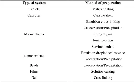
soluble drugs [80,88,89], drug targeting [90,91] and enhancement of peptide absorption [86,76,92]. Different types of chitosan based drug delivery systems are summarized in Table 1. The micro/nanoparticulate drug delivery systems offer numerous advantages over the conventional dosage forms. These include improved efficacy, reduced toxicity and improved patient compliance [93,94-96]. In the present section we have addressed the trends in the area of micro/nanoparticulate chitosan-based drug delivery systems. Literature of the past decade has been covered and results are evaluated.
Table 1 Chitosan based drug delivery systems prepared by different methods.
Type of system Method of preparation
Tablets Matrix coating
Capsules Capsule shell
Microspheres Emulsion cross-linking Coacervation/Precipitation Spray drying Ionic gelation Sieving method
Nanoparticles Emulsion-droplet coalescence
Coacervation/Precipitation
Beads Coacervation/Precipitation
Films Solution casting
Gel Crosslinking
1.4.1.1 Methods for the preparation of Chitosan micro/nanoparticles
Different methods have been used to prepare chitosan particulate systems. Selection of any of the methods depends upon factors such as particle size requirement, thermal and chemical stability of the active agent, reproducibility of the release kinetic profiles, stability of the final product and residual toxicity associated with the final product. However, selection of any of these methods depends upon the nature of the active molecule as well as the type of the delivery device.
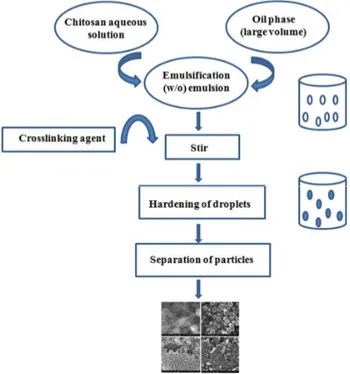
1.4.1.1.1 Emulsion crosslinking
This method utilizes the reactive functional amine group of chitosan to cross-link with the possible reactive groups of the cross-linking agent. In this method, a water-in-oil (w/o) emulsion is prepared by emulsifying the chitosan aqueous solution in the oil phase. Aqueous droplets are stabilized using a suitable surfactant. The stable emulsion is cross-linked by using an appropriate cross-linking agent to harden the droplets. Microspheres are filtered and washed repeatedly with alcohol and then dried [97]. By this method, size of the particles can be controlled by controlling the size of aqueous droplets. However, the particle size of final product depends upon the extent of cross-linking agent used while hardening in addition to speed of stirring during the formation of emulsion. This method is schematically represented in figure 3. The emulsion cross-linking method has few drawbacks since it involves tedious procedures as well as use of harsh cross-linking agents, which might possibly induce chemical reactions with the active agent. Also, complete removal of the un-reacted crosslinking agent may be difficult in this process.
Agnihotri et al. [98] have used the emulsion crosslinking method to prepare chitosan microspheres to encapsulate diclofenac sodium using three crosslinking agents viz, glutaraldehyde, sulfuric acid and heat treatment. Microspheres were spherical with smooth surfaces. The size of the microparticles ranged between 40 and 230 µm. Among the three cross-linking agents used, glutaraldehyde cross-linked microspheres showed the slowest release rates while a quick release of diclofenac sodium was observed by the heat cross-linked microspheres. Sankar et al. [99] prepared the chitosan-based pentazocine microspheres for intranasal delivery. Formulation parameters such as drug loading, polymer concentration, stirring speed during cross-linking and oil phase were altered to develop microspheres having good in vivo performance. In vivo studies indicated a significantly improved bioavailability of pentazocine. Application of in vitro data to various kinetic models indicated that these systems followed the diffusion controlled release kinetics.
Figure 3. Schematic representation of preparation of chitosan particulate systems by emulsion
cross-linking method
1.4.1.1.2 Coacervation/ Precipitation
This method utilizes the physicochemical property of chitosan since it is insoluble in alkaline pH medium, but precipitates/coacervates when it comes in contact with alkaline solution. Particles are produced by blowing chitosan solution into an alkali solution like sodium hydroxide, NaOH-methanol or ethanediamine using a compressed air nozzle to form coacervate droplets. Separation and purification of particles are done by filtration/centrifugation followed by successive washing with hot and cold water. The method is schematically represented in figure 4. Varying compressed air pressure or spray-nozzle diameter controlled the size of the particles and then using a crosslinking agent to harden particles can control the drug release. Chitosan microspheres loaded with recombinant human interleukin-2 (rIL-2) have been prepared by dropping of rIL-2 with sodium sulfate solution in acidic chitosan solution [100]. When protein and sodium sulfate solutions were added to chitosan solution and during the precipitation of chitosan, the protein was incorporated into microspheres. This method is devoid of cross-linking agent. The rIL-2 was released from microspheres in a sustained manner for up to 3 months. Efficacy of the systems developed was studied by using two model cells viz., HeLa and Lstrain cell lines.
Microspheres were taken up by the cells and rIL-2 was released from the microspheres. Chitosan–DNA nanoparticles have been prepared using the complex coacervation technique [101]. Important parameters such as concentrations of DNA, chitosan, sodium sulfate, temperature, pH of the buffer and molecular weights of chitosan and DNA have been investigated. At the amino to phosphate group ratio between 3 and 8 and chitosan concentration of 100 ng/ mL, the particle size was optimized to 100–250 nm with a narrow distribution. Surface charge of these particles was slightly positive with a zeta potential of 112 to 118 mV at pH lower than 6.0, and became nearly neutral at pH 7.2. The chitosan– DNA nanoparticles could partially protect the encapsulated plasmid DNA from nuclease degradation.
Figure 4. Schematic representation of preparation of chitosan particulate systems by
coacervation/precipitation method
1.4.1.1.3 Spray-drying
Spray-drying is a well-known technique to produce powders, granules or agglomerates from the mixture of drug and excipient solutions as well as suspensions. The method is based on drying of atomized droplets in a stream of hot air. In this method, chitosan is first dissolved in aqueous acetic acid solution, drug is then dissolved or dispersed in the solution and then, a suitable cross-linking agent is added. This solution or dispersion is
then atomized in a stream of hot air. Atomization leads to the formation of small droplets, from which solvent evaporates instantaneously leading to the formation of free flowing particles [102]. Various process parameters are to be controlled to get the desired size of particles. Particle size depends upon the size of nozzle, spray flow rate, atomization pressure, inlet air temperature and extent of crosslinking. This method is however more commonly used for the preparation of microparticles than for nanoparticles. Huang et al. [103] prepared chitosan microspheres by the spray-drying method using type-A gelatin and ethylene oxide– propylene oxide block copolymer as modifiers. Surface morphology and surface charges of the prepared microspheres were investigated using SEM and microelectrophoresis. Shape, size and surface morphology of the microspheres were significantly influenced by the concentration of gelatin. Betamethasone disodium phosphate-loaded microspheres demonstrated a good drug stability (less 1% hydrolysis product), high entrapment efficiency (95%) and positive surface charge (37.5 mV). In vitro drug release from the microspheres was related to gelatin content. Microspheres containing gelatin/chitosan ratio of 0.4–0.6 (w/w) showed a prolonged release up to 12 h. In another study [104], vitamin D2 (VD2), also called as ergocalciferol, was efficiently encapsulated into chitosan microspheres prepared by spray-drying method. The microencapsulated product was coated with ethyl cellulose. The sustained release property of VD2 microspheres was used for the treatment of prostatic disease [105].
1.4.1.1.4 Emulsion-droplet coalescence method
The novel emulsion-droplet coalescence method was developed by Tokumitsu et al. [106], which utilizes the principles of both emulsion cross-linking and precipitation. However, in this method, instead of cross-linking the stable droplets, precipitation is induced by allowing coalescence of chitosan droplets with NaOH droplets. First, a stable emulsion containing aqueous solution of chitosan along with drug is produced in liquid paraffin oil and then, another stable emulsion containing chitosan aqueous solution of NaOH is produced in the same manner. When both emulsions are mixed under high-speed stirring, droplets of each emulsion would collide at random and coalesce, thereby precipitating chitosan droplets to give small size particles. The method is schematically shown in figure 5. Gadopentetic acid-loaded chitosan nanoparticles have been prepared by this method for gadolinium neutroncapture therapy. Particle size depends upon the type of chitosan, i.e., as the % deacetylation degree of chitosan decreased, particle size increased, but drug content decreased. Particles produced using 100% deacetylated chitosan had the mean particle size
of 452 nm with 45% drug loading. Nanoparticles were obtained within the emulsion-droplet. Size of the nanoparticle did not reflect the droplet size. Since gadopentetic acid is a bivalent anionic compound, it interacts electrostatically with the amino groups of chitosan, which would not have occurred if a cross-linking agent is used that blocks the free amino groups of chitosan. Thus, it was possible to achieve higher gadopentetic acid loading by using the emulsion-droplet coalescence method compared to the simple emulsion crosslinking method.
Figure 5. Schematic representation of preparation of chitosan particulate systems by emulsion –
droplet coalescence method
1.4.1.1.5 Ionic gelation
The use of complexation between oppositely charged macromolecules to prepare chitosan microspheres has attracted much attention because the process is very simple and mild [107,108]. In addition, reversible physical cross-linking by electrostatic interaction, instead of chemical cross-linking, has been applied to avoid the possible toxicity of reagents and other undesirable effects. Tripolyphosphate (TPP) is a polyanion, which can interact with the cationic chitosan by electrostatic forces [109,110]. Bodmeier et al. [111] reported the preparation of TPP–chitosan complex by dropping chitosan droplets into a TPP solution, many researchers have explored its potential pharmaceutical usage [112-117]. In the ionic gelation method, chitosan is dissolved in aqueous acidic solution to obtain the cation of
chitosan. This solution is then added dropwise under constant stirring to polyanionic TPP solution. Due to the complexation between oppositely charged species, chitosan undergoes ionic gelation and precipitates to form spherical particles. The method is schematically represented in figure 6.
Ko et al. [118] prepared chitosan microparticles with TPP by the ionic cross-linking method. Particle sizes of TPP-chitosan microparticles varied from 500 to 710 nm with drug encapsulation efficiencies more than 90%. Morphologies of TPP-chitosan microparticles have been examined by SEM. As the pH of TPP solution decreased and molecular weight of chitosan increased, microparticles acquired better spherical shape having smooth surface. Release of felodipine as a model drug was affected by the preparation method. Chitosan microparticles prepared at lower pH or higher concentration of TPP solution resulted in a slower release of felodipine. With a decreasing molecular weight and concentration of chitosan solution, the drug release increased. The release of drug from TPP-chitosan microparticles decreased when the cross-linking time was increased. Xu and Du [119] have studied different formulations of chitosan nanoparticles produced by the ionic gelation of TPP and chitosan. TEM indicated their diameter ranging between 20 and 200 nm with spherical shape. FTIR confirmed tripolyphosphoric groups of TPP linked with ammonium groups of chitosan in the nanoparticles. Factors that affect the delivery of bovine serum albumin (BSA) as a model protein have been studied. These include molecular weight and deacetylation degree of chitosan, concentrations of chitosan and BSA, as well as the presence of polyethylene glycol (PEG) in the encapsulation medium.
Figure 6. Schematic representation of preparation of chitosan particulate systems by ionic gelation
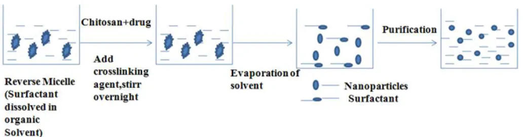
1.4.1.1.6 Reverse micellar method
Reverse micelles are thermodynamically stable liquid mixtures of water, oil and surfactant. Macroscopically, they are homogeneous and isotropic, structured on a microscopic scale into aqueous and oil microdomains separated by surfactant-rich films. One of the most important aspects of reverse micelle hosted systems is their dynamic behavior. Nanoparticles prepared by conventional emulsion polymerization methods are not only large (>200 nm), but also have a broad size range. Preparation of ultrafine polymeric nanoparticles with narrow size distribution could be achieved by using reverse micellar medium [120]. Since micellar droplets are in Brownian motion, they undergo continuous coalescence followed by re-separation on a time scale that varies between millisecond and microsecond [121]. The size, polydispersity and thermodynamic stability of these droplets are maintained in the system by a rapid dynamic equilibrium. In this method, the surfactant is dissolved in a organic solvent to prepare reverse micelles. To this, aqueous solutions of chitosan and drug are added with constant vortexing to avoid any turbidity. The aqueous phase is regulated in such a way as to keep the entire mixture in an optically transparent microemulsion phase. Additional amount of water may be added to obtain nanoparticles of larger size. To this transparent solution, a cross-linking agent is added with constant stirring, and cross-linking is achieved by stirring overnight. The maximum amount of drug that can be dissolved in reverse micelles varies from drug to drug and has to be determined by gradually increasing the amount of drug until the clear microemulsion is transformed into a translucent solution. The method is schematically represented in figure 7. Mitra et al. [122] have encapsulated doxorubicin– dextran conjugate in chitosan nanoparticles prepared by reverse micellar method. The surfactant sodium bis(ethyl hexyl) sulfosuccinate (AOT), was dissolved in n-hexane. This procedure produced chitosan nanoparticles encapsulating doxorubicin–dextran conjugate.
Figure 7. Schematic representation of preparation of chitosan particulate systems by reverse micellar
1.4.1.1.7 Seiving method
Recently, Agnihotri and Aminabhavi [123] have developed a simple, yet novel method to produce chitosan microparticles. In this method, microparticles were prepared by cross-linking chitosan to obtain a non-sticky glassy hydrogel followed by passing through a sieve as shown in figure 8. In the work by Agnihotri et al., a suitable quantity of chitosan was dissolved in 4% acetic acid solution to form a thick jelly mass that was cross-linked by adding glutaraldehyde. The non-sticky cross-linked mass was passed through a sieve with a suitable mesh size to get microparticles. The microparticles were washed with 0.1 N NaOH solution to remove the un-reacted excess glutaraldehyde and dried overnight in an oven at 40 8C. Clozapine was incorporated into chitosan before crosslinking with an entrapment efficiency up to 98.9%. This method is devoid of tedious procedures, and can be scaled up easily. Microparticles were irregular in shape, with the average particle sizes in the range 543–698 nm. The in vitro release was extended up to 12 h, while the in vivo studies indicated a slow release of clozapine.
Figure 8. Schematic representation of preparation of chitosan particulate systems by seiving method
1.4.1.2 Drug loading into Chitosan micro/nanoparticles
Drug loading in micro/nanoparticulate systems can be done by two methods, i.e., during the preparation of particles (incorporation) and after the formation of particles (incubation). In these systems, drug is physically embedded into the matrix or adsorbed onto the surface. Various methods of loading have been developed to improve the efficiency of loading, which largely depends upon the method of preparation as well as physicochemical properties of the drug. Maximum drug loading can be achieved by incorporating the drug during the formation of particles, but it may get affected by the process parameters such as
method of preparation, presence of additives, etc. Both water-soluble and water-insoluble drugs can be loaded into chitosan-based particulate systems. Water soluble drugs are mixed with chitosan solution to form a homogeneous mixture, and then, particles can be produced by any of the methods discussed before.
Water-insoluble drugs and drugs that can precipitate in acidic pH solutions can be loaded after the formation of particles by soaking the preformed particles with the saturated solution of drug. Diclofenac sodium, which precipitates in acidic pH conditions, has been loaded by the soaking method [98]. In this method, loading depends upon the swelling of particles in water. Percentage loading of drug decreased with increasing cross-linking due to decreased swelling. Water-insoluble drugs can also be loaded using the multiple emulsion technique. In this method, drug is dissolved in a suitable solvent and then emulsified in chitosan solution to form an oil-in-water (o/w) type emulsion. Sometimes, drug can be dispersed into chitosan solution by using a surfactant to get the suspension. Thus, prepared o/w emulsion or suspension can be further emulsified into liquid paraffin to get the oil-water-oil (o/w/o) multiple emulsion. The resulting droplets can be hardened by using a suitable cross-linking agent. Hejazi and Amiji [124] have prepared chitosan microspheres by ionic cross-linking and precipitation with sodium sulfate. Two different methods were used for drug loading. In method I, tetracycline was mixed with chitosan solution before simultaneous cross– linking and precipitation. In method II, drug was incubated with the pre-formed microspheres for 48h. Cumulative amount of tetracycline that was released from chitosan microspheres and stability of drug was examined in different pH media at 37 °C. Microspheres with a spherical shape having an average diameter of 2– 3 nm were formed. When drug was added to chitosan solution before cross-linking and precipitation, only 8% (w/w) was optimally incorporated in the final microsphere formulation. When drug was incubated with the pre-formed microspheres, a maximum of 69% (w/w) could be loaded. About 30% of tetracycline either in solution or when released from the microspheres was found to degrade at pH 1.2 in 12 h. Preliminary results of this study suggested that chitosan microspheres can be used to incorporate antibiotic drugs, which may be effective when administered locally in the stomach against H. pylori.
1.4.1.3 Drug release & release kinetics
Drug release from chitosan-based particulate systems depends upon the extent of cross– linking, morphology, size and density of the particulate system, physicochemical properties of the drug as well as the presence of adjuvants. In vitro release also depends upon
pH, polarity and presence of enzymes in the dissolution media. The release of drug from chitosan particulate systems involves three different mechanisms: (a) release from the surface of particles, (b) diffusion through the swollen rubbery matrix and (c) release due to polymer erosion. In majority of cases, drug release follows more than one type of mechanism. In case of release from the surface, adsorbed drug instantaneously dissolves when it comes in contact with the release medium. Drug entrapped in the surface layer of particles also follows this mechanism. This type of drug release leads to burst effect. He et al. [102] observed that cemetidine-loaded chitosan microspheres have shown burst effect in the early stages of dissolution. Most of the drug was released within few minutes when particles were prepared by spray drying technique. Increasing the cross-linking density can prevent the burst release. This effect can also be avoided by washing microparticles with a proper solvent, but it may lead to low encapsulation efficiency.
Drug release by diffusion involves three steps. First, water penetrates into particulate system, which causes swelling of the matrix; secondly, the conversion of glassy polymer into rubbery matrix takes place, while the third step is the diffusion of drug from the swollen rubbery matrix. Hence, the release is slow initially and later, it becomes fast. This type of release is more prominent in case of hydrogels. Al-Helw et al. [125] observed a high initial release of the drug in all the prepared formulations. Nearly, 20– 30% of the incorporated drug was released in the first hour. Release was dependent on the molecular weight of chitosan and particle size of the microspheres. The release rate from microspheres prepared from high molecular weight chitosan was slow compared to those prepared from medium and low molecular weight chitosan. This could be attributed to both lower solubility of high molecular weight chitosan and higher viscosity of the gel layer formed around the drug particles upon contact with the dissolution medium. The release within the first 3 h was fast (75– 95%) from microspheres within the size range of 250– 500 µm, but for particles in the size range of 500– 1,000 µ m, drug release was 56– 90% in 5 h. This was attributed to large surface area available for dissolution with a small particle size, thus favoring rapid release of the drug compared to larger microspheres.
Analysis of drug release data has several approaches. Ganza-Gonzalez et al. [126] analyzed the drug release data using the classic Higuchi equation [127]. Higuchi equation was used to describe the release of a solute from a flat surface, but not from a sphere [128], but the good fit obtained suggested that the release rate depends upon the rate of diffusion through the cross-linked matrix. Authors have also fitted the release data to equations developed by Guy et al. [129] to describe the diffusion from a sphere. The most commonly used equation for diffusion controlled matrix system is an empirical equation used by Ritger
and Peppas [130], in which the early time release data can be fitted to obtain the diffusion parameters,
ktn
M
Mt
=
∞
(1)Here, Mt/M∞ is the fractional drug release at time t, k is a constant characteristic of the
drug-polymer interaction and n is an empirical parameter characterizing the release mechanism. Based on the diffusional exponent [131], drug transport is classified as Fickian (n=0.5), Case II transport (n=1), non-Fickian or anomalous (0.5< n< 1) and super Case II (n>1).
Agnihotri and Aminabhavi [123] have analyzed the dynamic swelling data of chitosan microparticles using Eq. (1) to predict drug release from the water uptake data of the microparticles cross-linked with (5.0, 7.5 and 10.0)*10– 4 mL of glutaraldehyde/mg of chitosan. It was observed that as the cross-linking increases, swelling of chitosan microparticles decreases. Values of n obtained in the range of 0.160 to 0.249 indicating that the release mechanism deviates from the Fickian trend. The values of n are < 0.5 due to the irregular shaped particles and these decrease systematically with increasing cross-linking. In the swelling controlled release systems, drug is dispersed within a glassy polymer. Upon contact with biological fluid, the polymer swells, but no drug diffusion occurs through the polymer phase. As the penetrant enters the glassy polymer, glass transition temperature of the polymer is lowered due to relaxation of the polymer chains. Drug could diffuse out of the swollen rubbery polymer. This type of system is characterized by two moving boundaries: the front separating the swollen rubbery portion and the glassy region, which moves with a front velocity and the polymer fluid interface. The rate of drug release is controlled by the velocity and position of the front dividing the glassy and rubbery portions of the polymer. Jameela et al. [132] have obtained a good correlation fit for the cumulative drug released vs. square root of time, demonstrating that the release from the microsphere matrix is diffusion-controlled and obeys Higuchi equation [127]. It was demonstrated that the rate of release depends upon the size of microspheres. Release from smaller size microspheres was faster than those from the large size microspheres due to smaller diffusional path length for the drug and the larger surface area of contact of smaller particles with the dissolution medium.
1.4.2 Chitosan hydrogels
Chitosan hydrogels have been prepared with a variety of approaches. In each preparation chitosan is either physically associated or chemically cross-linked to form the
hydrogel. Our discussion below will focus on these two distinct hydrogel engineering approaches.
1.4.2.1 Physical association networks
In order to satisfy the requisite features of a hydrogel, the chitosan polymer network must satisfy two conditions: (1) inter-chain interactions must be strong enough to form semi-permanent junction points in the molecular network, and (2) the network should promote the access and residence of water molecules inside the polymer network. Gels that meet these demands may be prepared by non-covalent strategies that capitalize on electrostatic, hydrophobic, and hydrogen bonding forces between polymer chains [133,134]. Figure 9 shows the schematics of four major physical interactions (i.e. ionic, polyelectrolyte, interpolymer complex, and hydrophobic associations) that lead to the gelation of a chitosan solution.
Figure 9. Schematic representation of chitosan based hydrogel networks derived from different
physical associations: (a) networks of chitosan formed with ionic molecules, polyelectrolyte polymer and neutral polymers
Because the network formation by all of these interactions is purely physical, gel formation can be reversed. Tunable gel swelling behavior can be readily achieved in a physical gel by adjusting the concentration and nature of the second component used during the fabrication process. A chitosan-based physical gel can often be obtained by simply mixing the components that make up the gel under the appropriate conditions. These gels have a short
life time in physiological media, ranging from a few days to a month. Therefore, physical gels are good for short-term drug release applications. Because the gelation does not require any toxic covalent linker molecules, it is always safe for clinical applications. However, their widespread application is limited due to the weak mechanical strength and uncontrolled dissolution [135].
1.4.2.1.1 Ionic complexes
Thanks to the cationic amino groups of chitosan, ionic interactions can occur between chitosan and negatively charged molecules and anions. Ionic complexation of mixed charge systems can be formed between chitosan and small anionic molecules, such as sulfates, citrates, and phosphates [136,137] or anions of metals like Pt (II), Pd (II), and Mo (VI) [138,139]. These interactions can yield hydrogels with varying material properties that depend upon the charge density and size of the anionic agents, as well as the degree of deacetylation and concentration of the chitosan polymer.
Both anions and small molecules bind chitosan via its protonated amino groups, but metal ions form coordinate–covalent bonds with the polymer instead of electrostatic interactions [138,139]. Ionic complexation can be accompanied by other secondary interchain interactions including hydrogen bonding between chitosan's hydroxyl groups and the ionic molecules, or interactions between deacetylated chitosan chains after neutralization of their cationic charge [138,140]. These interactions can enhance the physical properties of the hydrogel, and can be modulated to express unique material properties, such as pH sensitivity.
1.4.2.1.2 Polyelectrolyte complexes (PEC’s)
While polyelectrolytes form electrostatic interactions with chitosan, they are different from the ions or ionic molecules used in ionic complexation in that they are larger molecules with a broad molecular weight range, such as polysaccharides, proteins and synthetic polymers. The associations between the chitosan polymer and polyelectrolytes are stronger than other secondary binding interactions like hydrogen bonding or van der Waals interactions. The advantages of this type of complex are significant. They are complexed without the use of organic precursors, catalysts, or reactive agents, alleviating the concern