ALMA MATER STUDIORUM - UNIVERSITÀ DI BOLOGNA
!
!
!
SCUOLA DI INGEGNERIA E ARCHITETTURA
!!!!!!!!!!!!!!!!!!!!!!!!!!!!!!!!!
CAMPUS DI CESENA
!
!
!
CORSO%DI%LAUREA%MAGISTRALE%IN%INGEGNERIA%BIOMEDICA%
!
!
!
TESI DI LAUREA
!
in!
!
Bioingegneria%molecolare%e%cellulare%LM%
!
!
DESIGN,!REALIZATION!AND!CHARACTERIZATION!!
OF!AUTOMATED!MILLIFLUIDIC!BIOREACTORS!
!FOR!INVESTIGATING!THE!MOLECULAR!EVOLUTION!
!OF!LYTIC!OR!LYSOGENIC!VECTOR!PHAGES!
!INFECTING!ENGINEERED!HOST!E.!COLI!
!
!
!
!
CANDIDATO:!
RELATORE:!
Fabio!Polesel!673501!
Chiar.mo!Prof.!Emanuele!D.!Giordano!
!
!
!
!
!
!
CORRELATORE:!
Prof.!Alfonso!Jaramillo!
!
!
!
!!
Anno!Accademico!2014/2015!
Sessione!I!
Alla mia famiglia,
a Marta
Un altro aspetto curioso della teoria dell'evoluzione
è che tutti pensano di capirla!
Table of contents
Introduction……….……….. 1
1.
Assisted continuous evolution (ACE): theoretical and experimental
aspects ………
……………..2
1.1. Quasispecies Theory: relation between information, self-replication and selection of biomolecules
………...………...……
….2
1.1.1. Self-replication concurs to the selection of molecular complexes and generation of Quasispecies
………...3
1.1.2. Wrong self-replication affects an assisted evolution process under a threshold error
……….…5
1.1.3. Virus: natural models for molecular evolution experiments
…..………6
1.2. Experimental strategies: natural and artificial selection
………
………
.….7
2.
State of the art: configuration, devices and technical solutions for
experimental molecular evolution………
………
………10
2.1. Viral continuous culture: the cellstat system
………
……….10
2.2. Molecular replicators evolution: the machine for mass-transfers
…...……12
2.3. “Small” Quasispecies evolution: the multi-channel machine for mass-transfers
…....………
……….14
2.4. Evolutive events “imaging”: the capillary reactor
……
………
….………15
3.
Methods and materials……….
……………
…………
……..18
3.1. Biological parts
………….
….………18
3.1.1. Escherichia coli bacteria: the "compartmental models for evolution"...18
3.1.2. Bacteriophages: the faster evolutionary replicator
………
..27
3.1.3. Bacterial and phage quantification
………...……….31
3.2. General principle of bacterial continuous culture
………
……
……
..35
3.2.1. Fermenter
……
…………
…………
.………
...……………
………36
3.3. Phage-assisted continuous evolution (PACE)
…
………
…………
..……..40
3.4. Evoprog Consortium and Jaramillo’s Lab research
………
…………
…...42
3.4.1. A technique for a lysogenic phagemid evolution into F+ E.coli cells…
42
3.4.2. Engineering T7 lytic phage towards a bacteriophage therapy ………..44
4.
Prototype and results…
………….
.……
…………...48
4.2. A preliminary mathematical model for describing a lytic bacteriophage
…
.51
4.3. Kinetic characterization of the host cell
……..………....…………..52
4.3.1. Growth curve of lysogenic phagemid infected and uninfected F+ E. coli
cells…
………
……………
………….52
4.3.2. Growth curve of lytic bacteriophage infected and uninfected F- E. coli
cells
…
…………
…………
.53
4.4. The prototype in chemostat and cellstat configuration: a basic machine for conducting ACE experiment
……….
………
….………
……………54
4.4.1. The prototype in switching configuration: a machine for a selective feedperiodically switched into common cellstat
…………
……………
…56
4.5. Relation between free M13 lysogenic phagemid end dilution rate
…
…..….57
4.5.1. Considerations about M13 phagemid experiment
…
……
………
……60
4.6. Relation between free T7 lytic bacteriophage phage end dilution rate…
…
.62
4.6.1. First example of long run experiment and free phage trend…
…….
.…65
4.6.2. Considerations about T7 phage experiments…
……………
.………..65
4.7. A preliminary experiment to demonstrate homologous recombination of T7 phage in continuous culture
……….67
5.
Conclusions and future directions………
…………...
.70
5.1. Limitations: Biofilm and spontaneous evolution of bacteria
……..
..……
…71
5.2. Future Directions
………..
..72
Bibliography…
………
…………
…………
…………
…...……74
Appendix A: equipment list
Abstract
This document presents a bioreactor intended for maintaining desired biological conditions that maximize the evolution of lytic (T7) or lysogenic (M13) of cloning vectors phages.
To this aim, the manuscript highlights concepts related to the Theory of
Quasispecies and the relationship between self-replication errors, and natural
or artificial selection pressures on populations of viruses: the model of system evolution.
Maintaining populations of virus means to provide them a substrate in which they replicate. Complex and expensive prototypes of dedicated machines have already been developed by other research groups for growing continuous populations of bacteria. The bioreactor, object of this work, is part of the European project Evoprog (Jaramillo’s Lab, University of Warwick), aiming to develop a General-Purpose Programmable Machine Evolution on a
Chip. This device, using existing phage technology and synthetic regulation, is
expected to produce biomolecules or biocomputational functionality two orders of magnitude faster than conventional techniques (PACE), while reducing consumable and overall costs.
This first protoype consists of one or more fermenters, where a bacterial cultuare is continuously grown under optimized conditions of concentration, pH and temperature; and a cellstat, a separate vessel, where only the replication of viruses happens. Both volumes are about few milliliters and properly interconnected to enable a continuous molecular screening experimental oriented.
The results of preliminary experiments to prove the reliability of the prototypes is presented here. The experiments conducted, indeed show the success of two viral continuous coltivations and the in vivo ricombination of engineerd lytic or lysogenic bacteriophages.
Limitations and future developments of the system are finally presented, in light of specific applications addressing the studies of a bacteriophage-based antimicrobial therapy.
Chapter 1
1. Assisted continuous evolution (ACE): theoretical and
experimental aspects
The evolutionary mechanism can be described by a system of differential equations which shows how the selection of biomolecules is an intrinsic property of a population of individuals able to self-replicate. This demonstrates that, molecular evolution can be investigated through targeted experiments of ACE where the natural biological molecules, that do not perfectly adapt to technological or medical aims, can be improved repeating cycles of mutation, selection and amplification.
We present here some mathematical models and experimental strategies, shown in literature, for describing those processes and effects involved during molecular evolution.
1.1 Quasispecies Theory: relation among information,
self-replication and selection of biomolecules
There are two most important classes of biological macromolecules in which the information is coded: polynucleotides and polypeptides 1. These have a common assembly principle: both are products of a linear polymerization where various functional groups are bound on a regular and repetitive 'skeleton'. This means that the information is linked to matter that is steadily degrading, so information would be lost if it was not preserved by a constant replication. However, the reproduction is not a entirely accurate event: the errors that occur during the replication process generate changes on the meaning of original information. The information content has to be then evaluated by its new coded functionality.
!!!!!!!!!!!!!!!!!!!!!!!!!!!!!!!!!!!!!!!!!!!!!!!!!!!!!!!!
1!Polypeptides and polynucleotides: A polypeptide is a long, continuous, and unbranched peptide chain.
Proteins consist of one or more polypeptides arranged in a biologically functional way, often bound to ligands such as coenzymes and cofactors. Peptides fall under the broad chemical classes of biological oligomers and polymers, alongside nucleic acids (DNA and RNA) made of nucleotides.!
The structure of these polymers, indeed, allows to produce a variability expressed by the equation (1), which describes the number N of different heteropolymers, whose length is v, that can be constructed from a number of different monomers:
N=μv (1)
Where: μ is 4 for the nucleic acids and 20 for natural proteins.
1.1.1 Self-replication concurs to the selection of molecular complexes
and generation of Quasispecies
Nucleic acids are the only known natural biopolymers that can serve as a template for its own replication. The ability that allows the interactions between their complementary constituents is an intrinsic property of the molecular structure of nucleic acids. Therefore, the base pairing allows the synthesis of a unique complementary copy of each polynucleotide.
The kinetics of the autoreplication can be expressed by differential equations, whose simplest case is represented by the following equation:
!!"!!
!" = ! !!!!! ! + !!!!!"!! ! − ∅!!!!!!!!!!!!!!!!!!!!!!!!!!!!!(2) with i,j= 1, .... , N, and
Wii=Ai Qi-Di≥0
where: i, variable index, representing all individual sequences in a population;
Wii, speed rate for the correct replication of the species i;
Ai, speed rate for the formation of the species i;
Qi, quality factor which represents the average probability for a faithful
copy of the sequence. Then, (1- Qi) is the corresponding probability of
abnormal reproduction;
Di, speed rate for the decomposition of the species i;
Wij, mutation coefficient, related to the formation probability of the
sequence i due to an erroneous reproduction of the sequence j;
ci(t), concentration of the sequence i at time (t);
∅i, flow that allows the control through an input and an output in an
open system.
competition of all individuals in a population and shows the following properties:
• mutability, wrong self-reproduction, appears when Qi < l e Wj ≥ 0;
• metabolism is linked to the irreversible formation and degradation of the molecules, values expressed here by Aici(t), Dici(t) e ∅i.
In such a kind of system, the phenomenon of the competition takes place if all the individuals i present in a population are considered. We define then:
xi(t), number of relative population;
Ei, excess of productivity of the species i;
!(t), mean of the excess of productivity, !! ! = !! !
!! ! ! !!= !!− !! then equation (2) can be re-written as
!"!(!)
!" = !!!− ! ! !! ! + !!!!!"!!(!) (3)
Equation (3) can be exactly solved thanks to a linear transformation [1] [2] [3]. It, thus results:
!"!(!)
!" = (ʎ!− ʎ(!))!!(!) (4)
where:!ʎ!, eigenvalues and yi, linear combinations of all the xi(t), are the
weighted coefficients of the individual mutants. These, are the contributions of the individual mutants weighed;
ʎ(t), the average value of the eigenvalues, corresponds to the average excessive productivity !(t) of the species i.
Therefore, according to equation (4),
- all the mutants distributions with ʎ! < ʎ(t) die;
- all the mutant distributions with ʎ! > ʎ(!)are amplified, increasing ʎ(t) up to reach its maximum speed value of replication Amax.
Increasing ʎ!, more and more sequences are removed from the competition so, only the mutant distribution that replicates more efficiently remains and this is the distribution with the greater eigenvalue ʎ!"#.
We call this behavior selection and this is an intrinsic property of the autocatalytic reproduction of any population of individuals. Consequently, this means that selection does not apply to a single molecular species that replicates itself, but rather it applies to situations in which there are groups of mutant. Distribution of mutants with higher efficiency replication behaves as an individual species, therefore is called Quasispecies [4] [3]. The Quasispecies, in this way, replaces the previous wild strain (WT).
1.1.2 Wrong self-replication affects an assisted evolution process under
a threshold error
During viral genome replication, errors are not statistically rare events. In these systems, many individual mutants appear quite commonly. The frequency of the production of a mutant by imprecise replication on the positions i of the genome long v, is expressed by the binomial theorem
!! = !! !!!!(1 − !)! (5)
Any nucleotide is incorporated in a certain position with a accuracy value expressed by a fidelity q, which is the geometric average of the individual (and position-dependent) error rates. Therefore, (1- q) represents the index of the mean error frequency. A frequency error of 0.1 is equivalent to a false incorporation (on average) every 10 positions.
On the other hand, the occurrence frequency of a certain mutant is also influenced by its replication rate. The extremal principle 2 (ie, results of a critical function), described by equation (4), considers this effect evaluating the presence of mutant distribution according to their fitness, or biological suitability: ability to reproduce themselves efficiently under specific environmental conditions. If the Quasispecies contains a master copy clearly dominant, and if we neglect the reverse mutation, it can be defined as
!! =! !!
!!(!!!!!!!!!) (6)
!!!!!!!!!!!!!!!!!!!!!!!!!!!!!!!!!!!!!!!!!!!!!!!!!!!!!!!!
2!The Extremal Principle is a methodology, a useful problem solving tactics, where a solution of a problem
is sought among possible candidates that satisfy some extreme conditions within the parameters of the problem.!
!o correlates the master sequence productivity, its rate of degradation and
over productivity average of all the competitors i. Āi≠0 e ̅Di≠0 are averages
values calculated on all the mutants that form the distribution, except for the master copy.
With !oQo ≥ 1 e Qo = qv, equation (6) leads to the following expression for the
limit informational capacity
!!"# ≈!"!!!!!! (6a)
Therefore, equation (6a) defines the maximum length of sequence that can be stably reproduced under the influence of the selection.
It also introduces that a principle of a threshold error has to be considered. A distribution of mutants just below this threshold has the largest possible genetic variation, even though remaining (meta) stable. However, these distributions are not symmetrical. They include many individual “neutral” or “almost neutral” 3 species, as well as more than one, or even no one, dominant species.
As a result of the above considerations, the mean sequences representing the “center of gravity” of the distribution could not be the most suitable or more frequent species, defined: WT strain. Also, the overcoming of the error threshold leads instead, to an accumulation of errors and thus to a random distribution of sequences, with complete loss of information.
Therefore, “evolutionary tests” for the selection of advantageous mutants, in known environmental conditions, should occur along the periphery of the distribution of the Quasispecies, where those mutants appear with sufficiently large occurrence frequencies !.
Several research groups has experimentally established that viruses behave as Quasispecies and operate near their error threshold [5] [6].
!!!!!!!!!!!!!!!!!!!!!!!!!!!!!!!!!!!!!!!!!!!!!!!!!!!!!!!!
3!Neutral species: a species’ niche encompasses all of the factors it requires for growth and reproduction
and how it impacts its environment. Neutral species have completely overlapped niches; they share the same niche and their fitness changes identically along an environmental gradient or niche axis. Greater niche differences correspond to less niche overlapping between species; species differ in their fitness at different points along an environmental gradient or niche axis.!
1.1.3 Virus: natural models for molecular evolution experiments
The study of the mechanisms that mimic the natural evolution should be conducted with simple, self-replicating species that involves only a few biochemical reactions. For this reason, S. Spiegelman [7] (1971) chose the virus as first simple replicators. Viruses are infectious particles, made of nucleic acid enclosed in a protein capsid. They do not have its own metabolic apparatus and therefore they are not autonomous living organisms. After the infection of a host cell and the injection of viral nucleic acid in the cell, the viral genetic information is read and translated and compete with the cellular DNA. Often, this process is fatal to the host cell because all its resources are used in the synthesis of new viral particles. Moreover, viruses show a surprising efficient replication, with rapid generation time, high population density and, due to the high error rate in the replication, rapid adaptation to the selective constraints.
The viruses are hence, ideally the best for molecular evolution experiments and this is the reason of their wide use.
1.2 Experimental strategies: natural and artificial selection
This section exposes the principles about the strategies experimentally adopted for conducting experiments about the comprehension of the molecular evolution, based on known properties of a population.
• Natural selection promotes individuals with more efficient reproductive systems. Accordingly, their fitness is manifested by a substantial advantage in growth. Exploiting this strategies, mutants with defined properties emerge under the influence of a selective pressure.
Some experiments have been conducted and proved that, molecular replicators and bacteriophage 4 exposed to various environmental constraints,
!!!!!!!!!!!!!!!!!!!!!!!!!!!!!!!!!!!!!!!!!!!!!!!!!!!!!!!!
4
Bacteriophages: or phage, is a virus that through a contraction mechanism injects its nucleic acid in a given parasited bacterium. Once injected, the phage genome can follow two paths:
- Lytic cycle (typical of bacteriophage T7): use the replication machinery of the host to produce new phage particles, up to the volume of the outbreak, at which time the cell will disintegrate for lysis. - Lysogenic cycle: however, the phage genome (usually λ or M13 phage) will integrate into a
have resulted in species with increased replicative efficiency. Starting from bacteriophage Qβ, the viral infections have been studied under the influence of antibodies that neutralize the protein coating of the phage [8]. In another experiment small replicative RNA could compete with the viral genome for the replication operated by the polymerase encoded by the phage [9]. In both experiments some mutant phages evolved and were able to avoid the antiviral attack which they were subjected.
• Artificial and non organized selection, without competition, in which mutantas with any desired property are promoted by an external interference.
The artificial selection, unlike natural selection, is based on the precise knowledge of the desired property, and therefore requires a method to select large populations according to the characteristics that have been defined for selection. A typical selection process begins with an extensive random mutagenesis 5 of a clone that presents considerable similarities to the desired characteristics. The resulting (library) of the mutants is then selected by an assay suitable to detect promising mutant. This process is repeated on the selected mutants again and again until one or more optimal individuals are obtained. The feature of this artificial selection procedure is the separation between growth (or amplification) and selection: in contrast to the natural system, where the rate of optimal reproduction always plays an important role in the selection, here it is possible to choose any selection criterion. One of the first experiments in which this strategy has been applied was devised by L. C. Tuerk and Gold [10] (1991).
Natural and artificial selection can also be combined for the aims of the experiments investigated, where amplification, or self amplification, of the individuals is a common step.
!!!!!!!!!!!!!!!!!!!!!!!!!!!!!!!!!!!!!!!!!!!!!!!!!!!!!!!!!!!!!!!!!!!!!!!!!!!!!!!!!!!!!!!!!!!!!!!!!!!!!!!!!!!!!!!!!!!!!!!!!!!!!!!!!!!!!!!!!!!!!!!!!!
every time the bacterial chromosome replicates, the genome of λ also replicates. A bacterium that contains a prophage is said "lysogenic".
5Mutagenesis is a process by which the genetic information of an organism is changed in a stable manner,
resulting in a mutation. It may occur spontaneously in nature, or as a result of exposure to mutagens. It can be achieved experimentally using laboratory procedures.
Chapter 2
2. State of the art: configuration, devices and technical solutions
for experimental molecular evolution
Using a theoretical basis of molecular evolution, devices were designed and developed to perform molecular evolution experiments where the mutation, selection and amplification processes are kept stable and performed sequentially.
Such instruments are able to 'mimic' the evolution process and have been used to 'build' functional macromolecules, nucleic acids or proteins, for technological or medical applications.
2.1 Viral continuous culture: the cellstat system
The competition in a population of individuals emerges when the total number of individuals is maintained constant and it is described by the equations [3] and [4]. A population can be maintained numerically constant keeping constant all the necessary resources. Therefore, when a variant, which is more suitable than the original WT appears, the new population can overtake the original one reaching a higher total concentration of individuals. Indeed, the evolutionary events are usually detected with the emergence of faster replication species.
When maintaining constant conditions, the application of any new selective pressure increases the visibility of the fitness of some individuals.
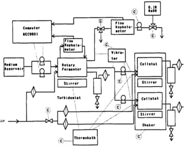
It is possible, in principle, to obtain the continuous cultures of viruses for evolutionary studies. However, viruses are parasites, they do not grow on their own, but they need a substrate of host bacteria to replicate. These bacterial host cells can be produced in a continuous culture bioreactor fermenter. The production of viruses can be realized by a device called cellstat and it can be divided in two mains units [11][12] (Fig. 1):
turbidity of the culture by a continuous measurement of absorbance and generates cultures of host cells with a constant physiological reproducibility in conditions of exponential growth phase;
2) cellstat reactors, where a steady flow of the host cell is infected with viruses, reaching steady state conditions and thereby producing constant amounts of phage, infected and not infected bacteria.
Figure 1: schematic diagram of the cellstat configuration system developed by Husimi et al. Fig. 6 (1989)
Maintaining separate bacterial and phage cultures avoids artifacts due to coevolution. If culture of the host and parasite are allowed into a single vessel this would inevitably lead to a bacterial population resistant to the virus, making the experiment meaningless. Rather, any bacteria potentially resistant to the infection has to be rapidly washed out from the cellstat system.
When a steady state is reached into the reactors, the evolution experiments can start, applying new and defined physical, chemical or biological selective pressures. The constraints applied to the system can lead to the natural selection of the phages, that in these conditions replicate more efficiently, and are therefore defined as the most suitable.
Thanks to this device it is possible to simulate viral infections of superior organisms for testing new antiviral strategies.
In a typical experiment conducted by Lindemann (1992), a phage Qβ has been exposed to the influence of monoclonal antibodies, directed against the
protein of the viral capsid, “simulating” then the immune response of a
superior organism to a viral infection. This has allowed some phages of the
population to escape the selective pressure, mutating the gene that encodes the capsid protein. The proteins analysis showed that these basic substitutions were expressed as altered antigenic epitopes on the viral surface. Later cultured mutants resistant to the antibody without selective pressure returned to be again a completely wild type phage.
In another experiment [13] a genetically modified phage (fd) for the expression of viral peptides on the surface [14] and containing a codified sequence for tetracycline resistance, was initially cultured into a cellstat without the antibiotic. As a result it lost the inserted gene encoding the tetracycline resistance. When, in a second experiment, the same phage was cultivated under the selective pressure of the antibiotic, the additional gene was stably inherited for several generations of phage.
Using continuous cultures of Qβ and fd phage allowed to demonstrate for the first time the mechanisms used by viruses to overcome antiviral therapy.
2.2 Molecular replicators evolution: the machine for
mass-transfers
Experiments that exploit the cellstat configuration allowed the study of the competitive growth of living organisms. Such competition can also be studied in vitro, via molecular replicating species. The most used replicator in molecular evolutionary experiments is Qβ constituted by the replicase6 of purified Qβ, which uses viral RNA as a template and nucleoside triphosphates as monomeric substrates. Spiegelman [15] (1965) was the first who, using this replicating system, applied constant conditions (i.e. similar to the cellstat). In the concept of transfers in series, a type of RNA is amplified to obtain a certain maximum concentration which is followed by a dilution of the molecular population in a new solution. In this way, the continuous alternation of phases of amplification and dilution simulates the characteristics described by the
!!!!!!!!!!!!!!!!!!!!!!!!!!!!!!!!!!!!!!!!!!!!!!!!!!!!!!!!
6RNA replicase: is an enzyme that catalyzes the replication of RNA from an RNA template. This is in
contrast to a typical DNA-dependent RNA polymerase, which catalyzes the transcription of RNA from a DNA template.
cellstat.
The molecular replicators are forced to evolve during the growth period, varying the growth conditions, for example, increasing the reaction temperature or adding substances. The products of the evolution can be analyzed after each transfer, providing information about the molecular mechanism of this process. If the external constraints are not applied, the speed of replication determines the composition of the population.
In general, it is important to execute evolution experiments in vitro under optimal conditions, in particular close to the error threshold, as suggested by the theory. Spiegelman’s experiments were conducted far below the error threshold of Qβ replicase. The result is that in these experiments the evolutionary process was rather slow.
Then, a machine able to execute faster and automatically experiments of mass-transfers (Fig. 2) has been built. The reaction vessels and the sample transporter are made of silver coated with biological inert material (gold).
Figure 2: overview of the machine for automatic mass-transfers developed at the Max Planck Institute for Biophysical Chemistry in in Göttingen (Germany). Strunk, Fig. 3
(1993) .
Starting from the initial position, the samples are transported to the measuring position, which is thermostatically controlled to an optimal growth temperature. The amplification reaction is triggered by a temperature jump. The concentration of the nucleic acids continuously produced, can be directly
196 G. Strunk, T. Ederhof/Bioph.vsical Chemisty 66 (1997) 193-202
chemistry and evolutionary biotechnology
(Koltermann & Kettling, this issue), the multichannel machine described here will have a number of addi- tional applications.
2. The single-channel serial-transfer machine The single-channel serial-transfer machine (STM) is a computer controlled apparatus with laser optics and a glass fiber fluorimeter, supplemented by the corresponding mechanics.
2.1. Optical Part
In order to measure fluorescence-based RNA concentrations in small sample volumes on-line with RNA growth, a sensitive glass fiber laser fluorimeter was constructed (Fig. 2). The 514 nm line of an 0.5mW argon-ion laser is coupled into a glass fiber with a diameter of 1 mm and a length of 1 m which leads the light beam to the measurement part of the machine. The fluorescence is collected in a front- surface-arrangement with a second glass fiber of the same kind. Because of this technical restriction ap- plied by usage of so-called Y-shaped fibers, the sig- nal in the emission branch contains a large fraction of reflected excitation light. Therefore, the light is passed through a nitrogen-cooled, double-banded interference filter (DAL-595.4 nm, Schott) with a high rejection at 514 nm and a high transmission at 600 nm before the fluorescence is measured by a photodiode. The signal is then visualized on a Keithley digital multimeter, and transmitted to a Digital PDP11/23 computer, where the data are col- lected with a data collection rate of I,7 set and com- pared with a predefined threshold value correspond- ing to a known RNA concentration. After the con- centration threshold has been reached, two electronic shutters block the light pathway, thereby stopping the measurement. Because fast-growing populations reach the threshold value in shorter times than the slow-growing ones, the system is self-adaptive with respect to the incubation times. This feature is of particular importance for the reproducibility of population size in different sample carriers.
Fig. 2: Optical system employed for fluorescence-based on
line detection of the RNA concentration.
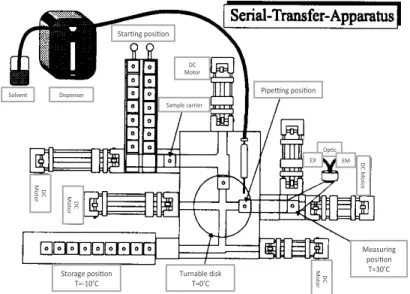
2.2. Mechanical part and experimental procedure In a typical serial-transfer experiment, several sample carriers, containing 20 ul of a solution that includes all components for the reaction except for the replicating RNA, are placed at the starting posi- tion of the machine (Fig. 3). They are frozen in order to prevent uncontrolled reactions. At the beginning of an experiment, the first carrier is inoculated with an RNA quasi-species containing about 10” mole- cules. Two motors, DCM(2) and DCM(3/6), move the inoculated carrier to the turnable disc which is
Fig. 3: Schematic diagram of the machine part of the Serial-
Transfer-Machine. Solvent( Star+ng(posi+on( Dispenser( Sample(carrier( DC( Motor( Pipe6ng(posi+on( Op+c( EX( EM( ( ( D C(Mo to r( Storage(posi+on( T=<10’C( Turnable(disk(T=0’C( Measuring( posi+on( T=30’C( DC( Mo to r( DC( Mo to r( DC( Mo to r(
followed by a fiber optic fluorometer. Ethidium bromide, in the reaction solution, intercalates into nucleic acids with a concomitant increase of fluorescence intensity which is measured. The number of photons emitted is proportional to the concentration of the RNA, so the intensity of the observed fluorescence controls the automatic pipetting device (through a central processor). As soon as RNA exceeds the set maximum concentration, a robot pipette removes an aliquot of the solution and transfer it to the next samples transporter which, then, goes to the measuring station.
The evolution of a Quasispecies of RNA under the selective influence of the TI and a ribonucleases7 was one of the first phenomena experimentally investigated with this device [16].
2.3 “Small” Quasispecies evolution: the multi-channel machine
for mass-transfers
As demonstrated, the cellstat and the mass-transfers machine are the appropriate tools to investigate the molecular evolution. Both the machines can be used to study the Quasispecies as global entities. Basically, a
Quasispecies can be diluted up to the level of subpopulations, or even to the
level of individual molecules, and each clone can be compartmentalized. In this way, the evolutionary adaptations of individual variants can be studied in parallel. Furthermore, the effect of an external constraints variety on mutants of a Quasispecies can be observed. For this purpose a machine that allows to study in parallel 96, or even 960 individual replicating clones has been built. This new robot enlarges the features of the previous machine for the mass-transfer, enabling evolutionary experiments assisted by the PCR 8.
The multi-channel machine for mass-transfers is made of three aluminum blocks, each one with 96 or 960 wells in which are placed the containers in
!!!!!!!!!!!!!!!!!!!!!!!!!!!!!!!!!!!!!!!!!!!!!!!!!!!!!!!!
7 Ribonucleases (Rnase) is a type of nuclease that catalyzes the degradation of RNA into smaller
components.
8 PCR: is a technology in molecular biology used to amplify a single copy or a few copies of of DNA across
several orders of magnitude, generating thousands to millions of copies of a particular DNA sequence. The method relies on thermal cycling, consisting of cycles of repeated heating and cooling of the reaction for DNA melting and enzymatic replication of the DNA. In the first step, the two strands of the DNA double helix are physically separated at a high temperature in a process called DNA melting. In the second step, the temperature is lowered and the two DNA strands become templates for DNA polymerase to selectively amplify the target DNA. The selectivity of PCR results from the use of primers that are complementary to the DNA region targeted for amplification under specific thermal cycling conditions.
which the reactions take place. The temperatures of these three metal blocks are individually controlled by a computer. The solutions containing the molecular systems are loaded into plastic containers with cavities that are adapted to the wells of the metal holder. These cells are covered, air-tight, with another plastic sheet and they can be moved from one block to another at different temperatures as defined by the PCR. In these blocks temperature changes can be obtained in few seconds. As in the experiments executed in the 'single channel’ machine, the concentrations of nucleic acids produced by PCR can be followed on line with a glass optical fibers fluorimeter of 960 channels. In addition, a robot is installed and it is capable of performing the necessary dilutions. The first experiments with this machine were conducted to investigate the influence of various chemicals on the amplification of DNA by the PCR. This machine can also be used to examine the artificial selection in the absence of competitive growth. Selections of new variants may be achieved by the simultaneous detection of 960 variants with assays that linked a fluorescent signal to a new or functionally optimized property, for example the incorporation of fluorescent analogs of nucleotides by a new mutant polymerase.
2.4 Evolutive events “imaging”: the capillary reactor
One of the common features of all the machines described so far is that the reaction systems investigated with these devices display a spatial homogeneity. This property allows the observation of the competition among individuals. J.S. McCaskill described a machine with a one-dimensional reaction environment that, unlike the instruments described hitherto, would create a spatial inhomogeneity which is advantageous to obtain additional information on the replicating system [17]. This is due to the effect of both reaction and diffusion, which determines a constant speed of propagation, unlike the simple diffusion described by the typical report squared distance on time [18] :
v ≈ 2√kD (7)
along a unidimensional “living space” simply depends on its rate of replication
k and diffusion coefficient D. A molecular replicator then spreads like a
“rambling cloud“.
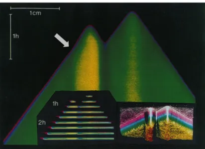
The device that realizes this idea was introduced by McCaskill and Bauer (1993). It is a reactor consisting of 144 parallel capillaries filled with the replication system and an intercalating dye to view the new synthetized RNA molecules. In this way it is possible to control the spatial progress in this environment. The individual molecules of RNA are the source of the colonies growing exponentially. After a short period of time, these clones can expand through replication and they can spread only along the capillary, since their growth is limited in the other two dimensions by the outer walls. This constraint therefore determines a wave of concentration that proceeds with constant speed. A CCD camera installed above the area of the capillaries can capture images at regular time intervals. Series of these images can then be combined as space on time images (fig. 3) to show the differences of evolutionary events and, also, the variations of individual colonies into the reactor.
Figure 3: plotting of a wave front of rapidly increasing RNA concentration propagates at constant velocity to the left to and right of the positions where two single RNA seeded the replication. A sharp increase in the front velocity, mark by the arrow, reflects a single evolutionary change in the replication rate of the RNA. Fig. 2
Thus, colonies originated from initial different molecules can be distinguished because their replication rate is matched to their expansion speed because clones with different rates of replication show, in the resulting images, different and characteristic gradients. Moreover, each evolutionary event associated with a change in the replicative rate alters the speed of advance of the front of wave that propagates, whose result is a change in the slope of the space-time curvature. The obvious advantage of the capillary reactor is the simplicity with which is possible to follow mutational events. After completing the experiment, the samples can be sequenced. In this way, the molecular strategies used to escape the constraints of the selection can be followed step by step.
Chapter 3
3. Materials and methods
Biological approaches and technical solutions for the development of this work are presented in this section.
The principles of the PACE (phage-assisted continuous evolution) technique and the innovations introduced by Jaramillo’s Lab [School of Life Sciences, University of Warwick, FP7 Evoprog: General-Purpose Programmable Machine Evolution on a Chip] are equally decribed.
3.1 Biological parts
3.1.1 Escherichia coli bacteria: the "compartmental models for
evolution"
The choice of Escherichia coli (E. coli) as a model system in biology derived from the extension of the general concept, at the genetic and molecular biology level, about uniformity of principles that govern living organisms. During the development of biochemical research in the thirties and forties, it became clear that all living organisms are formed, mostly, by the same chemical constituents. Also, mechanisms of production, transfer and use of energy, are largely mediated by ATP and the formation of high-energy phosphate bonds. With the development of molecular biology it became also clear that the storage and transfer of genetic information are essentially common to all living organisms.
E. coli, is a microorganism that grows easily in a simple culture media, containing any organic substance, as a source of carbon and energy, along with a few inorganic salts. Like many other bacteria, E. coli can grow within rather wide ranges of pH, osmotic pressure and oxygen tension. Finally it is attacked by numerous viruses.
All these features have made E. coli bacteria in practice, the model on which it is easier to conduct surveys and experiments (Fig. 4).
Figures 4: a| schematic drawing of a typical rod-shaped bacterium and its cell structure; b| a TEM section of a E. coli.
E. coli: the growth
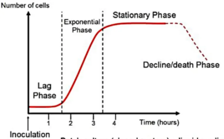
One of the reasons for the success of micro-organisms such as bacteria in the living world, is related to their rate of growth. An organism as E. coli, in favorable environmental conditions, is able to divide itself every twenty minutes: in other words its size doubles every twenty minutes and then divides by binary fission, in two equal halves. It follows that, to maintain this high rate of growth, Escherichia coli must have a correspondingly intense metabolism so the growth will be exponential until the environment will be suitable for the storage of the maximum rate of division (Fig. 5). This happens because E. coli has an high ratio of surface to volume. In fact, the speed of growth of an organism is typically limited by the rate of absorption of nutrients, such as oxygen, and the rapidity with which the catabolic products are eliminated. An increase to the maximum rate possible depends on various factors, such as temperature, pH, oxygen tension, osmotic pressure and nutrients in optimal concentration. An analysis of the necessary materials nutrient in Escherichia coli has highlighted that, although it can grow on a nutrient medium complex it
may also develop in a medium containing a single source of energy and carbon, together with other only inorganic nutrients; this is due to its biosynthetic ability to process all their chemical constituents starting from these simple constructive materials [19]. However, the study of the effect of changes in the type of nutrients, available during the growth, on the intracellular concentration of enzymes has led to an acquisition that was crucial for the development of molecular biology: some enzymes have no relationship with the environment, while others, called adaptive, are synthesized only in response to particular conditions. This ability to adapt to a great variety of different environmental conditions has been of considerable importance for biologists and it is the reason of a sharp distinction compared to many animal and plant cells that develop in very controlled environmental conditions.
A related topic with bacterial growth and that we will see later is the technique of continuous culture.
Figure 5: typical bacterial growth curve. Bacteria grown in a closed system (batch culture), exhibit these growth dynamics: cells initially adjust to the new medium (lag phase) until they can start dividing regularly by the process of binary fission (exponential phase). When their growth becomes limited, the cells stop dividing (stationary phase), until eventually they show loss of viability (death phase). Note the parameters of the x and y axes. Growth is expressed as change in the number viable cells vs time. Generation times are calculated during the exponential phase of growth. Time measurements are in hours for bacteria with short generation times
.
E.coli: genome structure
E. coli genome is a circular double strand of DNA. Spontaneous mutations occur with a frequency similar to that found in other living organisms and, as E. coli is haploid, are easily identified [20].
Therefore, thanks to the ability of E. coli to oppose a broad spectrum of environmental conditions stemming from relevant selective pressures, it is possible to isolate with relative ease a large number of different mutant. An evolutionary process that, under natural condition,s is carried over longer times happens in a lab just in the space of one night. For example, if a bacterium sensitive to penicillin mutates developing a resistance to the antibiotic, a WT population can be incubated in a culture medium containing an appropriate concentration of penicillin, so that a sufficiently large number of microorganisms may develop a resistant phenotype.
Significant pressures of this type can be present in nature: antibiotic resistance in clinical settings is one obvious example. This effect could also inevitably affect the continuous culture of a single bacterial strain, when not properly setup.
The doubt related to the spontaneous mutation were clarified only after 1940, thanks to the discovery of the mechanisms responsible for genetic recombination. These techniques are commonly used for biological investigations that require the presence of mutants.
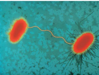
• Conjugation. J. Lederberg and EL Tatum in 1946 observed that if it were cultivated together two strains of Escherichia coli with different cultivation requirements, occurred a recombination of genetic determinants. This process requires physical contact between the two strains, one with the fertility factor (F+) and the other without it (F-). The conjugation is linked to the passage of DNA from the strain F+ to that F- (Fig. 6) [21].
Figure 6: Sex pilus connecting two E. coli cells during bacterial conjugation; on the left a F- cell is linked to a F+
cell on the right.
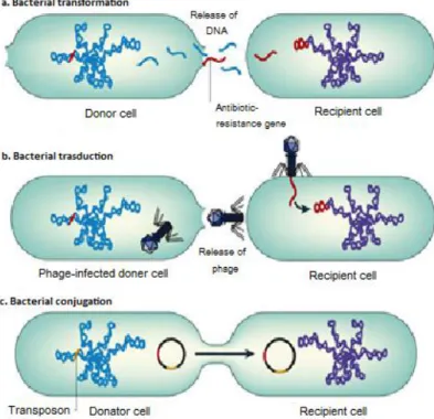
• Transduction. The phage head normally contains the phage genome. However, a small percentage may accidentally include a segment of the genome from a previous host (in a variable number of genes, up to 150). When a phage of this type attacks an E. coli, the DNA segment penetrates the new host and integrate, according to the normal processes of recombination, in the chromosome of the new host [22].
If in continuous culture, this phenomenon could then cause the co-evolution of species.
• Transformation. With one of the most important biological research of this century, RJ Dubos and OT Avery showed that in the pneumococcus, causative agent of pneumonia, genes can be transferred from one cell to another by fragments of purified chromosomal DNA. This discovery was the first real proof that DNA is the repository of genetic information. The process is accomplished in the pneumococcus with a speed much greater than in other bacteria and microorganism such as E. coli. Such differences in undergoing the transformation seem mainly related to the problem of penetration of a large molecule, such as DNA in the cytoplasm and then into the nucleus, since the cell wall and the cytoplasmic membrane normally behave like a real barrier against such a transfer. It is therefore difficult to
assess the importance of transformation in nature [23]. Figure 7 resumes all the above presented procedures.
Figure 7: a| Transformation occurs when naked DNA is released on lysis of an organism and is taken up by another organism. The antibiotic-resistance gene can be integrated into the chromosome or plasmid of the recipient cell. b| In transduction, antibiotic-resistance genes are transferred from one bacterium to another by means of bacteriophages and can be integrated into the chromosome of the recipient cell (lysogeny). c| Conjugation occurs by direct contact between two bacteria: plasmids form a mating bridge across the bacteria and DNA is exchanged, which can result in acquisition of antibiotic-resistance genes by the recipient cell. Transposons are sequences of DNA that carry their own recombination enzymes that allow for transposition from one location to another; transposons can also carry antibiotic-resistance genes. Nature Reviews | Microbiology, 2006.
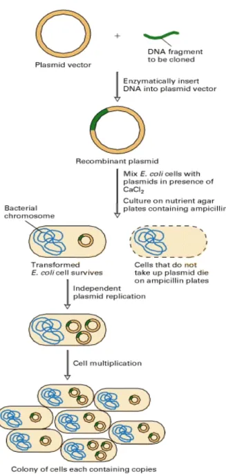
• Another mechanism to transform E. coli, exploits plasmids located outside of the main chromosome. Plasmids are circular fragments of double-stranded DNA that reproduce themselves within the cell; they possess many properties of a small chromosome and contain genetic information to control their replication and to ensure segregation of their copies in each daughter cell at the time of cell division. They differ, however, from a chromosome as they are much smaller and are not essential for normal vitality of the cell. Sometimes a plasmid can be integrated into the main chromosome of the
bacterial cell, indicated by the term 'episome'. The F factor, involved in conjugation is an example of a plasmid. Factors of resistance to drugs may be present in plasmids rather than in the main chromosome: a single plasmid may encode resistance to many commonly used antibiotics.
In lab, plasmids can be transferred remarkably easily from one cell to another and they can cause the rapid spread of an antibiotic resistance within a bacterial population (Fig. 8).
Figure 8: procedures of gene cloning into plasmid and transformation of E.coli bacteria.
3.1.2 Bacteriophages: the faster evolutionary replicator
During the second decade of the XX century FW Twort and FH d'Herelle gave a description of viral infections of the bacteria. They showed that bacterial cells can be infected and destroyed by filterable agents. These bacterial viruses were called bacteriophages or more simply phages. Although most phage present a more complex structure of the viruses of animals or plants, they have been studied deeply. The largest information on the mechanisms of phage reproduction and the influence of the host was obtained from viruses that infect E. coli: in particular from a group named T phages [24].
Phage: the structure
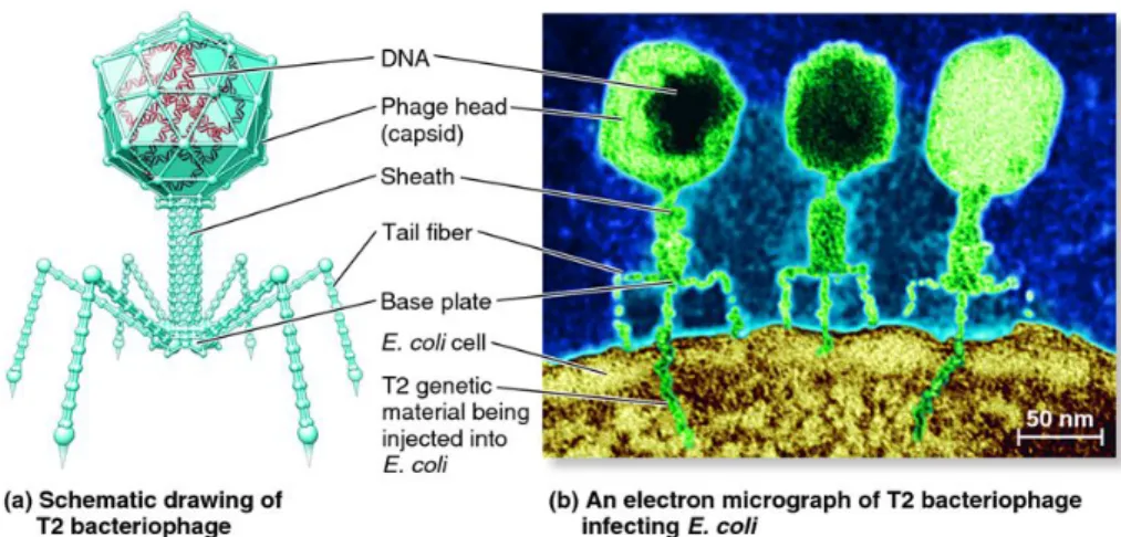
In the structure of the virus two main parts can be observed: a head and a tail. The head contains a single circular molecule of double-stranded DNA, or chromosome, whose molecular weight is 1.3 × 10⁸ Dalton. It is surrounded by an icosahedral capsid protein structure of approximately 125 nm long and 81 nm wide. More precisely, each half of the head is constituted by a half icosahedron and the two halves are connected by a short hexagonal prism. The capsid is formed from individual protein subunits or capsomeres, whose repetitive structure determines the crystal symmetry of the head.
The tail of the phage is a complex structure of about 95 nm in length. Its innermost component is formed by tubule protein subunits, in helical arrangement and covered by a sheath, constituted by other subunits, which extends from a collar to a plate baseline and has the ability to contract to less than half of its extension. Even the collar and the basal plate are composed of other protein subunits and on the plate there are small pins which have six long and thin caudal fibers (Fig. 9).
Figure 9: a| schematic drawing of a lytic T2 bacteriophage; b| an electron micrograph of T2 bacteriophage infecting an E. coli cell.
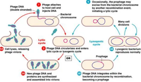
Phage multiplication: lytic or lysogenic cycle
The study of the events that occur in an infected cell can be distinguished into three phases, named: adsorption, multiplication, liberation. For describing this cycle, is necessary to consider separately two main categories of bacteriophages: virulent and temperate.
• Virulent bacteriophages, as T7 phage, contribute to the lytic cycle. It begins with a process of adhesion between the basal plate, the fibrils of the phage and the lipopolysaccharide of the bacterial cell wall. The degree of specificity of this process is equal to that which it has in the interaction of an antibody with the corresponding antigen.
The propagation of the virus depends solely by the presence of viral DNA in the host. This was confirmed by the observation that, if the permeability of the bacterial cell wall is increased with an appropriate treatment, the bacterium can be infected with DNA purified phage, with exactly the same result if it were infect by the whole virus [25].
It became also clear that the function of the protein components of the virus is to protect the nucleic acid during its passage from one cell to another and in facilitating a specificity of penetration into a host cytoplasm. In particular, after the absorption of the tail of the phage, its sheath is contracted which makes penetrate the central part of the tail across the
cell wall and the cytoplasmic membrane, thus coming in contact with the cytoplasm of the host. The penetration of the phage DNA into the host, determines an almost immediate suspension of the biosynthesis of DNA, RNA and proteins: the only de novo biosynthesis of these polymers is encoded by the phage genome. After a latency period, the synthesis of DNA resumes, but it is completely limited to the production of viral DNA. Then, the multiplication starts.
After the infection, only a small amount of RNA is processed and it seems that it is intended solely phage mRNA. It transcribes the information for the synthesis of specific proteins of the virus, which are produced according to a temporal sequence. The first are enzymes that participate in the production of phage DNA, then structural proteins necessary for the maturation of new phage particles are processed. Together with phage lysozyme they participate in the eventual lysis of the host cell. In consequence of all these processes, in the cytoplasm of the host, all the constituents of the particles of the mature virus are now accumulated. The first stage of the 'assembly' of new viruses consists in the condensation of the phage DNA into the structural elements with the shape of phage heads is induced by a special 'condensing' protein. This condensation is followed by the crystallization of the protein subunits of the head around the DNA, so as to form mature heads. Tail and the basal plate are then processed by the condensation of appropriate protein subunits and, finally, the tails and heads are combined. The final stage seems to consist in the synthesis and assembly of the fibrils of the tail. Each stage of the aggregation of a structural component is dependent on the completion of the previous stage. When the processing of new phage particles is complete, the lysis of the host cell occurs, followed by the liberation of some hundreds of phage particles that can infect other host cells.
Since the whole process of multiplication takes only ten to twenty minutes, the reproductive potential is clearly enormous and just limited by the availability of an adequate number of host cells.
takes an alternative algorithm than the previous named lysogenic cycle. The viral DNA enters the host as described, but instead of causing cell death by lysis, it is incorporated as a component of the host genome: the 'prophage'. It then replicates as if it was a part of the normal bacterial genome and only occasionally a lytic cycle of development breaks out spontaneously. The impact of multiplication step can be increased by some inducing agents. The presence of a prophage, as component of the bacterial genome, changes some host functionality: first, the lysogenic bacterium becomes immune from attacks by other phages, but even more important, there may be alterations of specific bacterial genotype, called lysogenic conversions, which don’t presents obvious relationship with the biological cycle of the phage. For example the surface antigens of the bacterium can be modified or it can be altered the toxin production [26]. Figure 10 shows the cycles steps.
Figure 10: Lytic and lysogenic cycle copyright for a virulent and temperate bacteriophage, respectively. Pearson education, 2004.
3.1.3 Bacterial and phage quantification
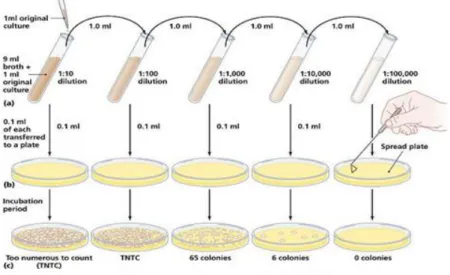
• Bacterial enumeration. In the study of microbiology, there are numerous occasions when it is necessary to either estimate or determine the number of bacterial cells in a broth culture or liquid medium. Determination of cell numbers can be accomplished by a number of direct or indirect methods.
Standard Plate Count (Viable Counts)
A viable cell is defined as a cell which is able to divide and form a population or colony. A viable cell count is usually done by diluting the original sample, plating aliquots of the dilutions onto an appropriate culture medium, then incubating the plates under proper conditions so that colonies are formed. After incubation, the colonies are counted and, from knowledge of the dilution used, the original number of viable cells can be calculated (Fig. 11). For accurate determination of the total number of viable cells, it is critical that each colony comes from only one cell, so chains and clumps of cells must be broken apart. However, since one is never sure that all such groups have been broken apart, the total number of viable cells is usually reported as colony-forming units (CFUs) rather than cell numbers. This method of enumeration is relatively easy to perform and is much more sensitive than turbidimetric measurement. Thus, the concentration of bacteria in the original suspension is calculated using the following formula:
!"# !" = !
!"#$%&!!"!!"#"$%&'!!"#$%&'!!"!!!!!!"#$%
!"#$%&!!"#$%&! !" ∙!"#$%"&'!!"#$%& (9)
A major disadvantage, however, is the time necessary for dilutions, platings and incubations, as well as the time needed for media preparation.
Figure 11: illustration of the protocol related to standard plate count (SPC) test. Pearson Education, 2006.
Turbidimetric Measurement
A quick and efficient method of estimating the number of bacteria in a liquid medium is to measure the turbidity or cloudiness of a culture and translate this measurement into cell numbers. This method of enumeration is fast and is usually preferred when a large number of cultures are to be counted.
Although measuring turbidity is much faster than the standard plate count, the measurements must be correlated initially with cell number. This is achieved by determining the turbidity of different concentrations of a given species of microorganism in a particular medium and then utilizing the standard plate count to determine the number of viable organisms per milliliter of sample. A standard curve can then be drawn, in which a specific turbidity or optical density reading is matched to a specific number of viable organisms. Subsequently, only turbidity needs to be measured.
Turbidity can be measured by an instrument such as a colorimeter or spectrophotometer (Fig. 12). These instruments contain a light source and a light detector (photocell) separated by the sample compartment. Turbid solutions such as cell cultures interfere with light passage through the sample, so that less light hits the photocell than would if the cells were not there. Turbidimetric methods can be used as long as each individual cell blocks or intercepts light. Thus, if the mass of cells becomes so large that some cells effectively shield other cells from the light, the measurement is no longer accurate.
Before turbidimetric measurements can be made, the spectrophotometer must be adjusted to 100% transmittance (0% absorbance). This is done using a sample of uninoculated medium. Percent transmittance of various dilutions of the bacterial culture is then measured and the values converted to optical density, based on the formula:
Figure 12: a| optical arrangements of nephelometry and turbidimetry; b| a commercial spectrophotometer.
Direct Microscopic Count
Petroff-Hausser counting chambers can be used as a direct method to determine the number of bacterial cells in a culture or liquid medium. In this procedure, the number of cells in a given volume of culture liquid is counted directly in 10-20 microscope fields (Fig. 13). The average number of cells per field is calculated and the number of bacterial cells ml-1 of original sample can then be computed. A major advantage of direct counts is the speed at which results are obtained. However, since it is often not possible to distinguish living from dead cells, the direct microscopic count method is not very useful for determining the number of viable cells in a culture.
Figure 13: illustration of the protocol related to direct microscopic count on petroff hausser counting chamber. Pearson Education, 2007.
Enumeration of phage particles. The plaque assay is a virological assay employed to count and measure the infectivity level of the bacteriophages. The basis of plaque assay is to measure the ability of a single infectious virus to form a “plaque” on a concurrent monolayer culture cells. Aliquots of diluted bacteriophage are mixed with host bacterial cells in several milliliters of soft agar, which are then spread onto agar plates containing media. The use of soft agar allows the phage to easily diffuse through the medium giving more consistent plaque formation. It also eliminates the problem of uneven absorption of the bacterial-phage solution into the hard agar that often caused uneven plaque formation on the plate [27].The bacteriophage adsorb onto the host bacterial cells, infect and lyse the cells, and then begin the process anew with other bacterial cells in the vicinity. After 6 – 24 hours, zones of clearing, plaques, are observable within the lawn of bacterial growth on the plate (Fig. 14).
Thus, the concentration of bacteriophage in the original suspension is calculated using the following formula:
!"# !" = !
!"#$%&!!"!!"#$%&'!!"#$%&'!!"!!!!!!"#$%
!"#$%&!!"#$%&! !" ∙!"#$%"&'!!"#$%& (11)
Plaque characteristics are related to the type of bacteriophage as well as other physical and chemical characteristics of the system in which the bacteriophage are grown [28].
3.2 General principle of bacterial continuous culture
Prior to its development, the bacterial culture was carried out in closed systems, a lot or batch, in which the microorganism was inoculated and cultured. The growth continues until the soil is not more suitable, due to the depletion of nutrients or to the accumulation of toxic products from bacterial growth itself. The culture, thus, enters in a stationary phase, where no evident growth occurs, and after a variable period of time extinction occurs.
Under conditions of continuous culture rather, the feeding of nutrients is continuous and controlled. Then, the culture is maintained in the same growth conditions even for long periods of time. All continuous flow systems consist essentially of a reactor into which reactants flow at a steady or dynamic rate and from where products emerge.
The factors governing their operation are:
the way in which material streams through the volume of reactor. This depends by the design of the configuration chosen.
the kinetics of the reaction taking place.
3.2.1 Fermenter
A fermenter is a culture vessel where microorganisms can be grown under suitable conditions. Sterile growth medium is fed into the culture vessel at a flow-rate (f) and culture emerges from it at the same rate. A device keeps the volume of culture in the vessel (v) constant. The contents of the vessel are sufficiently well stirred to approximate to the ideal of complete mixing, so that the entering growth medium is instantaneously and uniformly dispersed throughout the vessel.
Residence-times in such a culture vessel will be determined not by the absolute values of the flow-rate and culture volume but by their ratio which we call the dilution rate, D [h-1], defined as D=f/v, i.e. the number of complete volume-changes/hr. The mean residence-time of a particle in the culture vessel is thus equal to 1 / D.
For understanding which are the time characteristics of a continuous reactor, we assume that the bacteria in the culture vessel are not