UNIVERSITÀ POLITECNICA DELLE MARCHE
FACOLTÀ DI MEDICINA E CHIRURGIA
_______________________________________
SCUOLA DI DOTTORATO DI RICERCA XXIX CICLO
FACOLTA’ DI MEDICINA E CHIRURGIA
Corso di Dottorato in Salute dell’Uomo
Dipartimento di Scienze Cliniche Specialistiche ed Odontostomatologiche
THE UNFOLDED PROTEIN RESPONSE: A
LINK BETWEEN ENDOMETRIOID OVARIAN
CARCINOMA AND ENDOMETRIOSIS
Tutor: Chiar.mo
Prof. Andrea Ciavattini
Tesi di Dottorato di:
Alessandra Tozzi
Dedicated to my family, In Senigallia, Lima and Lugano
TABLE OF CONTENTS
TABLE OF CONTENTS 3
1. INTRODUCTION 4
1.1 EPITHELIAL OVARIAN TUMORS 6
1.1.1 INCIDENCE AND EPIDEMIOLOGY 6
1.1.2 PATHOLOGY 8
1.1.3 DIAGNOSIS 13
1.1.4 STAGING AND RISK ASSESSMENT 15
1.1.5 TREATMENT PLAN 16
1.1.6 FOLLOW-‐UP 21 1.2 ENDOMETRIOSIS AND OVARIAN CARCINOMA ASSOCIATED TO ENDOMETRIOSIS 22
1.2.1 ENDOMETRIOSIS 22
1.2.2 ENDOMETRIOSIS-‐ASSOCIATED OVARIAN CARCINOMA 30
1.3 ENDOPLASMIC RETICULUM STRESS 34
1.3.1 PERK 35
1.3.2 ATF6 36
1.3.3 IRE1 37
1.3.4 GRP78 37
1.3.5 HERPUD 2 38
1.3.6 UPR IN CANCER 39
2. MATERIALS AND METHODS 43
2.1 STUDY DESIGN 43
2.3 ENDOMETRIAL FROZEN SAMPLE PREPARATION 45 2.4 PARAFFIN EMBEDDED OVARIAN TISSUE SAMPLES PREPARATION 47 2.5 PCR ANALYSIS OF SYNTHESIZED cDNA 51 2. 6 GENE EXPRESSION ANALYSIS BY REAL TIME PCR 52
2.7 STATISTICAL ANALYSIS 55
3. RESULTS AND DISCUSSION 56
3.1 GENE EXPRESSION ANALYSIS OF UPR GENES IN ENDOMETRIOID CARCINOMA OF THE
OVARY 56
3.2 COMPARISON OF UPR GENE EXPRESSION AMONG ENDOMETRIOID OVARIAN
CARCINOMA, ENDOMETRIOSIC CYST AND ENDOMETRIAL TISSUE FROM ENDOMETRIOSIS
PATIENTS AND HEALTHY WOMEN 58
4. CONCLUSIONS 66
1. INTRODUCTION
Primary ovarian carcinomas represent more than 90% of malignant ovarian tumours (1), 3% of women carcinomas and the fifth cause of death for tumours in women. Malignant epithelial tumours (carcinomas) represent a heterogeneous group of distinct diseases, that are different in terms of epidemiological and genetic risk factors, precursor lesions, patterns of spread, and molecular events during oncogenesis, response to chemotherapy, and prognosis. Although the hypothesis of their development from a “neometaplasia” of superficial ovarian mesothelium cannot be excluded (1), recent investigations have demonstrated that a substantial number of cancers, traditionally thought to be primary ovarian tumors (particularly serous, endometrioid, and clear-cell carcinomas), originate in the fallopian tube and the endometrium and involve the ovary secondarily. According to this recent hypothesis, endometrioid ovarian carcinoma and clear-cell carcinoma originate from endometriosic cysts situated inside the ovaries.
Endometriosis is a complex benign and oestrogen-dependent disease, characterized by ectopic implants of endometrial tissue that induces a chronic inflammatory reaction. (2-3) Ectopic implants are especially detected in pelvis, affecting ovaries, peritoneum, utero-sacral ligaments, Douglas pouch and recto-vaginal septum; extra-pelvic findings are infrequent. Ovarian localization of endometriosis is found in 17-44% of endometriosis patients. (4) Endometriosis affects 5-10% of premenstrual women, but this percentage arises up to 17% of infertile women e 40-60% in patients suffering from dysmenorrhea (5) (6)
The possible transformation of endometriosic tissue in ovarian carcinoma has been postulated almost 100 years ago, the first author was Sampson in 1925. (7) It is estimated that the risk of developing endometriosis is doubles in endometriosis patients compared to the general population. (8) Nowadays several studies have been published on the definition of endometriosis as a risk factor for ovarian carcinoma and this
factors, such as early menarche, late menopause and nulliparity, and the same protective factors, such as tubal sterilization, hysterectomy, multiparity and oral contraception. (9) (10) Moreover they have common features, such as invasivity, neoangiogenesis, apoptosis reduction, local and at distance invasion and recurrence. (11) Genomic alterations represent an important risk factor for the development of this disease: some of these are in common both for endometriosis and for ovarian carcinoma, others are essential for the malignant transformation. Thus, although the correlation between endometriosis and ovarian carcinoma is clear, the exact mechanism through which the malignant transformation takes place is still not yet well established. Further studies are necessary to explain it.
The Unfolded Protein Response (UPR) is the response of the endoplasmic reticulum to external stress (12) and it is activated by the accumulation of unfolded or misfolded proteins inside the lumen. Recent studies have demonstrated the role of UPR in the neoplastic transformation process, since it determines the cellular survival in a hypoxic environment. This effect is mediated by the reduction of pro-apoptotic signaling, cellular metabolism modification and neo-angiogenesis. UPR activation can support cellular survival or cell apoptosis, in dependence of the milieu. Its activation in neoplastic cells plays a protective role against cellular death, that follows ER stress. The aim of this study is to analyze the expression profile of UPR genes in endometrioid ovarian carcinoma and to evaluate its possible involvement in the neoplastic progression of endometriosis.
1.1 EPITHELIAL OVARIAN TUMORS
1.1.1 INCIDENCE AND EPIDEMIOLOGY
The estimated number of new ovarian cancer cases in Europe in 2012 was 65 538 with 42 704 deaths (14). Incidence rate varies worldwide, being the highest in northern European countries. In the USA, there were 22.280 new diagnoses and 14.240 deaths from this neoplasm. (15). Ovarian cancer is the fifth most common type of cancer in women and the fourth most common cause of cancer death in women. The estimated lifetime risk for a woman developing ovarian cancer is about 1 in 54.
It affects mainly old, postmenopausal women, with median age at the time of the diagnosis at 63 years old (16). The exact cause of ovarian cancer is still yet unknown, but many efforts have been done to identify associated risk factors. Main contribute to develop ovarian cancer is related to woman’s reproductive history.
Increasing risk factors are the following: nulliparity, older age (>35 years) at pregnancy and first birth, early menarche, late menopause, postmenopausal hormone therapy, pelvic inflammatory disease and ovarian stimulation for in vitro fertilization (especially it can increase the risk of developing borderline epithelial tumors).
Protective factors are instead: multiparity, younger age at pregnancy and first birth (<25 years), use of the oral contraceptive pill, tubal ligation, breastfeeding and suppression of ovulation. All of these risk factors point to ovulation being correlated with the development of ovarian cancer. Further risk factors are obesity and possibly the use of talcum powder.
Family history (primarly patients having 2 or more first-degree relatives with ovarian cancer) plays a very important role in the development of ovarian cancer. Risk of ovarian cancer is more than doubled in women with a positive family history, compared to women with no family history. However, an identifiable genetic mutation, e.g. the
developing ovarian cancer and ≤85% risk of developing breast cancer. A BRCA 2 mutation increases the lifetime risk of ovarian cancer to 10%–20% and breast cancer risk of ≤85%. It has been demonstrated that women BRCA1 and BRCA2 mutated or members of families affected by Lynch Syndrome are shown to develop ovarian cancer ∼10 years earlier than women with non-hereditary ovarian cancer. Criteria to refer a patient affected by ovarian carcinoma for genetic testing are still not yet clear in literature and recommendation is made on the basis of a family history and ethnic background. It is however important to identify BRCA mutations, since it determines a different surgical and follow up management.
RISK FACTORS PROTECTIVE FACTORS
Family history of cancer Breast feading for at least 18 months
Genetic Syndromes Multiparous women
Early menarche, late menopause Late menarche, early menopause
Hormonal therapy with oestrogens for at least 5 years
Oral contraceptives
Nulliparous women Hysterectomy
High fat diet Low fat diet
Endometriosis Tube ligature
1.1.2 PATHOLOGY
Ovarian neoplasms consist of several histopathologic entities, the major one being represented by cancer of epithelial origin (∼90%). The World Health Organization recognizes the following distinct subtypes (17):
Figure 1: WHO Histologic Classification in the NCCN Guidelines for Ovarian Cancer Histopathologies. Epithelial ovarian tumors (85-95%)
Clinical trials have demonstrated that different histology is related to different clinical, pathological and prognostic features, thus determining a different therapeutic approach (18). Grade is an additional prognostic factor and is assigned (from 1 to 3) depending on the following tumor characteristics: architectural features, mitotic counts and nuclear atypia. (19). There is no single universally accepted grading system. Some use a two-tier staging (20). Because of the complexity of this subclassification, that has important impact on the therapeutic management, an expert in gynecological pathology is required.
Molecular pathogenesis
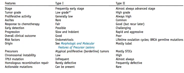
Ovarian cancer is nowadays recognized as a heterogeneous group of neoplasm, and many efforts have been done in the last few years to understand the initiating molecular events underlying the different tumor subtypes, in order to better characterize the mechanism of cancer development, the natural behavior and prognosis. According to recent morphological, immunohistochemical and molecular genetic achievements, a dualistic model of epithelial ovarian carcinogenesis has been more than a decade ago proposed. (21). It divides epithelial ovarian cancer (EOC) into two categories.
Type 1 cancers include low-grade serous, endometrioid, mucinous, clear-cell and malignant Brenner tumors and are characterized by low-grade and clinical indolence. Main molecular features for these tumors are: mutations of KRAS, BRAF, ERBB2, PTEN, PIK3CA and ARID1A and a relatively genetical stability.
These mutations occur early in the evolution of type 1 ovarian tumors and are also observed in borderline tumors and endometriosis. A stepwise sequence of tumor development is now well recognized from benign precursor extraovarian lesions (e.g. borderline tumor) to malignant lesions in type 1 cancer.
Clinically, they are generally indolent and confined to the ovary at presentation (stage I), thus having a better prognosis.
Type 2 cancers include high-grade serous, high-grade endometrioid, malignant mixed mesodermal tumors and undifferentiated tumors. Main molecular features for these
tumors are: TP53 mutations (97% of high-grade serous cancers were associated with a TP53 mutation) and BRCA1/2 mutation (22).
No clinical precursor lesion has been demonstrated for type 2 cancers. In recent years, accumulating evidence has shown that the majority of high-grade serous ovarian and peritoneal tumors originate in the fimbria of the fallopian tube (serous tubal intraepithelial carcinoma) (23, 24). These malignant cells then metastatise to the ovaries and the peritoneal cavity.
Clinically, type 2 cancers are aggressive and present in advanced stage (stage II-IV), being responsible for 90% of ovarian cancer deaths.
As far as it concerns endometrioid tumors, which are the topic of this thesis, there are two different entities. Low-grade endometrioid tumors are characterized by deregulation of PI3K/PTEN signaling (15-20% of cases) and deregulation of Wtn/β-catenin signaling (15-40% of cases), whereas TP53 mutations are not present. Inactivation of PTEN and activation of PIK3CA can lead to activation of the phosphatidylinositol3-kinase signaling pathway. Mutations of KRAS and BRAF are reported in less than 7% of cases. Microsatellite instability (loss of hMLH1, hMSH2, mutS, MSH6 and PSM2) has also been reported in up to 20% of cases. Conversely, high-grade endometrioid tumors are lacked Wnt/β-catenin or PI3K/PTEN signaling pathway defects and frequently present TP53 mutations, making this tumor more similar to poorly differentiated serous carcinoma (HGSC). Thus, in few cases a high-grade endometrioid carcinoma develops from a low-grade endometrioid carcinoma, but more frequently they represent disctinctive tumors. (21).
Endometrioid tumors are usually more likely to be early stage (stage 1) and low grade. They represent ∼10% of ovarian cancers.
At a molecular level, it is well estabilished in literature that endometrioid carcinoma shares the same genetic features with endometriosis, such as mutation of ARID1A (a tumor suppressor gene involved in cheratin remodeling) and deletion of PTEN, thus being endometriotic cysts (endometriomas) the origin of tumorigenesis. Moreover, the same molecular abnormalities have been observed in endometrium from endometriosis
This hypothesis is also supported by the evidence that tubal ligation determine a protective effect for endometrioid carcinoma of the ovary.
At present, molecular classification is at greatest interest, since emerging molecular genetic findings determine not only a more detailed characterization of the different subtypes of ovarian cancer, but also the possibility of early diagnosis and new target therapies.
The purpose of leately and future researches in ovarian cancer field is to detect tumors when they are still confined to the ovaries, in order to better assess the risk and to provide immediate therapy, that can reduce the high mortality of this disease. (26)
Figure 3: The revised dualistic model in the pathogenesis of ovarian epithelial cancer. The areas in individual histotypes reflect their relative prevalence. The inner cirle indicates the likely cell of origin of the different type I and type II neoplasms. The molecular pathway alterations that characterize each tumor subtype are summarized in the square boxes.
Table 2: Clinicopathologic and molecular features of Type I and Type II Ovarian Carcinomas *Clear cell carcinoma is not graded, but many consider the tumor as high-grade.
+Occasional progression to high grade can be observed.
1.1.3 DIAGNOSIS
Ovarian cancer is difficult to diagnose at early stage, when it is confined to ovaries, since it is predominantly asymptomatic or pauci-simptomatic. In fact, more than 70% of patients present with advanced disease, when symptoms are most commonly seen. Symptoms suggestive of ovarian cancer include: abdominal or pelvic pain, bloating, constipation, diarrhea, urinary frequency, vaginal bleeding, difficulty eating and fatigue. Physicians should be suspicious especially when these symptoms are new and frequent (> 12 day/month). In advanced ovarian cancer, ascites and abdominal masses lead to increased abdominal girth, nausea, anorexia, dyspepsia and early satiety. Respiratory symptoms may be developed when the disease extents across the diaphragm to the pleural cavities, producing pleural effusions. Clinical trials suggest that screening test based on symptoms are neither sensitive nor specific enough to diagnose early stage disease.
Thus, laboratory signs have been searched in order to help diagnosis. The first one is cancer antigen CA 125: it is elevated only in about 50% of patients with the International Federation of Gynecology and Obstetrics (FIGO) stage I disease, whereas it arises 85% of patients with advanced disease. Its utility to detect early disease is therefore questionable. Moreover is not specific for ovarian cancer and raised CA 125 levels may be found in non gynecological malignancies (e.g. breast, lung, colon and pancreatic cancer) and benign disease (e.g. endometriosis, pelvic inflammatory disease and ovarian cysts). Measurement of serum carcinoembryonic antigen (CEA) and CA 19–9 levels are required when it is unclear whether an ovarian mass is of gastrointestinal origin, or a primary mucinous ovarian tumor. Similarly, in these situations, colonoscopy and/or gastroscopy are sometimes considered, particularly when CA 125/CEA ratio is ≤25.
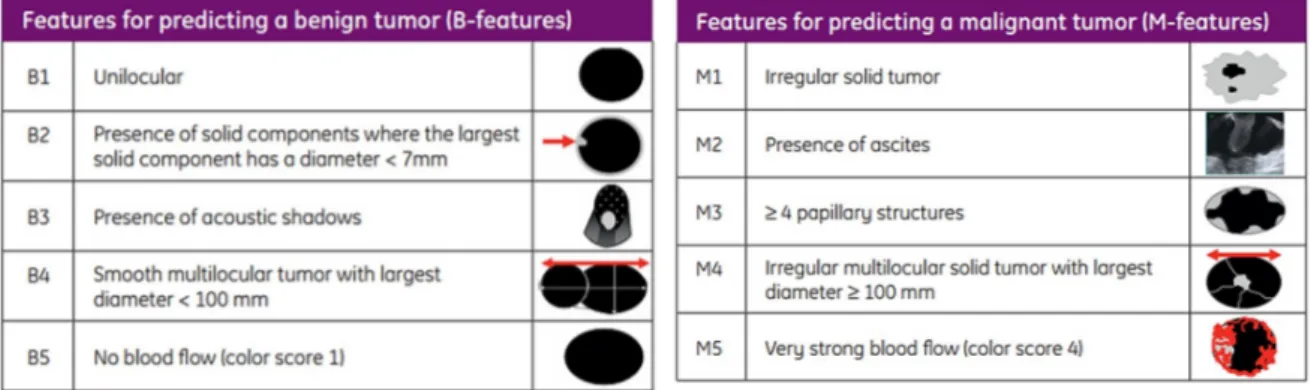
As far as it concerns image investigations, transvaginal and abdominal ultrasounds represent the first choice, since they allow the visualization and characterization of ovarian structures, thus improving the differentiation of malignant versus benign conditions, using noninvasive procedures. Anyway the preoperative assessment of an adnexal mass remains a major challenge for the gynecologist and several efforts have
been done to determine the most important variables to predict the likelihood of malignancy. The accurate characterization before any surgery on ovarian pathology is mandatory, in order to provide the proper treatment, because new treatment options are useful only if the preoperative diagnosis is correct. For example the prognosis worsens if during the operation, a cyst (that has been wrongly considered benign preoperatively) is broken, thus spreading malignant cells into the abdomen. The International Ovarian Tumor Analysis (IOTA) group produced some simple rules to standardize the ultrasound description of adnexal pathology. (27). Malignant feature, suggestive of ovarian cancer, are represented by: the presence of irregular multilocular solid tumor with largest diameter >100 mm, irregular solid tumor, presence of ascites, >4 papillary structures and very strong blood flow (color score 4). Conversely unilocular cyst, presence of solid components where the largest solid component has a diameter <7mm, presence of acoustic shadows, smooth multilocular tumor with largest diameter < 100 mm, no blood flow (color score 1) are predictive of a benign tumor.
Figure 4: Schematic representation of ultrasound feature for predicting benign or malignant tumors
If we compare 1) CA125 alone, 2) ultrasound with CA125 and 3) ultrasound alone, we can conclude that screening is in fact not effective, because the tumors with either an increased CA-125 level or an abnormal transvaginal ultrasound or both are mostly of patients with high stage ovarian cancer. Furthermore CA125 does not increase the detection of cancer over ultrasound alone and ultrasound results superior to CA125
Computed tomography (CT) scans are routinely used to determine the extent of disease and to aid in surgical planning. Imaging of the chest with CT or chest X-ray should be done to look for pleural effusions and disease above the diaphragm. A pleural effusion cannot be regarded as malignant and indicative of FIGO stage IV disease without confirmation of positive cytology. Magnetic resonance imaging (MRI) scans do not form part of routine investigations.
1.1.4 STAGING AND RISK ASSESSMENT
Ovarian cancer is classified primarily as stages I to IV using the FIGO (International Federation of Gynecology and Obstetrics) system. The new FIGO staging guidelines will combine staging for Fallopian tube carcinoma and ovarian cancer and will be effective on 1st January 2017. FIGO staging is necessary, because it represents, together with the tumor grading, the most powerful indicator of prognosis. It is defined during the operation, but also preoperative assessment with cross-sectional imaging (CT or MRI) is essential as it guides surgery and the pathway of intervention. Primary surgery remains the most common and preferred approach, but where this is deemed not feasible, an image-guided or laparoscopic biopsy should be carried out.
1.1.5 TREATMENT PLAN
Surgical management of early primary disease
Once early stage primary disease is diagnosed preoperatively, it should be submitted to appropriate surgical staging and cytoreduction. Initial surgery should include: peritoneal washings, ideally taken before manipulation of the tumor, bilateral salpingo-oophorectomy, hysterectomy, multiple peritoneal biopsies of all abdominal fields, at least infracolic omentectomy, appendectomy in case of mucinous histology and pelvic and para-aortic lymph node dissection up to the renal veins. This surgery allows adequate staging, in order to provide prognostic information and to define whether chemotherapy is needed.
In case of an incidental diagnosis of early stage primary disease, the intraoperative pathologic evaluation with frozen sections is suggested, in order to identify a malignant epithelial cancer and perform adequate surgical staging, without the need for a second operative procedure.
Surgical staging is mandatory for ovarian carcinoma, because it has been demonstrated that it can uncover occult advanced disease: Cass et al. showed that, in 96 patients with grade 3 tumors and gross disease confined to one ovary, 15% had microscopically positive lymph nodes (29)
As far as it concerns lymphadenectomy, a trend for improved progression-free survival (PF) and overall survival (OS) has been observed in patients undergoing this procedure, if compared to the control group, but the difference is not of statistic significance. (30) (31)
Thus, there are data showing upstaging with lymphadenectomy and other data showing that lymphadenectomy does not affect overall survival. However omentectomy and multiple biopsies of peritoneum (the most common sites of peritorneal implants) may upstage patients in approximately 30% of cases and may affect prognosis. Bulky lymph
When young women are affected by early stage disease and/or good-risk tumors and wish to preserve fertility, fertility-sparing surgery could be considered in early-stage disease. It consists of unilateral salpingo-oophorectomy, preserving the uterus and contralateral ovary. Comprehesive surgical staging should be performed to rule out occult higher stage disease.
For patients with stage IA or stage IC with unilateral ovarian involvement and favorable histology (mucinous, serous, endometrioid or mixed histology and grade 1 or 2), risk of recurrence seems to be related to higher incidence of extra ovarian spread observed in grade 3 tumors, rather than to a higher relapse rate in the preserved ovary (32). Thus, organ-preserving surgery is adequate in these patients, but only in combination with complete surgical staging, that includes a lymphadenectomy to exclude more advanced disease. Therefore, these patients should be carefully informed about their prognosis to enable them to make a personalized and thorough choice.
In advanced epithelial ovarian cancer, the aim is complete cytoreduction of all macroscopic visible disease, since this has been shown to be associated with a significantly increased OS and PFS (33-35). It includes: aspiration of ascites or peritoneal lavage, hysterectomy and bilateral salpingo-oophorectomy, all involved omentum shoud be removed, suspicious and/or enlarged nodes shoud be resected, if possible, bilateral pelvic and para-aortic lymph node dissection is recommended for those patients with tumor nodules, outside the pelvis, of 2 cm or less. Optimal cytoreduction is defined as less than 1 cm residual disease or resection of all visible disease. Thus, the more complete the debulking, the better the outcomes. It has been shown that residual tumor is a more powerful prognostic determinant than FIGO stage; A secondary interval debulking surgery after primary surgery with suboptimal cytoreduction and three cycles of chemotherapy has demonstrated improved survival in the European Organization for Research and Treatment of Cancer (EORTC) trial (34), but was not confirmed by another trial conducted by the Gynecological Oncology Group (GOG) (36).
A ‘second look’ diagnostic laparoscopy or laparotomy after completion of treatment to assess intraperitoneal status is obsolete and should not be carried out, as its impact on
In relapsed epithelial ovarian cancer, surgical cytoreduction is still debated. In literature, it has been shown that it determines a survival advantage only if one can achieve complete tumor resection (37, 38). Survival improves in patients with complete resection at first surgery, good performance status and no ascites. According to the European trial DESKTOP III [NCT01166737] and GOG 213 [NCT00565851], two prospective multi-center randomized trials evaluating, the value of surgery to improve palliation at later relapse is less clear. The largest multi-center retrospective analysis on tertiary cytoreduction (39) showed that residual tumors maintain a positive effect on survival even in the tertiary setting of epithelial ovarian cancer, reducing the impact of other well-established negative prognostic predictors of survival such as ascites, advanced FIGO stage and peritoneal carcinomatosis.
Adjuvant Chemotherapy
In patients with early-stage ovarian cancer, platinum-based adjuvant chemotherapy determines a better OS [hazard ratio (HR) 0.71; 95% confidence interval (CI) 0.53– 0.93] and PFS (HR 0.67; 95% CI 0.53–0.84) than management based on observation (40). This benefit is particularly confirmed in those patients at higher risk of recurrence (stage 1B/C grade 2/3, any grade 3 or clear-cell histology) (41). The optimal duration of treatment is discussed, since it has been proved that patients treated with three cycles therapy have no significant difference in terms of PFS and OS if compared to patients treated with six cycles therapy and receive significantly lower toxicity (42). It is not demonstrated that the addition of paclitaxel to carboplatin is superior, thus it is rational to consider single-agent carboplatin in case of intermediate and high-risk stage I disease.
In these cases, it is suggested to use a combination of paclitaxel and carboplatin, both administered intravenously every 3 weeks, for six cycles (43-45).
Targeted therapy
As it is explained above, the management of ovarian cancer is based on radical surgery and chemotherapy, but does not take in consideration the pathogenetic differences between type I and type II carcinomas, as they are described in the “dualistic model”. Nevertheless, this therapy has not substantially improved overall survival in > 50 years. Therefore new therapeutic strategies are required, to directly target the molecular pathways that are implicated in carcinogenesis. This could also help to develop screening, through the use of a panel of genes commonly mutated in ovarian carcinomas.
Type I tumors are effectively treated by removal of the affected ovary, since they mostly present when they are confined to the ovary. When they present at a more advanced stage and chemotherapy is required, usually they present chemoresistancy, since they have a slow proliferative activity. In this case, target therapy could be oriented towards the specific genetic mutations mentioned above, for example with molecules like kinase inhibitors. Another possible therapeutic strategy is the use of immune checkpoint inhibitors, since type I tmuors produce several neo-antigens that determine tumor-infiltrating lymphocites.
Type II tumors, conversely, are initially chemosensitive, but soon develop chemoresistancy. Studying mechanisms that lead to chemoresistancy can develop new therapeutic strategies. In this case, the goal of target therapy would be to find specific biomarkers for early detection of the tumor, when therapy will likely be more effective. (26)
Evaluating the response to treatment
Response to treatment can be evaluated both with CA125 measurement and using CT scan.
CT scan, performed at the beginning and in the middle of chemotherapy treatment, is able to detect residual of disease.
The disease is considered “partially-responsive” to front line treatment in case: a) CA125 does not reach the normal range before the end of chemotherapy, b) residual disease is visualized at CT.
1.1.6 FOLLOW-‐UP
Follow up comprises clinical examination and CA125 measurement and is usually scheduled as follows: every 3 moths for 2 years, then every 6 months in the fourth and fifth year or until progression occurs.
Serum CA125 progressive serial evaluation is the criteria that has been in the recent past clinically used to assess the relapse of disease. Two separate measurements one week apart are required. (46)
Actually, CA125 shows its highest utility during chemotherapy in order to evaluate the response to treatment, but its role after the completion of treatment is debated, since it does not determine a change in treatment.
There are studies in literature (47) showing that anticipating the beginning of second-line chemotherapy, based on CA125 elevation, not only determines no OS advantage, but also a decreased quality life and higher toxicity.
Thus, in clinical practice sever physicians no longer measure CA125 as part of the follow-up and decide the reintroduction of chemotherapy based on the appearance of symptoms rather than on the rising of CA125. The results of ongoing trials will determine whether surgery for relapse improves survival.
At last, imaging modality (CT scan and PET-CT) is used to detect disease relapse and possible additional sites of disease, in order to select patients for secondary debulking surgery. (48)
1.2 ENDOMETRIOSIS AND OVARIAN CARCINOMA ASSOCIATED
TO ENDOMETRIOSIS
1.2.1 ENDOMETRIOSIS
Endometriosis is a complex benign and oestrogen-dependent disease, characterized by ectopic implants of endometrial tissue that induces a chronic inflammatory reaction. (2, 3)
Ectopic implants are especially detected in pelvis, affecting ovaries, peritoneum, utero-sacral ligaments, Douglas pouch and recto-vaginal septum; extra-pelvic findings are infrequent. Ovarian localization of endometriosis is found in 17-44% of endometriosis patients. (4)
Endometriosis affects 5-10% of premenstrual women, but this percentage arises up to 17% of infertile women e 40-60% in patients suffering from dysmenorrhea (5, 6)
Among endometriosis patients, 50% suffer from major pelvic pain and 40-50% present with fertility problems. (49)
However the impact of endometriosis, according to recent studies (50, 51) is high in terms of productivity loss and decreased health-related quality of life, making it similar to other chronic diseases (diabetes, Crohn’s disease, rheumatoid arthritis).
It is most commonly diagnosed in 30-40 years old women, whereas it is uncommon under 20 years old.
Pathogenic theories
It is important to consider peritoneal endometriosis, ovarian endometriosis and recto-vaginal endometriosis as three different entities of pelvic endometriosis (52)
Peritoneal endometriosis is characterized by red peritoneal lesions, similar to eutopic endometrial tissue, suggesting that they represent the first phase of the primary implantation of endometrial glands and stroma. The colour turns from red to blue when an inflammatory reaction takes place in the implantation site, and from blue to white during the following fibrosis .
Recto-vaginal septum endometriosis is characterized by endometrial nodules, that have to be considered as adenomyomas, composed by smooth muscle cells with active glandular epithelium and slight stroma. Immunohistochemical analysis shows that these lesions are slightly differentiated and have no hormonal dependence, meaning that they are strongly related to Mullerian mesodermal epithelium.
Etiology is still yet unknown; the main risk factors are early menarche and late menopause. Three different theories have been postulated to explain the pathogenesis of ovarian endometriosis: the first one suggests that endometriomas originate from the retrograde menstruation through fallopian tubes, with consequent accumulation of menstrual debris in the ovarian surface and invagination of the cortex (Hughesdon and Brosen). (53, 54)
Donnez instead supports the hypothesis of invagination of the celomatic metaplasia of invaginated epithelial inclusions (5). According to Nezhat endometrioma results from the transformation of a functional cyst in endometriosis (55)
In endometriosis patients, pathogenesis seems to be related to immune system alterations with high production of cytokines, growth factors and angiogenetic products, thus determining attachment, growth and neoangiogenesis of endometrial tissue.
Several studies have demonstrated the impairment in the inflammation genes expression (56, 57)
Mainly involved molecular systems are those of adhesion molecules and proteoglycans, that play a central role in determining the anchorage of epithelial cells to basal membrane e of stromal cells to interstitial matrix, respectively. After adhesion of endometriosis cells to basal membrane, metalloproteinase system results hyper-activated (61), determining extracellular matrix degradation.
Furthermore a reduced activity of natural killer cells has been demonstrated, thus reducing immune response. It suggests that in endometriosis patients, macrophage have a poor capability of cleaning the pelvis from menstrual debris.
Recently an increasing production on pro-inflammatory cytokines, such as TNF-alpha, IL-8 and IL-10 has been detected (59).
Other involved systems are: aromatases, bleeding-associated endometrial factors, hepatocyte growth factor, 17 beta-hydroxysteroid dehydrogenasis (60), HoxA-10 and HoxA-11, leukemia inhibitory factor and progesterone receptors.
Diagnosis
Diagnosis is made through the identification of endometriosis during laparoscopic surgery and confirmed by histological analysis (61).
In 20-25% of cases it is asymptomatic and is diagnosed during a laparoscopy performed for unexplained infertility or for other indications. (62)
black blood, that can reach 15 cm diameter: they have thick wall and a dense haematic content, that have made them called “chocolate cysts”.
Peritoneal endometriosis appears with nodular or micro cystic lesions, surrounded by areas of scar retraction (62).
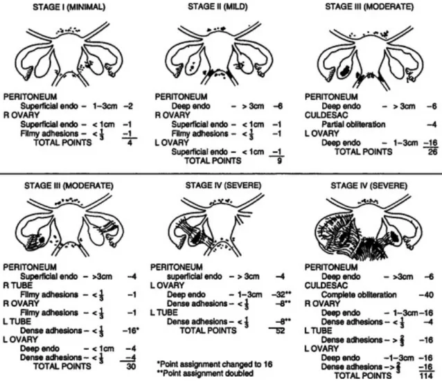
American Fertility Society divides endometriosis in four stages: minimal (stage I), mild (stage II), moderate (stage III) and severe (stage IV).
Therapy
The purpose of endometriosis management is alleviating pain associated to the disease, treating infertility and reducing implants.
The choice of the right treatment is personalized and depends on age, desire for pregnancy, severity of symptoms, position and extent of the lesions.
This can be achieved surgically or medically, although in most women a combination of both treatments is required. Long-term medical treatment is usually needed in most women.
Current medical treatments are based on two mechanisms of action: anti-inflammatory and hormonal. (63) Non-steroidal anti-inflammatory drugs (NSAIDs) are used commonly in women with dysmenorrhea, although there is not enough evidence to admit that they are effective in the treatment of endometriosis related pain, and there is lack of evidence to recommend one NSAID among the others. (64)
Hormonally active drugs act by blocking the ovarian function and creating a more stable hormonal environment. (63) Hormonal drugs currently used for the treatment of pain associated to endometriosis are hormonal contraceptives, progestogens and anti-progestogens, gonadotropin releasing hormone (GnRH) agonists and antagonists, and aromatase inhibitors. (65)
Hormonal contraceptives reduce pain associated to endometriosis, by oral, transdermal, or vaginal administration. (66-68) Progestogens (medroxyprogesterone acetate, oral or depot, dienogest, cyproterone acetate, norethisterone acetate, danazol, levonorgestrel intrauterine device) and anti-progestogens (gestrinone) are also recommended to reduce endometriosis-associated pain. (65, 69-71) GnRH agonists, with and without add-back therapy, are effective in the relief of endometriosis-associated pain, but can be associated with severe side effects. (72) There is insufficient evidence to recommend
pain associated to endometriosis. Clinical decision should take into consideration side effects, patient preferences, efficacy, costs, and availability. (65)
All the drugs with proven efficacy in the treatment of pain associated to endometriosis are hormonal drugs and have a contraceptive action. Endometriosis mainly affects women in their reproductive age; hence, these treatments can be inconvenient in the case of gestational desire. There is a need for new medications, effective in the treatment of pain, with an acceptable side effects profile, suitable for long-term use, with no contraceptive effect, and safe to use in the early pregnancy.
Among new hormonal drugs, the only ones approved for use in the treatment of pain associated to endometriosis are aromatase inhibitors. They seem to be effective in the relief of pain, and their use is recommended for hormonal treatment in women who do not respond to other treatments.
GnRH antagonists are still under study, with currently active Phase III RCT. They are expected to be as effective as GnRH agonists, but with easier administration.
More number of randomized trials should be developed in order to confirm SPRMs’ efficacy and long-term safety. (73)
Nevertheless, surgical therapy has a predominant role, compared to medical therapy, since it allows the definitive removal of lesions and reduces the risk of relapse. Moreover it allows the treatment of associated lesions, such as adhesions, that determines the anatomic alterations that worsen symptoms of pain and infertility.
It is offered when ovarian lesions are of large size or rapidly increasing , when there is a desire for pregnancy and when painful symptoms do not regress after medical therapy. Surgical excision of deep endometriosis is mandatory in presence of symptomatic bowel stenosis, ureteral stenosis with secondary hydronephrosis, and when hormonal treatments fail.
Surgical approach can be both laparoscopic and laparotomic. Thanks to laparoscopy, it is possible to diagnose, stage and treat the disease in one only step, and nowadays represents the gold standard for treatment.
In severe stages (III and IV), the laparotomic surgery allows an accurate research and evaluation of the extent of the disease in the abdominal and pelvic organs.
1.2.2 ENDOMETRIOSIS-‐ASSOCIATED OVARIAN CARCINOMA
The association between endometriosis and development of ovarian carcinoma has been long ago postulated. In 1925 Sampson (7) was first to describe the malignant evolution of endometriosis, proposing the following criteria for diagnosing: 1) coexistence of carcinoma and endometriosis within the same ovary, 2) a similar histological pattern and 3) exclusion of a second malignant tumor elsewhere. In 1953 Scott (74) added one other condition: the presence of benign endometriosis contiguous to the tumor.
Since then, several observations of the coexistence of endometriosis and cancer have been published. One study concerning endometriosis patients from 1969 to 1986 showed an overall relative cancer risk of 1.2 and relative risks for breast cancer, ovarian cancer and non-Hodgkin’s lymphoma to be 1.3, 1.9 and 1.8, respectively. (75, 76) Despite being itself a benign lesion, endometriosis has several features in common with invasive cancer and it has been shown that endometriosis patients are at a higher risk of undergoing malignant transformation and of developing epithelial ovarian carcinoma (EOC), especially when endometriosis is located to the ovaries. (77)
Though this is well established in literature, still there is a lack of understanding of the exact carcinogenic pathways by which endometriosis associated ovarian carcinoma (EAOC) develops.
Several pathways have been studied, such as those involved in oxidative stress, inflammation and hyper-oestrogenism. (78)
Risk factors of developing ovarian cancer for endometriosis patients were identified in: young age at the time of the diagnosis and duration of the disease. Adenomiosys, instead, did not seem to be related to an increased risk of ovarian cancer. Hysterectomy seemed to be a protective factor. (79)
since almost 60-80% of endometriosis associated ovarian cancer develops in presence of histologically demonstrated atypical ovarian endometriosis. (80) (81) Endometriosis is thus suggested to be in fact a premalignant condition. A summary of more recent cohort studies showed a standardized incidence ratio around 2 (82).
“Endometriosis-associated ovarian carcinoma” (EAOC) defines the following situations: 1) presence of ovarian cancer associated to endometriosis in the same ovary, 2) presence of cancer in one ovary and endometriosis in second ovary, 3) presence of ovarian cancer and pelvic endometriosis. Clear cell and endometrioid cancer are most main types. (83, 84)
Histological and clinical significant differences between EAOC patients and non-endometriosis-associated ovarian cancer have been demonstrated (85): the former has endometrioid or clear cell histology, is diagnosed earlier and has a better prognosis than the latter. (86)
However, the question of endometriosis being a prognostic factor for cancer survival is not clear. On one hand, some studies found no definitive association between the presence of endometriosis and survival (87). On the other hand, a large Swedish study found significantly better survival in women with endometriosis than for all malignancies combined. In the cases of breast and ovarian cancer, this survival was even more pronounced (88)
The underlying mechanism is probably related to estrogen stimulation, tissue damage related to repeated heavy menstruation, which result in molecular pathway changes. But no marker has been proved, so far, to be suitable for diagnosis and as a target for treatment.
Therefore, several molecular pathways have been studied in order to recognize the one responsible for malignant transformation of endometriosis.
Many hypothesize that changes in the expression of tumor suppressor genes and oncogenes occurring in the eutopic endometrium might lead to overgrowth of
endometrial foci outside the uterus (89, 90)
Loss of heterozygosity on p16 (Ink4), gut-associated lymphatic tissue (GALT), and p53 occurs are demonstrated in endometriosis. Activation of mutated K-ras gene is another important step in both genesis and progression of ovarian cancer. Alternation of p53 gene caused aberrant regulation of the H-ras protooncogene (91). Using a murine model of mutationally activated K-ras gene, it was demonstrated that these animals develop both endometrial lesions and the ovarian tissue. The following mutation blocking the expression of PTEN caused ovarian cancer (92). K-ras mutation may promote carcinogenesis of endometriosis leading to ovarian clear cell carcinoma (93).
Mutations in ARID1A and PIK3CA were first described in numerous cases of ovarian clear cell carcinoma, but later also in precursor endometriosis tissues. In addition, PTEN-PIK3CA-mTOR pathway was strongly implicated by finding PIK3CA mutation in up to 46% clear cell ovarian cancer (94). PIK3CA mutation is considered to be an early event in the development of endometriosis-associated ovarian clear cell adenocarcinoma.
The sequence of events leading from normal eutopic endometrium to endometriosis and subsequent ovarian cancer still remains hypothetic (95).
Some studies suggested that a histologically normal endometrium may bear genetic damage caused by iron-dependent oxidative stress (96). Some authors suggested that suppression of pre-apoptotic gene Bax and/or up-regulation of anti-apoptotic gene Bcl-2 can be involved in endometriosis and malignancies (97).
Genetic instability might lead to both endometriosis and ovarian cancer. It can include deactivation of some tumor-suppressing genes, changes in activity of enzymes involved in DNA repairs or mutations in genes such as GALT and GSTM. Similarly, mutations in tumor suppressive gene PTEN was often found both in endometric and cancer tissues (98). In addition, c-erbB-2 and p53 genes have been found to associate with
(100, 101).
In addition, inflammatory angiogenesis is implicated both in endometriosis and in EAOCs. Genetic polymorphisms of several genes have been demonstrated, especially involving intercellular cell adhesion molecule-1, interleukin (IL)-6 and IL-10 promoters, tumor necrosis factor-alpha, and nuclear transcription factor-κB (102). There are further studies and tests in literature concerning genetic factors such as loss of heterozygosity, K-ras, P53, and PTEN mutations or hepatocyte nuclear factor-1β (103). Genes involved in endometriosis and endometriosis-associated cancer appear to be those from the 1p36 region (104). The link between the endometriosis-associated ovarian carcinogenesis and oxidative stress-induced has also been tested (105). Biology aspects of ovarian cancer in endometriosis are summarized in the study of Mandai et al (106).
Understanding the mechanisms underlying this complex pathogenesis will help to better assess the risk of endometriosis to develop in ovarian cancer and to improve the therapeutic strategy both in endometriosis and in ovarian cancer.
Figure 9: Proposed development of low-grade endoemtrioid and clear cell carcinoma. Endometrial tissue, by a process of retrograde menstruation, implants on the ovarian surface to form an endometriotic
1.3 ENDOPLASMIC RETICULUM STRESS
The endoplasmic reticulum (ER) is an organelle responsible for homeostasis of intracellular calcium, for membrane lipids biosynthesis, folding and transport of proteins. Protein folding is a mechanism, which is extremely sensitive to cellular environment alterations. Changes in Ca2 + levels, changes in redox state, increased protein synthesis, pathogenic and inflammatory stimuli can all alter protein folding. When cellular environment changes, misfolded proteins accumulate and form aggregates that are toxic to cells: this condition is defined as “ER stress". The Unfolded Protein Response (UPR) is the system by which the ER responds to stress and is activated by the accumulation of misfolded proteins (107). UPR is a
Evolutionary conserved cytoprotective response that allows cells to adapt to ER stress. The activation of the UPR involves transient attenuation of protein synthesis, increased protein trafficking through the ER, increased protein folding and transport, activation of pathways involved in protein degradation (ERAD – ER Associated degradation). If these adaptive mechanisms are unable to solve the problem, the cells go through apoptotic pathway.
The activation of this pathway occurs not only in normal cells, but also in cancer cells. When misfolded proteins accumulate in the ER lumen, two key events are necessary for activation of UPR: first, these misfolded protein aggregates bind and sequester the chaperone BIP (immunoglobulin heavy chain binding protein), also known as Grp78 (107). Secondly, the consequent reduced levels of Grp78 is a clear pathway activation signal that induces the transcription of BIP, as well as other genes coding for chaperones (107, 108). This response takes place via the activation of three transmembrane receptors:
• Pancreatic ER kinase (PERK)
• Activating Transcription Factor 6 (ATF6) • Inositol-Requirng Enzyme 1 (IRE1).
Figure 10: Graphic representation of molecular pathways UPR-associated1.3.1 PERK
PERK (Pancreatic ER Kinase) is a type I transmembrane kinase that becomes activated by dimerization and autophosphorylation, after dissociation with Grp78. The activation of this kinase causes phosphorylation of eIF2α, responsible for protein translation inhibition (109). Activation of PERK-eIF2α reduces the global translation of mRNA, but on the other hand, increases the translation of various mRNA, including ATF4 and ATF5. ATF4 is a transcription factor that induces the expression of genes involved in ER function, in redox reactions, in stress response and protein secretion (110). ATF4 is also associated with CHOP
transcription factor that induce apoptosis ER-stress-mediated, both in vivo and in vitro (111). CHOP transcription is inhibited in the initial phases of the UPR activation, while it is induced when the stress become chronic. Therefore, under stress conditions, PERK is immediately activated and is function is to try to prevent cell damage and promote survival. If stress persists, ATF4 induces transcription of CHOP that induces programmed cell death (112).
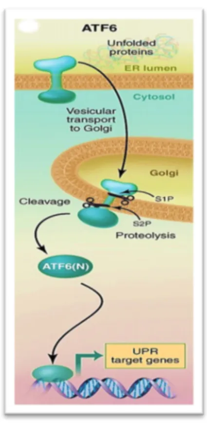
1.3.2 ATF6
The activating transcription factor 6 (ATF6) is a transmembrane glycoprotein, whose luminal domain detects protein misfolding. In mammals is present with two isoforms, α and β, both expressed ubiquitously in all the tissues. The cytoplasmic portion of ATF6 act as transcription factor because it contains a DNA binding domain. Following the Grp78 dissociation, ATF6 moves to the Golgi apparatus, where it is activated by a proteolytic cleavage by two serine proteases, sp1 and sp2 (113). Active ATF6 moves to the nucleus and it induces transcription of genes coding for the chaperones, which have an ER response element (ERSE) in their promoter (114). This determines an ER increased folding capacity, helping to restore initial homeostasis.
Figure 12: Graphic representation of ATF6
1.3.3 IRE1
IRE1 (Inositol Requiring Enzyme-1) is activated after detachment from GRP78, by dimerization and autophosphorylation. The activation of IRE1 is also affected the fluidity of the membrane, which is modified when oxidative stress occurs. XBP1 is the IRE1 substrate. In normal conditions XBP1 levels are very low; they increase when ER occurs due to ATF6 induction. In the presence of its substrate, IRE1 cut by splicing the XBP1 mRNA, forming its active form (XBP1s) that enters the nucleus and determines the activation of target genes (115, 116) that determine an increase degradation of misfolded proteins accumulated in the ER (117). Protein degradation could then represent the third stage of the response UPR, following translation block and increase of chaperone synthesis.
In addition, XBP1 overexpression induces many genes involved in the secretory pathway and determines the expansion of the ER. However IRE1α activation is attenuated in case of chronic stress, through a mechanism not fully established (117, 118). In addition to this mechanism, which promotes cell survival, IRE1 can also have a pro-apoptotic role by JNK74 kinase activation.
1.3.4 GRP78
Under normal conditions, the receptors (PERK, ATF6, IRE1) remain inactive through binding with the chaperone Glucose Regulated Protein 78 (Grp78). In stress conditions, Grp78 dissociates from them and determines the activation, inducing UPR. In the first
Figure 13: Graphic representation of IRE1
Subsequently, the activation of IRE1 appears to have a crucial role in setting up pro-apoptotic signals. If the ER stress persists, PERK and IRE1 pathways converge, enhancing their pro-apoptotic effect, mediated by CHOP and JNK60.
1.3.5 HERPUD 2
The process of destruction of misfolded proteins takes place in the cytosol, through the activation of ER-associated degradation (ERAD), that is responsible for degradation by the proteasome of misfolded protein of the endoplasmic reticulum (ER), that are there retro-traslocated. ERAD machinery is strictly associated with UPR: this mechanism of protein degradation helps cells to adapt to proteotoxic stress in the ER and therefore is critical for the life and death decision in cells under stress.
The product of gene HERPUD 2, protein Herp2, is implicated in several aspects of the endoplasmic reticulum stress response, since it is involved both in UPR and in ERAD, two pathways that are strictly connected one each other.
Herp is regulated by two of the branches of the UPR pathway: PERK mediated and IRE1/ATF6 mediated (119) as previously seen; Herp is involved in the retrotraslocation of proteins that takes place during ERAD formation (120). In those cells in which Herp expression is inhibited, the misfolded proteins are trapped into the ER: this event induces UPR activation. ER stress increases the production of UPR proteins, but also chaperonine and Herp expression (121-123).
Herp proteins family is therefore involved either before and after the UPR pathway. Thus, to test this pathway ,we decided to test the HERP family proteins.
In particular, literature shows that Herp-1 is inducible by ER stress, while Herp-2 is a constitutive expressed protein.
The gene that encodes for the protein is called HERPUD-2.
Recent computational analysis done in other disease models hypothesised that HERPUD expression is regulated by long non-coding RNA (lncRNA): in detail this
1.3.6 UPR IN CANCER
UPR activation has been found in many human diseases and in mouse models. Cell death is the physiological consequence of chronic stress of the ER, and is the key of the pathogenesis of many diseases, including metabolic diseases, inflammation, neurodegenerative diseases and cancer (107).
In particular, as far as it concerns cancer cells, they are characterized by rapid growth, that determines the development of an hypoxic microenvironment and the need of angiogenesis for the insufficient vascularization. Moreover, cancer cells are stimulated to produce large amount of proteins in a short time, therefore they are very dependent on the correct function of UPR system. UPR is also important in tumor pathology: it is indeed necessary for cancer cell growth in a hypoxic environment. The inactivation of PERK pathway, impairs cell survival in hypoxia (125). PERK also promotes the proliferation and growth of cancer cells, limiting the DNA damage from oxidative stress, through ATF4 (126). Thus, the PERK signaling cascade, phosphorylated eIF2α, ATF4 is essential for cancer proliferation.
The activation of UPR in cancer cells is due to intrinsic and extrinsic factors (127). The hyper-activation of oncogenes (such as HRAS, MYC, BRCA1 and PTEN) and the loss of tumor suppressor function, increases the synthesis and translocation of proteins in the endoplasmic reticulum, due to the high metabolic demand during neoplastic transformation (128-130). Consequently UPR pathway is activated to increase the protein folding capacity. In addition, the activation of UPR is required to promote the expansion of the ER for division and transmission to the cells during mitosis (131). In addition, the hostile environment caused by the rapid proliferation of tumor cells, determines a strong endoplasmic reticulum stress of cancer cells, which results in activation of UPR. In solid tumors, there is a hypoxic environment and a lack of nutrients, such as glucose, due to the rapid growth of the mass and thus poor vascularization.
! PERK pathway in carcinogenesis
PERK/phosphorylated eIFα/ATF4 pathway plays a key role in cancer cell survival. The inactivation of PERK, alter the possibility of cell survival hypoxic environment (125). PERK also promotes cell proliferation by limiting, through ATF4, DNA oxidative damage. The function of CHOP in oncogenesis is to date unknown, however it is repeatedly confirmed that the induction of CHOP in response to a prolonged ER stress, causes pre-malignant cell death, and prevent neoplastic progression (127). CHOP deletion increases the incidence of malignant lung tumors in mouse models KRAS-induced, suggesting an oncosuppressive role of CHOP (132).
➢ ATF6 pathway in carcinogenesis
The main ATF6 target is Grp78/BIP activation, which plays an important role in protein folding and assembly, in regulating Ca2 + levels in the ER and controlling the activation of transmembrane sensors of stress (127). It has been shown that Grp78/BIP activation in cancer cells protects them from apoptosis and from immune response (133). By contrast Grp78/BIP suppression inhibits tumor cell growth, metastases progression and development, both in vivo and in vitro (134, 135). Furthermore Grp78/BIP may be considered a marker of cell malignancy: in normal conditions is localized exclusively in ER, while in malignant cells, where it is hyper-expressed, can also be detected on the cell surface. In various tumor sites, such as lung, bladder, stomach and breast, overexpression of Grp78/BIP confers resistance to chemotherapeutic agents, as well as its suppression sensitizes cancer cells to pharmacological treatment (136).
➢ IRE1α pathway in carcinogenesis
IRE1α - XBP1 pathway is also important for cell survival and tumor growth in hypoxic environment, because it induces the transcription of proangiogenic factors, such as vascular and endothelial growth factors (127). In a glioma mouse model, IRE1α inhibition reduces of tumor growth, angiogenesis and blood perfusion (138). XBP1
the other hand, there are studies that demonstrate an oncosuppressive role of IREα/XBP1 pathway: in many human tumors was found IRE1α mutations (140), some of which result in a loss of kinase and endoribonucleasic function (141). In addition, the loss of XBP1 function promotes oncogenesis (142).
! GRP78 in carcinogenesis
In several tumors, a higher expression of GRP78 has been shown, such as hepatocellular carcinoma, gliomas, prostate and gastric cancer. (143-145, 135)
It allows tumor progression (146) (147), and determines therapy resistance (148). Since it is expressed in the cellular surface, it can suitably been considered as a target for cancer therapy, offering the possibility to use specific antibodies, as it has been studied in some tumor models. (149) (150)
2. MATERIALS AND METHODS
2.1 STUDY DESIGN
The study was performed using different histological samples: endometrioid carcinoma of the ovary, healthy ovary, endometriosis cysts, eutopic endometrium from endometriosis patients and healthy endometrium. The ovarian tissues derived from histological samples prepared for diagnostic purposes: the study analyzed the endometrioid carcinoma of the ovary as well as the healthy contralateral ovary from the same patient as control. Two kinds of samples were collected from patients with endometriosis: one from the eutopic endometrial tissue and the other from ectopic endometrial tissue, also called the endometriosis cysts. The control group consists of endometrial samples of patients subjected to hysteroscopy for diagnostic investigations related to other pathologies (polyps, fibroids).
From all the samples RNA was extracted and cDNA synthesis was performed by reverse transcription. cDNA was used for quantitative gene expression assays, made by Real Time PCR, analyzing genes belonging to the UPR pathway:
• ATF6 (Activating Transcription Factor 6) • GRP78 (Glucose Regulated Protein 78) • CHOP (DNA damage-inducible transcript 3) • XBP1s (spliced X-box binding protein 1)
• HERPUD1 (Homocysteine Inducible ER Protein With Ubiquitin Like Domain 1)
• HERPUD 2 (HERPUD family member 2)
2.2 SUBJECT RECRUITMENT CRITERIA AND COLLECTION OF SAMPLES Subjects participating at the study were recruited at the Obstetrics and Gynaecology Department of the Polytechnic University of Marche, Maternal and Child Hospital
Samples were divided into three groups: endometriosis patients (n = 6), healthy patients (n = 6) patients with ovarian endometrioid carcinoma (n = 6).
For each group inclusion, exclusion criteria and sampling method were summarised in Table 3. Endometriosis patients Healthy Patients (endometrial tissue) Patients with endometrioid carcinoma of the ovary
Inclusion criteria
Patients undergoing laparoscopic surgery for endometrial cysts removal with moderate/severe endometriosis diagnosis. Non pregnancy at the time of surgery
Patients in childbearing age undergoing laparoscopy (for infertility or ovarian cysts different from
endometriosis), where the presence of endometriosis is ruled out by laparoscopic inspection and histology
Patients undergoing surgical removal of bilateral ovarian with histological diagnosis of unilateral endometrioid carcinoma of the ovary . Not pregnancy at the time of surgery
Exclusion criteria
Hormonal therapy within 3 months before surgery Taking medications Patients suffering from hypertension, diabetes mellitus, kidney disease, proteinuria,
cardiovascular, hepatic, endocrine (thyroid, PRL, PCOS) and metabolic disorders
Advanced age Smoking and alcohol
Menopause Taking medications Patients suffering from hypertension , diabetes mellitus, kidney disease , proteinuria , cardiovascular, hepatic, endocrine (thyroid , PRL , PCOS) and metabolic disorders.
Presence of infection Diagnosis of adenomyosis has been through sonografy ruled out
Tumor presence in the contralateral ovary.
Sampling method
Collection of a cysts portion after laparoscopic removal, after pelvis peritoneal surfaces inspection to check the presence of any peritoneal implants. Tissue stored at -80 ° C.
Collection of scrap of endometrial tissue after resectoscopic
hysteroscopy or after biopsy using Novak cannula. Tissue stocked in nitrogen and immediately stored at -80° C.
Collection of scrap of endometrial tissue after resectoscopic hysteroscopy or after biopsy using Novak cannula. Endometrial tissue sample has been taken in the proliferative phase. Tissue stocked in nitrogen and immediately stored at -80° C.
Histological preparations from patients with endometroid ovarian carcinoma were selected and provided by the Section of Anatomic Pathology (Department of Biomedical Sciences and Public Health) of the Polytechnic University of Marche (Ancona). 8-10 10 µM thick sections from paraffin-embedded tissue, prepared for diagnostic purposes were used for the study.
From each patient it was collected two types of tissue: samples from endometrioid carcinoma of the ovary and healthy contralateral ovarian
2.3 ENDOMETRIAL FROZEN SAMPLE PREPARATION
Total RNA extractionPurification of total RNA from cells was performed in the endometrial tissue (≤30mg) using SV Total RNA Isolation System kit (Promega), using diethyl pyrocarbonate (DEPC) treated equipment.
In order to isolate RNA, 4 steps are essentials: tissue lysis, nucleoprotein complexes denaturation, endogene ribonucleasis inactivation (RNAse) and DNA and protein contaminants removal.
The SV Total RNA Isolation System combine the disruptive and protective properties of guanidine thiocynate (GTC) and β-mercaptoethanol to inactivate the ribonuclease present in cell extracts. GTC, in association with SDS, acts to disrupt nucleoprotein complexes, allowing the RNA to be released into solution and isolated free of protein. Dilution of cell extracts in the presence of high concentrations of GTC cause selective precipitation of cellular proteins to occur, while RNA remains in solution. RNA is then bound to the silica surface of the glass fibers found in the Spin Basket. DNAse treatment digest contaminating genomic DNA. The total RNA is finally eluted with nuclease-free water.
Quantity and purity of the total RNA obtained from the purification was tested reading the absorbance at 260 nm and 260/230 and 260/280 ratio, using Nanodrop instrument. We can summarize the protocol as it follows: the tissue, 30 mg, is lysed, adding to the sample 175 µl RNA lysis buffer and 350 µl di RNA dilution Buffer, and incubated at 70°C for 30 minutes. It is centrifuged for 10 minutes at 12.000-14.000 xg. In order to wash the product of lysis, 200 µl of ethanol 95% are added and are transferred in spin column assembly. One other centrifuge at 12.000-14.000 xg for 1 minute is done. Then, the liquid is eliminated from the spin basket and 600 µl of RNA wash solution are added, with another centrifuge at 12.000-14.000 xg for 1 minute. 50 µl of DNAse buffer, composed by 40µl yellow core buffer, 5µl 0.09M MnCl2 and 5µl di DNase I enzyme, are added to the membrane of the spin basket and they are incubated for 15
12.000-14.000 xg for 1 minute. 600 µl of RNA Wash solution (with ethanol) are added and centrifuged at 12.000-14.000 xg for 1 minute. This is repeated once again, adding 250 µl RNA Wash solution (with ethanol), and centrifuged at 12.000-14.000 xg for 1 minute. Finally, 100 µl Water of Nuclease-Free, are added to every column ad centrifuged at 12.000-14.000 xg for 1 minute. The flowthrough, that contains purified RNA, is taken. RNA is conserved at -70°C.
In every phase of RNA purification protocol, test tubes and pipette tips were used after treatment with dietil-pirocarbonate (DEPC). This treatment is necessary for RNAse inactivation.
C-DNA Synthesis
Retro-transcription is the mechanism through which cDNA is produced from RNA. This reaction is made using reverse transcriptase, an enzyme that is able to generate complementary DNA (cDNA) from an RNA template. DNA polimerases allow the progress of polymerase reaction of a polinucleotidic chain, complementary to a single DNA strand, used as a template. The enzyme catalyzes the formation of a phosphodiester bond between 3’OH group of deoxyribose of the last nucleotide to the phosphate at 5’ " 3’. For the reverse transcription reaction, several kinds of primer can be used: oligo(dT) primers, random primers or gene-specific primers. In this case, random primers were used. These primers are not selective and pair to every RNA molecule present, included the ones that do not contain poly(A) tail, and can be used for long inverse transcripts.
RNA was quantified and retro-transcribed to c-DNA using ImProm-II ™ Reverse Transcription System kit (Promega) using 2 µg RNA and random hexamers primes (P(n)6 (Promega). The optimized reaction buffer and the reverse transcriptase provided in the kit enable cDNA synthesis Volumes of the reaction used is 20µl. The heteroduplex cDNA/RNA formed was then directly amplified by PCR. Briefly 2µl of random primers were added to the 2µg of total RNA purified as a total volume of 12,2 µl of solution. The samples were then heated at 70°C for 5 min and then let cool on ice for another 5 min. Then the reaction mix with 5 µl M-MLV 5X BUFFER, 1 µl M- MLV RT (stock 200 units/µL), 0,6 µl Recombinant RNasin Ribonuclease Inhibitor (stock 40 units/µL), 1,25 10 mM dNTP Mix and water nuclease-free was added to the samples and incubated 1 hour at 37°C. The c-DNA obtained was stored at -20°C for further analysis.