205 ACTA oTorhinolAryngologiCA iTAliCA 2014;34:205-208
Rhinology
Endoscopic ultrasonic curette-assisted removal
of frontal osteomas
Curette per l’osso ad ultrasuoni per la rimozione degli osteomi del frontale
A. Bolzoni VillAret, A. SchreiBer, i. eSpoSito, p. nicolAiDepartment of otorhinolaryngology, University of Brescia, italy SummAry
indications for endoscopic resection of fronto-ethmoidal osteomas have been progressively expanded thanks to optimization of surgical expo-sure and the development of dedicated instruments. Curved cutting drills are still suboptimal to treat hard osseous neoplasms of the frontal si-nus. We present two patients affected by frontal osteoma treated with an endoscopic procedure using an ultrasonic bone curette. The ultrasonic bone curette may be considered an effective tool to reduce soft tissue manipulation, optimize surgical time and accelerate the healing process. however, the technique requires significant shape innovations to reach the lateral recesses and to manage pure intrasinusal lesions.
Key WordS: Frontal sinus • Sinunasal osteomas • Ultrasonic curette
riASSunTo
Le indicazioni alla chirurgia endoscopica nel trattamento degli osteomi fronto-etmoidali si sono progressivamente estese grazie all’otti-mizzazione dell’esposizione chirurgica ed allo sviluppo di una strumentazione dedicata. Le frese curve sono ancora subottimali nel tratta-mento di lesioni ossee eburnee del seno frontale. Presentiamo due pazienti affetti da osteoma frontale trattati con procedura endoscopica utilizzando la curette per osso ad ultrasuoni. La curette ad ultrasuoni può essere considerato un efficace strumento chirurgico per ridurre la manipolazione dei tessuti molli e per ottimizzare i tempi chirurgici e del processo di guarigione. Tuttavia è necessario migliorare la forma dello strumento per premettere di raggiungere i recessi più laterali e gestire lesioni localizzate interamente nel seno frontale.
PArole ChiAve: Seno frontale • Osteomi nasosinusali • Curette ad ultrasuoni
Acta Otorhinolaryngol Ital 2014;34:205-208
Introduction
Osteomas of the sinonasal complex are benign bony tu-mours often incidentally discovered in asymptomatic patients during head imaging or neuroimaging. Their lo-cation and dimension may cause recurrent sinusitis,
head-ache or orbital complaints 1-5.
Since the early 1990s, the indications for endoscopic resec-tion of fronto-ethmoidal osteomas have been progressively expanded, with a constant refinement in the definition of
contraindications and limits 6-14. The evolution of the
indi-cations 15-17 clearly reflects the optimization of the surgical
exposure (i.e. use of the contralateral nostril in a Draf III procedure) and the development of dedicated instruments. Curved drills are usually adopted in this surgical setting, although all are limited by low speed and efficacy. Straight and 20° high-speed drills can be used, even though the need for aspiration and the bone dust produced may limit visualization and increase the tediousness and length of the procedure. Furthermore, minimizing mucosal trauma is es-sential to facilitate healing, prevent crusting and infection of denuded bone, reduce scar tissue formation and avoid
stenosis of the frontal recess. Since 2008, during extended transnasal approaches to the skull base we have combined high-speed microdrill and sonic bone emulsification in selected cases. The ultrasonic bone curette (Sonopet
Ul-trasonic Aspirator, Stryker®, Kalamazoo, MI, USA) was
tested in two frontal osteomas to evaluate its cost efficacy and possible advantages in this specific setting. We selected small lesions located in the fronto-ethmoidal recess with-out complete filling of the frontal sinus. Preliminary evalu-ation of the device focused on the following endpoints: the traumatic impact of the device on surrounding mucosa, the balance among emulsification, irrigation, suction and endo-scopic view, the lack of good visualization of the tip of the instrument in the lateral aspect of frontal sinus, and finally the speed and effectiveness of the healing process.
Materials and methods
Case 1
A 50-year-old woman was seen for symptoms (nasal obstruction, rhinorrhoea, headache) related to chronic
A. Bolzoni Villaret et al.
206
rhinosinusitis. She had previously undergone several en-doscopic procedures at other institutions, with minimal improvement of symptoms. CT scan showed a radiodense mass suggestive for osteoma, occupying the right frontal recess with obstruction of the frontal drainage pathway (Fig. 2). Dishomogeneous ossification at the superior as-pect of the lesion and its ground-glass pattern suggested a reduced consistence in its upper part. She underwent endoscopic transnasal removal (Fig. 3A); the operative
time was about 2 hours and she was discharged on the 2nd
postoperative day. Follow-up nasal endoscopy at 1 and 6 months confirmed adequate and quick healing with mini-mal scar formation (Fig. 3B). She was free from symp-toms after 26 months of follow-up.
Case 2
This patient was a 40-year-old male with a clinical his-tory of recurrent frontal sinusitis resistant to conventional conservative treatment. CT scan revealed a hyperdense lesion occupying the right frontal sinus abutting into the frontal recess (Fig. 4). He underwent endoscopic trans-nasal removal (Fig. 5); the operative time was about 2
hours and he was discharged on the 2nd postoperative day.
Follow-up nasal endoscopy at 1 and 6 months confirmed adequate healing. He was free from symp-toms at 24 months after surgical procedure.
In both cases, endoscopic evalua-tion 1 month after surgery showed complete healing without signifi-cant oedema or scar deposition.
Surgical procedure
Both patients were positioned supine, with hyperextension of the head (Fig. 1). After topical decongestion and injection of the upper part of the uncinate process with adrenaline and mepivacaine, endoscopic examination directly demonstrated the inferior aspect of the lesion in the first patient. The extent of the surgical ap-proach has been tailored on a case specific basis (i.e. anterior ethmoidectomy, uncinectomy, middle turbinectomy, opening of an antero-superior septal win-dow). In case 1, once the inferior aspect of the osteoma was identi-fied to be covered by scarred mu-cosa, the ultrasonic bone curette
was introduced through the right nostril, running it over the endoscope; after blunt dissection of surrounding mu-cosa, the tip of the device was applied directly on the me-dial aspect of the lesion which was emulsified in 45 min under continuous close-up view. The residual lateral shell of bone was dissected and removed leaving the lamina papyracea intact.
In case 2, a type IIb Draf sinusotomy allowed exposure of the boundaries of the lesion which was gradually reduced
Fig. 1. Schematic drawing shows the position of the device running over the angled endoscope. Head extension improves the working angle.
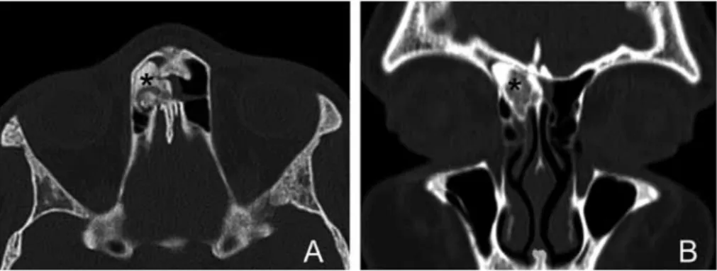
Fig. 2. Preoperative CT scan of case 1 shows an osteoma (asterisk) occluding the right frontal recess. Note the remodelled lamina papyracea (arrows) and the lesion attachment over the posteroinferior aspect of the frontal sinus. Axial (A) and coronal (B) plane.
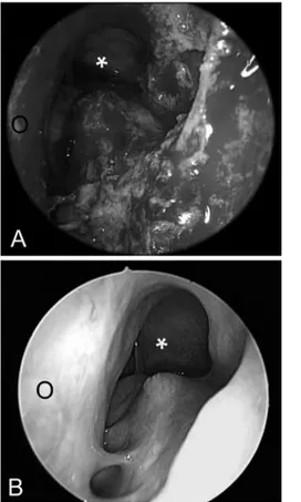
Fig. 3. Intraoperative and postoperative endoscopic views of case 1. A: after lesion removal the mucosa of the lamina papyracea is partially maintained. B: Post-operative examination at 6 months with angled tel-escope highlights complete healing with no stenosis of the frontal recess.
Endoscopic ultrasonic curette-assisted removal of frontal osteomas
207
with the use of the ultrasonic cu-rette in about 1 hour. The Spetzler
microclaw tip (Stryker®,
Kalama-zoo, MI, USA) was adopted in both cases allowing lesion emul-sification with only one side of the tip, maximally preserving the surrounding mucosa (Fig. 5).
Discussion
Even if an external approach to the frontal sinus still has a role in the treatment of osteomas, mul-tiple series published in the last 15 years support the efficacy and safety of an endoscopic approach through a transnasal corridor in properly selected
fronto-ethmoi-dal lesions 17. Moreover, the
in-creasing surgical experience and development of dedicated instru-mentation allow resection of se-lected frontal lesions even when
extended over the orbital roof 17.
The use of ultrasound curette in endoscopic transnasal procedures has been reported in the
litera-ture for inferior turbinoplasty 18,
sculpting of the nasal dorsum 19,
lateral orbital decompression 20
and removal of a fronto-ethmoidal osteoma 21. This
instru-ment delivers, in one hand piece, tissue fraginstru-mentation by rapid longitudinal motion, irrigation through coaxial flows around the tip to suspend fragmented tissue and cool the tip and aspiration with removal of fluid and fragmented tissue with a cannulated tip and suction.
In our Department, between 1996 and 2011, 20 patients un-derwent endoscopic transnasal removal of a frontal osteo-ma (17 frontal, 3 fronto-ethmoidal) with a mean operating time of 4.8 hours (1-12 hours; unpublished data). The het-erogeneity of cases and progressive evolution of the learn-ing curve are both factors influenclearn-ing the surgical time. We endoscopically approached these tumours with a type II or III Draf sinusotomy depending on the site and size of the le-sion. Standard endoscopic instruments were used to expose the caudal portion of the lesion; subsequently cavitation of the osteoma was performed using curve cutting drills to mobilize the peripheral fragments and minimize damage to surrounding tissues. In our preliminary experience, the ultrasonic bone curette does not increase the operating time in properly selected patients. Furthermore, the possibility to limit the working surface on one side of the device mini-mized mucosal damage, with subsequent easier care in the early postoperative course with almost no need to remove
Fig. 4. Preoperative CT scan of case 2 shows an osteoma (asterisk) located in the right frontal recess and inserted at the anterolateral aspect of the right cribriform plate. Axial (A) and coronal (B) plane.
Fig. 5. Intraoperative images of case 2. After lesion cavitation and dissection from surrounding structures, the residual shell of bone is further reduced with a four-hand technique (white asterisk indicates the frontal si-nus). B: close-up view. Note the mucosa surrounding the lesion (black asterisk), which is spared by the device.
granulation tissue or fibrin debris. During the procedure we never experienced slippage of the instrument, and its use was easy and straightforward compared to a traditional mi-crodrill since this device integrates irrigation and aspiration and requires no pressure over the working surface. Quick healing was documented during follow up with minimal scar formation (Fig. 3B). Further experience will be es-sential to confirm the low morbidity, efficacy, speed and cost-effectiveness of this device. However, two main fac-tors may contribute to a favourable application in transnasal approaches to the frontal sinus: the line of sight is improved since this device provides a bone emulsification-irrigation-suction mechanism in a single hand, and the oscillating energy of the working area is limited to a single side of the tip to prevent slippage. Moreover, minimal bone dust production and its constant aspiration consents continuous clear endoscopic view of the surgical field and the presence of dedicated tips improve the adaptability of the device to the working surface.
In contrast, the absence of curved tips designed specifi-cally for frontal sinus endoscopic surgery limits its use to properly selected cases, and the costs of the tips are not negligible, even if the main unit can be shared among dif-ferent departments of the same hospital.
A. Bolzoni Villaret et al.
208
Conclusions
Endoscopic surgery has a predominant role in the man-agement of benign tumours of the sinonasal tract. De-spite advances in image definition and instrumentation, visualization may still represent an issue, mainly during bone drilling in narrow spaces. State-of-the art curved cutting drills are still suboptimal to treat hard osseous neoplasms of the frontal sinus. Therefore, during endo-scopic transnasal removal of frontal sinus osteoma, an ultrasound bone curette can be considered an effective tool to reduce soft tissue manipulation, optimize surgi-cal time and speed the healing process. Furthermore, despite its straight configuration, this low profile device may be amneable for further developments and applica-tions far lateral along the coronal plane in the frontal sinus, but which will require significant shape innova-tions to reach the lateral recesses and manage pure in-trasinusal lesions.
References
1 Pagella F. Transnasal endoscopic approach to symptomatic sinonasal osteomas. Am J Rhinol Allergy 2012;26:335-9. 2 Viswanatha B. Maxillary sinus osteoma: two cases and review
of the literature. Acta Otorhinolaryngol Ital 2012;32:202-5. 3 Ciorba A, Aimoni C, Bianchini C, et al. Bilateral osseous
stenosis of the internal auditory canal: case report. Acta Otorhinolaryngol Ital 2011;31:177-80.
4 Dispenza C, Martines F, Dispenza F, et al. Frontal sinus os-teoma complicated by palpebral abscess: case report. Acta Otorhinolaryngol Ital 2004;24:357-60.
5 Bertoletti F, Capolunghi B, Bertolini G, et al. Giant osteoid osteoma of ethmoid sinus: role of functional endoscopic si-nus surgery. Acta Otorhinolaryngol Ital 2004;24:297-301. 6 Draf W, Weber R, Keerl R, et al. Current aspects of
fron-tal sinus surgery. Part I: Endonasal fronfron-tal sinus drainage in inflammatory diseases of the paranasal sinuses. HNO 1995;43:352-7.
7 Schick B, Steigerwald C, el Rahman el Tahan A, et al. The role of endonasal surgery in the management of fron-toethmoidal osteomas. Rhinology 2001;39:66-70.
8 Chiu AG, Schipor I, Cohen NA, et al. Surgical decisions
in the management of frontal sinus osteomas. Am J Rhinol 2005;19:191-7.
9 Castelnuovo P, Giovannetti F, Bignami M, et al. Open sur-gery versus endoscopic sursur-gery in benign neoplasm involv-ing the frontal sinus. J Craniofac Surg 2009;20:180-3. 10 Castelnuovo P, Valentini V, Giovannetti F, et al. Osteomas
of the maxillofacial district: endoscopic surgery versus open surgery. J Craniofac Surg 2008;19:1446-52.
11 Bignami M, Dallan I, Terranova P, et al. Frontal sinus osteo-mas: the window of endonasal endoscopic approach. Rhinol-ogy 2007;45:315-20.
12 Dubin MG, Kuhn FA. Preservation of natural frontal sinus outflow in the management of frontal sinus osteomas. Otolar-yngol Head Neck Surg 2006;134:18-24.
13 Seiberling K, Floreani S, Robinson S, et al. Endoscopic man-agement of frontal sinus osteomas revisited. Am J Rhinol Al-lergy 2009;23:331-6.
14 Ledderose GJ, Betz CS, Stelter K, et al. Surgical manage-ment of osteomas of the frontal recess and sinus: extending the limits of the endoscopic approach. Eur Arch Otorhi-nolaryngol 2011;268:525-32.
15 Georgalas C, Goudakos J, Fokkens WJ. Osteoma of the skull base and sinuses. Otolaryngol Clin North Am 2011;44:875-90. 16 Gil-Carcedo LM, Gil-Carcedo ES, Vallejo LA, et al. Frontal
osteomas: standardising therapeutic indications. J Laryngol Otol 2011;125:1020-7.
17 Turri-Zanoni M, Dallan I, Terranova P, et al. Frontoethmoi-dal and intraorbital osteomas: exploring the limits of the endoscopic approach. Arch Otolaryngol Head Neck Surg 2012;138:498-504.
18 Greywoode JD, Van Abel K, Pribitkin EA. Ultrasonic bone aspirator turbinoplasty: a novel approach for man-agement of inferior turbinate hypertrophy. Laryngoscope 2010;120(Suppl 4):S239.
19 Pribitkin EA, Lavasani LS, Shindle C, et al. Sonic rhino-plasty: sculpting the nasal dorsum with the ultrasonic bone aspirator. Laryngoscope 2010;120:1504-7.
20 Cho RI, Choe CH, Elner VM. Ultrasonic bone removal ver-sus high-speed burring for lateral orbital decompression: comparison of surgical outcomes for the treatment of thyroid eye disease. Ophthal Plast Reconstr Surg 2010;26:83-7. 21 Pagella F, Giourgos G, Matti E. Removal of a
fronto-ethmoi-dal osteoma using the sonopet omni ultrasonic bone curette: first impressions. Laryngoscope 2008;118:307-9.
Address for correspondence: Andrea Bolzoni Villaret, De-partment of Otorhinolaryngology, University of Brescia, Italy. Tel. +39 030 3995319. E-mail: [email protected]