Received: Ocrtober 17, 2018.
Accepted: April 3, 2019.
Pre-published: April 4, 2019.
©2019 Ferrata Storti Foundation
Material published in Haematologica is covered by copyright. All rights are reserved to the Ferrata Storti Foundation. Use of published material is allowed under the following terms and conditions:
https://creativecommons.org/licenses/by-nc/4.0/legalcode. Copies of published material are allowed for personal or inter-nal use. Sharing published material for non-commercial pur-poses is subject to the following conditions:
https://creativecommons.org/licenses/by-nc/4.0/legalcode, sect. 3. Reproducing and sharing published material for com-mercial purposes is not allowed without permission in writing from the publisher.
Correspondence:
PAOLO LUNGHI
[email protected]
Haematologica
2019
Volume 104(12):2465-2481
doi:10.3324/haematol.2018.208280
Check the online version for the most updated
information on this article, online supplements,
and information on authorship & disclosures:
www.haematologica.org/content/104/12/2465
Ferrata Storti Foundation
C
onsidering that Aurora kinase inhibitors are currently under clinical
investigation in hematologic cancers, the identification of molecular
events that limit the response to such agents is essential for
enhanc-ing clinical outcomes. Here, we discover a NF-
κB-inducing kinase
(NIK)-c-Abl-STAT3 signaling-centered feedback loop that restrains the efficacy of
Aurora inhibitors in multiple myeloma. Mechanistically, we demonstrate
that Aurora inhibition promotes NIK protein stabilization via
downregula-tion of its negative regulator TRAF2. Accumulated NIK converts c-Abl
tyro-sine kinase from a nuclear proapoptotic into a cytoplasmic antiapoptotic
effector by inducing its phosphorylation at Thr735, Tyr245 and Tyr412
residues, and, by entering into a trimeric complex formation with c-Abl and
STAT3, increases both the transcriptional activity of STAT3 and expression
of the antiapoptotic STAT3 target genes PIM1 and PIM2. This consequently
promotes cell survival and limits the response to Aurora inhibition. The
functional disruption of any of the components of the trimer
NIK-c-Abl-STAT3 or the PIM survival kinases consistently enhances the
responsive-ness of myeloma cells to Aurora inhibitors. Importantly, concurrent
inhibi-tion of NIK or c-Abl disrupts Aurora inhibitor-induced feedback activainhibi-tion
of STAT3 and sensitizes myeloma cells to Aurora inhibitors, implicating a
combined inhibition of Aurora and NIK or c-Abl kinases as potential
thera-pies for multiple myeloma. Accordingly, pharmacological inhibition of
c-Abl together with Aurora resulted in substantial cell death and tumor
regression in vivo. The findings reveal an important functional interaction
between NIK, Abl and Aurora kinases, and identify the NIK, c-Abl and PIM
survival kinases as potential pharmacological targets for improving the
effi-cacy of Aurora inhibitors in myeloma.
Introduction
Despite encouraging advances in therapy, multiple myeloma (MM) remains an
incurable disease due to complex genomic alterations, lower sensitivity to
chemotherapy of MM cells in the bone marrow microenvironment, and the
emer-gence of drug resistance.
1Recent genetic evidence has established a pathogenetic role for NF-
κB signaling
in MM.
2-4In particular, at various frequencies, MM cells harbor gain-of-function
mutations as well as loss-of-function mutations in genes encoding components of
the classical and the alternative NF-
κB pathways.
2-4Among these, mutations in the
genes encoding NF-
κB-inducing kinase (NIK) or its negative regulators TRAF2,
Functional interplay between NF-
κB-inducing
kinase and c-Abl kinases limits response to
Aurora inhibitors in multiple myeloma
Laura Mazzera,
1,2Manuela Abeltino,
1Guerino Lombardi,
2Anna Maria Cantoni,
3Roberto Ria,
4Micaela Ricca,
2Ilaria Saltarella,
4Valeria Naponelli,
1Federica Maria Angela Rizzi,
1,5Roberto Perris,
5,6Attilio Corradi,
3Angelo Vacca,
4Antonio Bonati
1,5and Paolo Lunghi
5,61
Department of Medicine and Surgery, University of Parma, Parma;
2Istituto Zooprofilattico
Sperimentale della Lombardia e dell’Emilia Romagna “Bruno Ubertini,” Brescia;
3
Department of Veterinary Science, University of Parma, Parma;
4Department of
Biomedical Sciences and Human Oncology, Section of Internal Medicine and Clinical
Oncology, University of Bari "Aldo Moro" Medical School, Bari;
5Center for Molecular and
Translational Oncology, University of Parma, Parma and
6Department of Chemistry, Life
Sciences and Environmental Sustainability, University of Parma, Parma, Italy
ABSTRACT
TRAF3, cIAP1, and cIAP2 lead to increased stability of NIK
and subsequent aberrant activation of the non-canonical
and canonical NF-κB pathways.
2-7In addition to regulating NF-
κB pathways, the NIK
sig-naling pathway has been demonstrated to crosstalk with
and activate other critical cancer-associated pathways
including the MAPK-ERK
8,9and JAK/STAT3.
10Moreover,
these pathways are highly interconnected at many levels,
and have been demonstrated to be often persistently and
simultaneously activated in many human cancers,
includ-ing myeloma.
11,12NF-
κB and STAT3 signaling can also be regulated by
c-Abl,
13,14a ubiquitously expressed non-receptor tyrosine
kinase that plays an important role in regulating critical
cellular processes, including proliferation, survival,
apop-tosis, differentiation, invasion, adhesion, migration, and
stress responses.
15,16The tyrosine kinase c-Abl has been reported to have
opposing and antagonistic functions in the regulation of
cell proliferation and survival depending on its subcellular
localization, phosphorylation state, and cellular context.
17In particular, activation of cytoplasmic c-Abl in response
to growth factors, cytokines and Src tyrosine kinases, can
promote mitogenic and survival signals,
17,18whereas
acti-vation of nuclear c-Abl in response to DNA damage can
negatively regulate cell proliferation and mediate
apopto-sis/necrosis.
15The subcellular localization of c-Abl is critically
con-trolled by binding with the 14-3-3 protein, which requires
the phosphorylation of c-Abl at an amino acid residue
Thr735.
19Wild-type c-Abl is localized both in the nucleus and
cytoplasm, in contrast to its oncogenic forms that are
localized exclusively in the cytoplasm. Oncogenic forms
of c-Abl exhibit enhanced kinase and transforming
activi-ties and play a critical role in the pathogenesis of chronic
and acute leukemias.
20MM cells display high levels of
nuclear c-Abl in response to ongoing DNA damage and
genomic instability.
21,22However, most of its nuclear tumor
suppressor functions are compromised because of the
dis-ruption of the ABL-YAP1-p73 axis.
21In MM and other hematologic and solid malignancies,
genomic instability, centrosome amplification and
aneu-ploidy have been associated with the overexpression of
Aurora kinases, a family of serine/threonine kinases that
play essential and distinct roles in mitosis.
23In addition to their mitosis specific substrates, Aurora
kinases have also been found to functionally interact with
proteins involved in critical cancer-associated pathways
including NF-
κB, STAT3 and DNA-damage response
path-ways.
24-27On the basis of these findings, Aurora kinases
have been considered as therapeutic targets for cancer and
Aurora kinases inhibitors (AKI) have been extensively
explored. These have shown encouraging pre-clinical and
early clinical activity in different cancer types either alone
or in combination with other agents.
25,28-31Unfortunately,
AKI have not proved to be sufficiently effective and/or
caused too many adverse side-effects in myeloma
patients, both when used as monotherapy or in
combina-tion with other targeted therapy agents.
29-31The poor
effi-cacy of AKI therapies in MM may, in part, be related to
the still undetermined drug-induced compensatory
mech-anisms occurring in both the MM cells and their
microen-vironment, and, consequently, to the lack of appropriate
mechanism-based combination therapies.
In this study, we demonstrate that pan-AKI generate
pro-survival signals in MM cells by inducing the
expres-sion/activation of the pro-survival serine/threonine
kinas-es PIM1 and PIM2
32through a NIK/c-Abl-mediated
activa-tion of STAT3, a cascade of molecular events that
conse-quently limit the response to pan-AKI. Our findings reveal
a novel functional interplay between NIK and c-Abl with
implications for treatment of MM. They therefore provide
the rationale for targeting c-Abl as a novel strategy to
enhance activity of Pan-AKI.
Methods
Reagents
Pan-AKI MK-0457 (Merck & Co. Rahway, NJ, USA); pan-AKI
PHA-680632 (Pfizer/Nerviano, Italy); pan-AKI AMG-900
(Cayman Chemical Company; Ann Arbor, MI, USA); NIK
inhibitor
isoquinoline-1,3(2H,4H)-dione
(Santa
Cruz
Biotechnology, Santa Cruz, CA, USA); proteasome inhibitor
bortezomib (PS-341) from Janssen-Cilag (Milan, Italy); c-Abl
inhibitors imatinib mesylate and nilotinib (Novartis
Pharmaceuticals, Basel, Switzerland). STAT3 inhibitor Stattic
(6-Nitrobenzo [b]thiophene-1,1-dioxide) and pan-PIM kinase
inhibitor SMI-4A
(5Z)-5-[[3-(Trifluoromethyl)-phenyl]-methyl-ene]-2,4-thiazolidinedione (Sigma-Aldrich, St. Louis, MO, USA).
Cell cultures
Cell cultures were: human myeloma cell lines (HMCL) OPM-2,
U266, RPMI-8226 and JJN3 (DSMZ, Braunschweig, Germany);
multidrug-resistant RPMI-8226/R5 HMCL was established as
pre-viously described;
33human bone marrow-derived stromal cell line
HS-5 (ATCC, Manassas, VA, USA). Primary MM cells from MM
patients and peripheral blood mononuclear cells (PBMC) of
healthy subjects were isolated and treated as described in the
Online Supplementary Methods. The study was approved by the
Ethics Committee of the University of Bari “Aldo Moro”
(identifi-cation n. 5143/2017), and all patients and healthy donors provided
informed consent in accordance with the Declaration of Helsinki.
Apoptosis assays, siRNA and plasmid transfections,
molecular and statistical analysis
These methods have been previously published
34and are
described in the Online Supplementary Methods.
Animal studies, histology, immunohistochemistry and
immunofluorescence
The animal study was approved by the Istituto Zooprofilattico
Sperimentale della Lombardia e dell’Emilia Romagna review
board (n. PRC 2009018). Five-week old non-obese diabetic (NOD)
severe combined immunodeficiency (SCID)
NOD.CB17-Prkdcscid/J (NOD-SCID) mice (Jackson Laboratory, Bar Harbour,
ME, USA) were maintained under the same specific pathogen-free
conditions. Histological, immunohistochemical and
immunofluo-rescent studies are described in the Online Supplementary Methods.
Results
Pharmacological blockade of Aurora kinases elevates
NF-
κB-inducing kinase protein levels through TRAF2
degradation
Although pan-AKI were able to prevent TRAIL-induced
canonical and non-canonical NF-
κB activation, they
proved to be only partially effective in reducing the basal
NF-
κB activity of MM cells.
25Based on these observations,
we formulated the hypothesis that NIK, a kinase capable
of activating both the alternative and classical
NF-κB pathways through IKKa/β phosphorylation,
2-4could interfere with the inhibitory effects of pan-AKI on
NF-
κB signaling.
24,25To investigate this hypothesis, we blocked Aurora
kinase activity with the pan-AKI MK-0457 or
PHA-680632,
25,29and monitored the impact on NIK levels in
HMCL with barely detectable (OPM-2), very low (U266),
low (8226 and R5), or high (JJN3) NIK expression.
2-4Interestingly, pan-AKI significantly increased NIK protein
levels in all the tested HMCL, although to varying degrees
depending on the cell line examined, with an average fold
increase ranging from 1.3 (U266) to 7.8 (OPM-2) (Figure
1A). Furthermore, consistent with previous studies
demonstrating that MM-microenvironmental interactions
induce reciprocal activation of NF-
κB in both cellular
com-partments,
35together with the fact that NIK stabilization
is a critical step for NF-
κB activation in MM cells,
2-4we
found that adherence of MM cells to HS-5 stromal cells
caused a significant accumulation of NIK protein in 4 of 5
HMCL tested (except JJN3) and also in the HS-5 stromal
cells, and that this increment was further enhanced by
pan-AKI treatment in both the co-cultured cell
popula-tions, MM and stromal cells (Figure 1B). Notably, pan-AKI
did not significantly affect NIK mRNA levels in MM cells
(Figure 1C), thereby suggesting that pan-AKI-induced NIK
protein accumulation in MM cells is mainly due to
post-translational rather than transcriptional regulation.
Given the critical role of TRAF2 and TRAF3 in
regulat-Figure. 1. Aurora kinases inhibition enhances NF-κB-inducing kinase (NIK) expression through TRAF2 degradation. (A) Western blot analysis of endogenous NIK, TRAF2 and TRAF3 proteins in multiple myeloma (MM) cell lines treated for 24 hours (h) with MK-0457 (0.4 μM) or PHA-680632 (1 μM); anti-Actin immunoblotting was performed as loading control. Bands were subjected to densitometric scanning using the TINA 2 software and the ratio of NIK to Actin, TRAF2 to Actin and TRAF3 to Actin was calculated. The relative fold change of protein levels was normalized with respect to the level of the untreated control, which was taken as 1, and is shown under each lane. Histogram represents the mean±Standard Deviation (SD) of six independent experiments (Tukey-Kramer test, °P<0.05, *P<0.005, **P<0.001). (B) MM cell lines were incubated with MK-0457 (0.4 μM) or PHA-680632 (1 μM) in presence or absence of human bone marrow-derived stromal cell line HS-5 (see Online Supplementary Methods). After 24 h, MM cell lines were separated from HS-5, lysed and subjected to western blot analysis to monitor the expression of NIK, TRAF2, TRAF3 and Actin as loading control. Bands were then subjected to densitometric analysis as described above and the relative fold change of protein levels was normalized with respect to the level of the untreated control in absence of HS-5, which was taken as 1, and is shown under each lane. In the same way, western blot and densitometry analysis of NIK, TRAF2 and TRAF3 were performed in HS-5 stromal cells separated from co-culture with OPM-2 and JJN3 cells. (Bottom) Changes (folds increase or decrease) in the levels of each protein relative to untreated control in absence of HS-5, which was taken as 1; the his-tograms represent the mean±SD of 3 independent experiments (Dunnett test, °P<0.05; *P<0.01; **P<0.005). (C) MM cell lines were incubated with MK-0457 (0.4 μM) or PHA-680632 (1 μM) and after 24 h RNA was purified and the expression levels of NIK and TRAF2 mRNA were determined by RT-qPCR in untreated (CTR), MK- and PHA-treated cells. The relative mRNA fold change in treated versus untreated cells was calculated by the 2^-ΔΔCTmethod. Results are expressed as mean±SD
of two independent determinations. Relative mRNA fold changes comprised between 0.5 and 2 (indicated with black lines) are not considered biologically relevant. (D) RPMI-8226 and 8226/R5 cells were transfected with siRNA against Aurora A and Aurora B (AURK A+B) or unrelated non-specific control siRNA. Forty-eight hours after siRNA transfection MM cell lines were subjected to western blot analysis to monitor the expression of Aurora A, Aurora B, TRAF2, NIK and Actin as loading con-trol. TRAF2 and NIK bands were then subjected to densitometric scanning and the number below each lane represents the relative amount of TRAF2 and NIK nor-malized to Actin. Protein expression under control siRNA conditions was set as 1 for comparison. (E) MM cell lines were electroporated with non-specific control siRNA or with TRAF2 siRNA. After 24 h, lysates from control or TRAF2 siRNA-transfected MM cells were subjected to western blot analysis to monitor the expression of TRAF2 and NIK; anti-Actin immunoblotting was performed as loading control. The number below each lane represents changes (folds increase) in the levels of NIK in TRAF-2 siRNA relative to control siRNA condition which was set as 1 for comparison.
A
B
C
ing cIAP1/2-mediated NIK proteasomal degradation,
2-7we
investigated the effects of pan-AKI on the protein
expres-sion of these NIK negative regulators. We found that
pan-AKI treatment induced a significant reduction in the
pro-tein levels of TRAF2 but not TRAF3 in all the tested
HMCL, either cultured alone (Figure 1A) or co-cultured
with HS-5 stromal cells (Figure 1B). Furthermore, pan-AKI
treatment did not modulate TRAF2 mRNA levels in MM
cells (Figure 1C), thereby indicating that its protein
expres-sion is not regulated at transcriptional levels by these
inhibitors.
Furthermore, small interfering RNA (siRNA)-mediated
knockdown of Aurora-A and -B recapitulated the ability of
pan-AKI to down-regulate the negative regulator of NIK,
TRAF2, and increase NIK protein levels (Figure 1D),
there-by confirming the significant role of Aurora kinases in
modulating NIK stability through TRAF2 in MM cells. On
the other hand, siRNA-mediated knockdown of TRAF2
led to NIK accumulation in all the HMCL studied (Figure
1E and Online Supplementary Figure S1), including those
with deletion or inactivating mutations of TRAF3 (U266,
8226 and 8226/R5) or bearing alterations in the
TRAF3-binding domain of NIK (JJN3),
2,3thereby
confirm-ing the important role of TRAF2 in regulatconfirm-ing NIK
degra-dation in MM.
2,4NF-
κB-inducing kinase attenuates the anti-tumor
activity of pan-AKI in multiple myeloma cells
We found that NIK inhibition by either the NIK
small-molecule inhibitor 4H-isoquinoline-1,3-dione (NIK-in)
36or
the NIK-specific siRNA significantly enhanced
pan-AKI-induced cell death in all the HMCL tested, either cultured
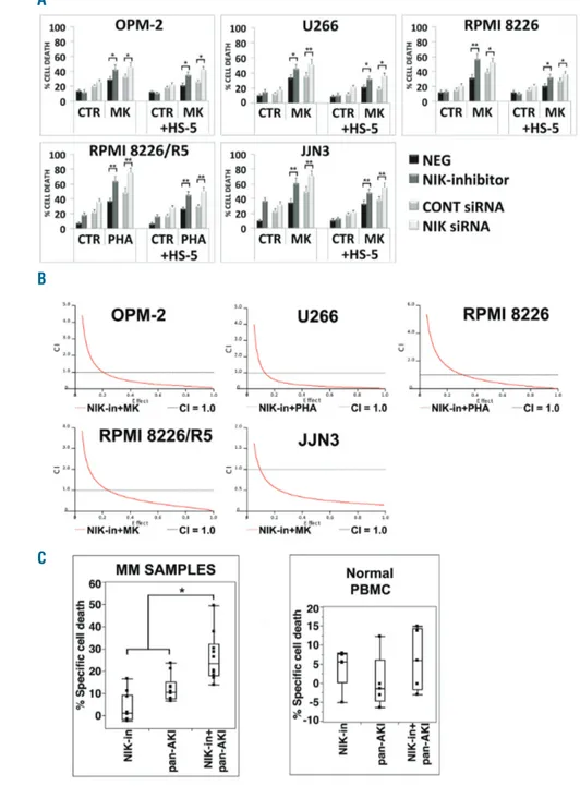
Figure 2. NF-κB-inducing kinase (NIK) inhibi-tion sensitizes multiple myeloma (MM) cells to pan-AKI-induced cell death. (A) MM cell lines were incubated with the NIK inhibitor (NIK-in) at 10 μM or were transfected with NIK siRNA and after 3 hours (h) MM cell lines were treated with MK-0457 (0.4 μM) or PHA-680632 (1 μM) in absence or presence of HS-5 cells (+HS-HS-5) (see Online Supplementary Methods). After 48 h, cell death was meas-ured by annexin-V labeling. Values represent means±Standard Deviation (SD) of four inde-pendent experiments. (Dunnett and Tukey-Kramer tests, *P<0.05; **P<0.01 vs. MK-0457 treatment). (B) MM cell lines were treat-ed sequentially with escalating doses of the NIK inhibitor (NIK-in) (1-20 μM) for 3 h and subsequently with MK-0457 (0.1-1 μM) or PHA-680632 (0.1-2 μM) alone or in combina-tion with the NIK inhibitor at a fixed ratio indi-cated in Online Supplementary Table S1. After 48 h, cell death was measured by annexin V labeling and the Combination Index values (CI) were calculated using the Chou-Talalay method and Calcusyn software, and the isobologram plots were constructed. CI<1.0 indicate synergism, CI=1.0 indicate additive effect, and CI>1.0 indicate an antagonistic effect. (C) CD138-purified plasma cells from ten patients with MM seeded in presence of HS-5 cells and peripheral blood mononuclear cells (PBMC) from five healthy volunteers were incubated with the NIK inhibitor (NIK-in) at 10 μM for 3 h and then were treated with MK-0457 (0.4 μM) or PHA-680632 (1 μM). After 24 h, cell death was measured by annexin-V staining or sub-G1 DNA content. Because of heterogeneous levels of basal cell death, the data of all ten primary samples and PBMC tested are expressed as % of specific cell death with the formula % Specific cell death = 100 x (induced cell death−basal cell death)/(100−basal cell death) and are shown in box plot format (median line in box delimit-ed by 25thand 75th) (*P<0.005 vs. either
treat-ment alone; Dunnett test).
A
B
Figure 3. NF-κB-inducing kinase (NIK) accumulation interferes with inhibitory activities of pan-AKI NF-κB. (A) siRNA silencing of NIK but not the non-specific control siRNA, led to a decrease in NIK protein expression. Three hours (h) after electroporation, multiple myeloma (MM) cell lines were treated with the pan-AKI MK-0457 (0.4 μM) or PHA-680632 (1 μM). After 48 h, MM cell lines were lysed and subjected to western blot analysis to monitor the expression of NIK, phospho-IKKa/β, phospho-NF-κB2 p100, phospho-IκBa and Actin as loading control. All western blotting results were evaluated by densitometric scanning, corrected with respect to Actin expression, and expressed relative to the value obtained with the corresponding control set as 1. The relative protein amount is reported below the lanes. (B) RPMI-8226 and 8226/R5 were stably transfected with an empty vector or with plasmid expressing NIK protein. Pools of stable clones (8226-NIK and 8226/R5-NIK) were obtained by selection with puromycin. Both expression vectors co-expressed GFP to monitor the transfection by flow cytometry. Plots represent GFP fluorescence of cells transfected with empty vector (Blank) or that encoding for NIK compared to non-transfected cells. (C) NIK overexpression enhances nuclear localization and DNA transactivation activity of NF-κB subunits. Stable clones of RPMI-8226 and 8226/R5 transfected with empty vector (Blank) or expressing NIK protein or untrans-fected cells were seeded at a density of 2x10⁵cells/mL. After 24 h cytoplasmic and nuclear extracts were prepared using the Active Motif's Nuclear Extract Kit.
Cytoplasmic cell lysates were immunoblotted against NIK, p-IKKa/β, p-NF-κB2, p-IκB-a and tubulin as marker of cytoplasmic separation as well as loading control; nuclear extracts were immunoblotted against NF-κB p65, NF-κB1 p50, NF-κB2 p52, RelB and histone H2B as nuclear loading control; bands were subjected to den-sitometric scanning and the number below each lane represents the relative amount of the indicated proteins normalized to tubulin or histone H2B expression. Graphs below represent DNA binding activity of the NF-κB p65, NF-κB1 p50, NF-κB2 p52 and RelB subunit from the same nuclear extracts (TransAM NF-κB ELISA kit); results were normalized to the untransfected control (Untr). Values represent mean±Standard Deviation (SD) of three separate experiments. (**P<0.01 vs. untransfected condition; Dunnett’ test). (D) NIK inhibition attenuates NF-κB signaling. siRNA silencing of NIK but not the non-specific control siRNA, led to a decrease in NIK protein expression. RPMI-8226-NIK and 8226/R5-NIK were electroporated with control siRNA or with NIK siRNA. After 24-h cytoplasmic and nuclear extracts from transfected and untransfected cells were prepared. Cytoplasmic cell lysates were immunoblotted against NIK, phospho-IKKa/β, p-NF-κB2 p100, p-IκB-a and tubulin as marker of cytoplasmic separation as well as loading control; nuclear extracts were immunoblotted against NF-κB p65, NF-κB1 p50, NF-κB2 p52, RelB and Histone H2B as nuclear loading control; bands were subjected to densitometric scanning and the number below each lane represents the relative amount of the indicated proteins normalized to Tubulin or Histone H2B expression. Graphs below represent DNA binding activity of the NF-κB p65, NF-κB1 p50, NF-κB2 p52 and RelB subunit from the same nuclear extracts; results were normalized to the untransfected control (Untr). Values represent mean±SD of three separate experiments. (*P<0.05 vs. control siRNA condition; Dunnett test).
A
B
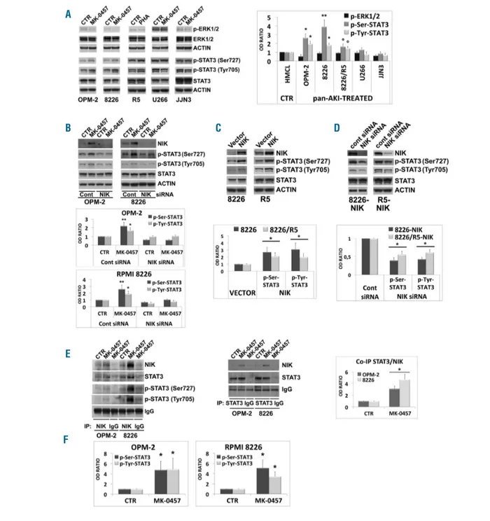
Figure 4. Pan-AKI-mediated accumulation of NF-κB-inducing kinase (NIK) induces STAT3 activation. (A) Wesern blot analysis of endogenous phospho-ERK1/2 (Thr202/Tyr204), ERK1/2 and Actin in MM cell lines treated with MK-0457 (0.4 μM) for 24 hours (h). The same lysates were prepared and immunoblotted against phospho-STAT3 (Ser727), phospho-STAT3 (Tyr705), STAT3 and Actin as loading control. Phospho-ERK1/2 and Ser727- and Tyr705-phosphorylated STAT3 were nor-malized to total ERK1/2 and STAT3 levels, respectively. In the graph, the phosphorylations under control conditions were set as 1 for comparison. In histogram are shown means±Standard Deviation (SD) of three independent experiments (°P<0.05, *P<0.01, **P<0.005 vs. control, Dunnett test). (B) OPM-2 and RPMI-8226 cells were transfected with NIK siRNA and after 3 h MM cell lines were treated with MK-0457 (0.4 μM). After 48 h whole cell lysates were prepared and immunoblot-ted against NIK, phospho-STAT3 (Ser727), phospho-STAT3 (Tyr705), STAT3 and Actin as loading control. Bands were subjecimmunoblot-ted to densitometric scanning. STAT3 phos-phorylations were normalized to overall STAT3 levels. STAT3 phosphos-phorylations under untreated control condition were set to 1. Histograms below represent the mean±SD of three independent experiments. (*P<0.01, **P<0.005, Tukey-Kramer test). (C) Wesern blot analysis of NIK, phospho-STAT3 (Ser727), phospho-STAT3 (Tyr705), STAT3 and Actin in stable clones of RPMI-8226 and RPMI-8226/R5 transfected with empty vector or with plasmid expressing NIK. All western blotting results were evaluated by densitometric scanning, and histograms represent the relative levels of phospho-STAT3 corrected with respect to Actin and normalized to STAT3 expression. In the graph below, STAT3 phosphorylations under control conditions (empty vector transfection) were set as 1 for comparison. Histogram repre-sents the mean±SD of five independent experiments. (*P<0.001 vs. empty vector, Dunnett test). (D) NIK expression of RPMI-8226-NIK and RPMI-8226/R5-NIK cells was inhibited by siRNA silencing; after 24 h cells were subjected to western blot analysis to monitor the expression of NIK, phospho-STAT3 (Ser727), phospho-STAT3 (Tyr705), STAT3 and Actin as loading control. Bands were subjected to densitometric scanning and STAT3 phosphorylations were normalized to total STAT3 levels. In the graph below, the relative fold change of protein levels was normalized with respect to the level of the control siRNA (cont), which was taken as 1. Histogram rep-resents the mean±SD of five independent experiments (*P<0.001 vs. untreated control, Dunnett test). (E) OPM-2 and RPMI-8226 cell lines were treated with MK-0457 (0.4 μM) and after 24 h of treatment were lysed and subjected to immunoprecipitation (IP) using anti-NIK antibody or anti-STAT3 antibody or control antibody (IgG) and immunoblotted (IB) with either NIK or STAT3 antibodies. Western blot results were subjected to densitometric scanning and the histogram on the right shows average quantification results±SD of the association NIK/STAT3 from 3 immunoprecipitations (*P<0.0001 vs. untreated control cells, Dunnet and Tukey-Kramer tests). Anti-NIK immunoprecititate Filters stripped and reprobed for phospho-STAT3 (Ser727), phospho-STAT3 (Tyr705) and subjected to densitometric analy-sis (F); histograms represent the relative levels of phospho-STAT3 corrected with respect to IgG and normalized to STAT3 expression. STAT3 phosphorylations under control conditions were set as 1. Histograms show average quantification results±SD of three independent blots (*P<0.005, vs. untreated control cells, Dunnett and Tukey-Kramer tests).
A
B
C
D
E
alone or in co-culture with HS-5 stromal cells (Figure 2A).
Importantly, NIK-in synergized with pan-AKI to kill MM
cells (Figure 2B and Online Supplementary Table S1).
Furthermore, adherence of MM cells to HS-5 stromal cells
conferred significant protection against pan-AKI-induced
cell death in the majority of the HMCL analyzed.
However, this protective effect was significantly reduced
by NIK inhibition (Figure 2A), thus confirming the
impor-tant role of NIK in the stroma-mediated pan-AKI
protec-tion. Finally, NIK-in significantly (P<0.005; n=10)
increased the cytotoxicity of pan-AKI in patient-derived
primary MM cells (Figure 2C and Online Supplementary
Figure S2A), with no significant differences in the response
rates between newly diagnosed (n=3) and relapsed (n=7)
patients (Online Supplementary Figure S3) but not on PBMC
from healthy individuals (Figure 2C and Online
Supplementary Figure S2B). These observations thereby
indicate that NIK plays an important role in the
respon-siveness of MM cells to pan-AKI. (Patients’ demographic
and clinical characteristics are summarized in Online
Supplementary Table S2).
It is also important to highlight the fact that treatment
of MM cells with the proteasome inhibitor bortezomib
(currently the standard of care for MM) caused a strong
accumulation of the NIK protein in the majority of the
HMCL analyzed (Online Supplementary Figure S4A) and its
chemical inhibition significantly enhanced the
anti-myelo-ma effects of bortezomib, thereby indicating that NIK can
influence the sensitivity of MM cells to this drug (Online
Supplementary Figure S4B).
NF-
κB-inducing kinase interferes with the inhibitory
activity of pan-AKI on NF-
κB-inducing kinase
To examine whether NIK accumulation induced by
pan-AKI counteracts their ability to inhibit NF-κB pathways in
MM cells, we blocked its function with a NIK-specific
siRNA and monitored NF-
κB activity in response to
pan-AKI. We found that in 4 of 5 HMCL tested (except
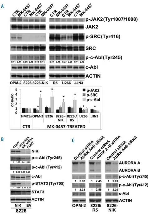
OPM-Figure 5. NF-κB-inducing kinase (NIK) accumu-lation induces c-Abl activation. (A) Western blot analysis of phospho-JAK2 (Tyr1007/1008), JAK2, phospho-SRC (Tyr416), SRC, phospho-c-Abl (Tyr245) and c-phospho-c-Abl kinases in multiple myelo-ma (MM) cell lines treated with MK-0457 (0.4 μM) for 24 hours (h); anti-Actin was performed as loading control. Bands were then subjected to densitometric scanning and levels of phospho-JAK2 (Tyr1007/1008), phospho-SRC (Tyr416), and phospho-c-Abl (Tyr245) were normalized to total JAK2, SRC, and c-Abl levels. Densitometric values of the ratio of phosphorylated c-Abl/total c-Abl are shown below the blots. The graph below represents the phosphorylation status of JAK2, SRC and c-Abl; changes (folds increase or decrease) in the levels of each phosphorylated protein relative to untreated control was taken as 1 (mean±Standard Deviation (SD) of 3 inde-pendent blots; °P<0.01, *P<0.005, vs. untreat-ed control cells, Dunnett test). (B) NIK expres-sion of RPMI-8226-NIK cells was inhibited by siRNA silencing; after 24 h transfected and untransfected cells were subjected to western blot analysis to monitor the expression of NIK, phospho-c-Abl (Tyr245), phospho-c-Abl (Tyr412), c-Abl, phospho-STAT3 (Tyr705), STAT3 and Actin as loading control. Protein expression of siRNA transfected 8226-NIK cells was compared to empty vector (EV) transfected control 8226 cells. Bands were then subjected to densitometric scanning: c-Abl and STAT3 phosphorylations were normalized to total c-Abl and STAT3 levels, respectively. The relative fold change of protein levels was normalized with respect to 8226-NIK untransfected condition, which was taken as 1 and are reported under each blot. (C) MM cell lines were transfected with siRNA against Aurora A and Aurora B (AURK A+B) or control siRNA. After 48 h transfected MM cell lines were sub-jected to western blot analysis to monitor the expression of Aurora A and B, phospho-c-Abl (Tyr245), phospho-c-Abl (Tyr412), c-Abl and Actin as loading control. c-Abl phosphorylations were subjected to densitometric scanning and were normalized to c-Abl levels. The relative fold change of protein levels was normalized with respect to the level of the untreated control, which was taken as 1, and is shown under each lane.
A
C
B
2 cells that have low NF-
κB index
3,4,25), NIK knockdown
reduced the basal phosphorylation/activation status of
IKKa and IKKβ (p-IKKa/β) and their respective
down-stream direct targets NF-
κB2/p100 and IκB-a (Figure 3A),
thus confirming that NIK affects not only the
non-canoni-cal but also the canoninon-canoni-cal NF-
κB pathway in MM cells.
2-4Notably, pan-AKI were ineffective (OPM-2) or only
par-tially effective (all the other HMCL analyzed) in
attenuat-ing NF-κB signalattenuat-ing
25(Figure 3A), and their reduced
inhibitory activity on NF-
κB signaling was closely linked
to NIK induction because its knockdown by siRNA
com-pletely abrogated the pan-AKI-induced NF-
κB activation
in OPM-2 as well as greatly enhanced the
pan-AKI-induced NF-
κB inhibition in all the other HMCL analyzed
(Figure 3A).
In support of these data, we found that experimental
overexpression of NIK in MM cells (Figure 3B) caused
enhanced phosphorylation of IKK
a/β, NF-κB2/p100 and
I
κB-a, and increased nuclear localization and DNA
bind-ing activities of the NF-
κB p65, p50, p52, and RelB
sub-units (Figure 3C). In contrast, NIK knockdown in these
NIK-over-expressing MM cells consistently and
signifi-cantly decreased their basal NF-
κB activity (Figure 3D),
thus confirming the important role of NIK in controlling
NF-
κB signaling in MM.
3,4NF-
κB-inducing kinase induction by pan-AKI activates
the STAT3 signaling pathway in multiple myeloma cells
Because NIK induction by pan-AKI was not associated
with an increased activation of NF-
κB pathways in 4 of 5
HMCL tested (except OPM-2), and yet NIK signaling has
been demonstrated to crosstalk at different levels with
other important prosurvival signaling pathways including
MEK-ERK and STAT3 pathways,
8-10we explored whether
NIK induction by pan-AKI affected these pathways in
MM cells.
Because NIK can phosphorylate MEK1 and thereby cause
activation of downstream MAPK ERK,
9we investigated
whether NIK induction by pan-AKIs is associated with
increased phosphorylation/activation of ERK in MM cells.
We found no significant change (U266, R5) or even a
decrease (OPM-2, JJN3) in ERK activity (p-ERK1/2) in the
pan-AKIs-treated HMCL (Figure 4A), thereby indicating
that NIK, stabilized by pan-AKI, does not act through this
pathway. Because STAT3 activity is regulated by two
independent phosphorylations, one occurring at Tyr705
and one at Ser727, which are both required for it to be
fully functional,
37we specifically analyzed the STAT3
(Tyr705) and STAT3 (Ser727) phosphorylation patterns
alongside with the overall protein expression levels. We
found that treatment with pan-AKI significantly increased
both Ser727 and Tyr705 STAT3 phosphorylation in
OPM-2, RPMI-8226 and 8226/R5, but not in U266 and JJN3
HMCL where no significant changes in p-Ser-STAT3 or a
decrease in p-Tyr-STAT3 phosphorylation were observed
(Figure 4A).
Notably, NIK knockdown in MM cells completely
abro-gated both Ser727 and Tyr705 STAT3 phosphorylation
induced by pan-AKI (Figure 4B), which would suggest that
NIK is involved in the pan-AKI-mediated STAT3
activa-tion. Confirming these data, we found that ectopic
expres-sion of NIK in MM cells caused enhanced
phosphoryla-tion of STAT3 in both serine and tyrosine residues (Figure
4C), whereas its depletion in these NIK-over-expressing
MM cells consistently and significantly (P<0.001)
decreased their basal STAT3 activity levels (Figure 4D).
In the light of evidence supporting reciprocal regulatory
mechanisms and crosstalk between the NIK and STAT3
proteins,
10we examined whether NIK exists in a complex
with STAT3 in MM cells. Co-immunoprecipitation
showed that STAT3 was associated with NIK and that this
association was significantly enhanced by pan-AKI
treat-ment of the cells (Figure 4E).
We also examined the Ser727 and Tyr705
phosphoryla-tion state of STAT3 that co-immunoprecipitated with NIK
and found that treatment with pan-AKI promoted a strong
increase in the phosphorylation of NIK-associated STAT3
in both serine and tyrosine residues (Figure 4E and F),
stressing the putative function of NIK in controlling
STAT3 activation.
Aurora kinases inhibitors induce a NF-
κB-inducing
kinase dependent cytoplasmic relocalization and
activation of c-Abl and promote the formation of the
NIK-c-Abl-STAT3 ternary complex in multiple myeloma
Given the high levels of tyrosine-phosphorylated
STAT3 that co-immunoprecipitates with the
serine/threo-nine kinase NIK in response to pan-AKI treatment, we
explored whether pan-AKI affect the Stat3 upstream
tyro-sine kinases JAK2, Src and/or c-Ab
l
38activity/expression.
We found that, depending on the HMCL examined,
pan-AKI caused a decrease or no significant changes in the
Tyr-phosphorylation/activity of JAK2 JAK2) and Src
(p-SRC) kinases (Figure 5A), whereas they were able to
sig-nificantly activate c-Abl in 4 of 6 HMCL tested (except
U266 and JJN3 in which no significant changes or a
decrease in c-Abl tyrosine-phosphorylation levels were
observed, respectively) (Figure 5A). A significant increase
(>3-fold) in p-c-Abl, but not in p-JAK2 and p-Src, was also
observed in untreated 8226-NIK as compared to untreated
8226 HMCL (Figure 5A, lane 5 vs. lane 3). This finding
links NIK to c-Abl signaling and, indeed, experimental
overexpression of NIK in MM cells causes enhanced
phos-phorylation of endogenous c-Abl on Tyr245 and Tyr412
residues (both commonly used as Abl activation
markers),
16,17as well as Tyr705 phosphorylation of STAT3.
Conversely, knockdown of NIK in these
NIK-over-expressing MM cells consistently and significantly
decreased their basal tyrosine-phosphorylation levels
(Figure 5B). Accordingly, abrogation of Aurora-A and -B
induced c-Abl phosphorylation at both Tyr245 and
Tyr412 residues in MM cells (Figure 5C).
As c-Abl may exhibit both pro- and antiapoptotic
func-tions depending on the subcellular localization (nuclear or
cytoplasmic),
15-18and its intracellular localization is
regulat-ed by phosphorylation of its Thr735 residue promoting
cytoplasmic sequestration by the 14-3-3 protein,
19we
explored whether pan-AKI affect Thr735 phosphorylation
and/or subcellular localization of c-Abl in MM cells in
which the pervasive DNA damage leads to a
predomi-nantly nuclear localization of c-Abl.
21,22As shown in Figure
6 and Online Supplementary Figure S5, endogenous
c-Abl was predominantly accumulated in the nucleus of
the MM cells,
21while pan-AKI were able to cause a
signif-icant translocation of c-Abl from the nucleus to cytoplasm,
thus elevating its cytoplasm/nucleus ratio in 4 of 5 HMCL
tested (except JJN3) (Figure 6). Notably, the
pan-AKI-induced cytoplasmic accumulation of c-Abl was
associat-ed with increasassociat-ed Thr735 phosphorylation of the
cyto-plasmic fraction of c-Abl (Figures 6 and 7A).
Both processes, Thr735 phosphorylation and
concomi-tant cytoplasmic accumulation of c-Abl, were closely
linked to NIK induction since its overexpression in MM
cells increased Thr735 phosphorylation of cytoplasmic
c-Abl (Figure 7A) and caused c-c-Abl to translocate from the
nuclear to the cytoplasmic compartment, whereas its
inhi-bition in these NIK-over-expressing cells reversed this
shuttling (Figure 7A). These data were further confirmed
by immunofluorescence analysis (Online Supplementary
Figure S6).
A closer examination revealed that NIK was diffused in
the cytoplasm with an accumulation around the nucleus
of the tumor cells treated with pan-AKI (Figure 7B), and its
overexpression also caused enhanced tyrosine
phosphory-lation of cytoplasmic c-Abl (Figure 7A), to elicit its
anti-apoptotic functions.
15-18Together with these results, siRNA-mediated
knock-down of NIK completely abrogated the pan-AKI-induced
Thr735 phosphorylation of c-Abl in OPM-2 and greatly
decreased the high basal c-Abl Thr735 phosphorylation in
the high NIK expressing JJN3 HMCL (Figure 7C).
Because pan-AKI can induce NIK accumulation and
comitant c-Abl activation, and both these kinases
con-verge on and activate the STAT3 pathway,
10,17we next
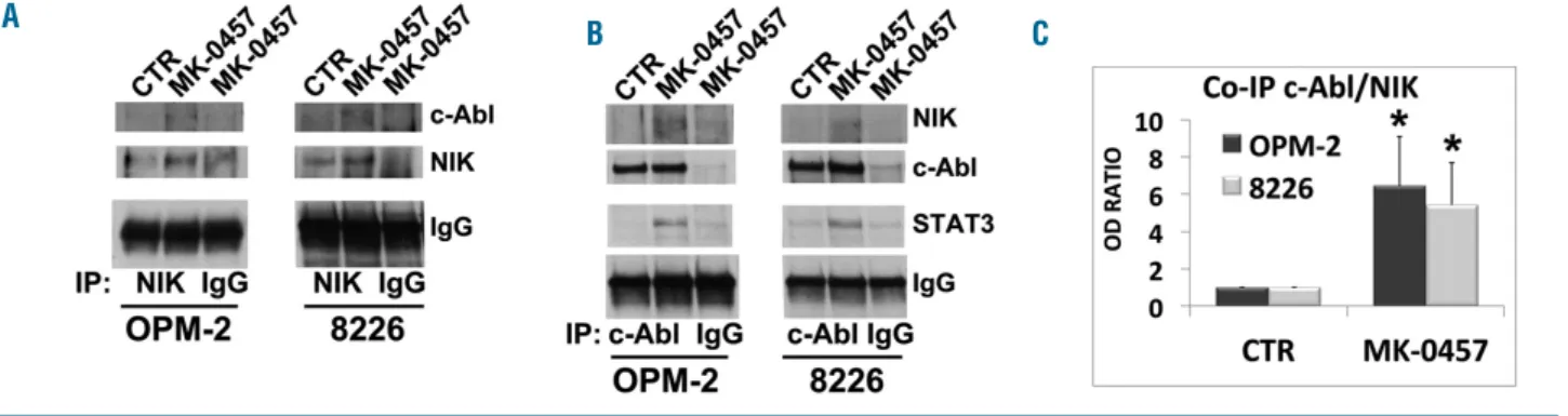
investigated whether c-Abl can form a heterotrimeric
complex with NIK and STAT3 in MM cells. As indicated
in Figure 8A and B, there was little if any detectable
inter-action of c-Abl and NIK in untreated MM cells. However,
exposure of MM cells to Pan-AKI led to an increase in the
association of c-Abl with NIK kinases that was at least a
3-fold higher than in untreated control cells (Figure 8C).
The interaction between NIK and c-Abl, and that
previ-ously shown between NIK and STAT3 (Figure 4E),
togeth-er with the fact that c-Abl can regulate the activation of
STAT3 in cancer cells,
17indicated that these three proteins
may form a trimeric complexes in pan-AKI-treated MM
cells. Accordingly, as shown in Figure 8B,
immunoprecip-itation of endogenous c-Abl from lysates of untreated or
pan-AKI-treated MM cells followed by STAT3
immunoblotting revealed that the pharmacological
block-ade of Aurora kinases induced a physical interaction of
c-Abl with STAT3, thus confirming that, in MM cells,
pan-AKI can promote the formation of the ternary complex
NIK-c-Abl-STAT3.
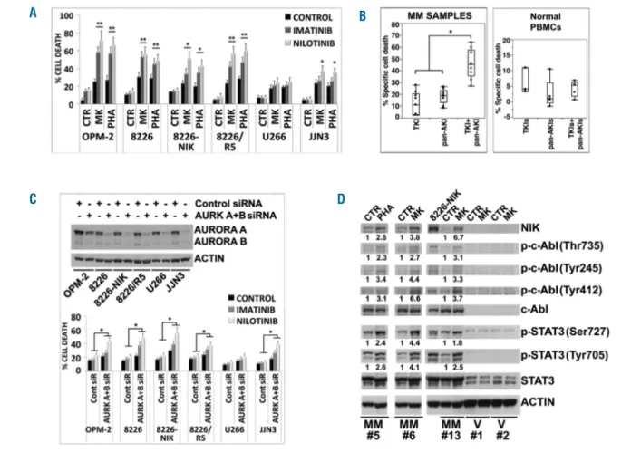
Pharmacological blockade of c-Abl sensitizes multiple
myeloma cells to pan-AKI
To examine the functional significance of the
pan-AKI-induced activation of c-Abl in MM cells we blocked its
function using the Abl kinase inhibitors imatinib or
nilo-tinib
20and monitored cell death in response to pan-AKI
treatment. Both imatinib or nilotinib significantly
increased the pan-AKI-induced cell death in the majority
of the HMCL as well as in patient-derived primary MM
cells, (P<0.005; n=9) (Figure 9A and B and Online
Supplementary Figure S7A and B), with no significant
differ-ences observed in the response rates of newly diagnosed
(n=4) versus relapsed (n=5) patients (Online Supplementary
Figure S8) and no effects seen in normal PBMC (Figure 9B
and Online Supplementary Figure S7C).
In agreement with these results, Aurora-A and -B
inhibi-tion by either Aurora A/B-specific siRNA or AMG-900,
23,29a potent and highly selective pan-AKI, significantly
enhanced the sensitivity of MM cells to c-Abl inhibitors
(Figure 9C, Online Supplementary Figures S9 and S10A and
B). Furthermore, c-Abl kinase inhibitors consistently
syn-ergized with pan-AKI to induce cell death in MM cells
(Online Supplementary Figure S11 and Online Supplementary
Table S3).
Remarkably, as observed in the majority of the HMCL
analyzed, treatment of cells isolated from MM patients
with Pan-AKI induced NIK accumulation, increased
Thr735, Tyr245 and Tyr412 phosphorylation of c-Abl and
Ser727 and Tyr705 phosphorylation of STAT3 (Figure 9D).
None of these conditions was observed in similarly
treat-ed PBMC from healthy donors.
Figure 6. Aurora kinases inhibitors induce a cytoplasmic relocalization of c-Abl. Multiple myeloma (MM) cell lines were treated with MK-0457 (0.4 μM) and after 24 hours (h) cytoplasmic and nuclear extracts were prepared. Equal amount of Cytoplasmic (cyto) and nuclear (nuc) cell lysates (10 μg) were immunoblotted against c-Abl, phospho-c-Abl (Thr735), β Tubulin and Histone H3 as loading control of cytoplasmic and nuclear fraction, respectively. Bands were subjected to densitometric scanning: cytoplasmic and nuclear blots were normalized to total β Tubulin and Histone H3, respectively. The densitometric analysisis is reported in the graphs below: the relative fold change of cytoplasmic or nuclear c-Abl and phospho-c-Abl (Thr735) levels was normalized with respect to control condition, which was taken as 1. The ratio of cytoplasmic to nuclear c-Abl and phospho-c-Abl (Thr735) protein expression (Cyto/Nuc) is shown. The c-Abl and phospho-c-Abl (Thr735) Cyto/Nuc ratio in untreated cells was set as 1. In the histograms are shown average quantification results±Standard Deviation (SD) of four independent blots (°P<0.05, *P<0.005, **P<0.001 vs. untreated control cells, Dunnett test).
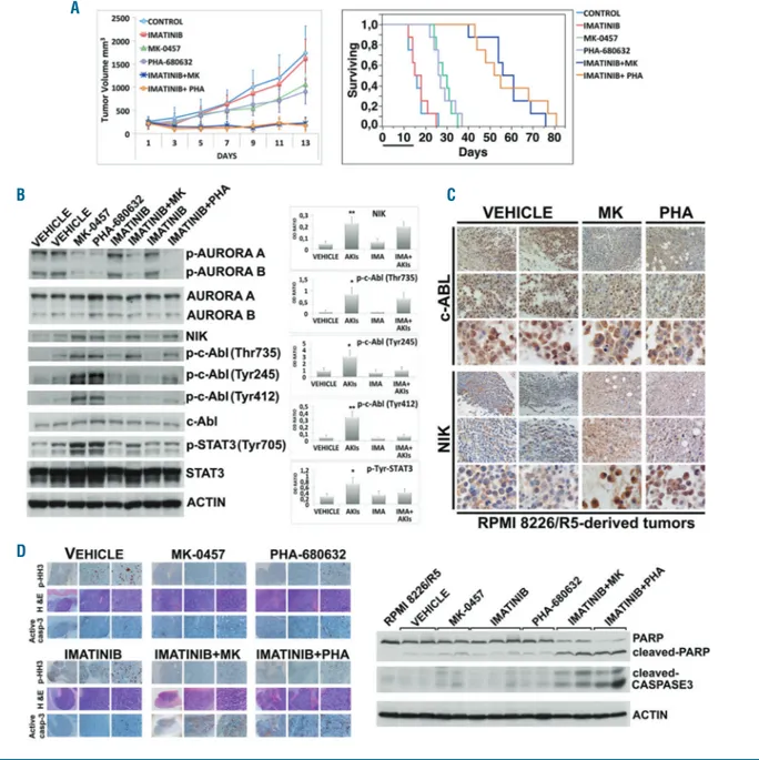
To verify that the anti-tumor activity of pan-AKI and
the synergizing effects of c-Abl inhibitors observed on
cul-tured/isolated MM cells could be reproduced in vivo, we
set up a multidrug-resistant xenograft mouse model of
human MM. Consistent with our in vitro results, imatinib
significantly potentiated the anti-tumor activity induced
by pan-AKI in this in vivo setting, while having no effect as
a single agent in vivo in a multidrug-resistant xenograft
mouse model of human MM (Figure 10A). Animal
sur-vival was also significantly improved in mice treated with
the combination imatinib/pan-AKI versus those that
received monotherapies or vehicle alone (P<0.0015)
(Figure 10A and Online Supplementary Table S4).
Immunobloting analyses on tumor masses harvested
after five days post treatment confirmed decreases in the
phosphorylation levels of Aurora kinases, enhancement of
Figure 7. Accumulted NF-κB-inducing kinase (NIK) activates cytoplasmic c-Abl.(A) Stable clones of RPMI-8226 transfected with empty vector (EV) or with plasmid expressing NIK (NIK) were treated with MK-0457 (0.4 μM) or NIK siRNA, respectively. After 24 hours (h) cytoplasmic and nuclear extracts were prepared and equal amount of Cytoplasmic (cyto) and nuclear (nuc) cell lysates (10 μg) were immunoblotted against NIK, phospho-c-Abl (Thr735), phospho-c-Abl (Tyr245), c-Abl, β Tubulin and Histone H3 as loading control of cytoplasmic and nuclear fraction, respectively. Bands were subjected to densitometric scanning: cytoplasmic and nuclear blots were normalized to total β tubulin and Histone H3, respectively. In the graph below the relative fold change of cytoplasmic or nuclear c-Abl, phospho-c-Abl (Thr735) and phospho-Abl (Tyr245) levels was normalized with respect to empty vector (EV) control condition, which was taken as 1. The ratio of cytoplasmic to nuclear Abl, phospho-Abl (Thr735) and phospho-Abl (Tyr245) protein expression (Cyto/Nuc) is shown. The Cyto/Nuc ratio of phosphorylated and non-phosphorylated c-Abl in empty vector (EV) control condition (CONT) was set as 1. In histogram are shown average quantification results±Standard Deviation (SD) of three independent blots [#P<0.01, °P<0.005, *P<0.001 vs. (EV) control condition, Dunnett test]. (B) Stable clones of RPMI-8226 transfected with empty vector (EV) untreated or treat-ed with MK-0457 (0.4 μM) and of RPMI-8226 expressing NIK (NIK) electroporattreat-ed with non-specific control siRNA (CONT) or with NIK siRNA were harvesttreat-ed after an incubation of 24 h for cytospins and stained for c-Abl or were formalin fixed and paraffin embedded in cytoblocks for NIK staining. The microphotographs shown are representative of similar observation in three independent experiments (20x, 40x and 100x original magnifications). (C) OPM-2 and JJN3 cells were transfected with non-specific control siRNA (Cont) or NIK siRNA and after 3 h multiple myeloma (MM) cell lines were treated with MK-0457 (0.4 μM). After 48 h whole cell lysates were prepared and immunoblotted against NIK, phospho-c-Abl (Thr735), c-Abl and Actin as loading control. Bands were subjected to densitometric scanning. Levels of Thr735-phosphorylated c-Abl were normalized to overall c-Abl levels and c-Abl phosphorylation under non-specific siRNA control condition was set to 1. Histogram below represents the mean±SD of three independent experiments. (°P<0.02, *P<0.005, **P<0.0005 vs. control siRNA condition, Dunnett test).
B
C
A
NIK protein, and increases in the Thr735, Tyr245 and
Tyr412 phosphorylation of c-Abl and Tyr705
phosphory-lation of STAT3 in the case of xenografted animals treated
with pan-AKI when compared to vehicle-treated controls
(Figure 10B). In addition, immunohistochemical staining
of tumor lesions for NIK and c-Abl revealed that also in
vivo pan-AKI were capable of causing cytoplasmic NIK
accumulation, which was most prominent around the
nucleus of the tumor cells (Figure 10C and Online
Supplementary Figure S12), whereas c-Abl was observed to
have been extensively translocated from the nucleus to
the cytoplasm (Figure 10C).
Finally, immunohistochemical analysis of tumor lesions
isolated from pan-AKI-treated animals consistently
revealed a significant reduction in the phosphorylation of
Histone H3 on Ser10 (Figure 10D), a protein known to be
a physiological substrate of Aurora kinases and a cellular
proliferation marker.
39This result would be consistent
with the retardation of tumor growth observed in
pan-AKI-treated versus vehicle-treated mice (Figure 10A).
Notably, combined imatinib and pan-AKI treatment
blunted the pan-AKI-induced tyrosine (but not threonine)
phosphorylation of c-Abl (Figure 10B) and increased the
levels of apoptosis (cleaved-PARP and -caspase-3 staining),
relative to that seen with monotherapies and vehicle alone
(Figure 10D); a result that agreed with the tumor
regres-sion and the improved survival rate observed in mice
treated with the imatinib-Pan-AKI combination therapy
(Figure 10A).
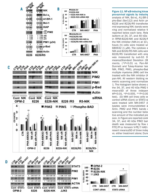
Pan-AKI-induced NF-
κB-inducing kinase accumulation
promotes survival signaling through PIM kinases
activation
Consistent with the fact that NIK can elicit pro-survival
signals in MM cells through activation of NF-κB and
STAT3 pathways, we found that experimental
overex-pression of NIK in MM cells caused the induction of the
antia-poptotic NF-
κB/STAT3 regulated genes Bcl-xL,
A1/Bfl-1, Mcl-1 and XIAP
40(Figure 11A), all of which
rep-resent important targets for sensitizing MM cells to
anti-cancer agents,
1including pan-AKI.
25NIK overexpression
was also associated with upregulation of PIM1 and PIM2
(Figure 11A), both oncogenic, constitutively active
serine/threonine kinases transcriptionally regulated either
by NF-
κB or STAT3, that mediate survival signaling
through the phosphorylation and inactivation of Bad
32,41(Figure 11A). In accordance with its role in controlling
anti-apoptotic signal transduction events, NIK
overexpres-sion protected MM cells from pan-AKI-induced cell death,
which was reversed by the chemical or genetic disruption
of NIK functions (Figure 11B).
We further found that in 5 of 7 HMCL tested (except
U266 and JJN3 cells), the pan-AKI-induced
NIK-stabiliza-tion was associated with enhanced levels of PIM1 and
PIM2 proteins, and phosphorylation of their direct
down-stream target Bad (Figure 11C and Online Supplementary
Figure S13); RNA interference-mediated knockdown of
NIK or the use of a NIK-inhibitor (NIK-in) prevented these
increments (Figure 11C), thus confirming the role of NIK
in PIM kinases induction in MM cells. Taken together
with our previous findings (Figures 4-8), the observations
also supported the existence of a NIK /c-Abl /STAT3 /PIM
/Bad signaling axis in pan-AKI-treated MM cells.
Consistent with the fact that STAT3 can regulate the
expression of PIM kinases,
32,41we found that its inhibition
by siRNA completely abrogated the pan-AKI-induced
PIM1 and PIM2 upregulation in OPM-2, RPMI-8226 and
RPMI-8226-NIK HMCL, and greatly decreased their basal
levels in JJN3 cells (Figure 11D).
Loss-of-function of STAT3 by either siRNA or the
small-molecule inhibitor STATTIC
42significantly enhanced the
pan-AKI sensitivity of MM cells (Figure 11D and Online
Supplementary Figure S14), thereby indicating that STAT3
activated by pan-AKI acted as a prosurvival, antiapoptotic
transcription factor in MM.
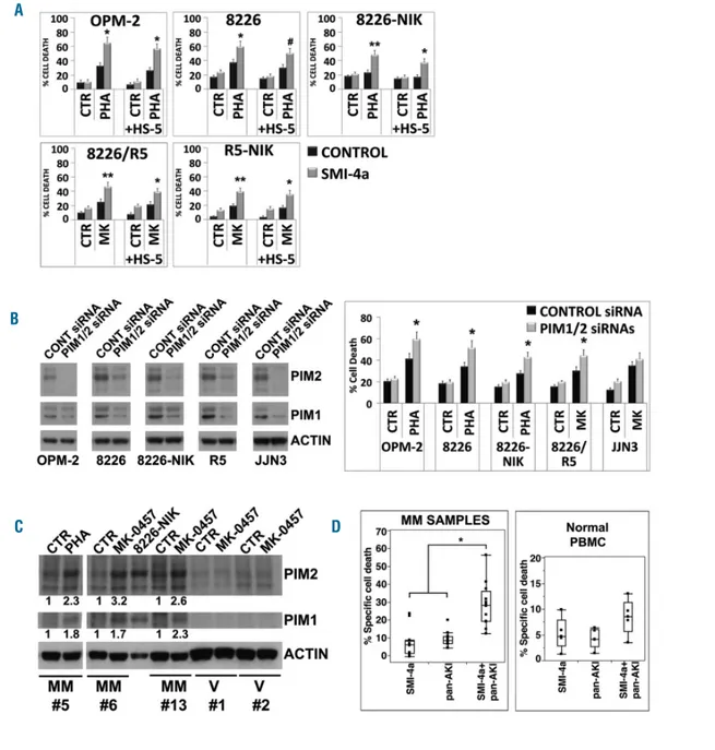
PIM kinases have been implicated in the regulation of
MM cell proliferation, survival, and drug resistance.
43Given this, we examined whether their inhibition affected
the responses of MM cells to pan-AKI. PIM1/2 inhibition,
by either the specific small-molecule inhibitor SMI-4a
44or
by PIM1/2-specific siRNA significantly increased the
pan-AKI-induced cell death in all the HMCL tested either
cul-tured alone or together with HS-5 cells, except for U266
and JJN3 (Figure 12A and B, and Online Supplementary
Figure S15), in which pan-AKI failed to increase PIM
kinas-es levels (Supplementary Figure S14).
Furthermore, treatment of patient-derived MM cells,
but not normal PBMC, with Pan-AKI led to an increment
Figure 8. Accumulated NF-κB-inducing kinase (NIK) physically interacts with c-Abl and contributes to the NIK-c-Abl-STAT3 prosurvival complex formation.OPM-2 and RPMI-8226 cell lines were treated with MK-0457 at 0.4 μM and after 24 hours (h) of treatment were lysed and subjected to immunoprecipitation (IP) using (A) anti-NIK or (B) anti-c-Abl or control antibody (IgG) and immunoblotted (IB) with either NIK or c-Abl antibodies. Anti-c-Abl immunoprecititate filters were stripped and reprobed for STAT3. (C) Western blot of anti-NIK and anti-c-Abl immunoprecipitates results were subjected to densitometric scanning and protein expression under control conditions was set as 1. The histogram shows average quantification results±Standard Deviation (SD) of the association c-Abl/NIK from three immunopre-cipitations (*P<0.001 vs. untreated control cells, Dunnett and Tukey-Kramer tests).
A
of PIM1/2 protein levels (Figure 12C), that significantly
(P<0.005; n=10) influenced the responsiveness of the cells
to pan-AKI, with similar response rates between newly
diagnosed (n=3) and relapsed (n=7) patients (Figure 12D
and Online Supplementary Figures S2A and S3), thereby
indicating that these kinases may significantly impact on
the susceptibility of MM cells to pan-AKI exposure.
Discussion
The critical role of NIK in regulating non-canonical and
canonical NF-
κB pathways in MM,
4-6together with the
fact that NIK and Aurora kinases can converge on
com-mon targets,
24-26prompted us to hypothesize that NIK
might interfere with and reduce or bypass the NF-κB
inhibitory effects exerted by pan-AKI on MM cells. In
sup-port of this hypothesis, we found that pan-AKI induce
NIK protein stabilization and that this depended on the
downregulation of the TRAF2 protein, one of the critical
NF-
κB negative regulators that, together with TRAF3,
form a molecular bridge that couples NIK to the NIK
K48-ubiquitin ligase cIAP1/2.
6,7We also found that TRAF2
reduction was sufficient to elevate NIK protein levels in
MM cells harboring alterations in the TRAF3-binding
domain of NIK or in TRAF3 itself, thus confirming that
TRAF2 can regulate NIK stabilization independent of
TRAF3.
4,45Although experimental overexpression of NIK led to a
marked activation of both NF-
κB and STAT3 pathways, its
induction by pan-AKI resulted in the activation of only the
STAT3 pathway, thereby suggesting that Aurora kinases
Figure 9. Pharmacological inhibition of Abl kinase enhances cytotoxicity induced by Aurora inhibition. (A) Multiple myeloma (MM) cell lines were incubated with ima-tinib and niloima-tinib at 2 μM for 3 hours (h), and were then treated with MK-0457 (0.4 μM) and PHA-680632 (1 μM). After 48 h the cell death was measured by sub-G1 DNA content and Annexin-V method. Values represent means±Standard Deviation (SD) of four independent experiments. (°P<0.05, *P<0.005, **P<0.001 vs. either treatment alone; Dunnett and Tukey-Kramer tests). (B) CD138-purified plasma cells from nine patients with MM seeded in presence of HS-5 cells and periph-eral blood mononuclear cells (PBMC) from five healthy volunteers were preincubated for 3 h with imatinib or nilotinib at 2 μM and then with MK-0457 (0.4 μM) or PHA-680632 (1 μM). After 24 h cell death was measured by annexin-V staining or sub-G1 DNA content. Because of heterogeneous levels of basal cell death, the data of all nine primary samples and PBMC tested are expressed as % of specific cell death with the formula % Specific cell death = 100 x (induced cell death−basal cell death)/(100−basal cell death) and are shown in box plot format (median line in box delimited by 25thand 75th(*P<0.005 vs. either treatment alone; Dunnett
test). (C) MM cell lines were transfected with siRNA against Aurora A and Aurora B (AURK A+B) or unrelated non-specific control siRNA (Cont) and after 48 h MM cell lines were subjected to western blot analysis to monitor the expression of Aurora A and Aurora B and Actin. Twenty-four hours after siRNA transfection, MM cell lines were treated with imatinib or nilotinib at 2 μM. After 48 h of treatment cell death was measured by flow cytometry analysis of Annexin-V staining or sub-G1 DNA con-tent. Values represent means±SD of three independent experiments. (°P<0.05, *P<0.005 vs. imatinib and nilotinib of Control siRNA conditions; Dunnett and Tukey-Kramer tests). (D) CD138-purified plasma cells from three patients with MM (samples MM#5, MM#6, MM#13) and PBMC from two healthy volunteers (samples V#1 and V#2) were incubated with MK-0457 (0.4 μM) or PHA-680632 (1 μM) and after 24 h were subjected to western blot analysis to monitor the expression of NIK, phospho-c-Abl (Thr735), phospho-c-Abl (Tyr245), phospho-c-Abl (Tyr412), c-Abl, phospho-STAT3 (Ser727), phospho-STAT3 (Tyr705), STAT3 and Actin. Bands were sub-jected to densitometric scanning and normalized to Actin. c-Abl and STAT3 phosphorylations were normalized to total c-Abl and STAT3, respectively. The relative fold change of protein levels was normalized with respect to the level of the untreated control, which was taken as 1, and is shown under each lane.
A
B
can significantly contribute to the basal NF-
κB activity of
MM cells and that their inhibition can partially
compen-sate for the NIK-induced activation of NF-κB pathways.
In MM, the pervasive DNA damage triggers constitutive
activation of the ATR/ATM-regulated DNA damage
response proapoptotic network which in turn leads to a
prominent and preferential nuclear localization of c-Abl.
Here, however, it is unable to induce apoptosis because of
disruption of the ABL-YAP1-p73 axis.
21The nuclear
accu-mulation of c-Abl in MM
21may explain its marginal role in
MM pathogenesis
46and the therapeutic inefficacy of c-Abl
inhibitors in monotherapy regimens or when used in
com-Figure 10. Pharmacological inhibition of Abl kinase improves the anti-myeloma effect of Aurora kinases inhibitors in vivo. (A) NOD-SCID mice were subcutaneously inoculated in the left flank with 107RPMI-8226/R5 cells. When tumor size reached approximately 250 mm3, mice were randomly assigned (n=12/group) to receive
vehicle alone, MK-0457 (50 mg/kg), PHA-680632 (50 mg/kg), imatinib (50 mg/kg twice daily), or the combination imatinib/MK-0457 or imatinib/PHA-680632 for two weeks. Results are tumor volume, mean±Standard Deviation (SD) mm3, plotted against time (P<0.001 imatinib/MK-0457 or imatinib/PHA-680632 vs. either
treatment alone; Dunnett test). Kaplan-Meier survival curve was evaluated from the first day of treatment until death or sacrifice (P<0.0015, Log-Rank test after Bonferroni correction, imatinib/pan-AKI-treated animals vs. either treatment alone). The black bar on the abscissa represents the 14-day period of treatment. (B) After five days of treatment, four mice from each treatment group were humanely killed, and the tumors were removed for assay. Tumor tissues from mice were har-vested and processed for western blot analysis to monitor phospho-Aurora A (Thr288), phospho-Aurora B (Thr232), Aurora A, Aurora B, NIK, phospho-c-Abl (Thr735), phospho-c-Abl (Tyr245), phospho-c-Abl (Tyr412), c-Abl, phospho-STAT3 (Tyr705), STAT3 and Actin as loading control. Bands were subjected to densitometric scanning and normalized to Actin. c-Abl and STAT3 phosphorylations were normalized to c-Abl and STAT3 levels, respectively. The blots shown are representative of similar observations in four different mice receiving the same treatment. Histograms show mean±Standard Deviation (SD) of densitometry results from four mice (*P<0.01, **P<0.001, Tukey-Kramer test). (C) Tumors from vehicle or pan-AKI treated mice were formalin fixed paraffin embedded and analyzed by immunohistochemical analysis of c-Abl and NIK. The microphotographs shown are representative of similar observation in four mice receiving the same treatment (20x, 40x and 100x orig-inal magnification). (D) RPMI-8226/R5-derived tumors were analyzed by immunohistochemical staining for phospho-Histone H3, hematoxylin and eosin (H&E), and cleaved caspase-3 (4x, 10x and 20x original magnification). The microphotographs shown are representative of similar observations in four different mice receiving the same treatment. Western blot analysis for PARP, cleaved-PARP, cleaved caspase-3 and Actin of representative mice from each treatment group; for comparison, cell lysate from RPMI-8226/R5 cells was loaded in the same gel.
A
B
C
bination with other agents for the treatment of MM.
47Instead, by inducing a NIK-dependent cytoplasmic
relocal-ization and activation of c-Abl, pan-AKI switch it from a
pro-apoptotic to a pro-survival factor, thereby turning it
into a potentially effective target for MM. In accordance
with this, we demonstrate here that c-Abl inhibitors
con-sistently increase the sensitivity of MM cells to pan-AKI in
different experimental settings and in patient-derived cells.
Our data identify NIK as a kinase responsible for
phos-phorylation of c-Abl at Thr735, which is critical for its
cytoplasmic retention, thereby indicating that NIK
influ-ences the subcellular localization of c-Abl in MM cells.
NIK, stabilized by pan-AKI, forms a trimeric complex
with c-Abl and STAT3 and, together with c-Abl,
con-tributes to the serine 727 and tyrosine 705
phosphoryla-tion of STAT3. NIK is also involved in the
tyrosine-phos-phorylation/ activation of c-Abl observed after pan-AKI
treatment, as supported by our genetic perturbation
exper-iments of NIK in MM cells. This recalls the fact that also
serine/threonine kinases, in addition to SRC family
kinas-es, may regulate the catalytic activity of c-Abl via direct
protein-protein interactions and/or by promoting
phos-phorylation of c-Abl on serine and/or threonine
residues.
16,17Moreover, pan-AKI failed to induce c-Abl and
STAT3 tyrosine phosphorylation in those HMCL (U266
and JJN3) in which the high basal activity of Src kinase
Figure 11. NF-κB-inducing kinase (NIK) accumulation promotes pro-survival signals by inducing PIM kinases. (A) Western Blot analysis of NIK, Bcl-xL, A1/Bfl-1, Mcl-1, XIAP, PIM2, PIM1, phos-pho-Bad (Ser112) and Actin proteins in stable clones of RPMI-8226 and RPMI-8226/R5 transfected with empty vector or with plas-mid expressing NIK; bands were subjected to densitometric scan-ning and normalized relative fold change in protein levels are reported below each lane. Relative protein levels of each PIM2 isoform at 34, 37, and 40 kDa are reported. (B) NIK expression in RPMI-8226-NIK and 8226/R5-NIK cells was inhibited using the NIK inhibitor (NIK-in) at 10 μM or by siRNA silencing; after 3 hours (h) cells were treated with MK-0457 (0.4 μM) and PHA-680632 (1 μM). The cytotoxic effects of NIK inhibition of 8226-NIK and 8226/R5-8226-NIK cells were compared to those of 8226 and 8226/R5 transfected with empty vector. After 72 h, apoptosis was measured by sub-G1 DNA content. Values represent means±Standard Deviation (SD) of three independent experi-ments. (*P<0.01 vs. Pan-AKI-treated NIK-expressing cells; Dunnett and Tukey-Kramer test). (C) Western blot analysis of NIK, PIM2, PIM1, phospho-Bad (Ser112) and Actin proteins in multiple myeloma (MM) cell lines transfected with NIK siRNA or treated with the NIK inhibitor (NIK-in) in presence or absence of pan-AKI. All western blotting results were evaluated by densito-metric scanning and normalized to the untreated control set as 1. The histogram below shows combined densitometric values of the 34, 37, and 40 kDa PIM2 bands. Histogram represents the mean±SD of three independent experiments. (°P<0.05, #P<0.01, *P<0.005, **P<0.001 vs. untreated control, Dunnett test). (D) MM cell lines cells were transfected with non-specific control siRNA (Cont) or STAT3 siRNA and after 3 h MM cell lines were treated with MK-0457 (0.4 μM). After 48 h whole cell lysates were immunoblotted against STAT3, PIM2, PIM1 and Actin. PIM2 and PIM1 bands were subjected to densitometric scanning and the number below each lane represents the rela-tive amount of the indicated proteins normalized to Actin expres-sion. In Figure are reported combined densitometric values of the 34, 37, and 40 kDa PIM2 bands. At the same time point cell death was measured by flow cytometry analysis of Annexin V-FITC/PI or Annexin V-PE/7-AAD staining. Values in the graph rep-resent means±SD of three independent experiments. (*P<0.005
vs. either treatment alone; Dunnett and Tukey-Kramer tests).