Microscopic features of mitochondria rejuvenation by amino acids
A. Stacchiotti *1 , G. Corsetti *1 , A.Lavazza 2 and R.Rezzani 11 Division of Human Anatomy, Department of Clinical and Experimental Sciences, Brescia University, Viale Europa 11,
25123 Brescia, Italy - * Corresponding authors: [email protected]; [email protected] Phone: +39 0303717481; Fax +39 0303717486
2 Istituto Zooprofilattico Sperimentale della Lombardia e dell’Emilia-Romagna, Via A.Bianchi 7/9, 25124 Brescia, Italy
Mitochondria represent the key site for using and delivering energy for any cell in metabolically active tissues or organs such as skeletal muscles, heart, brain, and kidney. Proper nutrition, and in particular caloric restriction imitation, is a fundamental tool to retard aging, being able to stimulate mitochondria biogenesis and function. In an attempt to limit progressive organ dysfunctions associated with aging and metabolic disorders, we adopted a nutritional strategy based on the long-term supplementation to rodents of a balanced branched-chain amino acid mixture. Moreover, we tested taurine, a non essential beta-amino acid dissolved in drinking water for two weeks, in rats given puromycin aminonucleoside to reproduce podocyte damage and proteinuria. Here we resume our main morphological findings obtained by light and ultrastructural analysis and morphometry on mitochondria, associated with fibrosis and nitrosactive stress. All experiments revealed the effectiveness of amino acids supplementation to improve mitochondria feature and to reduce nephrosis, suggesting that a diet based on a single amino acid or better a mixture, may represent a promising anti-aging strategy also for humans.
Keywords branched-chain amino acids; taurine; mitochondria; TEM
1. General remarks
Aging induces progressive organ malfunctions and chronic diseases (including atherosclerosis, cancer, diabetes, sarcopenia) that have a large impact on social and sanitary costs all over in the world, so preventive anti-aging strategies represent a crucial issue [1].
A key event that characterizes aging is the change in metabolism and the abnormal balance between anabolic and enhanced catabolic activities, which lead to inability to fulfill basal energetic requirements in fundamental organs like heart, skeletal muscles, brain [2, 3]. Mitochondria are the favourite centre of aerobic metabolism necessary to produce a large amount of energy, associated to adenosine triphosphate molecule (ATP) via the oxidative phosphorylation reaction [4]. This biochemical reaction is responsible for converting energy from several macronutrients, like amino acids or carbohydrates to ATP molecule, through a sequence of mitochondrial reactions, called “the respiratory chain”, that end with the reduction to water and adenosine diphosphate (ADP) phosphorylated to ATP [5].
It is accepted that aging is also “a mitochondrial affair”, linked to a less amount of vital and efficient mitochondria in organs that develop a metabolic impairment and inability to respond properly to whole organism demands [6, 7]. Mitochondria morphology varies with aging and structural mitochondrial alterations are involved in several key pathologies associated with aging [8]. Indeed the crucial importance of mitochondria in the longevity of cells is strengthened by the evidence that mitochondria are not static organelles but they change their shape and density by using fusion and/or fission of their intrinsic DNA (mtDNA) [9, 10, 11]. Short and long-term mitochondrial dysfunctions and altered mitochondria size and density have been detected after oxidative damage and in aged organs in rodents [12, 13, 14].
During senescence the major deposition of reactive oxygen species (ROS) progressively damage mitochondria by replacing normal mtDNA with altered mtDNA, in the so called “free radical theory of aging” [15, 16]. Proteomic studies on mitochondria have recently characterized many genes, transcription factors and proteins like sirtuins, specifically associated to mitochondria activity during aging [17, 18, 19]. In this article we wish to remark that, beside molecular approach, also microscopy has a crucial role in the basic and clinical research on mitochondriopathies and aging-associated disorders. In fact any morphological approach on mitochondria may reliably report on the energy-providing system in that organism [20].
Malnutrition and micronutrient paucity, frequent in senescence, selectively impair mitochondria structure and metabolism, and vitamins, minerals or proteins deficit often induces oxidative stress, mtDNA damage and irreversible mitochondria decay [21, 22]. In this scenario, a preventive and therapeutical nutritional approach may be necessary to retard the onset of aging-related diseases and in particular to preserve mitochondria feature and activity.
2. Caloric restriction and aging
Caloric restriction (CR), without malnutrition, represents one of the most effective tools to maintain mitochondria health and the proper energetic supply, so extending the life-span in different organisms [23-26].
People practicing CR introduce generally 20%-30% fewer calories than recommended, while they eat a nutrient dense diet to fulfill all adequate nutritional requirements [27-28]. So it is important to remark that CR does not mean necessary dietary restriction, but a suitable caloric intake.
There are several described methods to obtain CR aimed to achieve longevity, including alternate day fasting, or associate caloric restriction and expending energy with physical exercise, or restriction of specific groups of food [29]. Recent evidence suggests that CR and a reduction of glucose metabolism are able to extend the life-span in various lower organisms such as Drosophila melanogaster and Caenobarbitis elegans, by promoting controlled release of ROS and consequently an adaptive mitochondria hormesis or mitohormesis [30]. Therefore, according to this different perspective, CR causes a regulatory response to specific metabolic alterations during food deprivation or reduced food uptake [31]. Until recently ROS or nitrogen reactive species were considered to be toxic products of aerobic energetic metabolism in cells; nevertheless, there are increasing indications that experimental amplification of anti-oxidant defenses obtained by controlled oxidative stress might enhance the resistance to unavoidable deterioration linked to aging. The redox-state of cells and mitochondria integrity are the inner sensors that trigger aging-associated dysfunctions [32].
Although CR has beneficial modulatory effects and is able to stimulate mitochondria biogenesis in different animal models and humans [33], such nutritional regimen is hardly useful in the senescent people.
Indeed CR should be prolonged for long time (at least over 6 months) and this schedule is difficult to be followed in the elderly [34]. Thus, different research groups are working to resolve this problem by testing alternative formulations able to mimic CR with dietary limitation but no reduction in caloric intake, but the results are not convincing yet [35, 36].
In particular resveratrol, a plant-derived polyphenol present in grapes, or rapamycin, an immunosuppressive antibiotic, have been variably tested as promising anti-aging dietary supplements in rodents even if their real efficacy in humans is questionable [37, 38]. Other drugs that reduce insulin and circulating glucose, like metformin and a non-metabolizable glucose analogue, 2-deoxyglucose, have been proposed as potential CR inducing agents. However, the proper dose able to sustain a positive effect is variable in different organisms and, unfortunately, it results often toxic in rats [39].
Epigenetic mechanisms have been recently considered potential contributors to nutritional control of aging and longevity. In particular DNA methylation and chromatin modifications may influence the life-span [40]. CR stimulates specific genes called “vitagenes” that, recognizing the intracellular nutrient status and the functional mitochondria, encode for homeostatic stress proteins and thioredoxin system [41]. Finally CR directly stimulates mitochondria bioenergetics and longevity by interacting with regulatory pathways based on sirtuins and heat shock proteins [42, 43, 44, 45].
In Fig. 1 we summarize the impact of oxidant damage, associated with aging and life styles, on mitochondria quality control pathways.
Mt.number and integrity
Aging
& ROS Antioxidant agents, PhysicalAmino acids, CR, activity, Sympathetic nervous
system activity
Mt.Biogenesis Mt.number and integrity Mt.Biogenesis
Cell damage or death
Cell protection and survival
Fat diet, Sedentary, Smoke, Stress etc..
mtDNA
damage Mt Mt
Make a right choice
Fig.1 Consequences of aging and life style on mitochondria biogenesis, number and integrity. (ROS = reactive oxygen species; CR =
3. Amino acid mixture and aging
In eukaryotic organisms energy metabolism is sustained by a strict network of regulatory molecules, hormones, metabolites and nutrients that control both anabolic and catabolic pathways. Among all these controllers, dietary amino acids have been considered mainly anabolic effectors. Indeed, amino acids are the building blocks for the synthesis of structural proteins and other fundamental bioactive molecules, which include neurotransmitters (serotonin, dopamine, GABA), hormones (epinephrine, thyroxine), antioxidants (glutathione, melatonin). Several dietary regimens based on amino acid mixtures were demonstrated to improve metabolism by restoring altered protein content in damaged tissues like atrophic skeletal muscles in aged people [46, 47]. A recent nutritional study conducted on old rodents has demonstrated that increased splanchnic sequestration of amino acid during meals induced the paucity of net whole-body proteins synthesis and so sarcopenia that occurred during aging [48].
Traditionally amino acids have been classified as essential (EAA) or non essential (NEAA). EAA are called indispensable because their carbon skeletons must be provided only from an adequate nutrition, whereas NEAA are synthesized in a species-dependent manner, so dietary intake is not so necessary [49]. Actually nutritional indication on the use of NEAA in human diet requirements is still lacking [50]. Nevertheless the nutritional strategy that uses amino acids in the treatment of aging and age-related diseases is based on the human necessity to introduce a constant and adequate nitrogen supplementation for general metabolism [51]. Considering that in advanced protein diets is crucial the introduction of “functional amino acids”, i.e. amino acids like glutamine and arginine, able to properly stimulate specific metabolic pathways and ensure growing, health and reproduction in organisms [52], we dedicated many experimental studies to this topic.
In particular, we orally supplemented rodents with a balanced EAA mixture, able to provide 1.5 g/Kg/day amino acids contributing 6 kcal/kg/day, in drinking water for two or three months. Along with prolonged survival, enhanced mitochondrial biogenesis and amelioration in the morphology of both skeletal muscles and cardiomyocytes were observed [53, 54, 55]. Since functional mitochondria alterations and changes in their dynamic nature are still difficult to be visualized in vivo, we performed high resolution transmission electron microscopy (TEM), associated to morphometry, to describe and quantify aging changes in skeletal muscles and in the heart. Following EAA intake, we detected in aged quadriceps muscle both an increasing number of mitochondria and the presence of small size-mitochondria (about 43%), probably as consequence of biogenesis or due to the fission of pre-existing size-mitochondria. In the aged mice heart, we observed normal mitochondria and an increased density (about 40%), while cristae organization was regular and giant mitochondria, a hallmark of aging, absent after EAA supplementation (Fig. 2).
Aged Aged+EAA
Density of mitochondria (N°/1000u3)
0 50 100 150 200 250
adult aged aged+EAA heart muscle A B C ° * ° *
Fig.2 A) Density of mitochondria in heart and skeletal muscle according to groups. Aged animal show significant reduction of
mitochondria density compared to adult ones. On the contrary, the mitochondria density in aged EAA fed animals is higher than aged group and similar to those observed in adult animals in both heart and skeletal muscle samples; * p<0.05 vs adult; ° p<0.05 vs aged.
B-C) TEM micrographs showing heart mitochondria in aged (B) and aged EEA fed animals (C). The aged animals show fewer and
smallest mitochondria, whereas the mitochondria of EEA fed animals appear to be larger and numerous than aged (Scale bar 20μm).
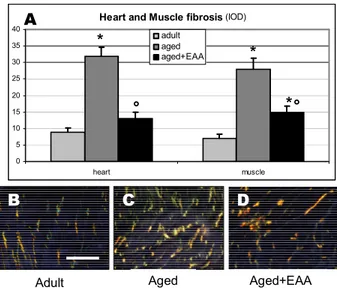
Considerable amelioration of fibrosis and less collagen deposition after EAA mixture supplementation were further demonstrated in muscles and heart by correlative light microscopy using Sirius red method (Fig. 3).
Heart and Muscle fibrosis (IOD) 0 5 10 15 20 25 30 35 40 heart muscle adult aged aged+EAA * * ° Aged+EAA Adult Aged B C D A * °
Fig. 3 A) Fibrosis degree in heart and skeletal muscle according to groups represented as Integrated Optical Density (IOD). The EAA
supplementation of aged animals reduces significantly the fibrosis degree similarly to adult ones in both heart and skeletal muscle * p<0.05 vs adult; ° p<0.05 vs aged. B-D) light micrographs taken by polarized light from heart showing the fibrosis in each groups. The aged plus EEA fed animals show very scarce fibrosis compared to aged ones (scale bar 20μm).
We remarked that the efficacy of EAA supplementation on mitochondria rejuvenation is organ specific, not beneficial in the liver and adipose tissue, and particularly beneficial in middle-age rodents that increased their average life-span [56]. If EAA addition stimulates organ-specific mitochondrial biogenesis and recovery, it may be useful and promising as adjuvant to maintain or prolong wellness in middle-age, so retarding unavoidable senescence.
Besides direct mitochondria observation and quantification, microscopy is helpful in indirect assessment of mitochondria physiology by visualization of selected enzymatic activities in tissues [57] and the expression of specific mitochondrial markers like chaperones HSP60 and GRP75 [58].
In middle-aged rat kidney we demonstrated that EAA supply potentiates chaperones immunostaining with respect to rodent fed normal diet [59]. The strengthening of chaperone activity is a crucial anti-aging strategy that makes mitochondria similar to juvenile conditions, so more able to react against ROS attacks. Indeed, mitochondrial chaperones are crucial in the proper folding and activity of respiratory chain enzymes, they are also able to mediate calcium flux inside mitochondria and represent the main sensor of oxidative stress [60]. Furthermore, inside mitochondria the activity of complex IV or cytochrome c oxidase (COX) is easily detectable by histochemistry. Considering that the catalytic subunits of COX are selectively encoded by mtDNA and are necessary for proper complex assembly, COX histochemistry is the gold standard method to indicate proper mitochondria status and changes during oxidative damage [12].
Moreover in the kidney the production of nitric oxide (NO) by different nitric oxide synthase isoforms may significantly influence hemodynamic and hypertension [61]. In our in vivo model we observed enhanced endothelial nitric oxide synthase (eNOS) in renal tubules in middle-aged rats after EAA supplementation, similar to younger rats and this finding well correlated with enhanced mitochondria biogenesis [62]. To strengthen the utility of long term EAA diet, we showed COX trend and eNOS immunostaining in the kidney of middle-aged (M-aged, 9 months old) rats plus or without supplementation compared with adult (4 months old) rats (Fig. 4).
M-Aged+EAA Adult M-Aged B C D IOD - Kidney 0 10 20 30 40 50 60 COX eNOS adult M-aged M-aged+EAA A * * ° °
Fig.4 A) Integrated optical density (IOD) of COX and eNOS in kidney according to groups. The EAA supplementation in
middle-aged (M-middle-aged) animals restores the COX and eNOS expression similarly to those observed in adult ones. * p<0.05 vs adult; ° p<0.05 vs aged. B-D) eNOS immunostaining in kidney according to groups. The M-aged+EEA fed animals show evident immunostaining like as adult ones (scale bar 20μm).
The results obtained indicated directly or indirectly mitochondrial rejuvenation after three months EAA intake and may be promising to suggest the prolonged use of specific nutrients in the prevention of aging-associated degenerative changes.
4. Taurine intake in puromycin aminonucleoside (PAN) nephrosis
Another nutritional rodent model described here is the introduction of the amino acid taurine, ad libitum via drinking water for two weeks, to rats presenting proteinuria and nephrosis induced by PAN, an adenosine derivative used as an antibiotic and anti-proliferative drug in humans [63]. The rationale of this study is due to our current interest in nutrition and previous experience in drugs inducing renal toxicity assessed in vivo and in vitro models [64, 65].
Taurine (TAU), known also as 2-aminoethanesulfonic acid, is a non-essential beta amino acid, which has many pleiotropic activities: it is not incorporated in proteins but works like a potent free radicals scavenger and osmo-regulator agent and prevents DNA damage at physiological concentrations [66-67].
TAU is present at high concentration in many organs like heart, brain, liver where it derives from others amino acids like methionine or cysteine [68].
Moreover it protects cellular membranes against oxidative damage and ischemia/reperfusion injury [69]. In the past, TAU has been successfully tested in rodents against chronic renal diseases such as streptozocin-induced diabetes, toxic nephropathies and hypertension [70, 71, 72].
The most used animal model to study nephrotic syndrome, and podocyte damage is PAN-injection in rats, whereas mice are generally adopted as transgenic models to study gene silencing [73, 74].
Recently it has been proposed to classify podocytopathies into four categories on the basis of the glomerular morphology and podocytes preservation: 1) minimal change disease (where podocyte number was unaffected); 2) focal segmental glomerulosclerosis; 3) mesangial sclerosis; 4) capillary collapse (high podocyte proliferation) [75].
A significant amount of our current knowledge on minimal change (MC) disease has come from experimental studies in rats injected with PAN, that develop podocyte foot effacement and loss of the electronegative charge of the glomerular barrier and reversible proteinuria [76].
It is necessary to remind that glomerular ultra-filtration barrier consists of three layers, starting from the blood flux: the endothelium with pores, the glomerular basement membrane and the visceral epithelial podocytes [77]. In particular podocytes represent a fundamental component of glomerular barrier, particularly vulnerable to injury: indeed they are high differentiated post-mitotic cells, similar to neurons, and unable to proliferate. Remarkably they do not restore after a severe stress and so glomerular filtration irreparably changes [78].
A recent wave of research indicates that quiescent post-mitotic podocytes undergo a metabolic dysfunction and in particularly mitochondrial deficit during experimental nephrosis and podocyte damage induced by aldosterone [79-80]. Indeed in humans, MC nephrosis, that affects mainly children, is generally treated by corticosteroids, which have adverse side-effects in long term schedule.
According to innovative studies by Clement [81] and Reisen [82], a new avenue to study the pathogenesis of MC nephrosis and its treatment is directed to abnormal podocytes that secrete a diffusible glycoprotein, called
Feeding rats with sialic acid precursors may increase ANGPTL4 sialylation and reduce proteinuria. A nutritional strategy with acid sialyc precursors, like N-acetyl-D-mannosamine, indicates an alternative way in the treatment of proteinuria linked to acquired MC disease. So novel nutritional strategies based on major sialylation, may alleviate the necessity to introduce large amount of steroids in organisms or even eliminate their necessity.
Other dietetic strategies based on antioxidants, like grape seed extract and vitamins, have been reported to ameliorate MC nephrosis and to prevent its progression into irreversible focal segmental glomerulosclerosis [83, 84]. If proteins inside the basement membrane of podocytes are directly involved in the charges distribution and selective transfer of elements from the blood, the continuous flux of materials requires metabolically active and efficient podocytes [85, 86].
Previous studies reported that podocytes hypertrophy and glomerular fibrosis in aging are ameliorated by caloric restriction in rats [87]. Morphological and molecular studies outline that it is essential to preserve mitochondria in podocytes for limiting the progression of acquired MC disease into unavoidable renal impairment [88, 89].
Indeed podocyte health has been considered not only a “morphologic” but also a “metabolic affair”, linked to many energy-consuming processes and therefore to the proper function of mitochondria, lysosomes and endoplasmic reticulum [90, 91].
To achieve this metabolic effect we tested TAU (1.5% in drinking water) one week before PAN (15 mg/hg) i.p. injected in adult rats and again another week after the antibiotic challenge. Proteinuria, measured by commercial kit in 24h collected urine, was significantly ameliorated by TAU administration versus PAN group together with mitochondrial chaperones expression within glomeruli. Among stress proteins analyzed, we focus here on mortalin/mt Hsp70/Grp75, a chaperone essential for delivery of mitochondrial enzymes, and strongly involved in the apoptotic response via the p53 regulation in nephrotoxic models [92]. Grp75 is down-regulated in aging kidney and in mercury nephropathy and influenced by nutritional antioxidant strategies [58, 63]. In Fig.5 we show microscopic feature of Grp75 immunolocalization in the rat glomerulus after TAU alone, PAN or combined PAN plus TAU regimens.
A B
C D
TAU PAN
PAN+TAU Saline
Fig.5 Grp75 immunolocalization in the rat glomerulus according to groups. (A) TAU administration did not vary significantly the
immunostaining compared to saline group (D). On the contrary PAN administration increased strongly the chaperone immunostaining (B). TAU administration in PAN treated animals attenuated significantly the chaperone signal (C), suggesting a protective role of TAU against oxidant damage. Scale bar 20μm
Remarkably in animals drinking TAU or normal tap water, used as control group, we observed almost similar weak brown reaction in the glomerular filter, while in contrast, after a single PAN injection, Grp75 staining significantly enhanced in the glomerulus. Finally after TAU plus PAN supplementation, the overall distribution of Grp75 was similar to controls. These findings might indicate that intense Grp75 is necessary to counteract ROS and oxidant damage induced by PAN [93] and probably pro-apoptotic changes as reported previously by Pedreanez et al. [94]. On the contrary, attenuated glomerular Grp75 signal indicates that the glomerular metabolism is not damaged anymore. Nevertheless it is important to outline that TAU is effective here on mitochondria probably as ROS scavenger because it is known that this beta-amino acid, devoid of a carboxyl group, is absent from cellular proteins and does not represent a direct source of energy.
In conclusion, a diet based on a specific amino acid alone or in a mixture may retain mitochondria rejuvenation in different experimental conditions and we believe that correlative light and electron microscopy based on histochemical, immunohistochemical and ultrastructural analyses play an important role in the modern nutritional research.
Acknowledgements The skillful contribution of Mrs. Claudia Romano and Mr. Giovanni Bozzoni in performing
immunohistochemistry and electron microscopy are gratefully acknowledged. Financial supports to the authors’ work derive from local grants (ex 60% MIUR 2012, A.S. and G.C.).
References
[1] Cavallini G, Donati A, Gori Z, Bergamini E. Towards an understanding of the anti-aging mechanism of caloric restriction. Curr Aging Sci. 2008; 1: 4-9.
[2] Hekini S, Guarente L. Genetics and the specificity of the aging process. Science 2003; 299: 1351-1354.
[3] Peterson K, Befroy S, Dufour D et al. Mitochondrial dysfunction in the elderly: possible role in insulin resistance. Science. 2003; 300: 1140-1142.
[4] Wallace D, Fan W, Procaccio V. Mitochondrial energetics and therapeutics. Annu Rev Pathol. 2010; 5: 297-348.
[5] Navarro A, Boveris A. The mitochondrial energy transduction system and the aging process. Am J Physiol Cell Physiol. 2007; 292: C670-C686.
[6] Ahmed N, Mandel R, Fain M. An emerging geriatric syndrome. Am J Med. 2007; 120: 748-753.
[7] Passos J, Zglinicki T. Mitochondrial dysfunction and cell senescence-skin deep into mammalian aging. Aging. 2012; 4: 74-75. [8] Seo A, Joseph A, Dutta D, Hwang J, Aris J et al. New insights into the role of mitochondria in aging: mitochondrial dynamics and
more. J Cell Sci. 2010; 123: 2533-2542.
[9] Dufour E, Larsson N. Understanding aging: revealing order out of chaos. Biochem Biophys Acta. 2004; 1658: 122-132. [10] Lopez-Lluch G, Irusta P, Navas P, de Cabo R. Mitochondrial biogenesis and healthy aging. Exp Gerontol. 2008; 43: 813-819. [11] Trifunovic A, Larsson N. Mitochondrial dysfunctions as a cause of ageing. J Int Med. 2008; 263: 167-178.
[12] Jendrach M, Mai S, Pohl S, Voth M, Bereiter-Hahn J. Short and long term alterations of mitochondrial morphology, dynamics and mtDNA after transient oxidative stress. Mitochondrion. 2008; 8: 293-304.
[13] Kerner J, Tutkaly P, Minkler P, Hoppel C. Aging skeletal muscle mitochondria in the rat: decreased uncoupling protein 3 content. Am J Physio Endocrin Metab. 2001; 281: E1054-E1062.
[14] Terman A, Kurz T, Navratil M, Arriaga E, Brunk U. Mitochondrial turnover and aging of long-lived postmitotic cells: the mitochondrial-lysosomal axis theory of aging. Antioxid Redox Signal. 2010; 12: 503-535.
[15] Finkel T, Holbrook N. Oxidants, oxidative stress and the biology of ageing. Nature. 2000; 408: 239-247.
[16] Chandel N, Budinger G. The cellular basis for diverse responses to oxygen. Free Radic Biol Med. 2007; 42: 165-174. [17] Salminen A, Kaarniranta K. SIRT1: a regulation of longevity via autophagy. Cell Signal. 2009; 21: 1356-1360.
[18] Trifunovic A, Wredenberg A, Falkenberg M et al. Premature ageing in mice expressing defective mitochondrial DNA polymerase. Nature. 2004; 429: 417-423.
[19] Hiona A, Sanz A, Kujoth G, et al. Mitochondrial DNA mutations induce mitochondrial dysfunctions, apoptosis and sarcopenia in skeletal muscle of mitochondrial DNA mutator mice. PLoS ONE. 2010; 5: e11468.
[20] Bereiter-Hahn J, Voth J. Dynamics of mitochondria in living cells: shape changes, dislocation, fusion and fission of mitochondria. Microsc Res Tech. 1994; 27: 198-219.
[21] Ozawa T. Genetic and functional changes in mitochondria associated with aging. Physiol Rev. 1997; 77: 425-464.
[22] Yang H, Yang T, Baur J, et al. Nutrient-sensitive mitochondrial NAD + levels dictate cell survival. Cell. 2007; 130: 1095-1107. [23] Mair W, Dillin A. Aging and survival : the genetics of life span extension by dietary restriction. Annu. Rev Biochem. 2008; 77:
727-754.
[24] Fontana L. Modulating human aging and age-associated diseases. Biochim Biophys Acta. 2009; 1790: 1133-1138.
[25] Roth L, Polotsky A. Can we live longer by eating less? A review of caloric restriction and longevity. Maturitas. 2012; 71: 315-319.
[26] Colman R, Anderson R. Non human primate calorie restriction. Antioxid. Redox Signal. 2011; 14: 229-239. [27] Fontana L, PartridgeL, Longo V. Extending healthy life span-from yeasts to humans. Science. 2010; 328: 321-326.
[28] Trepanowski J, Canale R, Marshall K, Kabir M, Bloomer R. Impact of caloric and dietary restriction regimens on markers of healthy and longevity in humans and animals: a summary of available findings. Nutrit J. 2011; 10: 107.
[29] Rochon J, Bales C, Ravussin E et al. Design and conduct of the CALERIE study: comprehensive assessment of the long term effects of reducing intake of energy. J Geronto A Biol Sci Med Sci. 2011; 66: 97-108.
[30] Ristow M, Schmeisser S. Extending life span by increasing oxidative stress. Free Radical Biology Med. 2011; 51: 327-336. [31] Ristow M, Zarse K. How increased oxidative stress promotes longevity and metabolic health: The concept of mitochondrial
hormesis (mitohormesis). Exp Gerontol. 2010; 45: 410-418.
[32] Sohal R. On W. The redox stress hypothesis of aging. Free Rad Biol Med. 2012; 52: 539-555.
[33] Smith D Jr, Nagy T, Allison D. Calorie restriction: what recent results suggest for the future of aging research. Eur J Clin Invest. 2010; 40: 440-450.
[34] Ahmed T, Haboubi N. Assessment and management of nutrition in older people and its importance to health. Clin Interv Aging. 2010; 5: 207-216.
[35] Ingram D, Zhu M, Mamczarz J, et al. Calorie restriction mimetic: an emerging research field. Aging Cell. 2006; 5: 97-108. [36] Longo V, Fontana L. Intermittent supplementation with rapamycin as a dietary restriction mimetic. Aging. 2011; 3: 1-2. [37] Baur J, Sinclair D. Therapeutic potential of resveratrol: the in vivo evidence. Nat Rev Drug Discov. 2006; 5: 493-506.
[38] Barger J, Kayo T, Vann J, et al. A low dose of dietary resveratrol partially mimics caloric restriction and retards aging parameters in mice. PLoS ONE. 2008; 3: e2264.
[39] Minor R, Smith D, Sossong A, et al. Chronic ingestion of 2-deoxy-d-glucose induces cardiac vacuolization and increases mortality in rats. Toxicol Appl Pharmacol. 2010; 243: 332-339.
[41] Calabrese V, Cornelius C, Cuzzocrea S, et al. Hormesis, cellular stress response and vitagenes as critical determinants in aging and longevity. Mol.Aspects Med. 2011; 32: 279-304.
[42] Guarente L. Mitochondria-A nexus for aging, calorie restriction, and sirtuins? Cell. 2008; 132: 171-176.
[43] Dancso B, Spiro Z, Arslan M, et al. The heat shock connection of metabolic stress and dietary restriction. Curr Pharmac Biotechnol. 2010; 11: 139-145.
[44] Vendelbo M, Nair K. Mitochondrial longevity pathways. Biochim Biophys Acta. 2011; 1813: 634-644.
[45] Weber T, Reichert A. Impaired quality control of mitochondria: Aging from a new perspective. Exp Gerontol. 2010; 45: 503-511.
[46] Fujita S, Volpi E. Amino Acids and Muscle Loss with Aging. J Nutr. 2006; 136: 2775-2805.
[47] Timmerman K, Volpi E. Amino acid metabolism and regulatory effects in aging. Curr Opin Clin Nutr Metab Care. 2008; 11: 45-49.
[48] Jourdan M, Deutz N, Cynober L, Aussel C. Features, causes and consequences of splanchnic sequestration of amino acid in old rats. PLoS ONE 2011; 6: e27002.
[49] Symons T, Schutzler S, Cocke T, et al. Aging does not impair the anabolic responses to a protein-rich meal. Am J Clin.Nutr. 2007; 86: 451-456.
[50] Elango R, Ball RO, Pencharz PB. Amino acid requirements in humans: with a special emphasis on the metabolic availability of amino acids. Amino Acids. 2009; 37: 19-27.
[51] Dioguardi F. Clinical use of amino acids as dietary supplement: pros and cons. J.Cachexia Sarcopenia Muscle 2011; 2: 75-80. [52] Wu G. Functional amino acids in growth, reproduction, and health. Adv.Nutr. 2010; 1: 31-37.
[53] Valerio A, D’Antona G, Nisoli E. Branched-chain amino acids, mitochondrial biogenesis, and healthspan: an evolutionary perspective. Aging. 2011; 3(5): 464-478.
[54] Corsetti G, Pasini E, D’Antona G, et al. Morphometric changes induced by amino acid supplementation in skeletal and cardiac muscles of old mice. Am J Cardiol. 2008; 101: 26E-34E.
[55] Pansarasa O, Flati V, Corsetti G, et al. Oral amino acid supplementation counteracts age-induced sarcopenia in elderly rats. Am J Cardiol. 2008; 101: 35E-41E.
[56] D’Antona G, Ragni M, Cardile A, et al. Branched chain amino acid supplementation promotes survival and supports cardiac and skeletal muscle mitochondrial biogenesis in middle-aged mice. Cell Metab. 2010; 12: 362-372.
[57] McKiernan S, Tuen V, Baldwin K, et al. Adult-onset calorie restriction delays the accumulation of mitochondrial enzyme abnormalities in aging rat kidney tubular epithelial cells. Am J Physiol Renal Physiol. 2007; 292: 1751-1760.
[58] Deocaris C, Kaul S, Wadhwa R. On the brotherhood of the mitochondrial chaperones: mortalin and heat shock protein 60. Cell Stress Chaperones 2006; 11: 116-128.
[59] Corsetti G, Stacchiotti A, D’Antona G, et al. Supplementation with essential amino acids in middle age maintains the health of rat kidney. Int J Immunopathol Pharmacol. 2010; 23: 523-533.
[60] Szabadkai G, Bianchi K, Varnai P, et al. Chaperone-mediated coupling of endoplasmic reticulum and mitochondrial Ca2+ channels. J Cell Biol. 2006; 175: 901-911.
[61] Kone B. Nitric oxide in renal health and disease. Am J.Kidney Dis. 1997; 30: 311-333.
[62] Nisoli E, Falcone S, Tonello C, Cozzi V, Palomba L, Fiorani M, Pisconti A et al. Mitochondrial biogenesis by NO yields functionally active mitochondria in mammals. Proc Natl Acad Sci USA. 2004; 101: 16507-16512.
[63] Stacchiotti A, Lavazza A, Rodella L, Rezzani R. Topicality of TEM in experimental nephrotoxicity. In: A.Mendez-Vilas and J.Diaz, eds. Microscopy Book Series 4. Microscopy: Science, Tecnology, Applications and Education. Formatex, Badajoz, Spain; 2010: Vol.2: 1245-1250.
[64] Stacchiotti A, Li Volti G, Lavazza A, et al. Different role of Schisandrin B on mercury-induced renal damage in vivo and in vitro. Toxicology. 2011; 286: 48-57.
[65] Rovetta F, Stacchiotti A, Cadei M, et al. ER signaling regulation drives the switch between autophagy and apoptosis in NRK-52E cells exposed to cisplatin. Exp Cell Res. 2012; 318: 238-250.
[66] Messina S, Dawson R Jr. Attenuation of oxidative damage to DNA by taurine and taurine analogs. Adv Exp Med Biol. 2000; 483: 355-367.
[67] Gupta R, Win T, Bittner S. Taurine analogues; a new class of therapeutics: retrospect and prospects. Curr Med Chem. 2005; 12: 2021-2039.
[68] Chesney R, Han X, Patters A. Taurine and the renal system. J Biomed Sci. 2010; 17: S4.
[69] Guz G, Oz E, Lortlar N, et al. The effect of taurine on renal ischemia/ reperfusion injury. Amino Acids 2007; 32: 405-411. [70] Trachtman H, Futterweit S, Maesaka J, et al. Taurine ameliorates chronic streptozocin-induced diabetic nephropathy in rats. Am
J Physiol. 1995; 269: F429-F438.
[71] Pushpakiran G, Mahalakshmi K, Viswanathan P, Anuradha C. Taurine prevents ethanol-induced alterations in lipids and ATPases in rat tissues. Pharmacol Rep. 2005; 57: 578-587.
[72] Hagar H, Etter E, Arafa M. Taurine attenuates hypertension and renal dysfunction induced by cyclosporine A in rats. Clin Exp Pharmacol Physiol. 2006; 33: 189-196.
[73] Pippin J, Brinkkoetter P, Cormack-Aboud F, Durvasula R, Hauser P, et al. Inducible rodent models of acquired podocyte diseases. Am J Physiol Renal Physiol. 2009; 296: F213-F229.
[74] Chugh S, Clement L, Macè C. New insights into human minimal change disease: lessons from animal models. Am J Kidney Dis. 2011
[75] Barisoni L, Schnaper H, Kopp J. A proposed taxonomy for the podocytopathies: a reassessment of the primary nephritic diseases. Clin J Am Soc Nephrol. 2007; 2: 529-542.
[76] Andreoli S. Reactive oxygen-molecules, oxidant injury and renal disease. Pediatr Nephrol. 1991; 5: 733-742.
[77] Haraldsson B, Nystrom J, Deen W. Properies of the glomerular barrier and mechanisms of proteinuria. Physiol Rev. 2008; 88: 451-487.
[78] Kriz W. Podocyte is the mayor culprit accounting for the progression of chronic renal disease. Microsc Res Tech. 2002; 57: 189-195.
[79] Yan K, Ito N, Kurayama R, et al. The struggle for energy in podocytes leads to nephrotic syndrome. Cell Cycle 11, 1504-1511, 2012.
[80] Zhu C, Huang S, Yuan Y, et al. Mitochondrial dysfunction mediates aldosterone-induced podocyte damage : a therapeutic target of PPARγ. Am J Pathol. 2011; 178: 2020-2031.
[81] Clement L, Avila-Casado C, Macè C, et al. Podocyte-secreted angiopoietin-like-4 mediates proteinuria in glucocorticoid-sensitive nephrotic syndrome. Nat Med. 2011; 17: 117-122.
[82] Reisen. Filtering new facts about kidney disease. Nat Med. 2011; 17: 44-45.
[83] Mattoo T, Kovacevic L. Effect of grape seed exctract on puromycin-aminonucleoside-induced nephrosis in rats. Pediatr Nephrol. 2003; 18: 872-877.
[84] Matsui I, Hamano T, Tomida K, et al. Active vitamin D and its analogue, 22-oxacalcitriol, ameliorate puromycin aminonucleoside-induced nephrosis in rats. Nephrol Dial Transplant. 2009; 24: 2354-2361.
[85] Mathieson P. Update on the podocyte. Curr Opin Nephrol Hyperthens. 2009; 18: 206-211. [86] Fogo A. The targeted podocyte. J Clin Invest. 2011; 121(6): 2142-2145.
[87] Wiggins J, Goyal M, Sanden S, et al. Podocyte hypertrophy, adaptation and decompensation associated with glomerular enlargement and glomerulosclerosis in the aging rat: prevention by calorie restriction. J Am Soc Nephrol. 2005; 16: 2953-2966. [88] Hagiwara M, Yamagata K, Capaldi R, Koyama A. Mitochondrial dysfunction in focal segmental glomerulosclerosis of
puromycin aminonucleoside nephrosis. Kidney Int. 2006; 69: 1146-1152.
[89] Sasaki S. Determination of altered mitochondria ultrastructure by electron microscopy. Methods Mol.Biol. 2010; 648: 279-290. [90] Ito N, Nishibori Y, Ito Y, et al. mTORC1 activation triggers the unfolded protein response in podocytes and leads to nephrotic
syndrome. Lab Invest. 2011; 91: 1584-1595.
[91] Yan K, Ito N, Nakajo A, et al. The struggle for energy in podocytes leads to nephrotic syndrome. Cell Cycle. 2012; 11: 1504-1511.
[92] Jiang M, Dong Z. Regulation and pathological role of p53 in cisplatin nephrotoxicity. J Pharmacol Exp Therapeut.2008; 327: 300-307.
[93] Liu H, Bigler S, Henegar J, Baliga R. Cytochrome P4502B1 mediates oxidant injury in puromycin-induced nephritic syndrome. Kidney Int. 2002; 62: 868-876.
[94] Pedreanez A, Rincon J, Romero M, Viera N, Mosquera J. Melatonin decreases apoptosis and expression of apoptosis-associated proteins in acute puromycin aminonucleoside nephrosis. Nephrol. Dial Transplant. 2004; 19: 1098-1105.