Diagnostic accuracy of first-trimester ultrasound in detecting
abnormally invasive placenta in high-risk women with
placenta previa
G. CALI
1, F. FORLANI
1, F. FOTI
1, G. MINNECI
1, L. MANZOLI
2, M. E. FLACCO
3, D. BUCA
4,
M. LIBERATI
4, G. SCAMBIA
5and F. D’ANTONIO
6,71Department of Obstetrics and Gynaecology, Arnas Civico Hospital, Palermo, Italy;2Department of Medical Sciences, University of Ferrara, Ferrara, Italy;3Local Health Unit of Pescara, Pescara, Italy;4Department of Obstetrics and Gynecology, University of Chieti, Chieti, Italy;5Department of Obstetrics and Gynaecology, Catholic University of The Sacred Heart, Rome, Italy;6Women’s Health and Perinatology Research Group, Department of Clinical Medicine, Faculty of Health Sciences, UiT-The Arctic University of Norway, Tromsø, Norway;7Department of Obstetrics and Gynaecology, University Hospital of Northern Norway, Tromsø, Norway
K E Y W O R D S: abnormally invasive placenta; diagnostic accuracy; first trimester; ultrasound
ABSTRACT
Objective To ascertain the diagnostic accuracy of
ultra-sound in detecting abnormally invasive placenta (AIP) during the first trimester of pregnancy (11–14 weeks’ gestation) in women at risk for this condition.
Methods This was a retrospective analysis of data
collected prospectively from women at risk for AIP based upon the presence of at least one prior Cesarean section (CS) and/or uterine surgery and placenta previa, who had ultrasound assessment for AIP at the time of the 11–14-week scan. The ultrasound signs explored in the present study were: loss of the clear zone, placental lacunae, bladder wall interruption and uterovesical hypervascularity. The potential of ultrasound and different ultrasound signs to predict the different types of AIP was assessed by computing summary estimates of sensitivity, specificity, diagnostic odds ratio (DOR) and positive (LR+) and negative (LR–) likelihood ratios.
Results One hundred and eighty-eight women with
placenta previa and at least one previous CS or uterine surgery were included in the study. All the explored ultrasound signs were associated significantly with the occurrence of AIP. Overall, when at least one ultrasound sign was used to make the diagnosis, ultrasound had a sensitivity of 84.3% (95% CI, 74.7–91.4%), specificity of 61.9% (95% CI, 51.9–71.2%), DOR of 8.6 (95% CI, 4.1–19.3), LR+ of 2.2 (95% CI, 1.7–2.9) and LR– of 0.3 (95% CI, 0.1–0.4) in detecting AIP. Using two ultrasound signs to label a case as positive increased the
Correspondence to: Dr F. D’Antonio, Department of Obstetrics and Gynaecology, University Hospital of Northern Norway, Department of Clinical Medicine, Faculty of Health Sciences, UiT – The Arctic University of Norway, Hansine Hansens veg 18, 9019 Tromsø, Norway (e-mail: [email protected])
Accepted: 3 March 2018
diagnostic accuracy in terms of specificity, although it did not affect sensitivity. Among the different ultrasound signs, loss of the clear zone had a sensitivity of 84.3% (95% CI, 74.7–91.4%) and a specificity of 81.9% (95% CI, 73.2–88.7%) in detecting AIP, while sensitivities for placental lacunae and bladder wall interruption were 78.3% (95% CI, 67.9–86.6%) and 75.9% (95% CI, 65.3–84.6%), respectively, and specificities were 81.0% (95% CI, 72.1–88.0%) and 99.1% (95% CI, 94.8–100.0%), respectively. The optimal combination of sensitivity and specificity was achieved when at least two imaging signs of AIP were used in the diagnostic algorithm.
Conclusions AIP can be detected from the first trimester
of pregnancy in women at risk for this condition, and ultrasound performed between 11 and 14 weeks’ gestation has an overall good diagnostic accuracy for detecting all types of AIP. However, these findings are applicable only to women with placenta previa and prior uterine scar. Copyright© 2018 ISUOG. Published by John Wiley & Sons Ltd.
INTRODUCTION
The rise in Cesarean-section (CS) rate observed during the last two decades has led to a large increase in the prevalence of abnormally invasive placenta (AIP)1.
AIP encompasses a spectrum of disorders characterized by various degrees of placental invasion through the myometrium and uterine serosa. It is associated with
a high burden of maternal morbidities such as severe life-threatening hemorrhage, need for blood transfusion, reoperation and damage to adjacent organs2–4.
Prenatal diagnosis of AIP is fundamental and it has been reported to improve outcome by allowing preplanned treatment in centers with a high level of surgical expertise5. Although the prenatal diagnosis of AIP is
commonly achieved during the second or third trimester of pregnancy, there are reports suggesting that signs of AIP are already present in early pregnancy6.
A recent systematic review exploring the diagnostic performance of first-trimester ultrasound in detecting AIP reported that signs of AIP can be detected in about 90% of women affected by these anomalies who are scanned during the first trimester of pregnancy6. Despite this, the
small sample size of included studies, heterogeneity in gestational age at assessment and explored ultrasound signs, and inclusion of only cases with confirmed AIP, with subsequent lack of information on specificity, does not allow extrapolation of robust evidence on the actual diagnostic performance of ultrasound in detecting AIP during the first trimester of pregnancy.
The aim of this study was to ascertain the diagnostic accuracy of ultrasound in detecting AIP during the first trimester of pregnancy in women at risk for this condition.
METHODS
This was a retrospective analysis of data collected prospectively from women at risk for AIP who were referred to our center between 2007 and 2017. These women were identified from an electronic database of the fetal medicine unit. Delivery details were retrieved from hospital maternity records, and operative notes were checked for details of operative findings and interventions performed.
Inclusion criteria were women with at least one prior CS and/or uterine surgery and placenta previa who had an ultrasound assessment for AIP at the time of the 11–14-week scan. Repeat assessments were performed in the second and third trimesters of pregnancy. Data regarding the presence of the different ultrasound signs of AIP were entered prospectively at the time of the original examination. Two examiners (G.C., F.D.A.), blinded to pregnancy outcome and pathology reports, analyzed all the stored images independently and labeled them according to the presence of different ultrasound signs suggestive of AIP.
The ultrasound signs explored in the present study (Figure 1) were7: (1) loss of the ‘clear zone’, defined as loss
or irregularity of the hypoechoic plane in the myometrium beneath the placental bed; (2) placental lacunae, defined as the presence of numerous (at least three) lacunae, often containing turbulent flow visible on gray-scale or color Doppler ultrasound; (3) bladder wall interruption, defined as loss or interruption of the bright bladder wall (hyperechoic band or ‘line’ between the uterine serosa and bladder lumen); and (4) uterovesical hypervascularity, defined as a striking amount of color Doppler signal
seen between the myometrium and posterior wall of the bladder, including vessels appearing to extend from the placenta, across the myometrium and beyond the serosa into the bladder or other organs, often running perpendicularly to the myometrium.
Ultrasound assessment was performed transabdomi-nally in all cases, while transvaginal ultrasound was limited to cases with a strong suspicion of AIP, cases in which transabdominal ultrasound did not allow overall good visualization of the bladder–uterine interface and retroplacental space or in cases of posterior placenta. All examinations were performed originally using a GE Volu-son 730 or GE VoluVolu-son E8 (GE Healthcare Italy, Milan, Italy) or a Samsung WS80A Elite (Samsung Healthcare Italy, Milan, Italy) ultrasound machine, equipped with a 4.0–6.0-MHz curved transabdominal or 5.0–7.0-MHz transvaginal transducer. When using color Doppler ultra-sound, the pulse-repetition frequency was initially set at 1.3 kHz, but was later lowered in order to identify the presence of placental lacunar flow.
The final diagnosis of the type of AIP was made after surgery and hysterectomy on the basis of the pathological examination of the removed uterus. Placenta accreta was diagnosed when anchoring placental villi were attached to the myometrium rather than to the decidua, but without completely invading it. Placenta increta was diagnosed when chorionic villi penetrated the myometrium, while placenta percreta was diagnosed when chorionic villi penetrated through the myometrium into the uterine serosa or adjacent organs4.
We planned a sensitivity analysis according to the depth of placental invasion and the number of prior CSs. For the purpose of this analysis, AIP was divided into two different subgroups: placenta accreta/increta and placenta percreta. STARD (Standards for Reporting of Diagnostic Accuracy Studies) guidelines for studies on diagnostic accuracy were followed8.
We investigated the potential association between AIP – overall and by degree of invasion (placenta acc-reta/increta or percreta) – and nine potential predictors, including four ultrasound signs (loss of the clear zone, pla-cental lacunae, bladder wall interruption and uterovesi-cal hypervascularity) and five other maternal/gestational characteristics (mother’s age, gestational age at birth, par-ity, number of previous CSs and previous uterine surgery). We first evaluated the prevalence of AIP by each potential predictor using standard univariate analysis: the chi-square test for categorical variables and the t-test and Kruskal–Wallis test for normally and non-normally distributed continuous variables, respectively (distribu-tion assessed using the Shapiro–Wilk test). Both number of previous CSs and overall number of detected ultra-sound signs (which was computed for each woman) were included in the analyses, both in their original (contin-uous) form and after dichotomization. The number of previous CSs was split into three dichotomous variables, each including women with one or no, two or three or more CSs; the overall number of ultrasound signs was
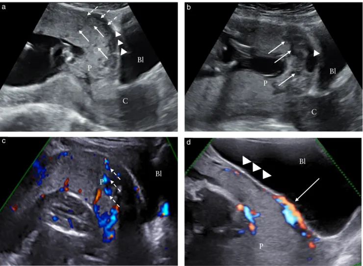
Bl Bl P P C Bl Bl P P C
Figure 1 Ultrasound signs of abnormally invasive placenta in first trimester of pregnancy (11–14 weeks’ gestation). (a) Transabdominal
ultrasound (TAS), showing anterior placenta (P) previa, in which several hypoechoic round areas (placental lacunae) can be detected in placental parenchyma (arrows) and irregularity of hypoechoic plane beneath placental bed (loss of clear zone) is present (dotted arrows), as well as interruption of hyperechoic line between uterine serosa and bladder (Bl) lumen (bladder wall interruption, arrowheads). (b) TAS at 13 weeks, showing placenta implanted above internal cervical os (major previa), in which several placental lacunae (arrows) and interruption of bladder wall can be detected. (c) Color Doppler imaging, showing presence of blood flow within lacunae (dashed arrows). (d) Color Doppler imaging, showing presence of blood flow at level of bladder wall (arrow); irregularity of retroplacental clear zone is also visible (arrowheads). C, cervix.
used to generate four dichotomous variables (women with no, one, two or three or more detected signs).
Stepwise forward logistic regression analysis was then used to identify potential independent predictors of AIP. All covariates were tested for inclusion in the final model, in which only those significant on univariate analysis were retained. To reduce potential overfitting, the overall number of covariates was limited to 1/10 of the successes in all phases of model building. The goodness-of-fit was checked using the Hosmer–Lemeshow test, and the predictive power assessed through C-statistics (area under the receiving–operating characteristics curve). Standard postestimation tests were used to check the final model for validity, performing multicollinearity and influential observation analysis (using standardized residuals, change in Pearson’s chi-square and deviance). There were no missing values, thus no missing imputation technique was adopted.
We finally estimated the potential of each ultrasound sign to predict AIP, computing summary estimates of
sensitivity, specificity, positive and negative likelihood ratios (LR+ and LR–) and diagnostic odds ratios (DOR). Similarly, we assessed the diagnostic accuracy of the presence of two or≥ three ultrasound signs in the same woman, and of one, two or≥ three previous CSs.
Statistical significance was defined as a two-sided P of < 0.05, and analysis was performed using Stata 13.1 (StataCorp, College Station, TX, USA, 2013).
RESULTS
One hundred and eighty-eight women with placenta previa and at least one previous CS or uterine surgery were included in the study. The prevalence of AIP was 44.2% (95% CI, 37.2–51.3%); among the different types of AIP, placenta accreta/increta occurred in 54.2% (95% CI, 44.6–64.5%) while percreta occurred in 45.8% (95% CI, 35.5–56.5%) of cases.
General characteristics of the population analyzed in the study are reported in Table 1. Compared with those
unaffected by AIP, maternal age (34.3± 4.2 vs 29.6 ± 5.3; P= 0.0001), parity (2 (2–3) vs 2 (0–2); P = 0.0001) and number of previous CSs (2 (1–2) vs 2 (0–2); P= 0.0006) were higher in affected women, and these differences persisted when the women were stratified according to the severity of placental invasion. On logistic regression analysis, maternal age (OR 1.2 (95% CI, 1.1–1.4)) and number of ultrasound signs detected on the scan (OR 8.4 per 1-unit increase (95% CI, 4.2–17.0 per 1-unit increase)) were associated independently with the occurrence of AIP (Table S1).
Table 2 reports the prevalence and risk of detecting the different ultrasound signs of AIP in women affected, compared with those unaffected, by AIP. When comparing the prevalence of the different ultrasound signs of AIP explored in the present study between the first (11–14 weeks) and the second/third trimesters of pregnancy, loss of the clear zone was detected in 84.3% of cases in the first and in 92.4% in the second/third trimesters of pregnancy (P= 0.104), while the corresponding figures for placenta lacunae, bladder wall interruption and uterovesical hypervascularity were
78.3% and 100% (P= 0.0001), 75.9% and 93.3% (P= 0.0013) and 50.6% and 81.0% (P = 0.0001), respectively. There was full agreement in the labeling of all images with regard to the type of ultrasound sign between the original examiner and the two researchers in this study.
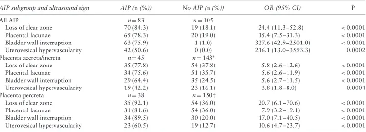
Loss of the clear zone was associated with a higher risk of AIP, with an OR of 24.4 (95% CI, 11.3–52.8) (Table 2); likewise, the presence of placental lacunae (OR 15.4 (95% CI, 7.5–31.3)), bladder wall interruption (OR 327.6 (95% CI, 42.9–2501.0)) and uterovesical hypervascularity (OR 216.1 (95% CI, 13.0–3593.3)) were more prevalent in women affected by AIP than in those unaffected by AIP who were scanned during the first trimester of pregnancy. When stratifying the analysis according to the severity of placental invasion, the strength of association between each of the explored ultrasound signs persisted (Table 2). However, in women affected by placenta percreta, there was a higher prevalence of each of the ultrasound signs suggestive of AIP than in controls. Table 3 reports the diagnostic accuracy of the different ultrasound signs in detecting AIP at the 11–14-week scan. Table 1 Characteristics of pregnant women at risk for abnormally invasive placenta (AIP), with univariate analyses comparing each
potential predictor in affected vs unaffected women, overall and according to severity of AIP
Characteristic Overall sample (n= 188) No AIP (n= 105) All AIP (n= 83) P* Placenta accreta/ increta (n= 45) P† Placenta percreta (n= 38) P‡ Maternal age (years) 31.7± 5.4 29.6± 5.3 34.3 ± 4.2 < 0.001 34.5± 4.6 <0.001 34.1± 3.7 <0.001 GA at delivery (weeks) 35.4± 2.3 36.3± 1.1 34.2 ± 2.9 < 0.001 35.1± 1.6 <0.001 33.3± 3.7 <0.001 Parous 46 (24.5) 22 (21.0) 24 (28.9) 0.234 14 (31.1) 0.212 10 (26.3) 0.502 Previous CS ≤ 1 75 (39.9) 52 (49.5) 23 (27.7) 0.003 14 (31.1) 0.048 9 (23.7) 0.007 2 71 (37.8) 31 (29.5) 40 (48.2) 0.010 21 (46.7) 0.06 19 (50.0) 0.029 ≥ 3 42 (22.3) 22 (21.0) 20 (24.1) 0.745 10 (22.2) 0.832 10 (26.3) 0.502
Previous uterine surgery§ 70 (37.2) 70 (66.7) 0 (0.0) — 0 (0.0) — 0 (0.0) —
Data are given as mean± SD or n (%). P calculated using chi-square test for categorical variables, and t-test and Kruskal–Wallis test for normally and non-normally distributed continuous variables, respectively. *All women diagnosed with AIP vs those without AIP. †Women diagnosed with placenta accreta or increta vs those without AIP. ‡Women diagnosed with placenta percreta vs those without AIP. §Other than Cesarean section (CS). GA, gestational age.
Table 2 Odds ratios (OR) for prediction of abnormally invasive placenta (AIP) for each ultrasound sign explored in present study of 188
high-risk women, overall and according to severity of AIP
AIP subgroup and ultrasound sign AIP (n (%)) No AIP (n (%)) OR (95% CI) P
All AIP n= 83 n= 105
Loss of clear zone 70 (84.3) 19 (18.1) 24.4 (11.3–52.8) <0.0001
Placental lacunae 65 (78.3) 20 (19.0) 15.4 (7.5–31.3) <0.0001
Bladder wall interruption 63 (75.9) 1 (1.0) 327.6 (42.9–2501.0) <0.0001
Uterovesical hypervascularity 42 (50.6) 0 (0.0) 216.1 (13.0–3593.3) 0.0002
Placenta accreta/increta n= 45 n= 143∗
Loss of clear zone 35 (77.8) 54 (37.8) 5.8 (2.6–12.6) <0.0001
Placental lacunae 34 (75.6) 51 (35.7) 5.6 (2.6–11.9) <0.0001
Bladder wall interruption 29 (64.4) 35 (24.5) 5.6 (2.7–11.5) <0.0001
Uterovesical hypervascularity 19 (42.2) 23 (16.1) 3.8 (1.8–8.0) 0.0004
Placenta percreta n= 38 n= 150†
Loss of clear zone 35 (92.1) 54 (36.0) 20.7 (6.1–70.6) <0.0001
Placental lacunae 31 (81.6) 54 (36.0) 7.9 (3.2–19.1) <0.0001
Bladder wall interruption 34 (89.5) 30 (20.0) 17.0 (7.1–40.5) <0.0001
Uterovesical hypervascularity 23 (60.5) 19 (12.7) 10.6 (4.7–23.7) <0.0001
Loss of the clear zone had an overall good diagnostic accuracy in detecting AIP, with a sensitivity of 84.3% (95% CI, 74.7–91.4%), specificity of 81.9% (95% CI, 73.2–88.7%), DOR of 23.8 (95% CI, 10.6–57.2), LR+ of 4.7 (95% CI, 3.1–7.1) and LR– of 0.2 (95% CI, 0.1–0.3); placental lacunae and bladder wall interruption had sensitivities of 78.3% (95% CI, 67.9–86.6%) and 75.9% (95% CI, 65.3–84.6%), respectively, while the corresponding values for specificity were 81.0% (95% CI, 72.1–88.0%) and 99.1% (95% CI, 94.8–100.0%). Finally, the presence of uterovesical hypervascularity alone had low sensitivity (50.6% (95% CI, 39.4–61.8%)) but high specificity (100% (95% CI, 96.6–100%)) in identifying AIP at the 11–14-week scan. When exploring the diagnostic performance of different ultrasound signs for detecting the different types of AIP during the first trimester, loss of the clear zone (sensitivity 92.1% (95% CI, 78.6–98.3%)), placental lacunae (sensitivity 81.6% (95% CI, 65.7–92.3%)), bladder wall interruption (sensitivity 89.5% (95% CI, 75.2–97.1%)) and uterovesical hypervascularity (sensitivity 60.5% (95% CI, 43.4–76.0%)) had a higher sensitivity for detecting placenta percreta than for detecting less severe types of AIP (Table 3).
The diagnostic performance of first-trimester ultra-sound in detecting AIP according to the number of imaging
signs used is also shown in Table 3. Overall, when using at least one sign, ultrasound had a sensitivity of 84.3% (95% CI, 74.7–91.4%), specificity of 61.9% (95% CI, 51.9–71.2%), DOR of 8.6 (95% CI, 4.1–19.3), LR+ of 2.2 (95% CI, 1.7–2.9) and LR– of 0.3 (95% CI, 0.1–0.4). Using two ultrasound signs to label a case as positive increased the diagnostic accuracy in terms of specificity although it did not affect sensitivity. When stratifying the analysis according to the severity of placental invasion, using at least one sign had sensitivities of 77.8% (95% CI, 62.9–88.8%) and 92.1% (95% CI, 78.6–98.3%) for pla-centa accreta/increta and percreta, respectively, but poor specificities (47.6% (95% CI, 39.2–56.1%) and 50.0% (95% CI, 41.7–58.3%), respectively). However, when using at least two ultrasound signs, specificity improved, to 75.5% (95% CI, 67.6–82.3%) and 76.7% (95% CI, 69.1–83.2%) for placenta accreta/increta and percreta, respectively (Table 3).
Finally, we explored which combination of ultrasound signs was associated with the optimal diagnostic accuracy in detecting AIP at the 11–14-week scan. For all types of AIP, loss of the clear zone together with placental lacunae or bladder wall interruption showed the best diagnostic performances, with respective sensitivities of 78.3% (95% CI, 67.9–86.6%) and 75.9% (95% CI, 65.3–84.6%), both having a specificity of 100% (95% Table 3 Diagnostic accuracy of different first-trimester ultrasound signs and number of detected signs for abnormally invasive placenta
(AIP) in 188 high-risk women during first trimester of pregnancy, overall and according to severity of AIP
AIP subgroup Sensitivity (95% CI) (%) Specificity (95% CI) (%) DOR (95% CI) LR+ (95% CI) LR– (95% CI) All AIP Ultrasound sign
Loss of clear zone 84.3 (74.7–91.4) 81.9 (73.2–88.7) 23.8 (10.6–57.2) 4.7 (3.1–7.1) 0.2 (0.1–0.3) Placental lacunae 78.3 (67.9–86.6) 81.0 (72.1–88.0) 15.1 (7.1–33.5) 4.1 (2.8–6.3) 0.3 (0.2–0.4) Bladder wall interruption 75.9 (65.3–84.6) 99.1 (94.8–100.0) 313.0 (48.5–13259.3) 79.7 (14.5–452.2) 0.2 (0.2–0.3) Uterovesical hypervascularity 50.6 (39.4–61.8) 100.0 (96.6–100.0) ∞ (25.8–∞) ∞ (14.3–∞) 0.5 (0.4–0.6) Number of signs ≥ 1 84.3 (74.7–91.4) 61.9 (51.9–71.2) 8.6 (4.1–19.3) 2.2 (1.7–2.9) 0.3 (0.1–0.4) ≥ 2 84.3 (74.7–91.4) 100.0 (96.6–100.0) ∞ (122.7–∞) ∞ (23.9–∞) 0.2 (0.1–0.3) ≥ 3 69.9 (58.8–79.5) 100.0 (96.6–100.0) ∞ (57.0–∞) ∞ (19.8–∞) 0.3 (0.2–0.4) Placenta accreta/increta Ultrasound sign
Loss of clear zone 77.8 (62.9–88.8) 62.2 (53.8–70.2) 5.7 (2.5–14.0) 2.1 (1.6–2.7) 0.4 (0.2–0.6) Placental lacunae 75.6 (60.5–87.1) 64.3 (55.9–72.2) 5.5 (2.5–13.2) 2.1 (1.6–2.8) 0.4 (0.2–0.6) Bladder wall interruption 64.4 (48.8–78.1) 75.5 (67.6–82.3) 5.5 (2.6–12.3) 2.6 (1.8–3.8) 0.5 (0.3–0.7) Uterovesical hypervascularity 42.2 (27.7–57.9) 83.9 (76.9–89.5) 3.8 (1.7–4.3) 0.7 (0.5–0.9) Number of signs ≥ 1 77.8 (62.9–88.8) 47.6 (39.2–56.1) 3.2 (1.4–7.1) 1.5 (1.2–1.8) 0.5 (0.3–0.8) ≥ 2 77.8 (62.9–88.8) 75.5 (67.6–82.3) 10.6 (4.6–26.7) 3.2 (2.3–4.4) 0.3 (0.2–0.5) ≥ 3 62.2 (46.5–76.2) 78.3 (70.7–84.8) 5.9 (2.7–13.1) 2.9 (1.9–4.2) 0.5 (0.3–0.7) Placenta percreta Ultrasound sign
Loss of clear zone 92.1 (78.6–98.3) 64.0 (55.8–71.7) 20.4 (6.0–108.7) 2.6 (2.0–3.2) 0.1 (0.04–0.3) Placental lacunae 81.6 (65.7–92.3) 64.0 (55.8–71.7) 7.8 (3.1–22.4) 2.3 (1.7–2.9) 0.3 (0.1–0.5) Bladder wall interruption 89.5 (75.2–97.1) 80.0 (72.7–86.1) 33.2 (10.7–138.5) 4.5 (3.2–6.3) 0.1 (0.05–0.3) Uterovesical hypervascularity 60.5 (43.4–76.0) 87.3 (80.9–92.2) 10.4 (4.3–25.7) 4.8 (2.9–7.8) 0.5 (0.3–0.6) Number of signs
≥ 1 92.1 (78.6–98.3) 50.0 (41.7–58.3) 11.6 (3.4–61.2) 1.8 (1.5–2.2) 0.2 (0.1–0.4)
≥ 2 92.1 (78.6–98.3) 76.7 (69.1–83.2) 37.5 (10.8–201.7) 4.0 (2.9–5.4) 0.1 (0.04–0.3)
≥ 3 79.0 (62.7–90.5) 80.7 (73.4–86.7) 15.3 (6.1–43.0) 4.1 (2.8–5.9) 0.3 (0.2–0.5)
CI, 96.6–100%) (Table S2). Loss of the clear zone and placental lacunae was the combination of ultrasound signs that predicted placenta accreta/increta most accurately, with a sensitivity of 75.6% (95% CI, 60.5–87.1%) and a specificity of 78.3% (95% CI, 70.7–84.8%). Finally, loss of the clear zone and either placental lacunae or bladder wall interruption showed the highest detection rates for placenta percreta, with sensitivities of 81.6% (95% CI, 65.6–92.3%) and 89.5% (95% CI, 75.2–97.1%) and specificities of 77.3% (95% CI, 69.8–83.8%) and 80.7% (95% CI, 73.4–86.7%), respectively (Table S2).
DISCUSSION
The findings of this study show that AIP can be detected from the first trimester of pregnancy in women at risk for this condition, and that ultrasound performed between 11 and 14 weeks’ gestation has an overall good diagnostic accuracy for detecting all types of AIP.
The major strengths of the study are the inclusion of a population with objectively recognized and homogeneous risk factors for AIP, stratification of the analysis according to the severity of placental invasion and assessment of specificity. Retrospective design, small sample size, lack of assessment of all the ultrasound signs suggestive of AIP reported in the published literature, lack of information from early first-trimester (5–10 weeks’ gestation) ultrasound and inclusion only of cases of AIP undergoing hysterectomy, represent the major limitations of the study. The results are applicable only to women with placenta previa and prior uterine scar because all cases of AIP in our population occurred in women with such risk factors. However, AIP can occur even in women with no classical risk factors for these conditions9.
Ultrasound has been shown to have an overall good diagnostic accuracy for detecting AIP, especially when different imaging signs are integrated with maternal and pregnancy characteristics in a multiparametric diagnostic algorithm10–12. Despite this, it has still to be ascertained
when to scan women at risk for AIP in order to detect more accurately these anomalies. Prenatal diagnosis of AIP is commonly performed during the second and third trimesters of pregnancy, but there are no robust data on first-trimester diagnosis. The present study shows that prenatal diagnosis of AIP is feasible during the first trimester of pregnancy at the time of the 11–14-week scan and that it has a good diagnostic performance, not only in detecting such disorders but also in diagnosing their severity. The main aim of first-trimester diagnosis of AIP would be to identify those women at high risk so that they can be referred to centers with expertise in the diagnosis and treatment of these disorders.
One of the major determinants of surgical outcome in women affected by AIP is the depth of placental invasion, with women affected by placenta percreta showing a greater frequency of surgical complications compared with those with placenta accreta or increta2. In the
present study, ultrasound assessment at 11–14 weeks
was able to identify about 90% of women affected by placenta percreta, showing that the optimal combination of sensitivity and specificity was achieved when predictive algorithms integrating loss of the clear zone and placental lacunae or bladder wall interruption were adopted. Furthermore, the overall diagnostic accuracy of ultrasound was higher in detecting placenta percreta than in detecting less severe types of AIP.
One of the most relevant issues when trying to diagnose AIP during the first trimester of pregnancy is which subset of women should be referred for assessment, because risk stratification for AIP in early pregnancy might not be completely clear. The major risk factors for AIP are placenta previa and previous CS2. However, first-trimester
diagnosis of placenta previa is not completely reliable, as a significant proportion of the placenta could move away from the cervix in the second and third trimesters of pregnancy. It might be hypothesized that only women presenting with major placenta previa, defined as that completely covering the internal cervical os, should be referred for ultrasound assessment. It has been reported that the distance of the placental edge to the cervical os may help in predicting placenta previa at delivery and that, if the placenta completely covers the internal cervical os, the chance of migration is low13.
Further large studies are needed in order to identify those women at higher risk for AIP who would benefit from early ultrasound screening for this condition and to ascertain whether combining first-, second- and third-trimester ultrasound with pregnancy characteristics and maternal risk factors could improve the diagnostic accuracy of prenatal ultrasound in detecting AIP and its variants.
REFERENCES
1. Timor-Tritsch IE, Monteagudo A. Unforeseen consequences of the increasing rate of cesarean deliveries: early placenta accreta and cesarean scar pregnancy. A review.
Am J Obstet Gynecol 2012; 207: 14–29.
2. D’Antonio F, Palacios-Jaraquemada J, Lim PS, Forlani F, Lanzone A, Timor-Tritsch I, Cali G. Counseling in fetal medicine: evidence-based answers to clinical questions on morbidly adherent placenta. Ultrasound Obstet Gynecol 2016; 47: 290–301.
3. Belfort MA. Placenta accreta. Am J Obstet Gynecol 2010; 203: 430–439. 4. Oyelese Y, Smulian JC. Placenta previa, placenta accreta, and vasa previa. Obstet
Gynecol 2006; 107: 927–941.
5. Silver RM, Fox KA, Barton JR, Abuhamad AZ, Simhan H, Huls CK, Belfort MA, Wright JD. Center of excellence for placenta accreta. Am J Obstet Gynecol 2015; 212: 561–568.
6. D’Antonio F, Timor-Trisch IE, Palacios-Jaraquemada J, Monteagudo A, Buca D, Forlani F, Minneci G, Foti F, Manzoli L, Liberati M, Acharya G, Cal`ı G. First-trimester detection of abnormally invasive placenta in high-risk women: systematic review and meta-analysis. Ultrasound Obstet Gynecol 2018; 51: 176–183.
7. Collins SL, Ashcroft A, Braun T, Calda P, Langhoff-Roos J, Morel O, Stefanovic V, Tutschek B, Chantraine F; European Working Group on Abnormally Invasive Placenta (EW-AIP). Proposal for standardized ultrasound descriptors of abnormally invasive placenta (AIP). Ultrasound Obstet Gynecol 2016; 47: 271–275.
8. Cohen JF, Korevaar DA, Altman DG, Bruns DE, Gatsonis CA, Hooft L, Irwig L, Levine D, Reitsma JB, de Vet HC, Bossuyt PM. STARD 2015 guidelines for reporting diagnostic accuracy studies: explanation and elaboration. BMJ Open 2016; 6: e012799.
9. Bailit JL, Grobman WA, Rice MM, Reddy UM, Wapner RJ, Varner MW, Leveno KJ, Iams JD, Tita AT, Saade G, Rouse DJ, Blackwell SC; Eunice Kennedy Shriver National Institute of Child Health and Human Development (NICHD) Maternal–Fetal Medicine Units (MFMU) Network. Mor-bidly adherent placenta treatments and outcomes. Obstet Gynecol 2015; 125: 683–689.
10. D’Antonio F, Iacovella C, Bhide A. Prenatal identification of invasive placentation using ultrasound: systematic review and meta-analysis. Ultrasound Obstet Gynecol 2013; 42: 509–517.
11. Pagani G, Cali G, Acharya G, Timor Trisch I, Palacios-Jaraquemada J, Familiari A, Buca D, Manzoli L, Flacco ME, Fanfani F, Liberati M, Scambia G, D’Antonio F. Diagnostic accuracy of ultrasound in detecting the severity of abnormally invasive placentation: a systematic review and meta-analysis. Acta Obstet Gynecol Scand 2018; 97: 25–37.
12. Rac MW, Dashe JS, Wells CE, Moschos E, McIntire DD, Twickler DM. Ultrasound predictors of placental invasion: the Placenta Accreta Index. Am J Obstet Gynecol 2015; 212: 343.e1–7.
13. Mustaf ´a SA, Brizot ML, Carvalho MH, Watanabe L, Kahhale S, Zugaib M. Transvaginal ultrasonography in predicting placenta previa at delivery: a longitudinal study. Ultrasound Obstet Gynecol 2002; 20: 356–359.
SUPPORTING INFORMATION ON THE INTERNET
The following supporting information may be found in the online version of this article:
Table S1 Logistic regression model evaluating potential independent predictors of abnormally invasive
placenta (AIP) in 188 high-risk women, overall and by degree of AIP
Table S2 Diagnostic accuracy of different combinations of ultrasound signs detected in first trimester in
diagnosing abnormally invasive placenta (AIP) in 188 high-risk women, overall and according to severity of AIP
Precisi ´on en el diagn ´ostico de la ecograf´ıa de primer trimestre para la detecci ´on de placenta invasiva en mujeres con alto riesgo por placenta previa
RESUMEN
Objetivo Determinar la precisi ´on en el diagn ´ostico de la ecograf´ıa para la detecci ´on de la placenta invasiva (AIP, por
sus siglas en ingl´es) durante el primer trimestre del embarazo (11–14 semanas de gestaci ´on) en mujeres con riesgo de presentar esta patolog´ıa.
M´etodos Este estudio fue un an ´alisis retrospectivo de datos recolectados de forma prospectiva de mujeres con riesgo
de AIP, determinado por la presencia de al menos una ces ´area previa (CS, por sus siglas en ingl´es) y/o cirug´ıa uterina y placenta previa, a las que se les hizo una evaluaci ´on ecogr ´afica para AIP al momento de la ecograf´ıa de la semana 11–14. Los marcadores ecogr ´aficos explorados en este estudio fueron: p´erdida de la zona clara, lagunas placentarias, interrupci ´on de la pared vesical e hipervascularidad uterovesical. El potencial de la ecograf´ıa y los diferentes marcadores ecogr ´aficos para predecir los diversos tipos de AIP se evaluaron mediante el c ´alculo de un resumen de las estimaciones de sensibilidad, especificidad, raz ´on de momios del diagn ´ostico (RMD) y los cocientes de verosimilitud positivos (LR+) y negativos (LR–).
Resultados Se incluyeron ciento ochenta y ocho mujeres con placenta previa y al menos una ces ´area previa o cirug´ıa
uterina. Todos los marcadores ecogr ´aficos empleados se asociaron significativamente con la aparici ´on de AIP. En general, cuando se utiliz ´o al menos un marcador ecogr ´afico para realizar el diagn ´ostico, la ecograf´ıa tuvo una sensibilidad del 84,3% (IC 95%, 74,7–91,4%), una especificidad del 61,9% (IC 95%, 51,9–71,2%), RMD de 8,6 (IC 95%, 4,1–19,3), LR+ de 2,2 (IC 95%, 1,7–2,9) y LR– de 0,3 (IC 95%, 0,1–0,4), para la detecci ´on de AIP. El uso de dos marcadores ecogr ´aficos para determinar un caso como positivo aument ´o la precisi ´on del diagn ´ostico en cuanto a la especificidad, aunque no afect ´o a la sensibilidad. Entre los diferentes marcadores ecogr ´aficos, la p´erdida de la zona clara tuvo una sensibilidad del 84,3% (IC 95%, 74,7–91,4%) y una especificidad del 81,9% (IC 95%, 73,2–88,7%) para la detecci ´on de AIP, mientras que las sensibilidades para las lagunas placentarias y la interrupci ´on de la pared vesical fueron 78,3% (IC 95%, 67,9–86,6%) y 75,9% (IC 95%, 65,3–84,6%), respectivamente, y las especificidades fueron 81,0% (IC 95%, 72,1–88,0%) y 99,1% (95% CI, 94,8–100,0%), respectivamente. La combinaci ´on ´optima de sensibilidad y especificidad se logr ´o cuando en el algoritmo del diagn ´ostico se utilizaron al menos dos marcadores ecogr ´aficos para AIP.
Conclusiones La AIP puede ser detectada desde el primer trimestre del embarazo en mujeres con riesgo de padecer esta
patolog´ıa, y la ecograf´ıa realizada entre las 11 y 14 semanas tiene, en general, una buena precisi ´on en el diagn ´ostico para detectar todos los tipos de AIP. Sin embargo, estos hallazgos son aplicables ´unicamente a mujeres con placenta previa y cicatriz uterina previa.