PISA
STUDIES ON THE ASSEMBLY OF
ROTAVIRUS’ VIROPLASMS
(Thesis submitted for the degree of Doctor Philosophiae)
(Perfezionamento in Genetica Molecolare e Biotecnologie)
Academic Year 2008-2009
Candidate: Roberta Contin
1 CONTENTS ... 1 ABSTRACT (1) ... 6 1 INTRODUCTION (1) ... 7 1.1 VIRUS CLASSIFICATION ... 8 1.2 VIRION STRUCTURE ... 9
1.3 GENOME STRUCTURE AND ORGANIZATION ... 11
1.4 VIRAL PROTEIN ... 13 1.4.1 STRUCTURAL PROTEINS ... 15 The core: ... 15 VP1 ... 15 VP3 ... 17 VP2 ... 17
The middle layer: ... 19
VP6 ... 19
The outer layer: ... 20
VP7 ... 20
VP4 ... 21
1.4.2 NON-STRUCTURAL PROTEINS ... 24
Essential role in virus replication cycle: ... 24
NSP2 ... 24
NSP5 ... 26
NSP4 ... 30
Controversial role in virus replication cycle: ... 32
NSP3 ... 32
2
NSP6 ... 35
1.5 ROTAVIRUS REPLICATIVE CYCLE ... 36
1.5.1 OVERVIEW ... 36
1.5.2 ATTACHMENT ... 38
1.5.3 PENETRATION AND UNCOATING ... 38
1.5.4 TRANSCRITPION ... 40
1.5.5 TRANSLATION ... 41
1.5.6 REPLICATION and PACKAGING ... 42
1.5.7 VIRUS ASSEMBLY AND RELEASE ... 45
1.6 PATHOGENESIS AND IMMUNITY ... 49
1.7 VACCINES ... 51
1.8 ROTAVIRUS REVERSE GENETIC ... 52
2 MATERIAL AND METHODS... 54
2.1 Cell culture ... 54
2.2 Virus propagation ... 54
2.3 Construction of plasmids... 54
2.4 Productions of antibody ... 56
2.5 Transient trasfction of MA104 cells ... 57
2.6 Cellular lysis... 57
2.7 Chemical DSP crosslinkig ... 58
2.8 Real-Time PCR ... 58
2.9 Immunoprecipitation, PAGE and Western Immunoblot analysis ... 58
2.10 λ-Phosphatase treatment of immunoprecipitates ... 59
2.11 Indirect immunofluorescence mycroscopy ... 60
2.12 DLPs CsCl purification ... 61
2.13 In vitro transcri ption assays ... 62
3
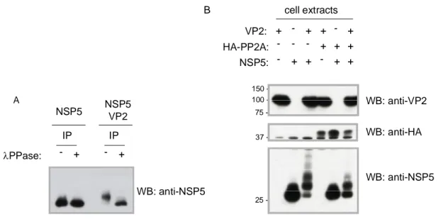
3.2 VP2 induces NSP5 hyperphosphorylation ... 65
3.3 Mapping NSP5 hyperphosphorylation induced by VP2 ... 67
3.4 Correlation between NSP5 hyperphosphorylation and VLS... 70
3.5 Towards viroplasms re-building:recruitment of different viral proteins into VLS .... 75
4 DISCUSSION (1) ... 81
ABSTRACT (2) ... 89
5 INTRODUCTION (2) ... 91
5.1 The Proteasome ... 91
5.1.1 The core particle (CP) ... 92
5.1.2 The regulatory particle (RP) ... 93
5.2 The ubiquitin-proteasome system (UPS) ... 95
5.3 The degradation process ... 97
5.4 UPS and virus ... 98
6 RESULTS (2)... 101
6.1 Intro ... 101
6.2 The striking observation ... 101
6.3 Proteasome activity is involved in Rotavirus infection ... 105
6.4 Proteasome inhibition affects viroplasms formation ... 107
6.5 Proteasome inhibition affects viroplasms growth ... 109
6.6 Proteasome inhibition affects production of viral particles ... 111
6.7 Viral proteins expression is not affected by proteasome inhibition... 112
6.8 Proteasome inhibition does not impaired viral polymerase activities ... 114
6.9 Viral infection affected by proteasome inhibitor is not due to IRF3 amounts ... 116
7 DISCUSSION (2) ... 118
4
LIST OF ABBREVIATIONS
3D three-dimensional aa amino acids
ATP adenosine triphosphate bp base pair
CPE cytopathic effect C-terminal carboxy-terminal DLP double-layered particle
DMEM Dulbecco’s modified Eagle’s medium DMSO dimethylsulfoxide
DSP Dithiobis(succinimidylpropionate) dsRNA double-strand RNA
EDTA ethylenediamine tetraacetic acid EGFP enhanced green fluorescent protein EM electron microscopy
ER endoplasmic reticulum
ERGIC ER-Golgi Intermediate Compartment FCS foetal calf serum
FITC fluorescein isothiocyanate GST glutathione-S-transferase HA hemagglutinin
HIT histidine triad
HRP horseradish peroxidase IFN interferon
IPTG isopropyl--D-thiogalactopyranoside IRF3 Interferone regulatory factor 3 λ-PPase lambda-phosphatase
MOI multiplicity of infection NSP nonstructural protein nt nucleotides
N-terminal amino-terminal ORF open reading frame
5 PAGE polyacrylamide gel electrophoresis PBS Phosphate buffered saline
h.p.i. Hours post infection p.t. post transfection
RdRp RNA-dependent RNA polymerase
RITC rhodamine isothiocianate
RNase Ribonuclease
siRNA small interfering RNA sn supernatant
ssRNA single-strand RNA TBS Tris buffered saline TLP triple-layered particle UTR untranslated region UV ultraviolet
VLP virus-like particle VLS viroplasm-like structures
6
ABSTRACT (1)
The processes that regulate Rotavirus replication are not fully understood and the lack of a reverse genetic approach represent an obstacle for the investigations in Rotavirus biology. Viroplasms are cytoplasmic structures that form soon after infection, and constitute the site of virus replication. Structural proteins like the viral RNA-dependent RNA -polymerase VP1, the capping enzyme VP3, the scaffolding protein VP2,and the middle layer VP6 localize in viroplasms; in addition, also the non-structural proteins NSP5 and NSP2 have been demonstrated to be essential components for viroplasm formation. Following the characterization of the interaction between NSP5 and VP1, we characterized the relationships between NSP5 and the structural protein VP2.
In this work, interaction of NSP5 with VP2 was investigated by coexpression of the two proteins in uninfected cells, which resulted in a strong hyperphosphorylation of NSP5 and in the formation of viroplasm like structures (VLS). The behaviour of NSP5 in the presence of VP2 is very similar to that induced by NSP2 and already described (1), (60). Therefore, a comparison between the phosphorylation degree of NSP5 and VLS formation induced either by VP2 or by NSP2 was conducted.
In both cases VLS formation was shown to assemble independently of the phosphorylation degree of NSP5, and to recruit the viroplasm-resident proteins VP1. However, VP6 (the protein forming the middle layer of the virion) was shown to be recruited only into VLS induced by VP2 (VLS(VP2i)), while it remains organized in tubular structures when VLSinduced by NSP2 (VLS(NSP2i)) were formed. Attempts to coimmunoprecipitate NSP5 and VP2 failed both from infected and co-transfected cells. However, promising preliminary results were obtained with a recently isolated monoclonal Ab specific for NSP5.
Altogether, these data showed that two different viral proteins induced the same kind of modifications in NSP5, suggesting that these modifications may have a fundamental role for virus replication. Moreover, these data suggest that NSP5 plays a key role in architectural assembly of viroplasms and in recruitment of the other viroplasmic proteins.
7
1 INTRODUCTION (1)
In the 1960s Rotavirus was discovered in animals, and in 1973 was first described in humans, when electron microscopy images reveal its presence in duodenal biopsies of children with acute gastroenteritis (19). Subsequently it was recognized as the most important cause of severe, dehydrating gastroenteritis in infant and young children worldwide. About 600.000 children die every year from Rotavirus, with more than 80% of all rotavirus related deaths occurring in resource-poor countries in south Asia and sub-Saharan Africa. Rotavirus related deaths represent approximately 5% of all deaths in children younger than 5 years of age worldwide. Furthermore, recent studies indicate that rotavirus causes approximately 39% of childhood diarrhea hospitalizations worldwide. The burden of rotavirus infection is not limited to the less-developed countries. Studies from the western European found that 50% of cases of gastroenteritis in children younger than 5 years old that were hospitalized were caused by Rotavirus infection, and, moreover, that these infections caused 230 deaths per year. In the United States Rotavirus is estimated to cause 20-60 deaths, and 55.000-70.000 hospitalizations per years, with very high health and social costs consequences (75).
According to all these statistical studies on Rotavirus infection, an effective vaccine program is necessary. Fortunately, following initials problems with the first attempts to develop a vaccine, in 2006 two new Rotavirus vaccine were licensed in the United States, the Europe Union and in many countries in Central and South America. The effectiveness of these two vaccines is well documented and still under investigation, in particular postmarketing surveillance studies are monitoring the impact of vaccine on different circulating strains of rotavirus, according to geographic distribution and selection pressure (75).
Rotavirus biology has been extensively studied in these past decades, and some infection mecchanisms have been elucidated, however much need to be learned in Rotavirus replication. In particular the goal of a reverse genetic system would answer to different questions related to rotavirus replication and to the role of different viral proteins during rotavirus infection, in order to identify a unique viral target that might be useful in developing therapies.
8
1.1 VIRUS CLASSIFICATION
Rotavirus is a non-envelope double stranded RNA virus that belong to the family of Reoviridae. The term Rotavirus derived from latin word rota, which means wheel, according to the distinctive morphologic appearance at electron microscopic analysis (58). The viral particle is large (1000 Å) and complex, it consists of three concentric protein layers (outer, intermediate, inner layer) surrounding the viral genome of 11 double strand RNA segments. The genome segments encode for 6 structural proteins (viral proteins, VPs VP1-4, VP6-7), that make up the virus particles, and 5 (or 6, depending on the strain) non structural viral proteins (NSPs, NSP1-5, NSP6) (58).
Rotaviruses are classified according to antigenic specificity. They are classified in groups, viruses of the same group share cross-reacting antigens serologically detected using monoclonal and polyclonal antibodies against VP6 (intermediate layer protein). Groups A-E have been clearly identified, two more groups F,G are likely to exist. VP6 mediates also subgroups specificity according to the exclusive reactivity of two VP6 specific monoclonal antibodies (subgroup I, and subgroup II).
The structural protein VP7 is the main component of outer layer, and it is associated with VP4, a structural protein that forms spikes that emerge from the VP7 shell. This particular organization induces neutralizing antibody responses that are the basis for the binary classification in serotypes mapping VP4, or VP7, and genotypes by sequence comparison, within each group. Serotypes determined by the glycoprotein VP7 are termed G (which stands for glycoprotein) and those defined by VP4 are named P (which stands for
protease-sensitive protein, which is the case for VP4). For VP7 classification,
neutralization assays and sequencing yeld to concordant results, so viruses are refer to their G serotype alone (G1, G2, G3 and so forth). On the contrary, for VP4 classification, serotypes do not correspond to genotypes and a dual system for P typing is used. P serotype are referred to by their serotype number (P1, P2, P3 ecc) while P genotypes are denoted in brackets (P[1], P[2] ecc). P genotyping is the most widely used method, since it is difficult to standardize VP4 neutralization assays. Currently, 19G and 24[P] types are known.(57, 58, 75).
Recently, it has been proposed a rotavirus classification system forall the rotavirus genes that recognizes phylogenetic relationships and defines genotypes based on percentage identity cutoff valuesfor each of the genes, which may have followed separate evolutionary paths. Such a classification system could be an important tool to elucidatehow rotaviruses
9
evolve over time,to identify reassortment events and gene constellationsshared by human and animal rotaviruses.(122)
1.2 VIRION STRUCTURE
The morphology of the complete rotavirus infective particle is peculiar, and by electron microscopy analysis it is possible to observe three types of particles in the cytoplasm of infected cells. The first type of particle is a triple-layered particle (TLP) that is the complete infective particle of about 100 nm in diameter and present a triple-layerd icosahedral protein capsid composed of an outer layer, an intermediate layer, and a inner core layer. The second type is the double-layered particle (DLP) formed when the outer layer of the TLP is missing, while the third type is the single-layered particle (or core) composed only by the innermost layer and seen infrequently, it lacks the genome and aggregates in the cytoplasm with other single-layerd particles (160) (210).
A particular characteristic of the virus structure is the presence of 132 large channels that extend over the two shells and link the outer surface with the inner core. Three types of channels (type I-III) can be distinguished based on their position and size (160).
The viral genome is packaged inside the inner core together with two viral proteins essential for viral transcription and replication: the viral RNA-dependent RNA-polymerase VP1 and the capping enzyme VP3 (140).
Herein a detailed description of virus structure is reported:
The outermost layer : the outer layer is mainly composed by the structural protein VP7 that is uniformly distributed and forms a smooth surface from which sixty spikes of VP4 hemagglutinin protein extend. VP7 is organized in trimers, with a total number of 780 VP7 molecules forming the outer shell organized in a T=13l (levo) icosahedral surface. The VP4 spikes is multi-domained with a radial length of about 200Å, and extend about 120Å from the surface of the virus. It interacts with two molecules of VP7 by extending inwards about 80Å into the virion outer layer, and inside it appears that VP4 interacts with six VP6 molecules that surround type II channel (209). The interaction between VP7 and VP4-VP6 implies that VP4 has a relevant role in maintaining the precise geometric organization between outer and middle capsids (210) (Fig.1A).
The intermediate layer : VP6 is the only component of the intermediate layer. 760 molecules of VP6 are arranged in trimers and localized in the local threefold axis of the
10
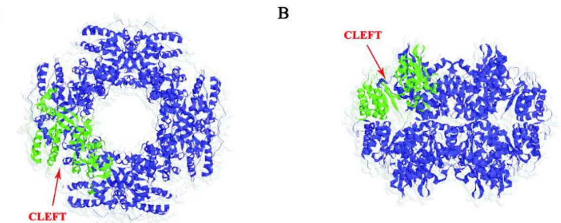
T=13 icosahedral lattice. VP6 trimers lie below VP7 trimers, so that the aqueous channel in the two T=13 layers are in register, and above the inner layer of VP2 (160). The VP6 molecule has two domains: the distal eight-stranded -sheet domain core makes contact with the VP7 layer, and the lower -elical domain that makes contact with the inner VP2 layer. The interacting surfaces (VP6-VP7 and VP6-VP2) expose the most conserved residues of VP6, suggesting that VP6 may play a major role in providing structural integrity to the rotavirus capsid. However, the lateral interactions between trimers that form the T=13 icosahedral organization, are not sufficient per sè to allow the closed shell. On the contrary, VP2 is able to form native like icosahedral shell, suggesting that VP2 layer provides a proper scaffold for the assembly of VP6 trimer into a T=13 icosahedral organization (210) (Fig.1A).
The innermost layer : the single layerd particle posses a T=1 symmetry and is composed of 120 VP2 molecules, arranged in dimers that surround the viral genome. VP2 protein interacts with both VP6 of the middle layer and the RNA genome, through the N-terminal residues that have RNA binding ability. N-terminal residues of VP2 are also involved in the anchoring to the inside surface of the VP2 layer, at the vertices of the icosahedral structure, heterodimer of RNA-dependent RNA-polymerase (RdRp) VP1 and capping enzyme VP3 (159). The RNA genome is well organized inside the core, as well. In particular the 25% of genome made internal dodecahedral structure in which the RNA double helices, interact closely with VP2 and are packed around the transcription complexes located at the vertices of the icosahedral structure. (Fig1B) (89)
The aqueous channels : in the mature infective particle there are three types of channels classified on the basis of positions and size. There are twelve type I channels located in the five-fold axes, sixty channels of type II at the six-coordinated position surrounding the fivefold axes, and sixty type III channels on the six-coordinated positions around the icosahedral three-fold axes. All three channels type are about 140Å in depth, and differ in width since type II and III reach 55Å in width at the outer surface of the virus, while type I channels are narrower (about 40Å) at the outer surface of the virus. In entering the viral particles, these channels constraints before to reach the maximum width, which is close to the surface of the inner shell. These channels are involved in importing the metabolites required for RNA transcription, and in particular type I channel in exporting the viral mRNA transcripts into the cytosol of the cells for subsequent viral replication processes (89).
11
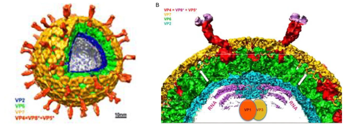
Figure 1: Rotavirus structure determined by cryo-EM. A) Cut-away view of the rotavirus TLP showing the outer layer (VP7 in yellow and VP4 in red), the middle layer (VP6 in green) and the inner layer (VP2 in blu). B) Cross-section of a rotavirus particle. The various protein components are colored as indicated in A). VP1 and VP3, the polymerase and mRNA capping enzyme, respectively, are anchored to the inside of the VP2 layer at the five-fold. White arrows show where the N-terminal arms of the outer-layer protein, VP7, clamp onto the underlying VP6. surrounding the enzymes VP1 and VP3 (in red), which (From Chen et al. PNAS 2009).
1.3 GENOME STRUCTURE AND ORGANIZATION
The viral genome consists of 11 segments of double stranded RNA, that range in size from 0.7 to 3.1 kB, and is enclosed inside the inner core of the virus. The viral segments are thought to be organized in the core with a dodecahedral symmetry: one copy of each segment interacts with one replication complex constituted by one molecule of RdRp VP1 and one of capping enzyme VP3 (159) (49).
The sequences from different rotavirus strains show common features in the structure of each segments (58):
Segments are A+U rich (58% to 67%).
5’-methylated cap sequences m7GpppG(m)GPy, uncapped minus strand lacking a -phosphate.
An open reading frame (ORF) that encodes the protein product of the gene, flanked by untranslated ragions (UTRs).
A set of conserved consensus sequences (CS) at the 5’-3’ UTRs.
The last four to five nucleotides of the 3’CS[(U)GACC] can function as translation enhancer.
There is no polyadenylation signal at the 3’-end of the mRNA segments.
12
The 5’ and 3’ UTRs of the 11 genome segments show considerable variation in length and sequence, this suggest that they are needed for their own translation and packaging. In contrast the UTRs of homologous segments are highly conserved among viruses belonging to the same group, in some cases more than the sequence of the ORF (142) (144).
In the case of group A rotavirus, that is the most important in term of human morbidity and mortality, the (+)strand RNAs of genome segments typically end with 5’CS, 5’–GGC(U/A)7
-3’, and the 3’CS, 5’-UGUGACC-3’ (49).
Computer modelling proposed that base-pairing in cis between 5’ and 3’ regions of mRNA, leads to the formation of panhandle structure from which the 3’CC extends in an un-paired tail (142),(34),(33). This stable structure allows the recognition by the viral RdRp that interacts with the 3’-CC and induces the formation of (–) strand initiation complex (145),(144). Indeed, the 3’-terminal CC have been shown to be crucial for the formation of the initiation complex of RNA replication (30),(32).
The 3’CS not only includes sequences that promote genome replication but also contains a determinant sequence required for efficient translation of rotavirus (+)RNAs. In particular the last four nucleotide of 3’CS [(U)GACC] are recognised by a dimer of the non-structural protein NSP3 (155),(154), that in turn is recognise by the eukaryotic translation initiation factor, eIF4GI (152),(151). This however, has been described below (see page 22).
Sequences enhancing replication are localized also in the 5’CS of the genome segments (145); however attempts to identify 5’-sequences recognised by the RdRp VP1 have been unsuccessful. It cannot be excluded that the 5’-recognition signals interact with VP1 or the core protein VP2, despite evidences from replication studies that the synthesis of (-)RNA depends on the interactions with viral protein of the replicase complex with both ends of viral segment (197). (Fig. 2)
The most interesting, and still obscure, aspect of rotavirus genome organization is related to the mechanisms that control replication and packaging of the 11 segments. Indeed the 11 segments should share similar cis-acting signals to be recognise and replicated by the same polymerase; moreover each segment should have a unique sequence signal in order to be distinguished from one another during the packaging. The packaging of the 11 dsRNA segments is mediated by a deep interaction between RNA and viral proteins. The viral proteins that participate in the encapsidation of the viral genome are still under identification. Obviously an important role is played by VP1, VP3 and VP2, but involvement of non-structural proteins is not to excluded (126), (58).
13
Figure 2: Schematic representation of a group A rotavirus plus-strand RNA. The conserved sequences at the 5’ and 3’ ends are indicated. Both sequences were shown to be essential for the formation of the minus-strand initiation complex. They are predicted to stably base-pair forming a panhandle structure. The dinucleotide GG indicated in purple is conserved within all groups of rotaviruses and the second G was shown to be essential for specific recognition by the polymerase VP1. Another recognition signal for VP1 is at the 3’UTR. Both signals are underlined. The sequence indicated in blue is a translation enhancer.
1.4 VIRAL PROTEIN
Although new functions for the different rotaviral proteins are continuously identified, their assignment with the 11 genome segments has been well established. The Rotavirus genome encodes for 6 structural proteins, that make up the viral particles, and 6 non-structural proteins, with exception of some strains that have 5 non-non-structural proteins, that are produced during the infection to maintain infective status.
The proteins of SA11, a simian strain, have been studied more thoroughly, since it was completed first and for this reason it is considered the strain of reference. The migration order/pattern of RNA segments could differ among different strain.
In figure 3 is reported the protein assignment to the different RNA segments of SA11 strain: RNA segments 1, 2, 3 encode for the core structural proteins VP1, VP2, and VP3; segment 6 encodes the middle layer viral protein VP6; segments 4 and 9 produce the outer layer proteins VP4, and VP7, respectively. The non-structural proteins NSP1, NSP2, NSP3, NSP4, are encoded respectively by segment 5, 8, 7, 10; while segment 11 encodes for NSP5, and in some strain in a different ORF for NSP6 (58).
ORF
5’ UTR 3’UTR
AUG
14
Figure 3: Gel PAGE of the 11 dsRNA segments of Roatvirus SA11. Genome segments are indicated on the left and the encoded proteins on the right.
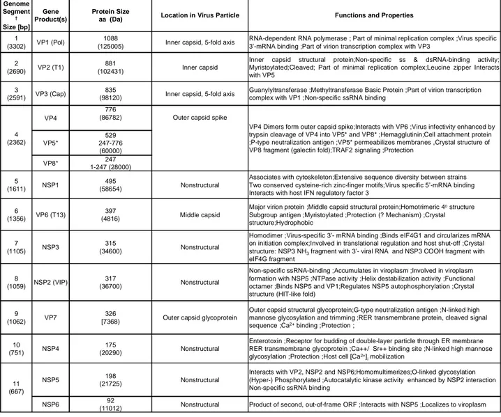
Table 1 summerizes and describes all Rotavirus proteins with their functions and properties.
Table 1: List of Rotavirus genes, relative proteins, functions and properties.
Genome Segment † Size [bp] Gene Product(s) Protein Size
aa (Da) Location in Virus Particle Functions and Properties
1
(3302) VP1 (Pol)
1088
(125005) Inner capsid, 5-fold axis
RNA-dependent RNA polymerase ; Part of minimal replication complex ;Virus specific 3’-mRNA binding ;Part of virion transcription complex with VP3
2
(2690) VP2 (T1)
881
(102431) Inner capsid
Inner capsid structural protein;Non-specific ss & dsRNA-binding activity; Myristoylated;Cleaved; Part of minimal replication complex;Leucine zipper Interacts with VP5
3
(2591) VP3 (Cap)
835
(98120) Inner capsid, 5-fold axis
Guanylyltransferase ;Methyltransferase Basic Protein ;Part of virion transcription complex with VP1 ;Non-specific ssRNA binding
4 (2362)
VP4
776
(86782) Outer capsid spike
VP4 Dimers form outer capsid spike;Interacts with VP6 ;Virus infectivity enhanced by trypsin cleavage of VP4 into VP5* and VP8* ;Hemagglutinin;Cell attachment protein ;P-type neutralization antigen ;VP5* permeabilizes membranes ,Crystal structure of VP8 fragment (galectin fold);TRAF2 signaling ;Protection
VP5* 529 247-776 (60000) VP8* 1-247 (28000)247 5 (1611) NSP1 495 (58654) Nonstructural
Associates with cytoskeleton;Extensive sequence diversity between strains Two conserved cysteine-rich zinc-finger motifs;Virus specific 5’-mRNA binding Interacts with host IFN regulatory factor 3
6
(1356) VP6 (T13)
397
(4816) Middle capsid
Major virion protein ;Middle capsid structural protein;Homotrimeric 4ostructure
Subgroup antigen ;Myristoylated ;Protection (? Mechanism) ;Crystal structure;Hydrophobic
7
(1105) NSP3
315
(34600) Nonstructural
Homodimer ;Virus-specific 3’- mRNA binding ;Binds eIF4G1 and circularizes mRNA on initiation complex;Involved in translational regulation and host shut-off ;Crystal structure: NSP3 NH3fragment with 3’- viral RNA and NSP3 COOH fragment with
eIF4G fragment
8
(1059) NSP2 (VIP)
317
(36700) Nonstructural
Non-specific ssRNA-binding ;Accumulates in viroplasm ;Involved in viroplasm formation with NSP5 ;NTPase activity ;Helix destabilization activity ;Functional octamer ;Binds NSP5 and VP1;Regulates NSP5 autophosphorylation ;Crystal structure (HIT-like fold)
9
(1062) VP7
326
[7368) Outer capsid glycoprotein
Outer capsid structural glycoprotein;G-type neutralization antigen ;N-linked high mannose glycosylation and trimming ;RER transmembrane protein, cleaved signal sequence ;Ca2+binding ;Protection ;
10
(751) NSP4
175
(20290) Nonstructural
Enterotoxin ;Receptor for budding of double-layer particle through ER membrane RER transmembrane glycoprotein ;Ca++/ Sr++ binding site ;N-linked high mannose glycosylation ;Protection ;Host cell [Ca2+]
imobilization
11 (667)
NSP5 (21725)198 Nonstructural
Interacts with VP2, NSP2 and NSP6;Homomultimerizes;O-linked glycosylation (Hyper-) Phosphorylated ;Autocatalytic kinase activity enhanced by NSP2 interaction Non-specific ssRNA binding
NSP6 92
15
In the follow paragraphs, the viral proteins are described more in detail starting from the structural proteins that form the viral particle, in particular from the proteins inside the core in contact with the viral genome, towards those forming the middle and the outer layer. Subsequently the non-structural proteins are taken in consideration, classified in those involved in viral replication and those with different role in viral morphogenesis.
1.4.1 STRUCTURAL PROTEINS The core:
VP1
VP1 is the viral RNA dependent RNA polymerase encoded by segment 1, and it functions as both, the transcriptase for mRNA synthesis, and the replicase for minus-strand RNA synthesis to generate genomic dsRNA.
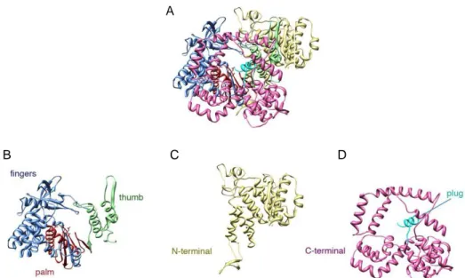
VP1 crystal structure has been recently resolved, revealing that it is a compact protein of about 70 Å in diameter. Three distinct domains were identified: an N-teminal domain (aa 1-332); a polymerase domain consisting in finger, palm, and thumb subdomains (aa 333-778) and a C-terminal “bracelet” domain (aa 779-1089) (118) (Fig.4).
N and C-terminal domains surround most of the polymerase domain creating a sort of cage with a large hollow center.
The polymerase domain has the six canonical motif A-F that are also present in RdRp of other viruses like reovirus λ3 (187), bacteriophage φ6 P2 (25), hepatitis C virus NS5B (3). The palm subdomain consists of three helices that support the four-stranded antiparallel -sheet and includes the conserved residues of the active site.
The finger subdomain comprises one side of the template entry tunnel, and has a three dimensional arrangement that is different of that observed for reovirus polymerase λ3. The thumb subdomain consist of a -strand followed by three -helices, a loop at the tip of thumb interacts with the tip of finger domain enclosing the catalytic site on the palm of the polymerase domain, and maintaining VP1 in a closed conformation (118).
The closed conformation is reinforced by the N-terminal domain that covers one side of the active domain, while C-terminal domain is situated around the exit tunnel for the dsRNA product of replication and for the minus RNA template of transcription. In particular aa1072-1089 form a -helical plug that extend into the template tunnel and reduces its diameter, so it is necessary to remove the plug for the exit of dsRNA (118).
16
Figure 4: Structure of the RdRp VP1. A)Ribbon diagram of the entire polypeptide chain. B) The fingers, palm, and thumb subdomains of the polymerase in blue, red, and green, respectively. C)The N-terminal domain is in yellow. D) the C-terminal bracelet domain, in pink; the C-terminal plug, in cyan. (118)
VP1 polymerase (separated by gradient centrifugation from the other components of open cores VP2 and VP3), when incubated alone with plus strand RNA does not show any replicase activity, indicating that the enzyme alone is an inactive form of the viral RdRp. However, when VP1 is incubated with (+)RNA and the core protein VP2, the synthesis of dsRNA activity takes place, proving that VP2 induces the conversion to the active form of VP1 (136). The ratio between VP1 and VP2 is also critical to obtain the maximal replicase activity. Stoichiometric analysis have demonstrated that the optimal ratio VP1:VP2 is 1:10 which is the ratio present at the vertices of rotavirus core. This suggests that not only the presence of VP2 is required to have an active polymerase but also that VP1 has to be collocated into a precise structure that is the pentamer unit of the core (141).
Electrophoretic mobility shift assays have shown that VP1 has a strong affinity for viral (+)RNAs due to the recognition of sequence signals located near the 3-end of RNAs. According to these assays it has been proposed a multistep interaction between the polymerase and (+)strand RNA: VP1 recognises the UGUGA sequence at 3’CS of (+)RNA in a VP2-independent manner, then VP1/+RNA complex interacts with VP2, (involving its N-terminal domain) that induces a conformational change on VP1 leading to the interaction with 3’CC portion to form the initiation complex. This model however does not take in consideration the requirement of VP2 also during the elongation step of (-)RNA synthesis. In addition, VP1 also interacts with other protein and in particular with the virus non-structural protein NSP5 and NSP2. (see NSP5 paragraph, page 25)
B C D
17
Interaction with NSP2 has been demonstrated by co-immunoprecipitation assays from infected and trancfected cells (96).
VP3
VP3 is the structural protein encoded by genome segment 3. It is the minor component of the virion core, there are 11-12 molecules per particle, and several studies provided evidences that VP3 is the viral guanylyltransferase, responsible for the capping of viral mRNAs. The protein has the intrinsic ability to bind GTP molecule in the absence of other viral proteins (31). Moreover, it has been demonstrated that VP3 has specific ability to bind ssRNA but no dsRNA, and also its 5’ capping activity was also shown to be non specific (138). VP3 interacts with the N-terminal of VP2 together with VP1 and the RNA segment, and is an essential components of trascriptase complex, but not of the replicase one, since baculovirus expressed core like particles composed only by VP1 and VP2, still possess replicase activity (141).
In addition to the capping function, VP3 seems to have another distinct role in the formation of replication intermediates, revealed by the analysis of mutants with a ts lesion in VP3. Thus it is supposed that VP3 would increase the rate of VP1-VP2 complexes assembly into replicase particles (201),(141).
VP2
The structural protein VP2 is the main component of the inner core of the virus. The core is made up by 120 copies of VP2, with two molecules of VP2, named VP2A and VP2B, in the icosaedral asymmetric unit. A recent CryoEM analysis for the identification of secondary structures revealed the presence of 28 distinct helices and four -sheet, where the -helices are well distributed in the structure, while three of the four -sheet are located in the lower part of the protein. VP2 shares common features in secondary structure with other viral inner core proteins like blutoungue VP3 (95). One characteristic feature of Rotavirus VP2 is helix 1, located at the the N-terminus, that is not present among other reovirus inner capsid proteins, and that extends towards the fivefold vertex and crosses over to an adjacent VP2 molecule. At the end of helix 1, another helix (helix 0) can be seen to cross underneath VP2, towards the fivefold vertex where it may interact with the transcription enzyme complex. However the resolution of this analysis does not permit to assign the helix to one or the other molecule of the dimer. Moreover interaction between
18
VP2 and VP6 layer has been resolved. Since there is a symmetry mismatch between VP2 and VP6 layers, different interactions between VP6 trimers and VP2 dimers have been described. In particular, the VP6 trimer that surround type I channel near the fivefold axes contacts the apical domain of both VP2A and VP2B; the VP6 trimer located at the threefold axes and near to type three channel makes contact only with VP2B; and other three VP6 trimers that surround the type II channels contact VP2A and/or VP2B. All these interactions strongly stabilize the double layer particle (112) (Fig.5).
Figure 5: VP2 pentamer (A) VP2A (pink) and VP2B (light green) subunits in the icosahedral asymmetric unit are shown. The VP6 trimers that sit atop VP2 subunits are indicated by triangles. The VP6 residues, as deduced from fitting of the VP6 crystal structure, that interact with VP2
are shown (112).
By analsysis of baculovirus-expressed recombinant proteins, it has been demonstrated that N-terminus of VP2 is required for the binding of VP1 and VP3, however, the synthesis of rotavirus dsRNA in vitro is sufficiently supported by particles formed by VP1 and VP2 (141). The N-terminal domain of VP2, contains also the RNA-binding domain and binds ssRNA more efficiently than dsRNA. This difference would have an important role during transcription, allowing the dsRNA genome able to move and to be read from VP1. Moreover, the low affinity of VP2 for dsRNA reveals that VP2 does not have a dominant role in the packaging of viral genome and involvement of other viral protein occurs (105). The VP2 N-terminal domain has also a structural role (as already describe): it contains the -helix1 that stabilizes the VP2 dimer allowing the pentamer organization (112).
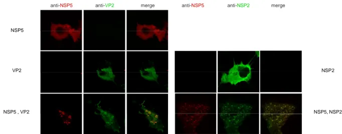
A weak interaction between VP2 and NSP5 has been reported in both mammalian infected cells and in transfected insect cells (18). The description of the interaction between NSP5 and VP2 will be discussed in this thesis.
19
The middle layer: VP6
VP6 is the most abundant protein of the viral particle. It is the main constituent of the intermediate layer of the triple layerd particle, in particular 780 molecules of VP6, organized in trimers surround the VP2 core with a icosahedral symmetry. VP6 integrates two principal functions of the virus: cell entry and transcription since it interacts with both, the components of the outer layer and with VP2, the core of the virus.
VP6 has two domains: an eight-stranded -sheet domain that interacts with VP7 layer, and a cluster of -helices that make contacts with the inner VP2 layer. The trimers of VP6 interact laterally to form the icosahedral structure and this contacts involved charged residues. The interacting surface with the other components of the triple layer particle involves conserved residues of VP6, the contacts with VP2 and VP7 involves principally hydrophobic residues. Although VP6 have the capacity to form trimers, stabilized by a Zn ion, it does not have the information to organize the different trimers into a closed shell. Indeed, when expressed alone VP6 easily forms helical tubes in the cytosol. So, the correct assembly of the middle layer is driven by the core, since VP2 alone has all the information to form native icosahedral shell around which VP6 trimers organize (Fig.5) (89).
Earlier biochemical studies revealed that none of the component of the DLPs are able to transcribe alone the viral genome, and that VP6, althought does not have any enzymatic activity, is essential for endogenous transcription. This hypothesis was confirmed using mutants, with an extra charge in the VP6-VP2 interface, that does not rescue the transcriptase activity of the reconstituted DLPs (29). Moreover, it has been observed that interfering with the conformational changes near the VP6-VP2 interface using monoclonal antibody (MAb) against VP6, affects viral transcription (192). A conserved -hairpin motif of VP6 extends inside a type I channel, used by the newly transcribed mRNA to exit the DLPs, and may play a role in the translocation of the mRNA. This suggests that the dynamics of VP6 itself and in the VP2-interface have an important role in mRNA viral transcription.
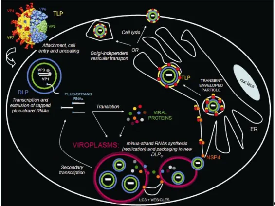
During rotavirus replication, VP6 is found in viroplasms, that are electrondense structures that form soon after infection and are the putative site of viral replication and core assembly. VP6 has active role in virus assembly, probably through the interaction with
20
NSP4, which may facilitate association to core particles, to form DLPs, during the exit of core particles from viroplasm (117).
The outer layer: VP7
VP7 is the main component of the outermost layer of rotavirus particles. It is a glycoprotein of about 38 KDa, that forms a smooth capsid where VP4 spikes associate. The shell has 760 copies of VP7 organized in 260 trimers with an icosahedral symmetry. The trimers are stabilized by two Ca2+ ions, bound at each subunit interface. The core of each subunit folds into two compact domains, a Rossmann fold domain (domain I) and a jelly-roll sandwich (domain II), with disordered N and C-terminal arms. The arms extend away from the compact core; the N-terminal arm moves towards the centre along the surface of VP6 subunit, so that the three arms of the VP7 trimer grip the VP6 trimer (Fig.1), and it is also involved intra-trimer contacts. The C-terminal arm of one subunit interacts with its counterpart of the other subunit, but most of its contacts appear to be within its trimer of origin (4, 35).
VP7 is involved in the entry of the virus into the cell by modulating VP4 rearrangements during attachment and penetration of the viral particle. While the appropriate levels of calcium help in maintaining the structural integrity of the VP7 layer, low calcium concentrations trigger VP7 conformational changes and subsequent dissociation of VP7 trimers (51). Since VP7 shell stably locks the spikes in the assembled virion, VP7 trimers dissociation would precede VP5* rearrangements, in order to promote virus penetration into the host cell .
According to measurement by cryo-microscopy the VP4 spikes located inwards the type II channels, have a diameter of 70Å, that fits more with the diameter of type II channel at the surface of VP6 layer (78Å) respect to that of VP7 layer (54-58Å). This suggest that, during the assembly of the virus, the spikes should first anchor to the VP6 layer about at the type II channel and then VP7 trimers would subsequently arrange above VP6 trimers to form stable triple layer particle (112).
VP7 has been identify as the viral protein that interacts with integrins. In particular, after the initial binding mediated by VP4 with sialic acid and integrin 21, VP7 is prompted to interacts with X2 and v3 integrins, and mediates the entry of the virus probably through an endocytic pathway (73). The low calcium concentration inside the endocytes may favour VP7 trimer dissociation and subsequent uncoating of TLPs to release DLPs.
21
During Rotavirus infection VP7 is localized in the ER where the virus morphogenesis is completed. It is found in the transient enveloped particle that the DLPs acquire in the budding process into the ER, together with NSP4 and VP4 (117). Silencing of VP7 induces accumulation of enveloped particles in the ER, a phenotype similar to depletion of calcium, suggesting that in order to displace the transient membrane surrounding the DLP, VP7 needs to trimerize (123).
VP4
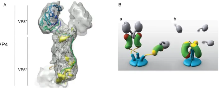
VP4 is encoded by segment 4, and is one of the two constituents of the outermost layer. It has an important role in virus attachment and entry into the host cell. Association of two or three, molecules of VP4 forms spikes that protrude from the smooth VP7 shell, and are located at the level of type II channels.
VP4 spikes are characterized by a globular domain that forms the head of the spike, and a central domain that forms the central body of the spike. Moreover, the spikes are anchored to a globular base that has been demonstrated to be part of VP4, and is linked to the central bodies with an elongated bridging domain (158).
In order to induce the attachment and penetration into the host cell VP4 needs to be cleaved by trypsin. The cleavage products are the globular domain (VP8*) that is involved in virus attachment, and the central body (VP5*) that is thought to mediate cellular penetration. Both the cleavage products remain associated to the viral particles.
Recently cryo-EM analysis gave evidence that spikes have both dimeric and trimeric structures.
VP5* is a well-ordered homotrimer with a C-terminal -helical triple coiled-coil, and a N-terminal globular domain. Each globular domain packs in a groove between the -helices of the other two subunits. The globular domain of VP5* have a core of eight stranded anti-parallel -sandwich, with two functional important -hairpin: one stabilizes the globular structure, while the other is involved in the rotavirus binding to the 21 integrins (52). VP8* is the head of VP4 protein, and is attached to the central body by about 25 amino acidic residues non structurally defined. However, hydrophobic interaction between VP5* and VP8* are present, as well. The central structural feature of VP8* domain is an 11-stranded anti-parallel -sandwich formed from a five-11-stranded -sheet, and a six-11-stranded -sheet with an interrupted top strand. The domain contained other structural elements like: intersheet loop containing a short -helix, a longer -helix at the c-terminus, and a
-22
ribbon. All these elements with a dense hydrophobic core between major structural elements suggest a rigid structure that does not undergo structural rearrangements during virus entry. Between the two -sheets lies the sialic acid bindig site, since rotavirus have a sialic acid-dependent mechanism to enter the host cell (53).
The spikes extend inward into the type II channels making contacts with VP7 trimers , and are anchored to the globular base located between the VP7 and VP6 layers. The base present a strong threefold symmetry with both -helices and -sheets secondary structures, and make symmetrical contacts with three trimers of VP6 (112).
Image reconstruction from electron cryomicroscopy of rotavirus particle have provided evidence that VP4 is subjected to a series of rearrangements, upon trypsin treatment, that allow the entry of the virus into the host cell. Before the processing by trypsin, the spikes are flexible structure visible by cryo-EM. Following trypsinization VP4 spikes are stabilized inducing a disorder-to-order transition, not shown for other viruses. The hypothesis is that, following trypsinization, they assume a dimeric appearance leaving the third subunit flexible, which, following an unknown triggering event, folds back to the dimer forming the trimeric structure and promoting cell membrane penetration and virus entry. VP8* is thought to dissociate from VP5* before o during the folding back rearrangment. (211). (Fig.6)
23
Figure 6: Structure of VP4 spike. A) Fitted VP8* and VP5*-t secondary structures are shown in the cryoEM density map of one of the dimeric subunits of the VP4 spike. The polypeptide chain is colored from the N (blue) to C (red) terminus. (112). B) Models of two VP4 conformations. (a)Two subunits form the spike visible in electron cryomicroscopy image reconstructions of trypsin-primed virions. A third subunit is flexible. VP8* is gray, the VP5* antigen domain is green bean-shape, with a red membrane interaction region and a yellow GH loop, the foot is blue, as is a protruding region that rearranges into the coiled-coil. Following trypsinization two of three spike dimerize living the third subunit flexible. (b)The putative post-membrane penetration state. Unknown trigger events induce VP8* released; the yellow parts of each subunit have joined in a -annulus; the -helical triple coiled-coil has zipped up; and the VP5* antigen domain has folded back. The models were produced by Digizyme, Inc.(211)
After the initial contact, which is mediated by VP8*, a second interaction with integrin 21 occurs. This interaction is mediated by the integrin binding motif DGE present in the -hairpin motif in the globular core of the VP5*. Moreover, VP5* is likely to be involved in binding to integrins 41 and 47 through its peptide sequence YGL (72, 74). Additional interactions in a post-attachment step, involving also VP7, occur and bind heat shock protein 70 (Hsc70) and other integrins v3 and x2 (76).
The role of VP4 during viral morphogenesis remains unclear. VP4 has been found at the plasma membrane associated to microtubules, and also detected in filamentous arrays. Infected cells treated with siRNA against gene4 still form viral particles, suggesting that VP4 is not essential for virus assembly or release of DLPs from the ER. More interestingly, the use of siRNA against gene 4 allowed to detect two different pools of newly synthetized VP4 inside the infected cells: one pool that rapidly associates with rafts at the plasma membrane and a second that associated with viral particle at the ER (45).
Within VP4 and in particular the N-terminal VP8* cleavage product, it has been identified a conserved TNFR-associated factor (TRAF) binding motif, that permits to bind TRAFs, a member of the family of adapter proteins involved in transducing signals generated by
VP8*
VP5*
VP4
a b
24
ligands of Tumor Necrosis factor (TNF), and NF-B transcription factor . This permit VP4to direct NF-B activation and the cellular response to viral invasion (106).
1.4.2 NON-STRUCTURAL PROTEINS Essential role in virus replication cycle: NSP2
NSP2 is protein of about 35KDa encoded by segment 8 of SA11 Rotavirus genome. Crystallographic analysis showed that the monomer consists of two domains, and it is organized in tetramer, that self-interact head-to-head to form a donut-shaped octamer with a central hole of about 35Å and deep grooves lined by basic residues at the periphery. NSP2 octamer possesses multiple activities: it binds ssRNA non-specifically and cooperatively, it has helix-destabilizing activity that is Mg2+ and ATP-independent (190); it has an associated Mg2+-dependent nucleoside-triphosphate phosphoidrolase (NTPase) activity and hydrolizes all four NTPs to NDP and Pi (188). Interestingly the NTPase activity
of NSP2 is associated to NSP2 phoshorylation transiently expresses in vivo. Since no phosphorylated NSP2 is found in infected cells, the phosphate group generated from hydrolysis of NTPs would rapidly transferred from NSP2 to another viral protein or removed by cellular phospahatases (191). The NSP2 monomer has two distinct domains (C-terminal, N-terminal domain) separated by a deep cleft involved in the biding and hydrolysis of NTPs. The C-terminal domain has a prominent twisted anti-parallel -sheet flanked by -helices that exhibits a HIT (histidine triad)-like motif, that is common among nucleotidyl hydrolases (90). In NSP2 the three histidine and a cluster of basic residues at the base of the cleft are probably involved in NTP binding and hydrolysis. In particular H225 was proposed as the catalytic residue of NSP2 since mutants H225A failed to promote dsRNA synthesis, without affecting viroplasm formation and the octameric structure of NSP2. This suggests that the triposphate activity is localized in the HIT motif and is involved in viral genome replication (104) (189) (Fig 7).
25
Figure 7: NSP2 octamer. A) and B), ribbon representation of the NSP2 octamer superimposed on a space-filling model. The 25-Å-deep cleft between the C- and N-terminal domains of one NSP2 monomer (green) oriented along 4-fold (A) and 2-fold (B) axes is indicated. Three histidines and a cluster basic residues at the base of the cleft are probably involved in NTP binding and hydrolisis.(200)
The N-terminal domain of NSP2 is mainly composed of -helices and it is possible to distinguish two sub-domains separated by a 24-residues basic loop. This loop lines the grooves that form on the surface of the octamer and, based to the concentration of charged residues at this level, they are supposed to be the site of ssRNA binding (191). At the same time, the loop exposed the electropositive residues at the entrance of the cleft that contains the HIT-like motif. The close proximity of grooves and clefts, allows the 5’-triphosphate end of the ssRNA, bound to the groove, to accommodate in the cleft where the HIT-like motif directs the cleavage of the - phosphoanhyidride bond at the 5’end of ssRNA. Thus, NSP2 protein shows an additional activity that is the RNA triphosphatase activity (RTPase) that utilizes the same HIT-like motif of the NTPase activity producing indistinguishable phosphorylated intermediates (200).
The binding of ssRNA, the helix-destabilizing activity, and the NTPase activity suggest that NSP2 might function as a motor that uses the energy derived from NTP hydrolysis to drive rotavirus dsRNA replication and packaging. In fact, in vivo complementation experiments revealed that the synthesis of dsRNA does not proceed without the hydrolysis of NTPs, despite formation of viroplasms. Thus, once the sub-cellular sites for replication and virus assembly form, NTPase activity of NSP2 takes place. It is been proposed that binding of nucleic to the cleft of the protein induces a structural rearrangements of NSP2 octameric structure, from a relaxed conformation to a more compact one, involving the ssRNA binding groove that allows the interaction with the 5’-phosphate of mRNA, switching from NTPase to RTPase activity, and to initiate of viral replication (189),(200).
The localization of NSP2 in viroplasms, where the replication and the packaging take place, is not surprising since it accumulates in a environment that contains the substrates
26
for both NTPase and RTPase activities. In particular, in the presence of RNA substrates, as in viroplasms, the RTPase activity would be anticipated inducing the switch from NTPase to RTPase that is an activity directly link with genome replication, traslocation and packaging. Early studies demonstrated the interaction of NSP2 with the viral RdRp VP1, VP2 and with partially replicated RNA, suggesting the active role of this protein in viral replication. This was further confirmed using siRNA specific for NSP2, that causes a complete inhibition in viroplasms formation, viral protein production and viral genome replication (180).
Interestingly, experiments preformed in transfected cells, revelead that NSP2 interacts with NSP5, leading to the formation of structures that resemble viroplasms of infected cells, named viroplasms like structures (VLS) where both proteins co-localize (60). This interaction has been demonstrated also with co-immunoprecipitation assays from both infected and co-trasfected cells. Moreover NSP2 has been demonstrated to induce NSP5 hyperphosphorylation, however VLS formation and NSP5 hyperphosphorylation appear not related events (1). However, part of this aspect is discussed in this thesis.
NSP5
NSP5 is encoded by the segment 11 of Rotavirus genome. It is a protein of 196-198aa with a high content of serine (21%) and threonine (4.5%). The protein is produced soon after the viral infection and initially was described as a protein of 26 kDa. Further studies showed that it is subjected to different post-translational modifications that involved both O-glicosylation and hyperphosphorylation, that, following Western blot analysis, is possible to separate into different isoform with two main bands at 26 and 28 kDa and a series of higher molecular weight bands spanning from 30 to 34 kDa.
Cytoplasmic O-glycosylation occurs by the addition of O-linked monosaccharide residues of N-acetylgluocosamine (O-GlcNAc) to serine or threonine residues of cytoplasmic or nuclear proteins. In particular O-GlcNAc of NSP5 occurs in both the 26 and 28 kDa forms of the protein, since N-acetylglucosaminitol was released from both isoforms following -elimination (67). The higher molecular weight isoforms showed almost no glycosylation. Indeed, in infected cells both the isoforms are labelled with [3H]glucosamine that is released upon -elimination, indicating that the 26 kDa isoform is not the first product of gene 11, and the protein is subjected to O-GlcNAc soon after its translation. This suggests that the O-Glcycosilation event would protect NSP5 from degradation and, since NSP5 is subjected to phosphorylation, it could also regulate the phosphorylation events (67).
27
The hyperphosphorilation events, involving NSP5, are not well clarified and still under investigation. Western blot analysis of extracts of infected cells treated with phospatases shows a disappearance of the higher molecular weight bands and the accumulation of the band at 26 kDa, confirming that the isoforms at higher molecular weight are due to hyperphosphorylation events during Rotavirus infection (2).
However the band at 26 kDa is still phosphorylated indicating that some of the phosphorylation sites are resistant to phosphatase treatment. The phosphorylation involved residues of serine and threonine as demonstrated by partial acid hydrolysis of NSP5 and a two-dimensional thin-layer electrophoresis of the obtained phospho-aminoacid (2). (Fig.8)
Figure 8: Hyperphosphorylation of NSP5 induced by NSP2. Anti-NSP5 Western immunoblot of cellular extracts of MA104 cellstransfected with pT7v-NSP5 or co-transfected with pT7v-NSP5 and pT7v-NSP2, as indicated. Where indicated, k-Ppase treatment of the extract was performed beforePAGE. Open and closed arrowheads indicate the NSP5 26 kDa precursor and phosphorylated forms, respectively (1).
NSP5 is able to multimerize, and the multimerization involved the C-terminal domain of the protein that have a predicted -helical structure, since NSP5 deletion mutants lacking the last 10aa, or the last 18aa, are not able to multimerize (195).
Several studies report that NSP5 has a low level of autokinase activity (56), that in any case is not sufficient to produce the higher molecular weight isoforms of the protein. In particular, a Mg2+-dependent triphosphatase activity of NSP5 has been identified and the N-terminal or C-terminal, or both, of NSP5 influence this activity. The triphospatase activity lead to the formation of -Pi products that could be released as free -Pi, or used to
produce low-level of autophosphorylated protein as detected by in vitro phosphorylation assays in the presence of [-32P] ATP (2), (12). These data suggest that other viral and/or cellular protein as phosphatases or kinases, would be necessary to obtain the hyperphosphorylated form of NSP5.
28
The NSP5 hyperphosphorylation has been proposed to be up-regulated by NSP2, an hypothesis supported by different observations: the in vivo hyperphosphorylation of NSP5 when it is co-expressed with NSP2 in uninfected cells (1); and the in vitro hyperphosphorylation when purified recombinant NSP5 and NSP2 are incubated in a phosphorylation assay. Moreover, it has been demonstrated a physical interaction between NSP5 and NSP2 by immunoprecipitation assays from both infected and co-trasfected cells. This interaction involves the N-terminal region of NSP5, and it is reinforced when both proteins are bound to RNA. This is suggested by UV treatment experiments, that allows RNA to crosslink to NSP2 (96) conferring a conformation that facilitate the interaction with NSP5 (1).
Recent studies have proved that NSP5 dimerized (in particular residues 66-188 are sufficient to drive the dimerization) and four dimers are able to interact with one NSP2 octamer, near the grooves, to form a stable complex detectable by cryoelectron microscopy (91). The mechanism that drive the hyperphosphorylation of NSP5 mediated by NSP2 is not known. One possibility is that the interaction between the two proteins induce conformational changes in NSP5 dimer that induce the activation of its (auto)-kinase activity. Alternatively, the fact that the dimer binds the groove of the NSP2 octamer, near its catalytic cleft, raised the hypothesis that the NTPase/NDP kinase activities of NSP2 could provide the phosphate moieties for NSP5 low (auto)-phophorilation (195) (202). This, however was not shown to be the case since NSP2 mutants that are NTPase defective induce NSP5 hyperphosphorylation (28).
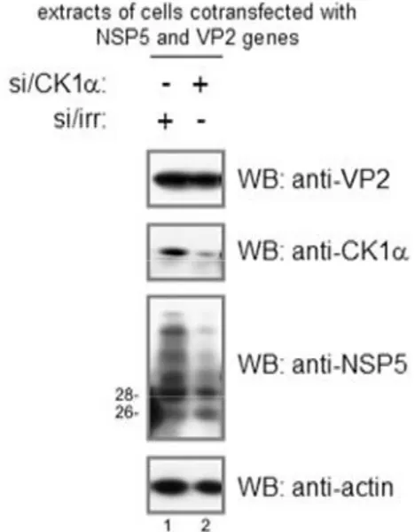
Attempts to map the regions involved in the hyperphosphorylation of NSP5 led to the identification of Ser67 as the residue responsible for initiation event of hyperphosphorylation. In particular, phosphorylation of this residue, more likely due to a cellular kinase, is the first step of a hierarchical process that leads to NSP5 hyperphosphorylation. Moreover it has been observed that Ser67 is localized in a consensus region for casein kinase I (CK1) phosphorylation. Indeed, in vitro phosphorylation assays with recombinant CK1, show phosphorylation of NSP5 wt, but no phosphorylation for a mutant NSP5, in which Ser 67 in mutated into Ala. (54).
The involvement of cellular CK1 in NSP5 hyperphosphorylation has been investigated using a small interference RNA specific for CKI. In particular the role of this kinase has been studied in the contest of the infection and co-expression of NSP5 and NSP2. In both cases, silencing of CK1 affected NSP5 hyperphosphorylation (26).
29
NSP5 has shown to be localized in viroplasms during rotavirus infection. More interestingly, recombinant NSP5 expressed in cells together with NSP2 induce the formation of particular structures resembling viroplasm of infected cells and for this reason called viroplasm like structure VLS (60). NSP5 and NSP2 have been found co-localized into VLS, and, although the interactions between NSP566-188 and NSP2 has been
characterized, the arrangements of the complex NSP5 dimer-NSP2 octamer to form VLS have not been identified, probably due to the lacking of NSP5 C-terminal region. However, it is has been proposed that C-terminal region of NSP5 in one NSP5-NSP2 complex may operate as a multimerizing domain, interacting with the C-terminal region of another NSP5-NSP2 complex. Since other reports show that also the N-terminal domain of NSP5 is involved (127), it is possible that a multi-step mechanism involving different regions of NSP5 controls NSP2-NSP5 interaction within VLS (195).(Fig 9)
Figure 9: VLS formation. Confocal immunofluorescence microscopy of cells co-expressing NSP2 and NSP5. (a) MA104 cells, either infected with rotavirus SA11 or co-transfected with NSP2 and NSP5 as indicated, were reacted simultaneously with anti-NSP2 (green) and anti-NSP5 (red). The rightmost panel is a superimposition of the two independently acquired images.
Several evidences showed that hyperphosphorylation of NSP5 is not related to VLS formation: i) NSP5 mutants with Ser67 mutated into alanine, that are not phosphorylated when expressed with NSP2, still form VLS (54); ii) in vivo inhibition of phosphatases in cells transfected with an NSP5 encoding plasmid results in a fully phosphorylated NSP5, but not in VLS formation (20) ; iii) an siRNA against CK1, the kinase involved in the phosphorylation of serine 67, inhibits NSP5 hyperphosphorylation, but not viroplasm formation (26). This observations are presented in results1 of this thesis.
Silencing the NSP5 expression in infected cells, with specific siRNA, abolishes viroplasms formation, indicating an important structural role of NSP5 during rotavirus infection.
Anti-NSP2 Anti-NSP5 merge
Rotavirus infection
30
Moreover, knocking down NSP5 in infected cells inhibits the accumulation of other viral proteins. Indeed the lack of viroplasms, as consequence of silencing NSP5, determines the inhibition of the assembly of new DLPs and the production of mRNA as a consequence of the secondary transcription. It has also been observed an inhibition of replication, revealing the relevant role of NSP5 in viral dsRNA production (27). This role was also supported by co-immunoprecipitation assays from infected and trasfected cells that showed that NSP5 strongly interacts with viral polymerase VP1. In particular, the last 48aa of NSP5 are involved in the interaction with VP1, and since the C-terminal region of NSP5 is also involved in dimerization, it is possible that a dimeric NSP5 is required for the association to VP1, with the binding region located just up-stream of the C-terminal tail. Moreover, this interaction is not weakened by the interaction of NSP5 with NSP2, since all three protein are immunoprecipitated in transfected cells and in particular they co-localized in VLS (6). On the contrary, VP1 impaired the ability of NSP2 to induce NSP5 hyperphosphorylation, probably sequestering NSP5, or blocking its conformational changing.
All the information collected about NSP5 features and functions in infected cells, provide new highlights to the important role of NSP5 during Rotavirus infection.
NSP4
The product of gene 10 is the non-structural protein NSP4, an ER-resident glycosylated protein that has been identified as the viral enterotoxin protein (216). The first protein product is a 20kDa protein, that upon glycosylation in the ER, becomes a polypepetide of about 28kDa.
NSP4 has two main domain: the N-terminal one that is anchored to the membrane of ER with three hydrophobic domains, and the C-terminal region, corresponding to the majority of the protein, that is oriented to the cytosol and exhibits all known NSP4 biological functions (44).
NSP4 has been extensively studied for its important role in virus morphogenesis and because it was first shown to be an enterotoxin that cause many symptoms of the viral infection (216).
A distinctive feature of NSP4 is its role as a receptor for DLP assembled into the viroplasms. About 20aa of the C-terminal region appear important for the binding of DLPs and their subsequent budding into the ER lumen (88). The receptor role of NSP4 is supported by the lower levels of TLP accumulated in cells treated with siRNA against NSP4 (117).
31
However knocking down NSP4 expression not only decreases the formation of triple layered particles as expected, but affects other Rotavirus activities: it induces increased levels of plus-strand RNAs, suggesting that NSP4 can be hypothesized to act as a feedback inhibitor in the infectedcell and to signal to the viral transcription system, when adequate plus-strand RNAs have been generated to allow productive infection (181). Moreover, NSP4 silencing does not affect the synthesis of the other viral proteins, normal amount of viral proteins are synthesized, however it induces failure of viroplasms maturation since NSP4 silenced cells show small viroplasms. Thus, the effect of the lack of NSP4 on viroplasms is not associated to a low amount of viroplasmic proteins, but it is likely to a defect on the traslocation of viroplasmic protein to the viroplasms. In particular a redistribution of VP2 and VP6 has been observed in NSP4 silenced infected cells: VP2 is more diffused in the cytoplasm rather than concentrated around viroplasms, and also VP6, is redistributed from viroplasms to fibers. This redistribution is thought to be mediated by the interaction of NSP4 C-terminal with VP6. Upon these observations, NSP4 is thought to create a cytoplasmic environment that promote the association of the different structural or non-structural proteins to form viroplasms (117).
Besides VP6, NSP4 has been found in oligomeric complexes with VP7 and VP4 in enveloped particles. The association of VP4, NSP4, and VP7 may represent sites on the endoplasmic reticulum membrane that participate in the budding of the DLPs into the lumen of the ER, where maturation to TLPs occurs (120).
The enterotoxin property of NSP4 has been associated to a 66aa cleavage product of NSP4 (NSP4112-175), that is secreted from infected cells early post infection. The trafficking
pathway that lead to the secretion of NSP4 peptide has been indentified as a nonclassical vesicular transport that bypasses the Golgi apparatus and involves the microtubule network, since treatment with nocodazole and cytochalasin D, but not with brefaldin A, impaired its secretion. The production of the enterotoxin is due to protease activity, however the proteases responsible for the cleavage of NSP4 have not been yetidentified (216).
The released cleavage product is available to bind to a putative receptor on the neighboring secretory cells to trigger the signal pathway that results in diarrhea (11),(131). Surface Plasmon resonance (SPR) associated to mass spectrometry identified two distinct functional domains on NSP4. In particular the NSP4114-130 domain binds to the MIDAS