Regulation of Notch signaling in the heart
by epigenetic modifications
PhD thesis in Molecular Genetics and Biotechnologies
Supervisor: Professor Mauro Giacca
Giulia Felician
Scuola Normale Superiore
2015
i
INDEX
INTRODUCTION ... 1
HEART DEVELOPMENT AND FUNCTION ... 1
IS THE HEART A POSTMITOTIC ORGAN? ... 5
CARDIAC REGENERATION IN MAMMALS ... 7
Stem cells and progenitor cells ... 9
Pluripotent stem cells ... 11
Epicardial stem cells ... 13
CARDIAC REGENERATION IN ZEBRAFISH ... 14
THE NOTCH RECEPTOR PATHWAY ... 17
Notch receptors and ligands ... 17
Notch signaling ... 19
Notch function ... 23
The role of Notch during heart embryogenesis and in the adult life ... 26
EPIGENETIC CONTROL OF GENE EXPRESSION ... 30
Histone modifications ... 30
Histone acetylation ... 31
Histone phosphorylation... 33
ii
Histone methylation ... 34
Arginine methylation ... 34
Lysine methylation... 35
Polycomb Group (PcG) proteins ... 38
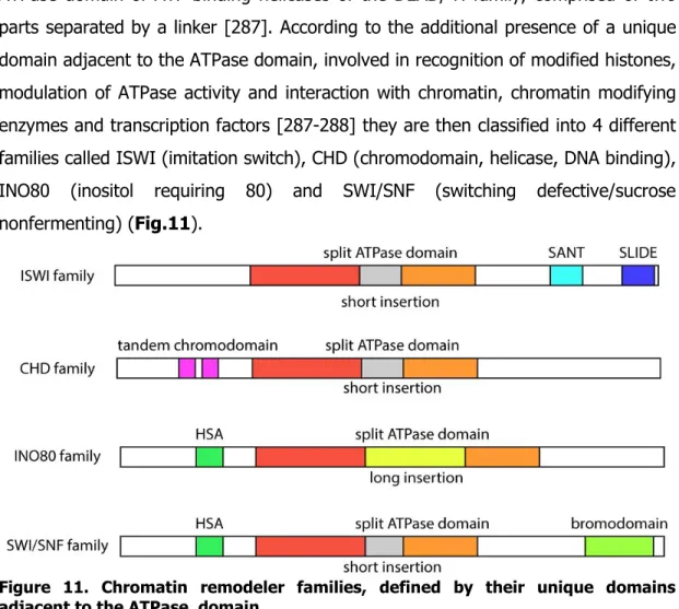
ATP-DEPENDENT CHROMATIN REMODELING COMPLEXES ... 41
DNA METHYLATION ... 45
EPIGENETIC REGULATION OF TRANSCRIPTION BY NOTCH ... 51
ADENO-ASSOCIATED VIRAL VECTORS ... 55
Adeno-associated viruses ... 55
AAV infection and viral life cycle ... 57
Adeno-associated viral vectors ... 59
RESULTS ... 63
THE DECREASE OF CARDIOMYOCYTE PROLIFERATION AFTER BIRTH IS PARALLELED BY A REDUCTION IN NOTCH SIGNALING ... 59
STIMULATION OF NOTCH PATHWAY BY AAV-MEDIATED GENE TRANSFER INDUCES NEONATAL CARDIOMYOCYTE PROLIFERATION IN VITRO... 68
AAV-MEDIATED NOTCH PATHWAY ACTIVATION DOES NOT STIMULATE HEART REGENERATION IN ADULT MICE AFTER MYOCARDIAL INFARCTION ... 80
AAV-MEDIATED NOTCH PATHWAY ACTIVATION DOES NOT INDUCE ADULT CARDIOMYOCYTE PROLIFERATION IN VITRO ... 83
NOTCH TARGET GENE PROMOTERS ARE METHYLATED AT THE DNA LEVEL IN ADULT CARDIOMYOCYTES ... 87
iii
MATERIALS AND METHODS ... 103 BIBLIOGRAPHY ... 119
SYNOPSIS
Understanding the molecular mechanisms regulating cardiac cell proliferation during the embryonic, fetal and adult life is of paramount importance in view of developing innovative strategies aimed at inducing myocardial regeneration after cardiac damage.
The Notch pathway plays a key role in the regulation of cardiomyocyte proliferation during mammalian embryonic life. Moreover, it is essentially involved in the cardiac regeneration process after injury in Zebrafish. Therefore, we assessed the efficacy of Notch pathway activation to sustain cardiac regeneration in a model of myocardial infarction in mice.
During early postnatal life, cardiomyocytes exit the cell cycle. We demonstrated that this event is paralleled by a decrease of Notch signaling and by the establishment of a repressive chromatin environment at Notch target genes, characterized by Polycomb Group protein 2-mediated silencing. The stimulation of the Notch pathway through Adeno-associated virus-mediated gene transfer of activated Notch1 or of the soluble form of the ligand Jagged1 prolonged the capacity of cardiomyocytes to replicate, which correlated with an increased rate of Notch target gene expression and the maintenance of an open chromatin conformation at Notch target gene promoters. However, the same vectors were ineffective in stimulating cardiac repair in a model of myocardial infarction in adult mice, despite efficient transgene expression. We identified the molecular cause of the lack of action of Notch signaling stimulation in adults in the increased DNA methylation at Notch target gene promoters, which correlated with permanent switch off of the Notch pathway. Our results confirm that the Notch pathway is an important regulator of neonata adults, due to the permanent epigenetic modifications at the DNA level at Notch responsive genes l
1
INTRODUCTION
HEART DEVELOPMENT AND FUNCTION
The heart is the first organ to form and to become functional in the embryo during its development. It is a complex organ composed of different muscle and non-muscle cell types (Fig.1): atrial/ventricular cardiac myocytes, conduction system cells, smooth muscle/endothelial cells of the coronary arteries and veins, endocardial cells, valve components and connective tissue. The major sources of precursor cells during heart development have been identified in cardiogenic mesoderm, cardiac neural crest and the proepicardial region.
Figure 1. Source of the different cellular components of the heart (adapted from [1]).
The genetic program responsible for heart development is evolutionarily conserved and is driven by a complex network of signaling molecules and tissue specific transcription factors, which control the activation of the genes responsible for heart morphogenesis (reviewed in [2-4]). Cardiac myocyte progenitor cells are already identifiable in the late gastrulation phase as an epithelial cell population in the
2
cranio-lateral mesoderm, the primary heart field, which gives rise to the cardiac crescent. The commitment of mesodermal cells to a cardiogenic fate strictly depends on the paracrine signaling between the endoderm, which secretes bone morphogenetic proteins (BMPs), positive regulators towards the cardiac lineage and the ectoderm, which secretes Wnt inhibitors [5-6]. The ultimate response to these molecules triggers the expression of specific sets of cardiogenic genes, which drive the morphogenetic events involved in heart development. In Drosophila, pro-cardiogenic signaling involves the transcription of the homeobox gene Tinman, necessary to activate the transcription of Mef2, which controls the differentiation of the precursors toward cardiomyocytes [7]. In vertebrates, Nkx2.5, the orthologue of Tinman, is expressed very early; its expression is cardio-specific and is maintained throughout the life. However, the factor does not appear to be necessary at this stage for cardiomyocyte specification, while mutant Nkx2.5 embryos die later during development due to abnormality in heart tube morphogenesis and left ventricle development [8]. Tinman/Nkx2.5 cooperates with the Gata family of transcription factors, important gene regulators of the cardiac developmental program. The cardiac crescent becomes organized as the linear heart tube, consisting of an inner layer of endocardial cells and an outer layer of myocytes held together by a dense extracellular matrix known as cardiac jelly. The linear heart acquires a rightward spiral form through a looping process, tightly controlled by an asymmetric axial signaling system. At this point, the cell fate of the precursors of the four cardiac chambers is already genetically determined.
Even if the genetic circuitry involved is not completely defined yet, the basic helix-loop-helix transcription factors Hand1 and Hand2 have been identified as important regulators of the development of the left and right ventricular segment respectively [2, 4]. At this stage, a second population of cardiac progenitors of splanchnic mesoderm is recruited; interestingly it seems that the main difference between the primary and the secondary heart field progenitor population is the timing of differentiation and not the expression pattern, since they both express similar
3
transcription factors [9]. These so-called secondary heart field progenitor cells contribute to right ventricle, atria and outflow tract formation. Newly formed cardiomyocytes start to secrete the extracellular matrix, resulting in the formation of the cardiac cushions, rapidly colonized by endocardial cells. Subsequently, a massive wave of myocardial proliferation triggers the formation of cardiac trabecolae.
The growth of each chamber results from a combinatorial signaling between cell layers. The endocardium secretes Neuregulins, the receptors of which are expressed by the myocardial layer; this signaling pathway is involved in the growth process of the developing ventricles [10]. At this stage, swelling of the cardiac cushion separates the cardiac tube into distinct chambers, causing septation.
Precursors of the cardiac valves arise from cardiac cushions: cells of endocardial origin migrate into the cardiac cushions and differentiate into fibrous tissue, responding to a complex signaling network, regulated at some extent by TGFβ family members [2, 11]. Other precursor cell populations migrate at this point to different areas of the developing heart to contribute to its final features: cardiac neural crest cells give rise to the vascular smooth muscle of the aortic arch and great vessels; epicardial cells, derived from the proepicardium, progressively envelop the developing heart; coronary precursor cells contribute to the coronary vasculature. From the epicardium, through epithelial to mesenchymal transition, a population of mesenchymal cells also arises, which contributes to the development of connective tissue, fibroblasts and smooth muscle cells of cardiac vessels [1]. Finally, the conduction system of the heart is essentially of myocardial origin: the decision of becoming conducting myocytes is regulated at the genetic level through the expression of a peculiar transcriptional network, while the cardiac ganglia innervating the conduction system are mainly derived from the neural crest [12]. The epicardium contributes to the conduction system through interstitial fibroblasts sparsely found between the mature cells of the conduction system (scheme of heart development in Fig.2).
4
Figure 2. Schematic representation of heart development (adapted from [1]).
The structure of the mature heart (Fig.3) reflects its embryonic development as a muscular tube. The right atrium receives venous blood, which enters the right ventricle through the tricuspid valve. From there, the blood is then pumped by the ventricle through the pulmonary artery to the lungs, where it is oxygenated; the oxygenated blood returns to the heart in the left atrium through the pulmonary veins and then it passes into the left ventricle through the mitral valve, from where it is pumped in the arterial vascular circuit in the body. Every heartbeat originates at the sino-atrial node located at the junction between right atrium and superior vena cava. The electrical impulse is propagated through the atria to the atrioventricular node and from there to the ventricle.
5
IS THE HEART A POSTMITOTIC ORGAN?
During the early postnatal life, a switch takes place between myocyte hyperplasia and hypertrophy. In humans, after withdrawal from the cell cycle, there is almost a threefold increase in the diameter of cardiomyocytes [13-14]. The paradigm of the heart as a postmitotic organ was established in the 1950s, when the first studies regarding heart growth were published [15-16]. First detection of mitotic figures in adult cardiomyocytes dates long back [17]. In more recent years, evidences of adult cardiomyocyte proliferation in human and rodent samples were obtained, showing that the mammalian heart maintains a mitotic activity even in adult organisms, where DNA duplication [18] and metaphasic chromosomes were detected in heart sections [19]. The detected proliferation rate was very variable in different studies, but consistently very low [20-21]. Recently, the attempt to precisely quantify cardiomyocyte turnover gave very different results according to the method used: using 14C dating, the Frisen group reported that cardiomyocytes are renewed with a
gradual decrease from 1% turn over annually at the age of 25 to 0.45% at the age of 75, therefore confirming a low proliferation rate [22]. A 20 fold higher turnover rate was calculated analyzing the incorporation of labeled nucleotides in cardiomyocytes [23], pointing to an underestimation of the number of proliferative myocytes in the previously reported low proliferation rates. Actually, the reported rate of proliferation by Bergmann and colleagues is in agreement with the known range of ploidy in human cardiomyocytes [24] and is consistent with the lack of significant regeneration after damage in the adult hearts. A variety of studies have identified proliferative cardiomyocytes dividing symmetrically throughout all the life span, albeit at a very low level; it has been proposed that the low turn-over due to adult cardiomyocyte proliferation is an important mechanism to maintain myocardial homeostasis [25-27].
6
The results are quite debatable also in animal models: in rodents, the analysis of a variety of mitotic markers in embryonic, neonatal, and adult phase has demonstrated robust cardiomyocyte proliferation in embryonic life only, followed mainly by binucleation of myocytes in the perinatal period, superimposable to their exit from the cell cycle [28]. The idea of a non proliferative adult heart is in agreement with the results of a recent study by Porrello and colleagues, who reported complete myocardial regeneration achieved through formation of new contractile cardiomyocytes in case of apical resection of the heart in 1-day old mice, while the formation of a fibrotic scar was prevailing when the ventricular resection was performed in 7-days old mice [29]. This evidence strongly supports the conclusion that the mammalian heart is endowed with an endogenous regenerative potential in fetal and early neonatal life only.
Discrepant results have been published regarding cardiomyocyte proliferation in the case of myocardial injury: Hsieh and colleagues identified an increased rate of cardiomyocyte proliferation after myocardial infarction in mice, possibly due to precursor cell proliferation [25]. A more recent study however indicated that the role of cardiac progenitors after injury is very limited [26]. The topic is really highly debated: besides the identification of the population (stem cells or differentiated cardiomyocytes) able to proliferate and eventually repair the heart in case of injury, the concept itself of the existence of a regenerative pathway in mammals still remains highly controversial. A recent study which did not detect any increase in the basal proliferation rate in case of myocardial infarction in mice have added further fuel to the controversy, even questioning the efforts to trigger any myocardial regeneration in mammals [27]. Finally, recent results by Naqvi and colleagues have also reopened the discussion on the proliferative potential of the postnatal heart in mammals, by showing that a second window of transient cardiomyocyte proliferation occurs post-natally in preadolescent (P15) mouse hearts, which appears to be regulated through the IGF-1/Akt pathway, known to modulate the early postnatal stages of heart development [30].
7
Although there are contrasting evidences about adult cardiomyocytes proliferation, what remains unquestionable is that the mammalian heart is unable to recover after massive cardiomyocyte loss, as in the case of myocardial infarction. Since cardiovascular diseases are the leading cause of mortality worldwide, among which ischemic heart disease is the most frequent disease condition, the strong need for therapies triggering cardiac regeneration still remains completely unmet.
CARDIAC REGENERATION IN MAMMALS
As depicted in Fig.4, cardiac regeneration has been approached in a variety of manners, from endogenous stem/progenitor cells proliferation stimulation to exogenous cell therapy, from stimulation of resident adult cardiomyocyte proliferation to prevention of cardiomyocyte apoptosis.
8
Antagonizing cell death and enhancing survival pathways in cardiomyocytes after an ischemic event are strategies which are feasible, but showing one major constraint: they are useful to prevent cell death, but cannot reconstitute cardiac muscle cell loss, which is one of the unsolved problems occurring after myocardial infarction [31]. Therefore major attention has been given to strategies which are aimed at reconstituting the cardiomyocyte population.
Accumulating evidences on adult cardiomyocyte division suggest that one of the main approaches to stimulate regeneration is to increase the number of dividing pre-existing cardiomyocytes. This task has been pursued manipulating different key players of the cell cycle or developmental signals known to act on cardiomyocytes. These include stimulation of Cyclin D2 [32] or Cyclin A1 [33], inhibition of p38 [34-35], stimulation of Periostin downstream signaling [36] or Neuregulin1-mediated ERBB2 pathway activation [37]. In vivo, some of these strategies have led to better functional outcomes in animal models, but the efficiency in promoting adult cardiomyocyte proliferation has been generally very limited, with a small number of mainly mononucleated adult cardiomyocytes completing cell division. Recently, miRNAs have been demonstrated to be an efficient, novel tool to achieve cardiomyocyte proliferation. Inhibition of the miR-15 family, which is involved in cell cycle arrest, has been shown in vivo to prompt adult cardiomyocytes to proliferate and improve functional outcome after myocardial infarction [38], while administration of miR-199a-3p and miR-590-3p were demonstrated to trigger cardiac regeneration, improving functional outcome after infarction and stimulating adult cardiomyocyte proliferation [39].
An alternative strategy to be considered to pursue heart regeneration is the formation of new myocytes from multipotent cells, resident progenitor cells committed to cardiac phenotype, stem cells present in the niches, or ex vivo transplanted cells.
9 Stem cells and progenitor cells
Historically, among adult stem cells, bone marrow derived cells (BMC) were the first population of stem cells reported to possess regenerative capacity in vivo. Orlic and collegues identified bone marrow cells able to transdifferentiate into cardiomyocytes and vessels once injected into the infarcted myocardium, providing significant functional benefit [40]. Similar results were concomitantly reported in other studies [41-42]. These results, however, have later been heavily questioned by a vast part of the scientific community [43-44]. Given the initial enthusiasm in the field, several clinical trials in patients with acute myocardial infarction or ischemic heart disease have been performed; transplantation of different subtypes of bone marrow-derived cells resulted in very different outcomes. The first clinical trial in which autologous mononuclear bone marrow cells were transplanted into the infarcted region claimed that the treated patients had better functional heart parameters due to the BMC-associated myocardial regeneration and neovascularization [45]. The subsequent clinical trials generated contrasting results, showing no significant improvement of heart function in the patients infused with autologous mononuclear bone marrow cells [46]. Several other trials have been performed later [47], and their results were collectively analyzed in 2012, reporting a modest but significant improvement of the left ventricular ejection fraction in the treated patients, together with a small beneficial effect in other left ventricular parameters [48]. More recently, other two clinical trials were performed, both reporting lack of effect of bone marrow mononuclear cell delivery [49-50].
Taking these evidences together, considering the differences in injection system, bone marrow cell preparation and evaluation of functional parameters, the overall result is that the injection of the cells is safe and feasible, even though the beneficial effect is very modest, possibly due to a paracrine secretion of angiogenic or pro-survival factors, which could stimulate cardiomyocyte pro-survival, or perhaps resident stem cells proliferation, therefore exerting a beneficial action of potential interest for the development of cell-free treatment [51].
10
Many different groups have described the existence of a pool of self-renewing, cardiac resident progenitor cells (CPCs) There is no unanimous agreement on the markers of this population: overlapping populations expressing Sca-1 [52], c-kit [53] or Abcg2 [54] have been described able to differentiate towards cardiomyocytes in vitro. One strategy to exploit this population for therapeutic approaches is its in vitro expansion. Studies on the c-kit+ population, which was reported to be present in humans, gave rise to controversial results: in an initial study, c-kit+ cells, isolated, expanded in vitro, differentiated toward cardiomyocytes, were transplanted in the context of myocardial infarction in rodents and were reported to support regeneration forming news cardiomyocytes [53, 55] or acting through a paracrine effect [56]. In other studies no transdifferentiation of c-kit+ positive cells to cardiomyocytes was detected [57-58]. The SCIPIO phase I clinical trial was performed injecting autologous CPCs in patients with ischemic cardiomyopathy [59-60]; the results seemed encouraging, suggesting that intracoronary infusion of these cells improved heart functional parameters in patients, therefore suggesting to proceed to a phase II study. Recently, however, the phase I study results have been questioned [61].
Another population of progenitor cells are the cardiosphere-derived cells, described as a population of stem cells isolated from biopsies, which can be expanded in vitro [62]. These cells were reported to exert beneficial effects when transplanted after myocardial infarction, thanks to their differentiation to new cardiomyocytes and to the secretion of factors exerting a beneficial paracrine effect [63]. Also in this case, there is controversy on the cardiomyogenic potential of these cells. One report has not confirmed their capacity to differentiate into cardiomyocytes and has proposed a fibroblast origin for these cells [64], while more recent data have indicated that their beneficial activity has essentially to be ascribed to their paracrine action [65]. Despite these controversies, cardiosphere-derived cells have been used for a clinical trial in patients with myocardial infarction, with reported beneficial results [66].
11 Pluripotent stem cells
Many attempts have also been performed to differentiate embryonic stem cells (ESC) and, more recently, reprogrammed induced pluripotent stem cells (iPS) to cardiomyocytes. The latter cell type is particularly appealing for regenerative purposes, since ESC-based therapies would be allogeneic, therefore requiring immunosuppression, while iPS cells would allow autologous transplantations. Several groups have successfully differentiated these cells into cardiomyocytes exhibiting intrinsic contractile activity and expressing cardiac transcription factors, but with myofibrillar organization typical of early-stage cardiomyocytes, therefore resembling immature cells [67-70].
In the context of an injured heart, ESC-derived cardiomyocytes were demonstrated to differentiate into immature cardiomyocytes, regenerate infarcted myocardium and achieve electromechanical integration with the surrounding tissue [71-73]. Not all the studies are concordant on the long term effect of the transplantation: it was also reported that, even in the case of graft survival, at longer time points the beneficial effect was not maintained [74]. Recent work by Murry and colleagues addressed the effect of human ESC-derived cardiomyocyte (hESC-CM) grafts in larger animal models. Non-human primates underwent myocardial infarction, followed by the injection of hESC-CMs. This study demonstrated for the first time the feasibility of the large-scale production of hESC-derived cardiomyocytes. The functional effect of the graft was evaluated, demonstrating that human derived cells provide re-muscularization to the infracted heart, showing electromechanical coupling with the host cardiomyocytes and perfusion by the host vasculature. The study revealed, however, the onset of nonfatal ventricular arrhythmias in the grafted primates, pointing to the need to deeper understanding the phenomenon, in order to achieve safe clinical translation to patients [75].
As far as iPS-transplantation is concerned, this now appears as a very promising avenue for the generation of cells capable of regeneration of various organs and tissues, including the heart. Indeed, pioneering studies have also indicated that
iPS-12
derived cardiomyocytes can engraft in the infarcted heart and provide therapeutic benefic in small animal models [76]. A relevant fear that might hamper further development of the iPS technology is the possibility that immature, non-differentiated iPS cells might give rise to the formation of teratomas. Indeed, a case of teratoma induced by the transplanted cells in the context of myocardial infarction, probably caused by incomplete differentiation of the cells, has been already reported [77].
A final interesting approach to achieve therapeutic formation of novel cardiomyocytes is the direct reprogramming of fibroblasts to become cardiomyocytes. A first attempt was performed to differentiate fibroblasts into cardiomyocytes using the iPS technology with a cocktail of 14 different transcription factors, out of which three (Gata4, Mef2C, c-Myc) were necessary to achieve the reprogramming of mouse cardiac fibroblasts, showing gene expression shifting from a fibroblast- to a cardiomyocyte-like profile. Even if the percentage of fully reprogrammed cells was around 1%, the reprogrammed fibroblasts were able to differentiate into cardiomyocytes when transplanted into mouse hearts [78].
Other strategies have been further applied to optimize the reprogramming: the so-called “Yamanaka cocktail” of genes (Oct4, Sox2, Klf4, c-Myc) and the addition of cardiogenic factor BMP4 were demonstrated to have a higher efficiency in converting mouse embryonic fibroblasts to cardiomyocytes [79]. A few studies also reported direct reprogramming in vivo. Using genetic lineage tracing experiments, resident non-myocytes were demonstrated to be reprogrammed into cardiomyocyte-like cells by in vivo local delivery of the 3 factors described by Ieda (Gata4, Mef2c and Tbx5), triggering also decreased infarct size and slightly better functional outcome in infarcted mice [80]. The authors believe that the in vivo administration of the reprogramming factors, which results in functional better outcome in the case of myocardial infarction, could be due to the higher efficiency of reprogramming achieved in the heart environment, compared to a Petri dish. The authors also hypothesized that some mechanisms, such as cardiac fibroblast activation block,
13
enhanced survival of cardiomyocytes, facilitated differentiation of cardiac progenitors or improved angiogenesis could contribute to the benefits observed upon expression of the reprogramming factors in the heart after myocardial infarction [81]. Finally, the reprogramming of cardiac fibroblasts to cardiomyocytes, both in vitro and directly in vivo, was also achieved using a combination of four microRNAs, which were able to induce direct cellular reprogramming in vitro, while the administration of the same microRNAs in vivo into the ischemic mouse heart results in the conversion of cardiac fibroblasts to cardiomyocytes [82]. Further improvement in the reprogramming process appears to be still needed, since often the reprogrammed cardiomyocytes show an immature phenotype [83] and the efficiency of the process is still remarkably low.
First attempts at reprogramming human fibroblasts were recently made by combining various cardiac transcription factors and/or microRNAs: the reprogrammed fibroblasts showed some sarcomere-like structures, calcium transients and a cardiomyocyte-like gene expression profile [84-85].
Epicardial stem cells
Considering the fundamental role of the epicardium during heart development and the variety of cell types that origin from this layer, epicardial progenitor cells (EPDC) in the context of heart injury have also been investigated. In the adult mouse heart, the epicardium overlaying the infarcted area is locally disrupted. In response to the injury, the surrounding epicardium undergoes a transient reactivation of the embryonic gene program [86]. The epicardium overlaying the infarcted areas is therefore regenerated in 3 days after the injury. This process is also paralleled by the formation of a thick layer of subepicardial mesenchyme above the infarcted area, originated by epicardial cells undergoing epithelial-mesenchymal transition (EMT), which contribute predominantly to fibroblasts, to a lesser extent to the coronary vasculature and possibly to cardiomyocytes [86]. This evidence demonstrates that a regenerative response is started, but is not effective. Recently,
14
it has also been reported that epicardium-derived cell activation, stimulated by myocardial injury, could substantially contribute to repair of the adult heart via regeneration of the coronary vasculature and of the myocardium vasculogenesis in the presence of Thymosin β4 (Tβ4) [87]; moreover Tβ4-activated EPDCs were reported to transdifferentiate into new cardiomyocytes, structurally and functionally integrated with the resident muscle, when the Tβ4 priming was performed prior to myocardial infarction [88], while no effect on EPDCs was detected when Tβ4 was administered after the injury, where EPDCs were contributing mainly to fibroblast population [89-90]. Finally, these cells were also reported to contribute to the reduction of infarct size through stimulation of angiogenesis and secretion of paracrine factors which can modulate the subepicardium compartment [91]. A deeper understanding of the potential of epicardial population is needed in order to evaluate the therapeutic potential of these cells in regenerative medicine.
CARDIAC REGENERATION IN ZEBRAFISH
It is widely known that organ regeneration can efficiently take place in lower vertebrates. In particular, Zebrafish has a high regenerative capacity since in adults amputated or injured tissues such as fins, maxillary barbel, retina, optic nerve, spinal cord, brain, pancreas, kidney and heart muscle can regrow [92].
The resection of up to 20% of the heart ventricle results in the immediate formation of a clot, replaced in a couple of days by fibrin deposition, followed by the production of new, viable and functional myocardium, reaching perfect recovery 60 days after the heart damage (Fig.5) [93]. Three different mechanisms have been proposed to explain which is the cell compartment responsible for the regeneration process: adult, contractile, differentiated myocytes could be stimulated to massively re-enter the cell cycle and reform the apex; regeneration could proceed through the recruitment of undifferentiated progenitor cells which are differentiating into new
15
cardiomyocytes; pre-existing cardiomyocytes could undergo “de-differentiation”, downregulating contractile genes, in order to create a population of less differentiated cells, able to proliferate and re-differentiate into cardiomyocytes [94]. Initially, it was proposed that regeneration was triggered by progenitor cells in the blastema, which started to express pre-cardiac markers and contractile genes and to proliferate. The authors speculated that injury-related signals are insufficient to stimulate massive adult myocardial cell proliferation, therefore progenitor cells contributed to the regeneration process [95]. This mechanism has recently been disproved by genetic fate mapping experiments, showing that the newly formed cardiomyocytes originate from the adult cells, which undergo partial dedifferentiation, detachment one from another and disassembly of the cytoskeleton, with no reactivation of the fetal gene program [96]. Non myocardial cells play a role in the regeneration process, as they create a suitable environment for myocardial proliferation, expressing a variety of factors, among which Raldh2, a retinoic acid-synthesizing enzyme, shown to be necessary for a correct injury-response to occur [97].
Figure 5. Schematic representation of Zebrafish heart regeneration after ventricular resection [98].
16
The Notch signaling pathway, through the Zebrafish orthologue Notch1b, was demonstrated to be involved in this heart regeneration process. As in the case of fin regeneration, the expression of Notch1b dramatically increases the day after the amputation and declines two weeks later. The Notch receptor ligand DeltaC parallels the expression pattern of Notch1b. The discovery that the Notch pathway is involved in heart regeneration was particularly interesting, since this pathway is not involved in the genetic program leading to heart development in Zebrafish, pointing out the existence of a specific regenerative genetic program [99]. In a recent study, fate mapping experiments demonstrated that, in the case of ventricular ablation, atrial cardiomyocytes transdifferentiate into ventricular myocytes upon activation of the Notch signaling cascade, since the block of Notch activation impedes the atrial to ventricular transdifferentiation [100]. Recent work has further confirmed the central role of the Notch pathway in the heart regeneration process. Following amputation of the Zebrafish ventricular apex, Notch expression is activated both in the epicardium and in the endocardium and suppression of Notch signaling profoundly impairs cardiac regeneration and induces scar formation at the amputation site; interestingly, the block of Notch signaling in the epicardium and endocardium resulted in decreased proliferation of the cardiomyocyte compartment, where Notch expression was not reactivated upon injury [101]. These results suggest the existence of a complex signaling network downstream Notch activation able to drive the whole regeneration process.
17
THE NOTCH RECEPTOR PATHWAY
Notch receptors and ligandsIn mammals, the Notch receptor family is composed of 4 type-1 transmembrane proteins (Notch1, -2, -3, -4), while there is only one receptor in flies (Notch) and two in C. elegans (LIN-12 and GLP-1) (Fig.6). The extracellular domain is composed by 29 to 36 epidermal growth factor (EGF) repeats followed by a negative regulatory region (NRR) composed of 3 cysteine-rich Lin12-Notch repeats (LNR) and a heterodimerization (HD) domain. For the interaction with the signal-sending cell, EGF repeats 11-12 are required, while repeats 24-29 prevent the interaction of the receptor with its ligand on the same cell [102-103]. Many of the EGF repeats can bind calcium ions, thus regulating Notch affinity for its ligand and signaling efficiency [104-105]. The LNR motifs and the heterodimerization domain act as negative regulators, preventing receptor activation in the absence of the ligand. The Notch transmembrane domain (TMD) is followed by the intracellular domain, composed by a RAM motif (RBP-Jk association module) involved in the binding of Notch with the transcription factor CSL/RBP-Jk through a high-affinity binding module centered on a conserved WxP motif, followed by an unstructured region containing a nuclear localization signal (NLS), 7 ankiryn repeats involved in protein-protein interaction and an evolutionary divergent transactivation domain (TAD), which recruits transcriptional activators as Mastermind-like and histone acetyltransferase complexes and contains a PEST domain regulating Notch stability and degradation [106-107]
.
18
Figure 6. Structure of Notch receptors in flies, mammals and C. elegans (adapted from [106]).
There are 5 Notch ligands in mammals: Jagged1 and -2, Delta-like1, -3, -4; in Drosophila they are called Serrate and Delta, and LAG-2 in C. elegans (Fig.7). They are type-1 transmembrane proteins sharing common features in the extracellular domain, composed by an N-terminal DSL (Delta/Serrate/LAG-2) motif, specialized EGF repeats called DOS (Delta and OSM-11-like proteins) domain involved in receptor binding, and EGF repeats, some of which required for the interaction with the Notch receptors (EGF repeats 11 and 12); only in Jagged1 and -2 there is an additional cysteine-rich region involved in receptor binding specificity. The short intracellular domain is more variable, contains a PDZ domain and is involved in downstream signaling [108].
19
Figure 7. Structure of Notch ligands Jagged/Serrate and Delta in flies and mammals (adapted from [106]).
Notch signaling
Notch protein is synthesized as a peptide of 300 kDa which undergoes glycosylation at different sites of the extracellular domain: the EGF repeats are glycosylated by an fucosyltransferase which adds fucose to serine or threonine residues; the O-fucosylation sites can be further modified by N-glycans by the Fringe protein. Fringe glycosylation can affect the ligand-binding activity [109] and could also enhance the cleavage occurring in the Golgi (S1 cleavage), mediated by the Furin-convertase; in mammals, this cleavage leads to a heterodimeric protein with an extracellular subunit of 180 kDa and a transmembrane domain of 120 kDa, non covalently associated. The heterodimer is kept inactive through a tight interaction between the LNRs and the heterodimerization domain. Upon ligand binding, the receptor undergoes a conformational change which results in the exposure of the cleavage site for an ADAM protease (ADAM10, Kuzbanian or TACE, tumor necrosis factor converting enzyme), 12 amino acids before the transmembrane domain (S2 cleavage) [110]. The cleaved extracellular domain of Notch bound to the ligand is cleared from the membrane through trans-endocytosis into the signal sending cell [111]. The truncated form of Notch resulting from S2 cleavage is the substrate for the subsequent S3 and S4 cleavages operated by γ-secretase, a
multicomponent-20
intramembrane complex composed by Presenilin, Nicastrin, PEN2 and APH1 [112]. γ-secretase processes Notch at two different sites, eventually leading to the release, into the cytosol, of the Notch intracellular domain (NICD), which is able to migrate to the nucleus [113-114]. In the nucleus, NICD binds CSL (CBF/RBP-Jk in mammals, Su(H) in flies, LAG-1 in C. elegans; I will refer to it as RBP-Jk from now onward) transcription factor [115]. In the absence of NICD, RBP-Jk behaves as a transcriptional repressor, binding to histone-deacetylases and other corepressors, keeping the chromatin in a transcriptional silent state [112]. It has been hypothesized that NICD has a stronger affinity to bind RBP-Jk compared to the repressors [116], but this has not been definitively proven. Another model suggests that NICD and the repressors compete for binding to RBP-Jk [116]. After the interaction with NICD, RBP-Jk is converted to an activator of transcription; the complex formed by NICD, RBP-Jk and MAML (Mastermind-like) recruits transcription factors and therefore activates transcription of the target genes, mainly the Hes (Hairy/Enhancer-of-split) and Hey (Hairy/Enhancer-Of-Split Related With YRPW Motif) families of basic helix-loop-helix transcription factors [117-118]. These proteins bind their target sequence on DNA as homo- or heterodimers and act by repressing transcription of their target genes in different ways. The binding of these transcription factors to their cognate binding sites can cause the recruitment of other corepressors, such as Groucho, which can recruit histone deacetylases, therefore causing transcriptional repression through alteration of local chromatin structure; alternatively, they can directly bind to other bHLH factors forming non functional heterodimers [119-120]. Moreover, the NICD-RBP-Jk complex is able to directly activate transcription of several targets, such as Cyclin D1, Cyclin D3, p21, glial fibrillary acidic protein, Myc, Nodal, PTEN, EphrinB2, smooth muscle actin. Interestingly, several of the genes regulated by Notch have a role in the maintenance of a proliferative status [119, 121]. After activation of target gene transcription, NICD is rapidly degraded: MAML couples the activator role with the degradation of NICD, promoting NICD phosphorylation, since the complex composed
21
of MAML-SKIP-RBP-Jk can recruit the nuclear kinase CycC/CDK8 which hyperphosphorylates Notch TAD and PEST domains [122-123]; the phosphorylated PEST domain is recognized by the ubiquitin-ligase Fbw7/Sel10, resulting in NICD ubiquitin-mediated proteasomal degradation [124-126]. Moreover, acetylation has been demonstrated to regulate the stability of the NICD: in endothelial cells, Sirt1, a member of class III deacetlyases, was discovered to deacetylate and therefore destabilize NICD, triggering its degradation, therefore antagonizing the establishment of the transcriptional activator complex [127].
Recent evidence suggests that endocytosis can modulate Notch trafficking, and therefore its activity. In Drosophila, mutants having defects in endocytosis, recycling, vesicular sorting or multivesicular body formation show defects in Notch signaling [128-129]. A typical way of regulation of receptor internalization is protein monoubiquitination, which can target several residues in the Notch intracellular domain. HECT-type E3 ubiquitin ligase Nedd4 and Itch in mammals (Suppressor of Deltex in Drosophila) can act on NICD as negative regulators. These proteins are involved in a regulatory mechanism to prevent inappropriate ligand-independent activation of Notch signaling, targeting NICD to the lysosomal degradation pathway [130-131]. Besides the involvement of the intracellular trafficking machinery in keeping Notch inactive, endocytosis could also have an active role in NICD release and signaling. Deltex, a ring finger-type ubiquitin ligase, is thought to counteract the effect of Nedd4 and Itch, positively regulating Notch signaling; the factor promotes Notch sorting from the endosomal compartments, escaping the lysosomal degradation [132-133]. Moreover, Notch monoubiquitination followed by endocytsosis is an absolute requirement for γ-secretase to process the receptor. Actually, the initial paradigm of the γ-secretase complex acting on the cell surface has been radically revised, since the multi-subunit protease complex has been visualized embedded in the membranes of the endocytic vesicles, consistent with the evidence showing that it has an optimal activity at a low pH, as it is in the endocytic compartment [134]. These mechanisms of Notch trafficking could have a role in
22
protecting the cell from accidental firing of the Notch pathway: since very little amount of NICD is required to activate the downstream pathway, this could be generated in a ligand independent manner; the continuous ubiquitination and degradation of the receptors at the cell surface could be a way to regulate the steady-state level of the Notch protein [130]. The possible role of endocytosis in the control of the ligands has also been investigated, since E3 ubiquitin-ligases Neuralized and Mind Bomb were found to be required for ligand internalization, causing higher activity of the ligands on the cell surface [135-136]. Interestingly, the Notch extracellular domain undergoes trans-endocytosis into the ligand-expressing cells, dissociating from the transmembrane domain which undergoes further processing [137]. Several hypotheses have been proposed to explain the link between ubiquitination, endocytosis and ligand activity: ligand endocytosis could generate a pulling force on the extracellular domain of the Notch receptor, therefore triggering the exposition of the cleavage site for ADAM proteases; ligand ubiquitination could promote its clustering and therefore a more robust Notch activation, or the trafficking in the endocytic compartment could allow post-translational modifications on the ligands improving their activity [138] (schematic representation of Notch pathway in Fig.8).
23
Figure 8. Notch trafficking and signaling pathway (adapted from [139]).
Notch function
Notch signaling is highly evolutionary conserved and plays a crucial role in the embryonic development of flies, worms and mammals. Its signaling pathway regulates many processes of cell fate decision; in the context of binary cell fate decision, Notch regulates lateral inhibition between adjacent cells, which means that a population of cells with equivalent developmental potential will signal to each other through inhibitory reciprocal Notch signaling. By the amplification of differences in the expression levels of either ligand or receptor within the cell population, one of the cells will acquire a higher level of the ligand amplified by
24
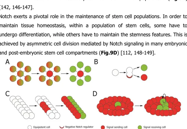
feedback regulatory loops, therefore inhibiting the neighboring cells to adopt the same fate using Notch signaling [140-143] (Fig. 9A). Another case of binary cell fate occurs when both sister cells express ligand and receptor but, due to asymmetric segregation of cell fate determinants which can negatively regulate Notch signaling occurred prior to mitosis, the signal is polarized and the cells undergo different fates. One of the most important Notch-inhibitory factors is Numb, a phosphotyrosine binding domain adaptor protein able to bind Notch and to prevent its activation promoting the receptor degradation (Fig.9B) [142, 144-145].
Notch signaling is also involved in the boundary specification through inductive signaling. This mechanism of signaling results in the creation of a new cell type after cell-cell interactions at the boundary between distinct cell populations (Fig.9C) [142, 146-147].
Notch exerts a pivotal role in the maintenance of stem cell populations. In order to maintain tissue homeostasis, within a population of stem cells, some have to undergo differentiation, while others have to maintain the stemness features. This is achieved by asymmetric cell division mediated by Notch signaling in many embryonic and post-embryonic stem cell compartments (Fig.9D) [112, 148-149].
Figure 9. Binary cell fate decision (A and B), inductive signaling (C) and stem cell pool mantainance (D) regulated by Notch signaling pathway.
25
More in detail, the importance of Notch signaling in the stem cell context has been widely reported in the nervous system, in Drosophila and in mammals. In Drosophila, the Notch signaling is responsible for the fate decision of individual cells among an equipotent cluster in the ectoderm, specified to become neuronal progenitors [150]. During embryonic development in mice, Notch stimulates precursor cell proliferation and inhibits neural differentiation promoting glial cell fate, while later it stimulates differentiation of astrocytes and inhibits terminal differentiation to oligodendrocytes [151]. In adults, it exerts a fundamental role in maintaining the stem cell pool present in the subventricular zone and in the subgranular zone of the brain. Here, it regulates the cell cycle exit of neural stem cells. Blocking Notch results in the exhaustion of the stem cell pool, with an increased differentiation into transient amplifying cells and neurons [152-153]. This mechanism might exert a role in the expansion of the neural stem cell pool after injury [154].
In the intestine, stem cells reside in the Lieberkühn crypts. Here, Notch plays two independent roles. It regulates stem cell proliferation, since its activation triggers the amplification of stem/progenitor cell pool through the action of its target gene Hes1 which blocks the expression of cyclin dependent kinase inhibitors [151, 155]. Moreover, it inhibits the differentiation toward secretory cells and favors the differentiation toward the absorptive phenotype via the negative regulation on the transcription factor Atoh1, which promotes secretory cell fate commitment [156-157].
During muscle development, a pool of progenitors exit from the cell cycle and undergo differentiation expressing specific transcription factors and forming multinucleated myotubes. Satellite cells expressing the Pax7 transcription factor are localized adjacent to muscle fibers under the basal lamina, where they remain quiescent. This compartment is crucial in the regenerative process after injury, since satellite cells are driven back into the cell cycle and induce myotube proliferation. In vitro, activation of Notch signaling leads to the prevention of multinucleated
26
myofiber formation, therefore suggesting that Notch activation blocks differentiation [158-159]. Loss of Notch signaling is accompanied by the loss of the satellite cell population since the pool of progenitors undergoes exhaustion [160]. Moreover, the satellite cells fail to assume the correct position, pointing to a role of Notch signaling also in the homing of stem cells to the correct niche [161]. In the adult, Notch pathway was demonstrated to be active in quiescent satellite cells, where it probably represses the key transcription factor necessary for terminal differentiation [162-163]. In the case of injury, Notch is down-regulated and the satellite cells exit from the quiescent state to differentiate and contribute to regeneration [164].
Finally, Notch signaling has been demonstrated to play a pivotal role in hematopoietic stem cell compartment in the embryo, where it is essential for generating hematopoietic stem cells (HSCs) from endothelial cells in the aorta-gonad-mesonephros regions [165]. In the adult, the role of Notch is more controversial. Notch signaling has been reported to affect self-renewal, proliferation and differentiation of adult HSCs, since the treatment with its ligand increases HSC expansion in vitro [166-167] and overexpression of active Notch preserves/expands hematopoietic progenitors in vivo [168-169]. Opposite to these results, several genetic studies could not identify any crucial role for Notch signaling in HSC maintenance or proliferation [170-171], supporting the idea that it is not necessary for homeostasis under steady-state conditions, while it is a potent tool to expand HSCs in vitro.
During somitogenesis, Notch also regulates temporal synchronization of the development of a group of cells: expression of the downstream gene Hes1 oscillates and Notch, while not triggering the oscillation itself, controls the synchronization of the oscillating signals [172-173].
The role of Notch during heart embryogenesis and in the adult life
Notch signaling exerts different important roles during cardiac morphogenesis. In the initial phase of development, Notch signaling acts as an inhibitor of
27
differentiation towards the cardiomyocyte fate. In different models, such as
Xenopus, chick embryo or mouse embryonic stem cells, the Notch pathway is able to inhibit cardiac differentiation and myocardial gene expression, blocking mesodermal commitment and cardiac differentiation [174-177]. Furthermore, expression of activated Notch in cardiac mesoderm leads to abnormal morphogenesis due to impaired cardiomyocyte differentiation and hyperplasia of the atrioventicular cushions [178]. Since Notch is involved in binary fate decision, prevention of cardiogenic commitment could result in commitment to another lineage. Indeed, embryonic stem cells receiving a positive Notch signal undergo neurectodermal transition and neural specification and become incapable of adopting a cardiogenic fate [179-180]. The exact mechanism by which Notch inhibits cardiogenesis is not completely defined. Notch target Hey transcription factors are able to block cardiac gene transcription either interacting (and thus inhibiting) the cardiac activator Gata4 or its target genes or binding the Gata4 responsive promoters [181-182]. Moreover, Notch can inhibit Mef2C, which marks the cardiac and skeletal muscle lineages during mouse embryogenesis [162], by either physically binding to it thus impeding its transcriptional ability, or competing for members necessary to form the Mef2C transcriptional complex [183]. During cardiac development, different Notch receptors are expressed with a distinct local and temporal pattern: Notch1 and -2 are mostly expressed in the developing heart, Notch3 is restricted to smooth muscle and Notch4 to the endothelium of the vascular system [184-185]. The ligands also have a , in the ventricular trabecolae and in the atrial myocardium, while Delta-like1 and Delta-like4 are expressed respectively in the endocardium and in the cardiac crescent and later in the ventricular endocardium [186]. Notch and its ligand are highly expressed in the non-myogenic dorsolateral domain of the primary heart field, where they act by suppressing cardiogenesis [174]. Loss of function studies have demonstrated that Notch is not necessary in the first phases of heart development, such as heart field specification and induction of cardiac mesoderm, since complete lack of Notch signaling due to RBP-Jk mutation is not lethal at this stage [187]. The
28
first defect linked to Notch impaired signaling is the random looping of the heart [188]. Defective Notch signaling also results in defective EMT during the formation of cardiac cushions. This process is essential for the correct development of the endodermal cardiac cushions, from where cardiac valves and atrial and ventricular septa will develop. Notch1 mutants show a defective induction of EMT, with very few migrating cells, lacking mesenchymal morphology [189-190]. The effect of Notch signaling in this process is probably due to Notch target genes, which are able to regulate the expression of specific metalloproteases required for cell migration and to the Notch-mediated negative regulation of cadherin expression, enabling the cells to invade the cardiac jelly [189-190]. During ventricular trabeculae formation, Notch1 is expressed in the endocardium and triggers cardiomyocyte proliferation through two different pathways: it stimulates the production of Neuregulin1 by the endocardium, which allows the transition of primitive myocardial epithelium to trabecular and compact myocardium, and it stimulates BMP10 production in cardiomyocytes, which positively regulates their proliferation [191]. The importance of the Notch pathway in cardiac morphogenesis is highlighted by the strong phenotype of the gain- and loss-of function mutants of the several key molecules of the pathway as well as by the features of the Alagille syndrome, a human autosomal disorder characterized by hepatic, cardiac, skeletal and eye malformation due to Jagged1 mutation. In the cardiovascular system, the abnormalities include ventricular septal defects and hypetrophy of the right ventricle due to underlying pulmonary stenosis [192]. In addition, Notch can also control differentiation of committed mesodermal progenitors and of cardiac precursor cells into cardiomyocytes, reinforcing the conclusion that the Notch pathway is able to control cardiogenesis at multiple steps of the differentiation process, from the mesodermal versus neuroectodermal commitment to myocyte differentiation and proliferation [178-179, 193].
In the adult heart, several members of Notch family are expressed at different levels and Notch signaling is detectable in cardiac non-myocytes and rarely in
29
cardiomyocytes [186]. It is mainly involved in the maintenance of adult heart tissue integrity. Given the role of Notch in preserving the stem cell pool in several tissues, Notch has been hypothesized to play an important role in cell-to-cell contact signaling between accessory cells and cardiac precursors in putative stem cell niches. In particular, it has been proposed that Notch could be involved in keeping the precursor population quiescent, while maintaining the proliferation and expansion of undifferentiated cardiac precursors cells, eventually leading them from the immature phenotype to the compartment of amplifying myocytes [194-195], also regulating their proliferation and expansion [196].
Several studies have analyzed the role of the Notch signaling in vivo, in the context of cardiac regeneration, reporting a variety of pro-regenerative roles. In a model of myocardial injury, Notch signaling was reported to promote cardiomyocyte survival [197] and contribute to protective signaling, interacting with the c-Met/Akt pathway [198]. It was also reported to inhibit excessive fibrosis in a model of myocardial infarction [197] and of pressure overload, through activation of Notch signaling in heart stromal cells [199]. In a model of aortic constriction, it could play a role in the mobilization of epicardial cells, which contribute to the resolution of fibrosis; in addition, these cells also display a modest differentiation potential towards cardiomyocytes [200].
Finally, Notch has also been proposed to play a role in the mobilization of different population of stem cells, including bone marrow-derived stem cells in the case of myocardial infarction [201], and cardiac precursor cells [194, 199, 202].
Several reports have assessed the role of Notch in the tuning of cardiomyocyte proliferation. Reports from our laboratory have shown that Notch1 drives proliferation and expansion of neonatal rat cardiomyocytes [196]. Moreover Notch ICD was reported to activate Cyclin D1 transcription and also to promote its nuclear localization, therefore stimulating cardiomyocyte cell cycle progression [203], although recent evidence has shown incomplete cardiomyocyte proliferation [197]. In differentiated cardiomyocytes, Notch inhibits hypertrophy, probably by impeding,
30
through its target gene Hey2, the pro-hypetrophic activity of Gata4. Therefore, Notch might maintain cardiac tissue homeostasis by limiting the extent of the cardiac hypertrophic response [181-182, 204].
EPIGENETIC CONTROL OF GENE EXPRESSION
In eukaryotic cells, nuclear DNA is wrapped around proteins to form the chromatin. The primary protein components of chromatin are histones, assembled in octamers to form nucleosomes. This structure ensures chromatin compaction and it can undergo many modifications regulating most of DNA-related processes, such as transcription, recombination, DNA repair, replication. The modifications can be covalent modifications of the histones, modulation of DNA accessibility through chromatin complexes called chromatin remodelers, or modification on the DNA itself by the addition of methyl groups on the cytosines in the context of CpG dinucleotides. Given the complexity of the topic, in this introduction I will mainly describe the chromatin modifications impacting on transcription.
Histone modifications
Chromatin structure is highly complex and impressively dynamic. The nucleosome and its histone core, which were once thought to be static and highly stabilized by the 14 non-covalent bonds occurring between DNA and histones, actually play an integral role in directing some elements of transcriptional specification. The histone octamers forming nucleosomes are characterized by 15-30 amino-terminal residues which protrude from the nucleosome to form the so-called “histone tails” [205]. More than 60 different residues mainly residing in the histone tails have been demonstrated to be modified by covalent binding of various functional groups. The most common additions are acetylation, methylation, phosphorylation and ubiquitination of residues, which can act combinatorially, according to the so-called “histone code” [206]. The best characterized mechanisms through which histone
31
modifications influence chromatin are the disruption of the contacts between nucleosomes and DNA, since many modifications result in the change of the net charge of the nucleosome and in the loosening of the coil, and the recruitment of non-histone proteins, which can combinatorially bind to the modified histones (reviewed in [207]).
Histone acetylation
Histone acetylation is a highly dynamic process known since the 1960s [208]. The class of enzymes responsible for histone acetylation are the Histone Acetyl Transferases (HATs): they utilize AcetylCoA as a cofactor to transfer an acetyl group to the ε-amino group of lysine, therefore neutralizing its positive charge. HATs are divided into 2 families, type-A and type-B HATs. Type-B HATs are mainly cytoplasmic and are able to acetylate newly synthesized histones (K4 on histone 3 and K12 on histone 4), prior to their assembly to form the nucleosome. Type-A HATs are divided into 3 subfamilies according to their sequence homology: GNAT (Gcn5-related N-acetyltransferase), MYST (MOZ, Ybf2/Sas3, Sas2, and Tip60) and CBP/p300; they have a limited substrate specificity, since they are able to add acetyl groups to many different lysine residues. The modified residues are mainly in the N-tail of the histones, and the neutralization of the positive charges given by the acetylation destabilizes the interaction between the nucelosomal proteins and the DNA [207]. There are also additional sites for acetylation within the globular structure of histones, facing the DNA major groove, such as K56 on histone 3 acetylated by Gcn5 in humans, which results in the disruption of the electrostatic interactions between DNA and histone cores, starting DNA unwrapping [209-210]. Acetylation of both histones 3 and 4 has been identified as a mark for active chromatin and HATs activity has been characterized in this context. Gcn5 is a part of an activator complex called SAGA (Spt-Ada-Gcn5 acetyltransferase) and is able to acetylate H3 and H2B, while Esa1, part of nucleosome acetyltransferase H4 complex
32
(NuA4), preferentially acetylates H4 and H2A. Both complexes are recruited by specific activators to the promoters of genes that are transcriptionally active [211]. The cooperation between HATs and chromatin remodeling complexes has been described in several cases, since acetylated histones are known to be binding sites for other proteins involved in gene regulation, such as the SWI/SNF remodeling complex, which is able to bind and acetylate histones and is involved in nucleosomal mobilization [212]. Tip60 has been identified together with Brg1 (a SWI/SNF chromatin remodeler family member) as a factor affecting the regulation of several developmental genes [213]. Moreover, the presence of other histone modifications such as H3K4 methylation (in the promoter) and H3K36 methylation (in the coding region) can also play a role in HAT recruitment, since proteins able to recognize methylated residues also take part in the formation of HAT-containing complexes [214].
The enzymes responsible for the reverse reaction, namely the removal of the acetyl groups, are named Histone Deacetylases (HDACs), and their action restores the positive charge of the lysine, therefore stabilizing the chromatin structure. HDACs are divided into four classes: class I, II and IV share some sequence homology and need Zinc as a cofactor; class I and II are homologous to yeast (sc)Rpd3 and scHda1, class IV is composed only by HDAC11, class III (also called Sirtuins) are homologous to scSir2 and are NAD+ dependent (extensively reviewed in [215-216]). HDACs not only act on chromatin, but are involved in the tuning of multiple intracellular signaling pathways, showing low substrate specificity. At the transcriptional level, HDACs are able to induce repression of transcription not only by restoring the net positive charge of the lysine residues, therefore reducing the affinity of histones for several transcription factors, but also increasing the affinity for transcriptional repressors, such as proteins containing the SANT domain, which recognizes unmodified histones [207, 214, 217].
To conclude, paradoxically, HATs are also found at inactive gene promoters, where they probably prime the genes for activation [218] and HDACs are also important in
33
active genes, since they are required to ensure proper transcription initiation and to commit genes to rapid repression [214, 219]. The pattern of histone tail acetylation can also regulate chromatin compaction dynamics, with a direct effect on chromatin structure, since there are evidences that acetylation in H4K16 inhibits the formation of the compact 30 nm fibers [220-221].
Histone phosphorylation
Histone phosphorylation is a highly dynamic process occurring on serine, threonine and tyrosine residues present in the N-tail of histones, able to modify the net charge of the chromatin thanks to the transfer of a negatively charged phosphate group on the side chain of the target amino acid. Several cellular kinases have been found to bind chromatin, suggesting their active role in chromatin phosphorylation [222]. Phosphorylated residues in the histone tails behave as docking sites for signaling effectors and adaptors, as 14-3-3 proteins, which have been characterized as downstream effectors [223]. Histone phosphorylation plays a role in different processes, including gene expression, DNA damage response and chromatin compaction (reviewed in [224]).
Regarding the regulation of gene expression, H3S10, T11 and S28 phosphorylation is associated to Gcn5-dependent histone acetylation, strongly supporting the existence of a crosstalk between acetylation and phosphorylation to promote transcription activation [224]. In some cases, activation of the transcription can be achieved through an allosteric regulation of the protein complexes present on the chromatin. As an example, H3S28 phosphorylation can be correlated to transcription activation through the displacement of the Polycomb repressive protein complex, which acts on the upstream amino acid (H3K27) in the histone tail. Moreover, H3Y41 phosphorylation activates transcription by disrupting chromatin binding by the repressive protein HP1 [224].
34 Histone ubiquitination
Ubiquitination, most frequently monoubiquitination, is a covalent modification occurring on lysine residues of the histone proteins assembled as octamers in the nucleosomes, as well as in the linker histone H1. The best characterized is the role of H2AK119 and H2BK120 (K123 in yeast) ubiquitination [207]. H2A-ubiquitin ligases are often found in transcriptional repressor complexes, as it is the case for Ring1, a member of gene silencing-related Polycomb Group protein 1, discussed later in details [225], while H2B ubiquitination is associated to active gene expression, promoting transcriptional activation and elongation [226-227].
Histone methylation
Histone methylation consists in the transfer of a methyl group from S-adenosylmethionine to the side chains of lysine or arginine residues; this modification does not affect the net charge of chromatin, but creates docking sites for different combinations of interacting proteins, able to modulate the downstream signaling. This chemical modification shows an additional level of complexity, since lysine residues can be mono-, di- and trimethylated, while arginines can be mono-, symmetrically or asymmetrically dimethylated [207]. This allows a wide combination of interactors to be recruited and be responsible for modulating the transcriptional cascade.
Arginine methylation
There are two classes of arginine methyltransferase (PRMTs): type-I PRMTs are responsible for monomethylation and asymmetric dimethylation, while type-II PRMTs are responsible for monomethylation and symmetric dimethylation [228]. PRMTs are recruited to promoters through specific transcription factors [229] and are able to methylate not only histones but transcription factors and coactivators as well, therefore impacting on a wide range of effector molecules. The role of arginine methylation can be linked to either active or repressed transcription. In the case of
![Figure 1. Source of the different cellular components of the heart (adapted from [1])](https://thumb-eu.123doks.com/thumbv2/123dokorg/4788875.48798/9.918.146.761.555.774/figure-source-different-cellular-components-heart-adapted.webp)
![Figure 5. Schematic representation of Zebrafish heart regeneration after ventricular resection [98]](https://thumb-eu.123doks.com/thumbv2/123dokorg/4788875.48798/23.918.133.763.179.810/figure-schematic-representation-zebrafish-heart-regeneration-ventricular-resection.webp)
![Figure 6. Structure of Notch receptors in flies, mammals and C. elegans (adapted from [106])](https://thumb-eu.123doks.com/thumbv2/123dokorg/4788875.48798/26.918.261.631.167.581/figure-structure-notch-receptors-flies-mammals-elegans-adapted.webp)
![Figure 7. Structure of Notch ligands Jagged/Serrate and Delta in flies and mammals (adapted from [106])](https://thumb-eu.123doks.com/thumbv2/123dokorg/4788875.48798/27.918.222.668.166.413/figure-structure-notch-ligands-jagged-serrate-mammals-adapted.webp)

![Figure 13. Schematic representation of mechanisms involved in DNA methylation and demethylation [307]](https://thumb-eu.123doks.com/thumbv2/123dokorg/4788875.48798/54.918.143.748.318.601/figure-schematic-representation-mechanisms-involved-dna-methylation-demethylation.webp)
![Figure 15. RBP-Jk mediates activation or repression of Notch signaling and interacts with different protein complexes in two opposite processes (adapted from [112])](https://thumb-eu.123doks.com/thumbv2/123dokorg/4788875.48798/61.918.242.649.168.439/mediates-activation-repression-signaling-interacts-different-complexes-processes.webp)
![Figure 20. Recombinant AAV production system (adapted from [407]).](https://thumb-eu.123doks.com/thumbv2/123dokorg/4788875.48798/69.918.256.634.164.542/figure-recombinant-aav-production-adapted.webp)
