1 Giuseppe Rubini MD, 1 Cristina Ferrari MD, 1 Alessandra Cimino MD, 1 Margherita Fanelli Stat,
1 Corinna Altini MD,
1 Angela Gaudiano MD,
1 Anna Giulia Nappi MD,
1 Valentina Lavelli MD,
1 Artor Niccoli Asabella MD
1. Nuclear Medicine Unit, DIM, University of Bari Aldo Moro , Policlinic of Bari, Bari, Italy
Keywords: Acute pulmonary embolism
- Lung scintigraphy perfusion - Emergency service - Emergency
Corresponding author: Lavelli Valentina, MD Piazza G. Cesare 11, 70124 Bari, Italy Tel: +39 080 5592913 Fax: +39 080 5593250 [email protected] Rece ved: 2 July 2019 Accepted: 20 September 2019
How often suspected pulmonary embolism is diagnosed and
its main diagnostic characteristics, in an emergency nuclear
medicine service? Four years experience
Abstract
Objective: Acute pulmonary embolism (APE) is an emergency condition and its treatment must be im-mediate. Nevertheless, the diagnosis of APE is di cult because its symptoms and risk factors are not spe-ci c. We present our 4 years experience on this subject. Subjects and Methods: We retrospectively stu-died 2178 lung perfusion scintigraphies (LPS). Of them 1846 were performed to patients suspected for APE admitted to the emergency departments of the University Polyclinic of Bari and examined immedi-ately by our Nuclear Medicine Department. Contingency tables and odds ratio (OR) were used to estimate the relation between symptoms, risk factors, D-dimers dosage, other imaging diagnostic tools and LPS re-sults. Results: Lung perfusion scintigraphy was positive for APE in 309/1846 (16.7%) patients which then were treated successfully. In 89.5% of these, 309 patients D-dimer dosage was previously examined and was increased in 97.7% of them, but was not predictive of APE (OR=1.04, P=1). Among all symptoms, a low diagnostic capacity was found for cough (OR=1.25, P=0.066) and for chest pain (OR=0.95, P=649). On the contrary, dyspnea was a signi cant symptom correlated with positive LPS (OR=1.78, P<0.001). The pre-sence of risk factors was predictive of positive LPS and positively correlated with the number of positive
2 oglin
lesions in LPS. x l =6.472, P=0.011). Lung perfusion scintigraphy positive for APE were signi cantly asso-2
ciated with computed tomography pulmonary angiography and/or chest X-ray results (x =9.618, P= 0.022). Conclusion: Lung perfusion scintigraphy could early diagnose APE in 16.7% of the cases (referred to our Nuclear Medicine Emergency Service) and exclude APE in 83.3% of these cases. Immediate treat-ment or release of these patients from the emergency departtreat-ment was thus possible. LPS has a key role in the early diagnosis but even more in exclusion of APE, optimizing the management of patients who do not require admission to intensive care. Our four-year and large-scale experience, based on clinical and re-source optimization, support the need of Nuclear Medicine Units to perform LPS as emergency in on-call 24 hrs service.
Hell J Nucl Med 2019; 22(3): 187-193 Epub ahead of print: 7 October 2019 Published online: 30 October 2019
Introduction
A
cute pulmonary embolism (APE) is a relatively common emergency, with a yearlyincidence in Western countries of 0.5 per 1000 inhabitants; in Italy there are about 65000 new cases/year [1]. Acute pulmonary embolism is de ned as a sudden oc-clusion of a pulmonary artery mainly caused by thrombus-derived embolus that develops in the veins of the lower extremities venous system, in the presence of deep vein thrombosis (DVT) [2]. Other non-thrombotic conditions, such as cancer and sepsis, can be linked to the development of APE [3].
Thromboembolic risk factors linked to APE are primary and secondary. The primary risk factors are due to coagulation factors de cit; the secondary risk factors are trauma, ne-urological diseases, age, central venous catheter, venous thrombosis, smoke, pregnancy, surgery, neoplasm, cardiac failure, obesity and drugs (contraception and chemotherapy) [4-6].
Age above 40 years is related to APE onset and risk increases with advancing age. How-ever, it is important to keep into account that APE may also arise in patients without risk factors.
Acute pulmonary embolism is considered one of the most important clinical emergen-cies because it has a high risk of mortality and morbidity. Its diagnosis is di cult because it may remain asymptomatic or show symptoms not speci c [7] thus emerges the need for early diagnosis of APE, preferably during the rst hour. Multidetector computed to-mography pulmonary angiography (CTPA) is the gold standard for APE diagnosis, but the low patient s compliance reduces its feasibility to perform, especially in emergency
conditions. Lung perfusion scintigraphy (LPS) is a nuclear me-dicine procedure often used for the diagnosis of APE [8]. Fur-thermore, LPS is a simple, fast and cheap examination, with-out contraindications and can be performed in all emergency patients when needed. In particular, the high speci city of LPS has been recognized as a valid criterion for excluding APE [2].
The aim of this study was to describe our experience for using LPS in the diagnosis and management of patients with suspected APE and thus to indicate the importance for ha-ving an emergency Nuclear Medicine LPS Service.
Subjects and Methods
Patients population
We have performed 2178 LPS between 2012-2016 in pa-tients with suspected APE, who were admitted to the Uni-versity of Bari Polyclinic emergency departments and were then referred to us, to be examined in our emergency LPS Service. From these patients 332 were omitted because we-re younger than 18 years, or because their clinical condition showed that they were not nally APE suspected patients, as their physicians nally decided. Thus only 1846 patients APE suspected were studied.
The following variables were recorded for each patient: age, gender, pregnancy status, hospitalization department, risk factors, symptoms and previous clinical and instrumen-tal evaluation (D-dimer dosage, Chest X-rays and/ or CTPA). The suspicion of APE was based on: presence of clinical symptoms such as chest pain, dyspnea and cough; altered D-dimer dosage results; pathological instrumental imaging results (Chest X-rays and/ or CTPA), performed within 24 ho-urs before the LPS [9].
For each patient, an individual informed consensus was obtained allowing us to use all data for research purposes.
Diagnostic exams
The perfusion scintigraphies were acquired using gamma OPTIMA NM/CT 670 and OPTIMA NM/CT 640 camera (GE Medical System, West Milwaukee, WI, USA) after the
intrave-99m nous injection of 185-370MBq technetium-99m ( Tc)-labelled macroaggregated albumin particles. Lung perfu-sion scintigraphy was performed according to the EANM guidelines with planar technique. Anterior, posterior, right and left posterior oblique ad right and left anterior oblique projections were acquired using with 256x256 matrix and 500-700kcounts per projection.
We used the following two criteria recommended by the Prospective Investigative Study of Acute Pulmonary Embo-lism (PISA-PED) in order to interpret LPS: a) The presence of single or multiple wedge-shaped perfusion defects, b) the size of the lesion which corresponded to that of the lobar, segmental, or subsegmental region of the lung [10]. Two ex-pert nuclear medicine physicians retrospectively revised all patients data. Di cult cases were reported by consensus. D-dimer testing was performed with MDA D-D-dimer
(bio-Mérieux Inc., Durham, NC, USA), a quantitative and rapid latex agglutination assay. D-dimer dosage of more than 500 ng/mL was considered suspicious of APE. All CTPA were per-formed with a multidetector scanner (MX 8000, Philips Me-dical System, Cleveland, OH, USA). Based on a standard pro-tocol we performed two consecutive acquisitions after in-jection of contrast medium and during the arterial pulmo-nary and also later phases. The clinical suspicion of APE was evaluated using validated scales as Wells or Geneva, based on the presence of one or more of the following factors: For the Wells score these factors were: Previous APE and deep venous thrombosis, heart rate >100bpm, surgery or im-mo-bilization, haemoptysis or active cancer. For the Geneva Sco-re the following factors aSco-re also included: The pSco-resence of lower limb pain and age > 65 years [11].
Statistical analyses
Patients demographic data as age, sex, type of exams re-quested ( emergency call or 24h person call service ) and previous diagnostic examinations were studied using data from the acceptance or hospitalization department (Figure 1) and LPS results. Comparisons among patients quantita-tive variables were performed by one-way ANOVA or Stu-dent s t-test for unpaired data. For qualitative variables Chi-square test was performed. We evaluated the LPS data in re-lation to symptoms, risk factors and results of instrumental tests by contingency tables followed by Chi-square test or Odds Ratio (OR). The value P<0.005 was considered statisti-cally signi cant. Chi-square for trend was performed when requested.
Results
On the 1846 examinations performed in our department, 1157 of cases were performed as 24h person call service . Most of these requests came from Emergency Unit (70.1%). The distribution of LPS during 24h person call service in relation to the department of origin is reported on Figure 1, showing a signi cantly di erence between these
depart-2
ments ( =116.063, P<0.001). Distribution of gender and sex for each group of all 1846 patients is described in Table 1.
Figure 1. Distribution of all 1157 patients studied as they were submitted to us
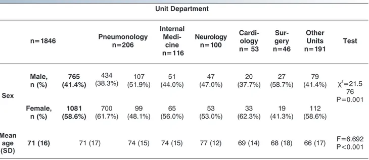
Distribution of patients in relation to sex was signi cantly di erent among Units of origin. Women more frequently were submitted to us from the departments of Cardiology and the Emergency Unit while men from the department of
2
Surgery (x =21.576, P=0.001) (Table 1). Patients mean age was 71 years (S.D±16) and was signi cantly di erent among the di erent departments of origin, with the oldest patients coming from the Neurology department (F=6.962, P<0.001) (Table 1). Data about the performance of previous instru-mental tests (chest X-rays and/or CTPA) and D-dimer dosage in all 1846 patients are reported on Table 2.
Patients had already by 93.2% performed chest X-rays and/or CTPA (Table 2). D-dimer values were increased (>500ng/mL) in 1625 of 1664 patients in which was previously performed (97.7%), (D-dimers mean value was 4448.59±191.84ng/mL).
Lung perfusion scintigraphy was positive for APE in 309/ 1846 (16.7%) patients [176 female (57%) and 133 male (43%)]
that consequently were promptly treated by their clinicians, with hospitalization in intensive care unit, and negative in 1537/1846 (83.3%) patients. Patients with negative LPS were dismissed and advised to return for follow-up within 2-3 we-eks.
The frequency of LPS negative was statistically signi cant 2
for all origin departments (X = 20.04, P=0.003). The highest percentage of positivity was found in patients from the Cardi-ology department (28%), followed by NeurCardi-ology (22%), with statistically signi cant di erences between the other depar-tments (Table 3).
The number of risk factors positively correlated with the 2
number of positive LPS (x =6.472, P=0.011) (Table 4 and Fi-lin
gure 2). The most frequent risk factor was arrhythmia and/or heart disease present in 608/1846 patients followed by pre-vious surgery (326/1846), and deep venous thrombosis (278/1846). However, the only risk factor predictive of positive LPS was venous thrombosis (OR=1.57, P=0.004) (Table 5).
Table 1. Demographic characteristics of the 1846 patients studied in relation to the departments of origin. Unit Department n=1846 Pneumonology n=206 Internal Medi-cine n=116 Neurology n=100 Cardi-ology n= 53 Sur-gery n=46 Other Units n=191 Test Sex Male, n (%) 765 (41.4%) 434 (38.3%) (51.9%)107 (44.0%)51 (47.0%)47 (37.7%)20 (58.7%)27 (41.4%)79 2 χ =21.5 76 P=0.001 Female, n (%) 1081 (58.6%) 700 (61.7%) 99 (48.1%) 65 (56.0%) 53 (53.0%) 33 (62.3%) 19 (41.3%) 112 (58.6%) Mean age (SD) 71 (16) 71 (17) 74 (15) 74 (15) 77 (12) 69 (14) 68 (18) 66 (17) F=6.692P<0.001
Table 2. Previous tests of all 1846 patients performed in the other departments. Unit Department n=1846 Emer-gency n=1134 Pneumo-nology n=206 Internal Medi-cine n=116 Neurology n=100 Cardi-ology n= 53 Sur-gery n=46 Other Units n=191 Test Previous CT/Chest X-ray 1721 (93.2%) 1060 (93.5%) 202 (98.1%) 112 (96.6%) 82 (82.0%) 48 (90.6%) 40 (87.0%) 177 (92.7%) 2 χ =33.277 P<0.001 2 χ =88.288 P<0.001 D-dimers dosage 1664 (90.1%) 1063 (93.7%) 184 (89.3%) 106 (91.4%) 86 (86.0%) 48 (90.6%) 30 (65.2%) 147 (77.0%)
The most often referred symptoms in our patient were dyspnea (1322/1846), chest pain (726/1846) and cough (420/1846). In 49.2% of patients was present only 1 sym-ptom, in 33.4% 2 symptoms, in 5.9% all 3 symptoms while 11.5% of patients had no symptoms but they presented one or more risk factors.
A signi cant association was observed between positive LPS and dyspnea (OR=1.78, P<0.001), conversely chest pain and cough were not associated with positive LPS (OR=0.95, P=649 and OR=1.25, P=0.066, respectively). D-dimers mean value was 4448.59ng/mL (S.D. 191.84ng/mL). Even if, the increased D-dimer dosage was not statistically associated with LPS results (OR=1.04, P=0.57), we observed that the D-dimer mean value was higher in patients with positive LPS
(6886.615±650,18ng/mL) than in patients with negative LPS (3986,773±189.71ng/mL). This di erence was statistically signi cant (t=-5.58, P<0.0001).
Chest X-rays and/or CTPA were negative for APE in 24.3% out of the 27.4% of patients suspected for APE, in 19.5% of non-speci c cases for APE with pleural e usion and in 28.8% of non-speci c with in ammatory interstitial diseases. Lung perfusion scintigraphies were positive in 13.6% of patients with negative chest X-rays and/or CTPA and in 21.3% with suspected chest X-rays and/or CT. Furthermore, was positive in 16.4% of cases with pleural e usion, in 15.2% with chronic obstructive pulmonary disease and in 16.6% with pulmo-nary in ammatory interstitial diseases. The remnant 16.9% of patients with positive lung pulmonary scintigraphy didn't Table 3. Departments of origin in relation to LPS results.
Department of origin (n=1846)
LPS positive for APE (n=309)
LPS negative for APE
(n=1537) Test P Emergency (n=1134) 178 (15.7%) 956 (84.3%) 2 χ =20.044 P=0.003 Pneumology (n=206) 43 (20.8%) 163 (79.2%) Int. Medicine (n=116) 21 (18.1%) 95 (81.9%) Neurology (n=100) 22 (22%) 78 (78%) Cardiology (n=53) 15 (28.3%) 38 (71.7%) Surgery (n=46) 8 (17.4%) 38 (82.6%) Other Units (n=191) 22 (11.5%) 169 (88.5%)
Table 4. Number of risk factors in relation to LPS results.
n=1846 LPS positive for APE
(n=309)
LPS negative for APE
(n=1537) Test P Risk factors 2 χ =6.472lin P=0.011 none 628 94 (15.0%) 534 (85%) 1 950 161 (17.0%) 789 (83%) 2 250 53 (21.2%) 197 (78.8%) 3 or more 18 6 (33.3%) 12 (66.7%)
Table 5. Risk factors in relation to LPS results.
Risk Factors LPS positive for APE LPS negative for APE Test P Arrhythmia/Heart disease 608 114 (18.8%) 494 (81.2%) 2
χ =1.8 0.180
Surgery 326 60 (18.4%) 266 (81.6%) 2
χ =0.488 0.485
Deep venous thrombosis 278 64 (23.1%) 214 (76.9%) 2
χ =8.187 0.004
Trauma/Fractures 155 22 (14.2%) 133 (85.8%) 2
χ =1.004 0.316
Drugs 77 16 (20.8%) 61 (79.2%) 2
performed previous diagnostic examination but had pre-sence of severe symptoms and risk factors.
Figure 2. Relationship of positive LPS and the number of risk factors.
Discussion
Since APE may be completely of partly symptomatic, its diagnosis can be incidental and sometimes challenging [12]. Mortality in untreated cases is about 30%. Early diag-nosis and adequate antithrombotic therapy reduce morta-lity to 2%-8% [11].
The high number of examinations performed in our hos-pital and especially in its Emergency Units re ects the high prevalence of suspicion for APE. Furthermore, our Emergen-cy Nuclear Medicine Service con rmed that APE is more fre-quently diagnosed in females and older patients, because the presence of comorbidity and risk factors for APE incre-ase with age. In 90% of cincre-ases, chest X-rays and/or CTPA and D-dimers dosage were performed before LPS, even if they are not speci c tools for APE diagnosis. The distribution of APE suspected patients by Hospital Care Unit in our study con rmed the fundamental role of LPS especially in emer-gency scenarios.
The high number of negative LPS performed by our LPS Service demonstrated that LPS is a key factor for early diag-nosis and for selecting the patients that need emergency treatment.
Our results con rm the observation of Miniati et al. (2010), which report very high speci city of LPS (97.7%) [10].
The presence of one or more risk factors was fundamental in the diagnosis of APE. Our study in fact, shows that LPS po-sitive is statistically correlated with the number of risk fac-tors. Deep venous thrombosis was, as others have found, the major risk factor for APE [11]. In addition, in most patients, dyspnea, chest pain, cough and syncope can supported the diagnosis of APE [12].
Dyspnea is considered the most frequent symptom of APE. It may be paroxysmal or severe in central APE, and mild and transient in small peripheral APE. Worsening dyspnea, in patients with pre-existing heart diseases, may be the only symptom suggestive of APE [13]. In our study population,
dyspnea was present in 71.6% of cases and was the only symptom signi cantly correlated with positivity LPS, con r-ming the necessity to not underestimate it.
Chest pain is also frequent in patients with suspected APE, caused by pleural irritation, due to distal embolism. In cen-tral pulmonary embolism, chest pain may imitate angina, possibly re ecting ventricular dysfunction, eventually as-sociated with ischemia. For this reason, patients with chest pain should be more frequently investigated with LPS.
Tests as chest X-rays and/ or CTPA and D-dimers dosage are usually performed in patients with suspected APE, even if they are not speci c to discriminate patients with APE. In fact, increase in D-dimer concentration is seen in several conditions such as DVT, cancer, in ammation, bleeding, tra-uma, surgery and (tissue) necrosis. Despite the elevated ne-gative predictive value of D-dimers dosage, its positive pre-dictive value is low and elevated D-dimer dosage does not help to con rm APE [14]. Our study con rms that the incre-ased value of D-dimer (>500ng/mL) is not predictive of APE but shows that D-dimers mean values are statistically higher in patients with positive LPS. In fact, as reported in literature, D-dimer levels are related to the simultaneous activation of coagulation and brinolysis in the presence of acute throm-bosis [15,16].
Chest X-rays allowed recognition of pulmonary embolism in sporadic cases while in most of the cases showed a non-speci c pattern non-speci cally in cases of pleural e usion or in-ammatory interstitial diseases, so integration with LPS is necessary to con rm the diagnosis of PE [17], as shown in Fi-gure 3.
Currently, multidetector CTPA is the examination of cho-ice for studying pulmonary vasculature in patients with sus-pected APE, thanks to the improvement of spatial and tem-poral resolution of the gamma camera and the quality of ar-terial opaci cation. Computed tomography pulmonary an-giography allows a panoramic view of the whole chest and visualization of di erent diseases like atelectasis, broncho-pulmonary foci, hemorrhagic foci and emphysema [18-19].
On the contrary, CTPA has a signi cantly higher exposure to ionizing radiation than the LPS. The absorbed dose du-ring CT is always about 10mSv vs 1mSv dudu-ring LPS [20] and needs the administration of medium contrast, not always possible, especially in emergency conditions as for patients with renal failure and contrast medium allergy. Computed tomography pulmonary angiography allows to diagnose large pulmonary embolism, but has a relatively low sensiti-vity (<80%) for sub-segmental pulmonary thromboembo-lism and cannot provide information about the hemodyna-mic e ect of emboli or vascular stenosis, responsible of lung perfusion alterations [21, 22]. Furthermore, the use of CTPA can diagnose a great number of APE cases but does not re-duce mortality. It may be that CTPA over-diagnoses APE [23, 24]. Figure 4 indicates the key role of LPS in the noninvasive assessment of APE.
Perfusion scans can be combined with ventilation studies to improve speci city: in APE, ventilation is usually to be nor-mal in hypoperfused segments (mismatch pattern) [25]. Ventilation scan requires a longer preparation and execu-tion time and active patient collaboraexecu-tion. These features do
not make it suitable to be performed in emergency. Both planar and tomographic acquisitions can be performed for a better case evaluation (of the perfusion scans) without fur-ther administration of a radiopharmaceutical and thus with-out ionizing radiation exposure. The availability of modern SPET/CT technology allows to obtain acquisition of tomo-graphic images associated with morphology, thus incre-asing diagnostic accuracy of APE but has a longer applica-tion time and greater exposure to radiaapplica-tion that makes it su-itable only in more complicated cases [26].
Currently, no diagnostic imaging protocol has been uni-versally adopted for the diagnosis of APE in emergency con-ditions. Lung perfusion scintigraphy can be preferably used in young or pregnant females or in renal diseases [27, 28]. According to the Italian legislation, considering the diag-nostic levels of reference, LPS has a very low irradiation ex-posure and so it can also be performed in pregnancy. It does not have side e ects, it is well tolerated and it takes about 15
minutes to be performed, so movement artifacts do not oc-cur and sedation of the patient is not necessary [29, 30]. Ba-sed on these advantages, we use LPS in the Emergency Nuc-lear Medicine Service of our hospital.
In our opinion, it is important that high-quali cation hos-pitals have a nuclear medicine emergency unit to provide LPS timely [31].
In conclusion, LPS has many advantages such as to be a
simple, quick and inexpensive examination; it does not re-quire preparation and has no side e ect so it can be perfor-med in all types of patients including pregnant women, po-lytraumatized and complicated patients (renal failure and contrast allergy). All these advantages make LPS suitable to be performed as Emergency Nuclear Medicine Service.
Our four year and large-scale experience related to a metropolitan area suggests that in patients with suspected APE, LPS has a key role in the early diagnosis, permitting to select a very low percentage of pts that need adequate Figure 3. A LPS of a female of 68 years old, with dyspnea, caugh and DVP as a risk factor. The value of D-dimer was 2014 g/L (n.v. <500) and chest CT showed signs of in ammatory intestitial desease. Irregular uptake was detected in both lungs due to chronic parenchymal disease, negative for APE.
Figure 4. A 70 years old patient a ected by multiple myeloma with dyspnea and chest pain, 18 hours after orthopedic surgery for femur trauma. Emergency LPS perfor-med in on-call 24hrs service, showed a perfusion defect on the anterior segment of the right upper lobe (red arrow) and superior lingular division (green arrow), positive for APE.
but even more in the exclusion of APE, optimizing the mana-gement of pts who do not require admission to intensive ca-re unit with high costs and limited availability.
Based on high incidence of APE, the di culty of its clinical diagnosis (non-speci c symptoms, numerous risk factors) and given the great demand and relevance of LPS, our study highlights how it's strongly recommended for Nuclear Medicine Units to perform LPS as emergency in on-call 24 hrs service.
Bibliography
1. Rubini G, Niccoli Asabella A, Stabile Ianora AA et al. Acute pul-monary embolism: comparison and integration of perfusion lung scintigraphy with multislice spiral CT. Radiol Med 2007; 112: 174-84.
2. Pelletier-Galarneau M, Zannier E, Zuckier LS, Le Gal G. Referral Pat-terns and Diagnostic Yield of Lung Scintigraphy in the Diagnosis of Acute Pulmonary Embolism. Thrombosis 2017; 2017: 1623868. 3. Heit JA, Silverstein MD, Mohr D et al. Risk factors for deep vein
thrombosis and pulmonary embolism: a population- based case-control study. Arch Intern Med 2000; 160: 809-15.
4. Anderson FA, Spencer FA. Risk factors for venous thromboem-bolism. Circulation 2003; 107: I9-I16.
5. Rogers MA, Levine DA, Blumberg N et al. Triggers of hospitalization for venous thromboembolism, Circulation 2012; 125: 2092-9. 6. Lane DA, Mannucci PM, Bauer KA. Inherited thrombophilia: Part 2.
Thromb Hamost 1996; 76: 824-34.
7. Cohen AT, Agnelli G, Anderson FA et al. Venous thromboembolism (VTE) in Europe. The number of VTE events and associated mor-bidity and mortality. Thromb Haemost 2007; 98: 756-64.
8. Costa AF, Basseri H, Sheikh A et al. The yield of CT pulmonary angi-ograms to exclude acute pulmonary embolism. Emergency Ra-diol 2014; 21: 133-41.
9. Wells PS, Anderson DR, Rodger M et al. Excluding pulmonary em-bolism at the bedside without diagnostic imaging: management of patients with suspected pulmonary embolism presenting to the emergency department by using a simple clinical model and d-dimer. Ann Intern Med 2001; 135: 98-107.
10. Miniati M, Pistolesi M, Marini C. Value of perfusion lung scan in the diagnosis of pulmonary embolism: results of the Prospective In-vestigative Study of Acute Pulmonary Embolism Diagnosis (PISA-PED). Am J Respir Crit Care Med 1996; 154(5): 1387-93.
11. Guidelines on the diagnosis and management of acute pulmo-nary embolism. Eur Heart J 2008; 29: 2276-315.
12. Miniati M, Prediletto R, Formichi B. Accuracy of clinical assessment in the diagnosis of pulmonary embolism. Am J Respir Crit Care Med 1999; 159: 864-71.
13. Wells PS, Ginsberg JS, Anderson DR et al. Use of a clinical model for safe management of patients with suspected pulmonary em-bolism. Ann Intern Med 1998; 129: 997-1005.
14. Di Nisio M, Squizzato A, Rutjes AW et al. Diagnostic accuracy of
D-dimer test for exclusion of venous thromboembolism: a syste-matic review. J Thromb Haemost 2007; 5: 296-304.
15. Stein PD, Hull RD, Patel KC et al. D-dimer for the exclusion of acute venous thrombosis and pulmonary embolism: a systematic review. Ann Intern Med 2004; 140: 589-602.
16. Righini M, Aujesky D, Roy PM. Clinical usefulness of D-dimer de-pending on clinical probability and cuto value in outpatients with suspected pulmonary embolism. Arch Intern Med 2004; 164: 2483-7.
17. Daftary A, Gregory M, Daftary A. Chest radiograph as a triage tool in the imaging-based diagnosis of pulmonary embolism. Am J Ro-entgenol 2005; 185: 132-4.
18. Lake DR, Kavanagh JJ, Ravenel JG. Computed tomography and pulmonary embolus: a review. Semin Ultrasound CT MR 2005; 26: 270-80.
19. Scialpi M, Rebonato A, Cagini L et al. Split-Bolus Single-Pass Multi-detector-Row CT Protocol for Diagnosis of Acute Pulmonary Em-bolism. Iran J Radiolog 2016; 13(1): e19844.
20. Raja AS, Greenberg JO, Qaseem A et al. Evaluation of patients with suspected acute pulmonary embolism: best practice advice from the Clinical Guidelines Committee of the American College of Physicians. Annals of Internal Med 2015; 163: 701-11.
21. Van Strijen MJ, De Monye W, Kieft GJ. Diagnosis of pulmonary em-bolism with spiral CT as a second procedure following scintigra-phy. Eur Radiol 2003; 13: 1501-7.
22. Members ATF, Konstantinides S, Torbicki A. 2014 ESC guidelines on the diagnosis and management of acute pulmonary embo-lism. Eur Heart J 2014; 35: 3033-69.
23. Anderson DR, Kahn SR, Rodger MA. Computed tomographic pul-monary angiography vs ventilation-perfusion lung scanning in patients with suspected pulmonary embolism: a randomized controlled trial. J Amer Med Assoc 2007; 298(23): 2743-53.
24. Molaee S, Ghanaati H, Safavi E et al. Computed Tomography Pul-monary Angiography for Evaluation of Patients With Suspected Pulmonary Embolism: Use or Overuse. Iran J Radiol 2015; 12(3): e22383.
25. Alderson PO. Scintigraphic evaluation of pulmonary embolism. Eur J Nucl Med 1987; 13: S6-10.
26. Perrier A, Desmarais S, Miron MJ. Noninvasive diagnosis of venous thromboembolism. Lancet 1999; 352: 190-5.
27. Washington L, Goodman LR, Gonyo MB. CT for thromboembolic disease. Radiol Clin North Am 2002; 40: 751-71.
28. Burge AJ, Freeman KD, Klapper PJ, Haramati LB. Increased diagno-sis of pulmonary embolism without a corresponding decline in mortality during the CT era. Clin Radiol 2008; 63: 381-6.
29. Niccoli Asabella A, Cimino A, Altini C et al. Lung Perfusion Imaging in Tetralogy of Fallot: A Case Report. Mol Imaging Radionucl Ther 2018; 27: 146-8.
30. Niccoli Asabella A, Stabile Ianora AA, Di Palo A et al. Lung scintigra-phy in pediatric patients with congenital malformations. Recenti Prog Med 2013; 104: 442-5.
31. Ferrari C, Cimino A, Bianco G et al. The impact of lung perfusion scintigraphy in the emergency management of patients with sus-pected pulmonary embolism. Hell J Nucl Med 2017; 20 Suppl: 166.