Journal of Surgical Case Reports, 2017;7, 1–4 doi: 10.1093/jscr/rjx135
Case Report
C A S E R E P O R T
A rare diaphragmatic hernia with a delayed
presentation of intestinal symptoms following
spleno-distal pancreatectomy: a case report
Giancarlo Pansini
1
, Giovanni Pascale
2,
*, Ilaria Pigato
2
, Enzo Malvicini
2
,
Dario Andreotti
2
, Annalisa Caruso
2
, Rocco Stano
3
, and Savino Occhionorelli
3
1
Department of Morphology, Surgery and Experimental Medicine, Section of Clinical Surgery, University of
Ferrara, Ferrara 44124, Italy,
2Department of Morphology, Surgery and Experimental Medicine, University of
Ferrara, Ferrara 44124, Italy, and
3Department of Morphology, Surgery and Experimental Medicine, Section
of Emergency Surgery, University of Ferrara, Ferrara 44124, Italy
*Correspondence address. Department of Morphology, Surgery and Experimental Medicine, University of Ferrara, Ferrara 44124, Italy. Tel:+39-053-223-7107; Fax: +39-053-223-7226; E-mail: [email protected].
ABSTRACT
Acquired diaphragmatic hernia, non-related to trauma, is a very rare condition. It can constitute a therapeutic problem and the surgical solution is not always immediately clear. We report the case of a 73-year-old woman with a history of spleno-distal pancreatectomy for a neuroendocrine tumour performed in 2009, who came back to Emergency Room 2 years later, complaining of abdominal pain. Chest radiography and computed tomography were performed; they showed a diaphrag-matic hernia with visceral migration into the thorax. The diaphragdiaphrag-matic defect was surgically repaired and the patient had an uneventful post-operative recovery.
INTRODUCTION
Diaphragmatic hernias (DH) are rare conditions and can be clas-sified into congenital hernias, traumatic hernias and iatrogenic hernias. Congenital DHs depend on anomalous embryonic development with a prevalence of 1.7–5.7 per 10 000 births [1].
Acute secondary diaphragmatic herniations are rare; no more than 5% of patients with abdominal trauma reports traumatic diaphragmatic injury. The literature includes descriptions of DH following laparoscopic cholecystectomy, laparoscopic hepatect-omy, esophagecthepatect-omy, gastrecthepatect-omy, gastric fundoplication, laparo-scopic gastric banding, radiofrequency ablation of liver lesions, splenectomy, nephrectomy and spleno-pancreatectomy, as well
as after living donor liver transplant and video-assisted thoraco-scopic surgery for partial resection of the lung [2–5].
The diaphragmatic damage may be silent or show up with symptoms of intestinal obstruction, immediately after the sur-gery or many years later. Acquired DH non-related to trauma is an even rarer presentation and it is very difficult to diagnose [6]. In this case report we describe an acquired DH occurring after spleno-pancreatectomy for a neuroendocrine tumour.
CASE REPORT
In April 2011, a 73-year-old woman with history of left-pancreatectomy and splenectomy for a neuroendocrine tumour
Received: May 15, 2017. Accepted: June 21, 2017
Published by Oxford University Press and JSCR Publishing Ltd. All rights reserved. © The Author 2017.
This is an Open Access article distributed under the terms of the Creative Commons Attribution Non-Commercial License (http://creativecommons.org/ licenses/by-nc/4.0/), which permits non-commercial re-use, distribution, and reproduction in any medium, provided the original work is properly cited. For commercial re-use, please contact [email protected]
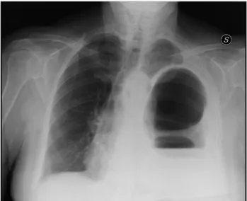
(2009) was admitted to the emergency department with abdom-inal cramps, nausea, without vomit or dyspnea. Her anamnesis was negative for asthma, respiratory distress, blunt or pene-trating trauma or conspicuous weight loss. Abdominal examin-ation showed a diffusely painful protruding abdomen, without signs of peritonitis and Blumberg sign was negative. Chest radiographs (Fig.1) showed a raised left hemidiaphragm, with bowel herniation into the lower half of the left hemi-thorax.
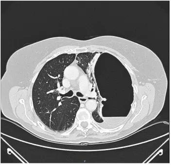
Thoraco-abdominal computed tomography (CT) showed the herniated bowel (transverse colon), translocated through a defect of about 7 cm in the left hemidiaphragm (Figs 2–4), a rightward deviation of the mediastinal structures and an atel-ectasis of the left lung (Fig.5). The left colon had the appear-ance of a volvulus.
No signs of concern emerged from the follow-up for the neu-roendocrine tumour until the last check, in particular no tumour recurrence was detected and there were no signs or symptoms of a diaphragmatic defect. During the follow-up a CT and
ultrasonography (US) were performed, both with negative out-comes. The only post-operative issue was a pancreaticfistula that was conservatively treated and that spontaneously resolved some months later. The patient underwent explorative laparotomy, reduction of the left DH, resection of the necrotic transverse colon with direct stapled side-to-side colic anastomosis, diaphragmatic suture and thorax drainage (Figs 6 and7). The diaphragmatic defect was repaired with interrupted suture. The recovery was complicated by ipsilateral pleural effusion and obstinate constipa-tion, though the patient was discharged on the fourteenth post-operative day, apparently in good health. The post-post-operative course was uneventful. One month later a thoraco-abdominal CT scan was performed, with no signs of recurrent diaphragmatic hernia. The oncologic follow-up was negative and the patient is still alive and apparently in good health.
Figure 1: Chest X-ray showing left colon herniation.
Figure 2: CT scan image showing left colon herniation.
Figure 3: 3D CT reconstruction.
Figure 4: 3D CT reconstruction. 2
|
G. Pansini et al.DISCUSSION
Acquired DHs may present with acute or chronic symptoms, or they may be detected accidentally during workup.
Acute presentation of a DH depends on the size and nature of the herniated organ. Epigastric or chest pain and dyspnea are typical symptoms due to the effects of pressure. If the stomach or bowel are obstructed, there will be episodes of vomiting.
Diagnosis could be challenging because of the unspecific nature of the presenting symptoms.
Acute presentation with strangulated or obstructed viscera needs for an urgent course of action and repair and thus a quick and accurate diagnosis. In the absence of strangulated or obstructed viscera, acquired DH or iatrogenic DH may be diag-nosed months or years after the initial surgery [7]. Chronic pain with or without respiratory changes may be the only presenting complaints. Chest radiographs are the best screening examin-ation, but an abnormality is detected in only 50% of patients [8].
CT scan, magnetic resonance imaging and US may be help-ful to make the right diagnosis. More invasive investigations might include laparoscopy or thoracoscopy evaluation.
Iatrogenic DHs may be due to damage by a grasping ment, electrocautery and interference by the suction instru-ment used to evacuate irrigation fluid. The problem is associated in particular with the energy released by these instruments, especially the ultrasonically activated scissors, which can expose a weak point in the diaphragm [2].
In our case, the DH was diagnosed 2 years after the original surgery and after a pancreaticfistula spontaneously resolved. It has been suggested that this delay in presentation is a result of the gradual enlargement of small tears in the diaphragm that go unnoticed during surgery. This develops slowly under the stress of increased intra-abdominal pressure associated with coughing and straining, and in our case a previous post-operative pancreaticfistula and obstinate constipation probably contributed to the pathogenesis. However, there are no irrefut-able evidences supporting this hypothesis, only several clues.
Surgery represents the first choice of treatment. While urgent surgery is frequently needed for the treatment of the symptomatic DHs, asymptomatic DHs may be performed days or months later, according to the patient’s status. Small dia-phragmatic defects are usually treated by primary repair with non-absorbable sutures. If the diaphragmatic defect is large or the muscles weak, synthetic grafts should be used because the primary repair could cause excessive tension [9]. In our case, we avoided the use of a graft because of the contamination due to the bowel resection in urgent surgery.
Surgery can be performed through of laparotomy or thora-cotomy. Some authors consider thoracotomy the best elective surgical approach for the correction of anatomical chest defects in absence of any abdominal pathology [3]. The patients who underwent thoracotomy experienced the longest stay, with a greater need for post-operative mechanical ventilation than those approached abdominally. In our case we preferred the abdominal incision and the DH was definitively repaired with direct non-absorbable suture.
Trans-abdominal surgery is suggested in acute or unstable patients to allow examination of intra-abdominal organs, dissec-tion of adhesions or exposure of the ischemic area of the bowel. Laparoscopic repair performed in patients with good general medical conditions is a suitable and safe procedure for the treat-ment of DH and it’s used especially in repairing left DH [8,10]. Figure 5: CT of chest showing defect on left side of the diaphragm and colon
herniation in the thoracic cavity.
Figure 6: Intra-operative image of trasverse colon herniation.
Figure 7: Intra-operative diaphragmatic defect.
CONFLICT OF INTEREST STATEMENT
None declared.
REFERENCES
1. Garne E, Haeusler M, Barisic I, Gjergja R, Stoll C, Clementi M. Euroscan Study Group. Congenital diaphragmatic hernia: evaluation of prenatal diagnosis in 20 European regions. Ultrasound Obstet Gynecol 2002;19:329–33.
2. Suh Y, Lee JH, Jeon H, Kim D, Kim W. Late onset iatro-genic diaphragmatic hernia after laparoscopy-assisted total gastrectomy for gastric cancer. J Gastric Cancer 2012; 12:49–52.
3. Singh M, Singh G, Pandey A, Cha CH, Kulkarni S. Laparo-scopic repair of iatrogenic diaphragmatic hernia following radiofrequency ablation for hepatocellular carcinoma. Hepa-tol Res 2011;41:1132–6.
4. Perwaiz A, Mehta N, Mohanka R, Kumaran V, Nundy S, Soin AS. Right-sided diaphragmatic hernia in an adult after
living donor liver transplant: a rare cause of post-transplant recurrent abdominal pain. Hernia 2010;14:547–9. 5. Fukami T, Konoeda C, Kitano K, Sakamoto M, Sano A,
Yoshida Y, Mura T, Nakajima J. Iatrogenic diaphragmatic hernia following partial resection of the lung via video-assisted thoracoscopy. Kyobu Geka 2010;63:1151–4.
6. Bisgaard C, Rodenberg JC, Lundgaard J. Spontaneous rupture of the diaphragm. Scand J Thorac Cardiovasc Surg 1985;19:177–80. 7. De Meijer VE, Vles WJ, Kats E, Den Hoed PT. Iatrogenic
dia-phragmatic hernia complicating nephrectomy: top-down or bottom-up? Hernia 2008;12:655–8.
8. Asencio JA, Demetrios D, Rodriguez A. Injury to the dia-phragm. In: Feliciano DV, Moore EE, Mattox KL, eds. Trauma. 3rd ed. Stamford: Appleton & Lange, 1996;461–85.
9. Yildirim B, Ozaras R, Tahan V, Artis T. Diaphragmatic Morgagni hernia in adulthood: correct preoperative diagno-sis is possible with newer imaging techniques. Acta Chir Belg 2000;100:31–3.
10. Edye M, Salky B, Posner A. Sac excision is essential to adequate laparoscopic repair of paraesophageal hernia. Surg Endosc 1998;12:1259–63.