UNIVERSITA’ DEGLI STUDI DI CATANIA
FACOLTA’ DI MEDICINA E CHIRURGIA DOTTORATO DI RICERCA IN
RICERCA MULTIDISCIPLINARE AVANZATA NEI TRAPIANTI
Direttore Prof. Pierfrancesco Veroux
Dott. Giuseppe Giuffrida
Laparoscopic Living donor nephrectomy.
Transperitoneal and Retroperitoneal Tech
nique.
Safety and efficacy
TESI DI DOTTORATO
Relatori: ● Chiar.mo Prof. M. Veroux ● Dr. C. Puliatti
Chapter 1
Introduction
Pag. 3
Chapter 2
Background
Pag. 7
Chapter 3
Materials and Methods
Pag. 9
Statistical Analysis Pag. 10 Patient Demography Pag. 10 Surgical Procedure Pag. 11 Post operative Management Pag. 24
Chapter 4
Results
Pag. 26
Chapter 5
Discussions
Pag. 29
Chapter 5
Conclusions
Pag. 36
References
Pag. 39
__________________________________________________Chapter 1
Chapter 1
Introduction
Kidney transplantation is considered the best replacement treat ment for patient with end stage renal failure. Thanks to the con tinue commitment of the scientific community massive im provements has been done since the first successful kidney transplant performed in 1954 by Joseph Murray at the Peter Bent Brigham Hospital in Boston from the identical Herrick twins.
We all know that thanks the efforts done to improve the surgical techniques, the management and the immunosuppression ther apy, the short term survival of both patients and graft has largely improved. However the long term results have not significantly changed over the last 30 years.
The transplant community has also to face day by day the con stant shortage of organs and increase demand for organs to be transplanted. Over the last decades different strategies have been developed with the intent of increasing the donor pool.
The most profitable strategies include the utilisation and optim isation of organs from the “Extended Criteria Donors” and ex pansion of living donors (LD) program.
Kidney transplantation (KTx) from living donors has shown to have the best graft and patient survival with an half life for the graft approaching 20 years (UNOS data).
__________________________________________________Chapter 1
During the last two decades there have been a constant increase of LD programs around the world and especially in USA and north Europe. This increase in number of LDKTx has been asso ciated with improvement of surgical techniques, looking mainly to develop a less invasive, but equally safe, procedure.
Miniinvasive surgical techniques for the LD nephrectomy have taken place over the classical open nephrectomy. Among the miniinvasive techniques we can recognise three different ap proaches, the miniincision open nephrectomy, the fully laparo scopic technique and the hand assisted laparoscopic technique. All these miniinvasive approaches, when compared with the classical open nephrectomy, have been associated with shorter convalescence time and increased quality of life for the donor and equally effective results for the recipient [1,2,3].
Within the laparoscopic techniques the hand assisted laparo scopic live donor nephrectomy (HALDN) is widely the most
The reasons why this is the most used techniques are several, the intaoperative tactile sensation that allow finger exploration and dissection, immediate bleeding control with direct pressure and short warm ischemia time and not lastly shorter learning curve for the surgeon [4, 5].
The retroperitoneoscopic techniques has been developed to try to minimise further more the potential complications of this type of surgery offering the intrinsic advantages of the conventional transperitoneal approach and potentially lower risk of intraperi toneal complications.
__________________________________________________Chapter 2
Chapter 2
Background
Laparoscopic donor nephrectomy has been developed to pro mote organ donation in living kidney transplantation, alleviating morbidities associated with conventional open surgery, and it is nowadays an accepted option supported by many studies report ing its excellent results with safety [3].
The HALDN can be performed either with a transperitoneal or with a retroperitoneal approach, both techniques have advant ages and disadvantages.
The nephrectomy with retroperitoneal approaches has been shown to have encouraging perioperative and functional out comes [4,5]. Compared to standard transperitoneal donor nephrec
tomy, the retroperitoneoscopic technique potentially has the ad vantage of facilitating a direct access to the hilum and avoids
The major disadvantage of this technique is represented by the limited working space.
In this single centre review we compared both techniques to as sess the efficacy and safety of retroperitoneoscopic living donor nephrectomy focusing on the itraoperative and perioperative outcomes in patients/grafts receiving this technique.
__________________________________________________Chapter 3
Chapter 3
Materials and Methods
This is a single centre retrospective analysis of 92 consecutive LD nephrectomies (LDN) performed at the Royal London Hos pital in the period between July 2011 and July 2013.
We divided the LDN in two groups, group A (n=34) including donors that received a retroperitoneoscopic hand assisted live donor nephrectomy (RHADN) and group B (n=58) including donors that received laparoscopic hand assisted donor nephrec tomy with transperitoneal approach (LHADN).
Statistical Analysis
The data analysed were:
· Warm Ischemia Time (WIT) · Length of operative time · Length of postoperative stay
· Incidence of intra and post operative complications
The aim of the study was to demonstrate the safety and efficacy of these techniques and to compare the outcomes.
Patient demography
There were no significant differences within the two groups on the base of age and gender.
In the retroperitoneal group the number of right nephrectomies was significantly higher (Table 1) while the incidence of vascu lar anomalies was similar in the two groups.
__________________________________________________Chapter 3
Transperitoneal (58) Retroperitoneal (34) PValue Age range (median) 25 – 75 (49) 22 – 61 (44)
Gender ratio M/F (%) 33/25 (56.9%/43.1%) 24/10 (70.6%/29.4%) 0,2662 Side L/R (%) 55/3 (94.8%/5.2%) 25/9 (73.5%/26.5%) < 0.0001
Table. 1
Surgical procedures RHALDN
Donor Position: the patient was placed in the standard right (or
left ) fullflank 90° position with straight operative table.
Hand-port placement: through a 78 cm Pfannenstiel incision
for either left or right nephrectomy.
Laparoscopic port placement. Two ports were used for left
nephrectomies, first 12mm port (camera port) positioned between the anterior superior iliac spine and the umbilicus on the midclavicular line, second port (working port) positioned
between the 12th rib and the superior border of the iliac bone on the anterior axillary line.
For right nephrectomies three ports were utilised. First 12mm port (camera port) positioned between the anterior superior iliac spine and the umbilicus on the midclavicular line, second port (working port) positioned between the 12th rib and the umbil icus on the midclavicular line, when procedure requested the re traction of the peritoneal sac, and the third 5 mm port
(assistant port) was positioned on the junction between the um bilical line and the mid axillary line (Fig. 1).
__________________________________________________Chapter 3
Fig.1 Incision position for RHALDN and LHALDN. Ports positioning for left and right nephrectomy (same position for both transperitoneal and retroperitoneal ap proaches)
Operative procedure: The surgeon and assistant stood on the
front side of the patient.
A 7 to 8 cm Pfannenstiel skin incision was made and through this wound, the recti muscles were separated and the fascia was exposed, and then bluntly detached by hand dissection from the muscular layer. The retroperitoneal space was bluntly made with gentle movement of the operator hand.
The GelPort (Fig. 2) was placed through the Pfannenstiel in cision.
Fig. 2 GelPort (Applied Medical, CA, USA) , hand assist sealing device permits the access of the hand to the surgical field, and rapid conversion to open surgery if necessary, while preserving the pneu moperitoneum/pneumoretroperitoneum.
__________________________________________________Chapter 3
The camera port (12 mm blunt tip port) was inserted at the mid point between the anterior superior iliac spine and the umbilicus on the midclavicular line. The position of the first port did not differentiate in both left or right nephrectomy.
The pneumoretroperitoneum was created and maintained by the insufflations of carbon dioxide at 10 – 12 mmHg.
The 30° endoscope was inserted through this port and the ret roperitoneal space was visualised.
Further blunt hand dissection of the retroperitoneal space from the iliac to the costal margin was performed, and the peritoneal sac was gently dissected medially from the muscle layer.
When enough retroperitoneal space was achieved the second 12 mm port was positioned between the12th rib and the superior border of the iliac bone on the anterior axillary line during the left nephrectomy.
When performing a right nephrectomy the working port was in stead placed between the 12th rib and the umbilicus on the mid clavicular line (Fig. 3)
Fig. 3. Port positioning during a left transperitoneal laparoscopic hand assisted donor nephrectomy (LHADN)
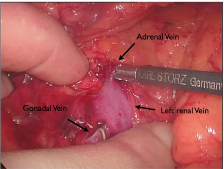
We first identified the proximal ureter and the gonadal vein at iliacs level. The gonadal vein was therefore followed up leading us to the renal vein (or to the inferior border of the IVC in case of right nephrectomy) (Fig. 4)
__________________________________________________Chapter 3
Fig. 4. Intraoperative picture: Left renal vein, Gonadal vein (already transected), Adrenal vein.
Secure sealing and transection of lumbar, gonadal and adrenal veins was performed (Fig. 4, 5) using vessel sealing systems (LigaSure, Covidien, Colorado, USA) (Fig. 6a).
As the dissection was progressed, the pulsation of the renal artery was identified.
The renal artery was dissected free using Harmonic scalpel (Ethicon) (Fig. 6b) and diathermy hook.
Fig. 6a: Ligasure Fig. 6b: Harmonic
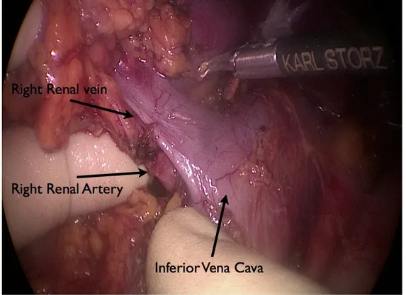
For the right nephrectomy, the renal artery was mobilized to the retrocaval site, and the renal vein was dissected down to the margins of the inferior vena cava (Fig. 7).
__________________________________________________Chapter 3
Fig. 7: Intraoperative picture: dissection of the right kidney hilum
The Gerota's fascia and the perirenal fat were dissected on the posterolateral surface using the Harmonic scalpel.
Once the kidney was completely dissected free a further mobil ization of the vessels was performed using Harmonic scalpel and diathermy hook to achieve maximum length of the vessels itself (Fig. 8).
Fig. 8: Intraoperative picture: Complex hilum anatomy: Ureter, 1 main renal vein, 1 accessory vein, 3 renal arteries.
The ureter was transected below the level of the iliac vessels and the distal ureter was closed with titanium clips (Fig. 9).
__________________________________________________Chapter 3
A linear cutting endovascular articulated endoGIA 35 mm (Ethicon) was then used to staple the artery and then the vein. The graft was immediately extracted from the GelPort and handed to the second surgeon, responsible for the kidney im plant, that was ready to rapidly cut the stapler line and reper fused the kidney with cold perfusion fluid (Soltran).
LHALDN
Donor Position: the patient was placed in a 60° right (or left )
flank position with the operative table broken at the umbilical level.
Hand-port placement: midline supraumbilical or transverse
subumbilical incision (78 cm) for either left or right nephrec tomy.
Laparoscopic port placement. Two ports were used for left
nephrectomies, first 12mm port (camera port) positioned between the anterior superior iliac spine and the umbilicus on the midclavicular line, second port (working port) positioned between the 12th rib and the superior border of the iliac bone on the anterior axillary line.
For right nephrectomies three ports were utilised. First 12mm port (camera port) positioned between the anterior superior iliac spine and the umbilicus on the midclavicular line, second port (working port) positioned between the 12th rib and the umbil icus on the midclavicular line, when procedure requested the re traction of the peritoneal sac, and the third 5 mm port
(assistant port) was positioned on the junction between the um bilical line and the mid axillary line (Fig. 1).
__________________________________________________Chapter 3
Operative procedure: The surgeon and assistant stood on the
front side of the patient.
Through the sub or supra umbilical incision the peritoneal space was reached. The camera and working port were inserted as pre viously described for the RHADN.
The large bowel was mobilised dissecting the paracolic gutter along the line of Toldt, from the left sigmoid to the splenocolic ligament for the right nephrectomy and from the caecum to the transverse colon for the right.
With the medialization of the dissected colon the retroperitoneal space was reached and the mobilisation ad dissection of the kid ney and the hilum proceeded as described for the RHALDN.
Post operative Management
Patients were encouraged to take fluids by mouth soon after they recovered from general anaesthesia.
Patient controlled analgesia (PCA) with parenteral narcotics (Fentanyl, Morphine) was started immediately after the surgery in recovery and discontinued on the first postoperative day.
Parenteral painkillers were replaced with oral medication (tra madol, oxycodone, or paracetamol) with parenteral analgesia supplements, if required.
Prophylaxis for deep venous thrombosis (low molecular weight Heparin, subcutaneous injection, prophylactic dose) was routinely used after few hours from the surgery if no concern of potential bleeding was raised.
The recovery was fast tracked by early mobilization starting from day one post op.
__________________________________________________Chapter 3
Donors fast track recovery program included also fluid and light meal intake few hours after the operation and deep breathing ex ercises.
Indwelling bladder catheter was removed on the first postoperat ive day.
IV fluids were also discontinued on day 1 post op and the pa tients were allowed to take regular oral fluids and meals.
The majority of patients were discharged on the third postoper ative day.
Chapter 4
Results
In total, 92 donors nephrectomy were included in our series. LDN was effectively and safely completed in 92 donors (100%) and o conversion to open surgery was required.
The operative time, despite numerically inferior in the retroperi toneal group (75 to 132 mins median 102), was not signific antly different from the operative time in the transperitoneal group (94 170 mins median 114) (Tab. 2).
The first warm ischemia time (WIT), measured as the time with in the clamping of the renal vessels and the start of the reperfu sion with cold preservation fluid was not statistically significant in the two groups (median 97 sec LHALDN vs. 96 sec RHALDN) (Tab. 2).
__________________________________________________Chapter 4
Transperitoneal (58)
Retroperitoneal
(34) PValue Operative time (median)
mins 94 – 170 (114) 75 – 132 (102) 0,9230 Warm Ischemia Time (medi
an) sec 79 146 (97) 60 130 (96) 0,4664 Length of Stay (median)
day 1 7 (3) 1 5 (3) 1.000
Tab. 2
Length of stay (3 days) was identical in the two groups of pa tients while the major complication rate (10.3% vs. 2.9%) was higher in the transperitoneal group. The reason of the fact the in cidence of major complications was not statistically significant is very likely due to the relatively small number of patients in cluded in the two groups (Tab. 3).
The incidence of minor complications such as urinary tract in fections, pulmonary infections or wound infections was identical in the two group and was managed with home
antibiotic therapy and therefore did not account as a cause of prolonged hospital stay or reason for readmission (Tab. 3).
Transperitoneal (58) Retroperitoneal (34) P-Value Minor Complications (Tot) 9 (15.5%) 7 (20.6%) 0,5765 Wound Infection 4 4 0,4615
Urinary Tract Infection 3 2 1.000
Pulmonary Infections 2 1 1.000 Major Complications (Tot) 6 (10.3%) 1 (2.9%) 0,2534 Incisional Hernia 3 0 0,2934 Bowel Injury 1 0 1.000 Intestinal Obstruction 1 0 1.000
Rotator Cuff Pain 1 1 1.000
__________________________________________________Chapter 5
Chapter 5
Discussion
The benefits of LD KTx are well recognised, because is associ ated with a lower incidence of delayed graft function, longer graft survival, and shorter recipient and donor hospital stay. The major disincentives to live donation are the postoperative mor bidity and the prolonged recuperation period.
Open live donor nephrectomy requires a long flank incision that is associated with significant postoperative and chronic pain and longer hospital stay for the donor. Wound complications include infection and hernia formation in 9% of donors [3,7]. In up to 25%
of donors, chronic incisional pain, wound “diastasis” or bulging has been reported, and return to normal activity may not occur for as long as 6 weeks to 8 weeks after nephrectomy [3,7].
Since 1995 when Ratner et al performed the first Laparoscopic donor nephrectomy (LDN), this procedure has now become the gold standard and has replaced the open technique of donor nephrectomy in most centres as it results in a short convales cence time, increased quality of life and better cosmetic results
[3,8].
In some early series however intraoperative safety has been de bated, as severe complications occur incidentally, but this may be part of the learning curve for this type of operation [8,9].
At the beginning of the laparoscopic experience the nephrec tomy was performed electively with full laparoscopic approach only for left kidney with single vascular anatomy. Nowadays has been widely demonstrated that any kidney fit for transplant ation, left or right, can be procured by the laparoscopic method despite the presence of anatomical variations [10].
__________________________________________________Chapter 5
In 1998 Wolf described the handassisted approach to make it more approachable from the majority of transplant surgeon also not familiar with laparoscopic surgery. In fact the hand assisted technique is easier to master, showing a shorter learning curve for the surgeon, and is also safer because of the handguided surgery while it maintains the benefits of endoscopic techniques
[4,5,11].
The ability to use the operator hand that allows direct tactile sensation and facilitate retraction and dissection of the structure is a clear advantage of this technique along with the better and prompt control of bleeding with direct pressure.
The intraperitoneal approach is at the moment the more utilised technique mainly because of the easiest access to the intraperi toneal space and wider space achieved in comparison with the retroperitoneal method.
There are not many controlled randomised trials or single centre report that are comparing the RHALDN and the LHADN. In our series we demonstrate that both approaches are safe and overall there are no statistically significant differences in terms of out comes and complications. The only difference with statistic sig nificant is represented by the presence of an higher percentage of right nephrectomy in the RHALDN group compared to the LHALDN group.
Has we know the incidence of vascular anomalies is quoted between 5 to 30% in different series. In our experience this in cidence was between 1520% and there was no difference between the two groups.
In our experience different surgeons performed both procedures so there is bias on the choice of a specific method.
__________________________________________________Chapter 5
In general we felt that the direct approach to the renal hilum and to the vascular structures of the kidney, allowed by the retroperi toneal access, can be a potential advantage in case of complex vascular anatomy and this is more evident on the right nephrec tomy where there is an easy access to the retrocaval space and then to the renal artery. This is very useful in case of early bi furcation or multiple vessels.
In previous experiences potential donors with abdominal surgery were often not considered for the laparoscopic approach due to the extensive adhesions. The retroperitoneal technique may be considered the elective approach for this type of patients, since it allow the surgeon to operate in a space surgically “virgin” avoiding long and potentially dangerous dissection of the in traabdominal adhesions.
One more advantage of the RHLDN is tht the mobilization of the intraperitoneal organs is not needed, possibly leading to few er incidence of postoperative ileus and damage to these organs. The downside of the technique is represented by the limited working space and the presence of few anatomical landmarks
[6,7] .
Several study demonstrates also that compared with miniin cision open donor nephrectomy, laparoscopic donor nephrec tomy (LDN) is considered costeffective reducing not only the length of stay in hospital for the donor but also reducing the time required for the donor to go back to normal social and working life.
In addition of the clinical advantages, such us better and longer graft functions, less morbidity and quicker recovery for the
__________________________________________________Chapter 5
donor, the live donation has also a clear economical benefit over dialysis and deceaseddonor transplantation [12,13,14].
Chapter 6
Conclusions
Living donor transplantation has been demonstrated to be the best option for patient on end stage renal failure.
The laparoscopic donor nephrectomy represents the gold stand ard technique in many centres and the hand assisted approach is the most used, combining the safety of handguided surgery with the benefits of endoscopic techniques.
Although laparoscopic transperitoneal approaches is superior to retroperitoneoscopic surgery in acquiring a wide surgical field and anatomical orientation, damage to abdominal organs and postoperative ileus is encountered only in small a fraction of donors treated retroperitoneally.
__________________________________________________Chapter 6
In our series, although we have small size numerical groups that cannot show statistical differences, no postoperative ileus was observed in the retroperitoneal group suggesting that the current retroperitoneoscopic approach is associated with less incidence of postoperative ileus. The incidence of the overall complica tions was also lower , but no statistically different in the ret roperitoneal group. The incidence of incisional hernias in our series is also numerically different in the two groups.
Overall there was no damage to the organ retrieved recorded and the outcome of the recipients was satisfactory and not different in both groups, confirming the efficacy and the safety of the pro cedure.
The hand assisted LDN offers to the donors all the advantages of the laparoscopic surgery and in particular the RHALDN has
got an intrinsic advantage over the conventional LHALDN be cause of the potentially lower risk for early and late donor in traperitoneal complications and can be electively used in pa tients with previous abdominal surgery avoiding potentially dan gerous dissection of adhesions.
Several studies supported the superiority of the LDN over open and miniincision nephrectomy, but more randomised controlled trials are needed in the future to prove the superiority between the different laparoscopic donor nephrectomy techniques.
References
1. Gill IS, Carbone JM, Clayman RV, Fadden PA, Stone MA, Lucas BA, McRoberts JW. Laparoscopic live-donor nephrectomy. J Endour ol. 1994;8(2):143–148. doi: 10.1089/end.1994.8.143.
2. Jacobs SC, Cho E, Dunkin BJ. Laparoscopic donor nephrectomy: current
role in renal allograft procurement. Urology. 2000;55(6):807–811. doi:
10.1016/S00904295(00)005252.
3. Buell JF, Lee L, Martin JE, Dake NA, Cavanaugh TM, Hanaway MJ, Weiskit tel P, Munda R, Alexander JW, Cardi M, Peddi VR, Zavala EY, Berilla E, Clippard M, First MR, Woodle ES. Laparoscopic donor nephrectomy vs.
open live donor nephrectomy: a quality of life and functional study. Clin
Transplant. 2005;19(1):102–109. doi: 10.1111/j.13990012.2004.00308.x. 4. Wolf JS Jr, Moon TD, Nakada SY. Hand assisted laparoscopic
nephrec-tomy: comparison to standard laparoscopic nephrectomy. J
Urol. 1998;160(1):22–27. doi: 10.1016/S00225347(01)630167.
5. Wolf JS Jr, Tchetgen MB, Merion RM. Hand-assisted laparoscopic live
donor nephrectomy. Urology. 1998;52(5):885–887. doi: 10.1016/S0090
4295(98)003896.
6. Dols LF, Kok NF, d'Ancona FC, Klop KW, Tran TC, Langenhuijsen JF,
Terkivatan T, Dor FJ, Weimar W, Dooper IM, Ijzermans JN. Randomized
8. Santosh A. Olakkengil, M. MinInvSu, M. Mohan Rao. Evolution of
minim-ally invasive Surgery for Donor Nephrectomy and Outcomes. JSLS. 2011
AprJun; 15(2): 208–212. doi: 10.4293/108680811X13071180406637 PM CID: PMC3148873
9. Ratner LE, Ciseck LJ, Moore RG, Cigarroa FG, Kaufman HS, Kavoussi L.
Laparoscopic live donor nephrectomy.Transplantation. 1995;60:1047–1049.
10. Cho HJ, Lee JY, Kim JC, Kim SW, Hwang TK, Hong SH. How safe and
effective is routine left hand-assisted laparoscopic donor nephrectomy with multiple renal arteries? A high-volume, single-center experience.
Department of Urology, The Catholic University of Korea, Seoul, Korea. 11. Slakey DP, Wood JC, Hender D, Thomas R, Cheng S. Laparoscopic living
donor nephrectomy: advantages of the hand-assisted method.
Transplantation. 68:581–583.
12. Peters TG. Living kidney donation: overcoming the financial disincentives. Contemp Dial Nephrol. 1996;17:22–25.
13. Yair Lotan, Matthew T. Gettman, Claus G. Roehrborn, Margaret S. Pearle, Jeffrey A. Cadeddu. Laparoscopic Nephrectomy is Cost Effective
Compared with open Nephrectomy in a Large County Hospital. JSLS.
2003 AprJun; 7(2): 111–115. PMCID: PMC3015483
14. Klop KW, Kok NF, Dols LF, D’Ancona FC, Adang EM, Grutters JP, IJzermans JN. Department of Transplant Surgery, Erasmus University Medical
roperitoneoscopic versus standard laparoscopic donor nephrectomy: a randomized study. Transplant Proc. 2012 Dec;44(10):29137. doi:
10.1016/j.transproceed.2012.04.038. Epub 2012 Sep 15.
15. Slojewski M, Myslak M, Domanski L, Pabisiak K, Jasiczek A, Sulikowski T, Sienko J, Ciechanowski K, Ostrowski M, Sikorski A. Program of
laparoscopic living-donor nephrectomy with retroperitoneoscopic access - a Polish single-center experience - success or disappointment? Transplant
Proc. 2012 Jun;44(5):121821. doi: 10.1016/j.transproceed.2011.12.079.
16. Dong J, Lu J, Zu Q, Yang S, Guo G, Ma X, Li H, Zhang X. Department of Urology, Chinese PLA General Hospital, Beijing, 100853, China.
Retroperitoneal laparoscopic live donor nephrectomy: report of 105 cases.
Transplantation. 2013 Jul 27;96(2):1705. doi: 10.1097/TP.0b013e318296ca25.